1
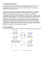
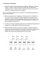
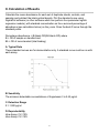
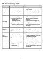
RayBio® Human/Mouse/Rat
Angiotensin II Enzyme Immunoassay
Kit
Catalog #: EIA-ANGII, EIAM-ANGII, EIAR-ANGII
User Manual
Last revised December 1, 2015
Caution:
Extraordinarily useful information enclosed
ISO 13485 Certified
3607 Parkway Lane, Suite 100
Norcross, GA 30092
Tel: 1-888-494-8555 (Toll Free) or 770-729-2992, Fax:770-206-2393
Web: www.RayBiotech.com, Email: [email protected]
1
Table of Contents
Section
Page #
I.
Introduction
3
II.
General Description
4
III.
How It Works
4
IV.
Storage
5
V.
Reagents
5
VI.
Additional Materials Required
6
VII.
Reagent Preparation
A. Preparation of Plate and Anti-Angiotensin II Antibody
B. Preparation of Biotinylated Peptide (Item F)
C. Preparation of Standards
D. Preparation of Positive Control
E. Preparation of Samples
F. Preparation of Wash Buffer and HRP-Strep
6
6
7
8
9
9
10
VIII.
Assay Procedure
10
IX.
Assay Procedure Summary
11
X.
Calculation of Results
A. Typical Data
B. Sensitivity
C. Detection Range
D. Reproducibility
E. Assay Diagram
12
12
12
12
12
13
XI.
Specificity
14
XII.
Select Publications
14
XIII.
Troubleshooting Guide
15
Please read the entire manual carefully before starting your experiment
2
I. Introduction
Angiotensin, a key player in the renin-angiotensin system, is a peptide hormone
that causes vasoconstriction, increased blood pressure, and release of aldosterone
from the adrenal cortex. It is derived from the precursor molecule angiotensinogen
produced in the liver.
Angiotensin II is formed from Angiotensin I, which is removed of two terminal
residues by the enzyme Angiotensin-converting enzyme (ACE). Angiotensin II acts
as an endocrine, autocrine/ paracrine, and intracrine hormone. Angiotensin II is
degraded to angiotensin III by angiotensinases that are located in red blood cells
and the vascular beds of most tissues. It has a half-life in circulation of around 30
seconds, while in tissue, it may be as long as 15-30 minutes.
The effect of obesity on Angiotensin II has recently been reported. Obese patients
show heightened renal vasodilation to blockade of the renin-angiotensin system,
suggesting deficits in vascular responses to angiotensin II. This may due to
increased reactivity of renal vasoconstriction to ANG II.
Angiotensin II has been associated with a number of important physiological
processes in heart, brain, adrenal gland and kidney. For cardiovascular effect,
Angiotensin II is a potent direct vasoconstrictor, constricting arteries and veins and
increasing blood pressure. It is also the most important Gq stimulator of the heart
during hypertrophy. For neural effects, Angiotensin II increases thirst sensation
(dipsogen) through the subfornical organ (SFO) of the brain, decreases the
response of the baroreceptor reflex, and increases the desire for salt. It increases
secretion of ADH in the posterior pituitary and secretion of ACTH in the anterior
pituitary. For adrenal effects, Angiotensin II acts on the adrenal cortex, causing it to
release aldosterone. For renal effects, Angiotensin II has a direct effect on the
proximal tubules to increase Na+ absorption.
3
II. General Description
The RayBio® Angiotensin II Enzyme Immunoassay (EIA) Kit is an in vitro
quantitative assay for detecting Angiotensin II peptide based on the competitive
enzyme immunoassay principle.
In this assay, a biotinylated Angiotensin II peptide is spiked into the samples and
standards. The samples and standards are then added to the plate, where the
biotinylated Angiotensin II peptide competes with endogenous (unlabeled)
Angiotensin II for binding to the anti-Angiotensin II antibody. After a wash step, any
bound biotinylated Angiotensin II then interacts with horseradish peroxidase (HRP)streptavidin, which catalyzes a color development reaction. The intensity of the
colorimetric signal is directly proportional to the amount of captured biotinylated
Angiotensin II peptide and inversely proportional to the amount of endogenous
Angiotensin II in the standard or samples. A standard curve of known concentration
of Angiotensin II peptide can be established and the concentration of Angiotensin II
peptide in the samples can be calculated accordingly.
III. How It Works
4
IV. Storage
The entire kit may be stored at -20°C to -80°C for up to 6 months from the date of
shipment. For extended storage, it is recommended to store at -80°C. Avoid
repeated freeze-thaw cycles. For prepared reagent storage, see table below.
V. Reagents
Component
Size / Description
Storage / Stability
After Preparation
Angiotensin II Microplate
(Item A)
96 wells (12 strips x 8 wells) coated with
secondary antibody.
1 month at 4°C*
Wash Buffer Concentrate
(20X) (Item B)
25 ml of 20X concentrated solution.
1 month at 4°C
Standard Angiotensin II
Peptide (Item C)
2 vials of Angiotensin II Peptide. 1 vial is enough
to run each standard in duplicate.
The first standard:
2-3 days at 4°C
Additional dilutions:
Do not store
Anti-Angiotensin II Polyclonal
Antibody (Item N)
2 vials of anti-Angiotensin II.
1 month at 4°C
5X Assay Diluent B (Item E)
15 ml of 5X concentrated buffer. Diluent for both
standards and samples including serum, plasma,
cell culture media or other sample types.
1 month at 4°C
Biotinylated Angiotensin II
Peptide (Item F)
2 vials of Biotinylated Angiotensin II Peptide, 1
vial is enough to assay half the plate.
2-3 days at 4°C
HRP-Streptavidin
Concentrate (Item G)
600 µl 100X concentrated HRP-conjugated
streptavidin.
Do not store and
reuse
Positive Control (Item M)
1 vial of Positive Control.
2-3 days at 4°C
TMB One-Step Substrate
Reagent (Item H)
12 ml of 3,3,5,5'-tetramethylbenzidine (TMB) in
buffer solution.
N/A
Stop Solution (Item I)
8 ml of 0.2 M sulfuric acid.
N/A
*Return unused wells to the pouch containing desiccant pack, reseal along entire edge.
5
VI. Additional Materials Required
1.
2.
3.
4.
5.
6.
7.
8.
9.
10.
11.
Microplate reader capable of measuring absorbance at 450 nm
Precision pipettes to deliver 2 µl to 1 ml volumes
Adjustable 1-25 ml pipettes for reagent preparation
100 ml and 1 liter graduated cylinders
Absorbent paper
Distilled or deionized water
SigmaPlot software (or other software which can perform four-parameter
logistic regression models)
Tubes to prepare standard or sample dilutions
Orbital shaker
Aluminum foil
Plastic wrap
VII. Reagent Preparation
Keep kit reagents on ice during reagent preparation steps.
A. Preparation of Plate and Anti-Angiotensin II Antibody
1. Equilibrate plate to room temperature before opening the sealed pouch.
2. Label removable 8-well strips as appropriate for your experiment.
3. 5X Assay Diluent B (Item E) should be diluted 5-fold with deionized or distilled
water.
4. Briefly centrifuge the anti-Angiotensin II antibody vial (Item N) Then add 50 µl
of 1X Assay Diluent B to the vial to prepare the antibody concentrate. Pipette
up and down to mix gently.
5. The antibody concentrate should then be diluted 100-fold with 1X Assay
Diluent B. This is your anti-Angiotensin II antibody working solution, which will
be used in step 2 of Assay Procedure (Section VIII).
Note: The following steps may be done during the antibody incubation procedure
(step 2 of Assay Procedure)
6
B. Preparation of Biotinylated Angiotensin II (Item F)
5. Briefly centrifuge the vial of Biotinylated Angiotensin II (Item F) before use.
6. See the image below for proper preparation of Item F. Transfer the entire
contents of the Item F vial into a tube containing 5 ml of 1X Assay Diluent B.
This is your Working Stock of Item F. Pipette up and down to mix gently.
The final concentration of biotinylated Angiotensin II will be 40 pg/ml.
a. Second Dilution of Item F for Standards: Add 2 ml of Working Stock Item
F to 2 ml of 1X Assay Diluent B. The final concentration of biotinylated
Angiotensin II will be 20 pg/ml.
b. Second Dilution of Item F for Positive Control: Add 100 µl of Working
Stock Item F to 100 µl of the prepared Positive Control (Item M). (See
section D for Positive Control preparation) The final concentration of
biotinylated Angiotensin II will be 20 pg/ml.
c. Second Dilution of Item F for samples: Add 125 µl of Working Stock Item
F to 125 µl of prepared sample (see section E for sample preparation).
This is a 2-fold dilution of your sample. The final concentration of
biotinylated Angiotensin II will be 20 pg/ml.
7
C. Preparation of Standards
7. Label 6 microtubes with the following concentrations: 1000 pg/ml, 100 pg/ml,
10 pg/ml, 1 pg/ml, 0.1 pg/ml and 0 pg/ml. Pipette 450 µl of biotinylated
Angiotensin II Item F working solution (prepapred in step 6a) into each tube,
except the 1,000 pg/ml (leave this one empty).
It is very important to make sure the concentration of biotinylated Angiotensin II is
20 pg/ml in all standards.
8. Briefly centrifuge the vial of Angiotensin II Standard (Item C). Pipette 8 µl of
Item C and 792 µl of 20 pg/ml biotinylated Angiotensin II working solution
(prepared in step 6a) into the tube labeled 1000 pg/ml. Mix thoroughly. This
solution serves as the first standard (1000 pg/ml Angiotensin II standard, 20
pg/ml biotinylated Angiotensin II).
9. To make the 100 pg/ml standard, pipette 50 µl of the 1000 pg/ml Angiotensin II
standard into the tube labeled 100 pg/ml. Mix thoroughly.
10. Repeat this step with each successive concentration, preparing a dilution
series as shown in the illustration below. Each time, use 450 µl of biotinylated
Angiotensin II and 50 µl of the prior concentration until the 0.1 pg/ml is
reached. Mix each tube thoroughly before the next transfer.
8
D. Positive Control Preparation
11. Briefly centrifuge the Positive Control vial (Item M).
12. Refer to step 6b. This is a 2-fold dilution of the Positive Control. The final
concentration of biotinylated Angiotensin II should still be 20 pg/ml.
The Positive Control is a mouse serum sample sample that serves as a system
control to verify that the kit components are working. The resulting OD will not be
used in any calculations; if no positive competition is observed please contact
RayBiotech Technical Support. The Positive Control may be diluted further if
desired, but be sure the final concentration of biotinylated Angiotensin II is 20 pg/ml.
E. Sample Preparation
13. If you wish to perform a 2-fold dilution of your sample, proceed to step 6c. If
you wish to perform a higher dilution of your sample, dilute your sample with
1X Assay Diluent B before performing step 6c.
EXAMPLE (to make a 4-fold dilution of sample):
a. Dilute sample 2-fold (62.5 µl of sample + 62.5 µl of 1X Assay Diluent B.).
b. Perform step 6c (125 µl of working solution Item F + 125 µl of sample
prepared above).
The total volume is 250 µl, enough for duplicate wells on the microplate.
It is very important to make sure the final concentration of the biotinylated
Angiotensin II is 20 pg/ml.
Note: Optimal sample dilution factors should be determined empirically, however you
may reference below for recommended dilution factors for serum: Human=2X
Mouse=2X Rat=2X.
If you have any questions regarding the recommendended dilutions you may contact
technical support at 888-494-8555 or [email protected].
9
F. Preparation of Wash Buffer and HRP
14. If Item B (20X Wash Concentrate) contains visible crystals, warm to room
temperature and mix gently until dissolved.
15. Dilute 20 ml of Wash Buffer Concentrate into deionized or distilled water to yield
400 ml of 1X Wash Buffer.
16. Briefly centrifuge the HRP-Streptavidin vial (Item G) before use.
17. Dilute the HRP-Streptavidin concentrate 100-fold with 1X Assay Diluent B.
VIII. Assay Procedure
1. Keep kit reagents on ice during reagent preparation steps. It is recommended
that all standards and samples be run at least in duplicate.
2. Add 100 µl of Anti-Angiotensin II Antibody (Item N) (See Reagent Preparation
step 3) to each well. Incubate for 1.5 hours at room temperature with gentle
shaking (1-2 cycle/sec). You may also incubate overnight at 4ºC.
3. Discard the solution and wash wells 4 times with 1X Wash Solution Buffer (200300 µl each). Washing may be done with a multichannel pipette or an automated
plate washer. Complete removal of liquid at each step is essential to good assay
performance. After the last wash, remove any remaining Wash Buffer by
aspirating or decanting. Invert the plate and blot it against clean paper towels.
4. Add 100 µl of each standard (see Reagent Preparation Section C), Positive
Control (see Reagent Preparation Section D) and sample (see Reagent
Preparation Section E) in appropriate wells. Be sure to include a blank well
(Assay Diluent only). Cover wells and incubate for 2.5 hours at room
temperature with gentle shaking (1-2 cycles/sec) overnight or at 4ºC.
5. Discard the solution and wash 4 times as directed in Step 3.
10
6. Add 100 µl of prepared HRP-Streptavidin solution (see Reagent Preparation
step 7) to each well. Incubate for 45 minutes at room temperature with gentle
shaking. It is recommended that incubation time should not be shorter or longer
than 45 minutes.
7. Discard the solution and wash 4 times as directed in Step 3.
8. Add 100 µl of TMB One-Step Substrate Reagent (Item H) to each well. Incubate
for 30 minutes at room temperature in the dark with gentle shaking (1-2
cycles/sec).
9. Add 50 µl of Stop Solution (Item I) to each well. Read at 450 nm immediately.
IX. Assay Procedure Summary
1. Prepare all reagents, samples and standards as instructed.
2. Add 100 µl anti-Angiotensin II to each well. Incubate 1.5 hours at room
temperature or overnight at 4ºC.
3. Add 100 µl standard or sample to each well. Incubate 2.5 hours at room
temperature or overnight at 4ºC.
4. Add 100 µl prepared Streptavidin solution. Incubate 45 minutes at room
temperature.
5. Add 100 µl TMB One-Step Substrate Reagent to each well. Incubate 30 minutes
at room temperature.
6. Add 50 µl Stop Solution to each well. Read at 450 nm immediately.
11
X. Calculation of Results
Calculate the mean absorbance for each set of duplicate stands, controls, and
samples and subtract the blank optical density. Plot the standard curve using
SigmaPlot software (or other software which can perform four-parameter logistic
regression models), with standard concentration on the x-axis and percentage of
absorbance (see calculation below) on the y-axis. Draw the best-fit curve through the
standard points.
Percentage absorbance = (B-blank OD)/B 0-blank OD) where
B = OD of sample or standard and
B0 = OD of zero standard (total binding)
A. Typical Data
These standard curves are for demonstration only. A standard curve must be run with
each assay.
B. Sensitivity
The minimum detectable concentrations of Angiotensin II is 2.62 pg/ml.
C. Detection Range
0.1-1,000 pg/ml
D. Reproducibility
Intra-Assay: CV<10%
Inter-Assay: CV<15%
12
E. Assay Diagram
Recommended Plate Layout:
Key:
Blank = Buffer Only
Total Binding = Biotin-Angiotensin II only
Standard 1 = 1000 pg/ml
Standard 2 = 100 pg/ml
Standard 3 = 10 pg/ml
Standard 4 = 1 pg/ml
Standard 5 = 0.1 pg/ml
Pos Control = Biotin with Item M
13
XI. Specificity
This kit can theoretically detect all active angiotensins, including ANGI, ANGII, ANGIII
and ANGIV. However, it does not detect inactive angiotensinogen.
Cross Reactivity: This EIA kit shows no cross-reactivity with any of the cytokines
tested: Ghrelin, Nesfatin, NPY and APC.
XIV. Publications Citing This Product
1. Dhar, Priyanka, Shashi B. Singh, and Vijay K. Sharma. "Autonomic Cardiovascular
Responses in Acclimatized Lowlanders on Prolonged Stay at High Altitude: A Longitudinal
Follow Up Study." PLoS One. PLoS One, 03 Jan. 2014. Web.
Species: Human
Sample Type: Plasma
2. Hao L., Wang X., Cheng J., You S., Ma S., Zhong X., Quan L., Luo B. The upregulation of
endothelin-1 and downregulation of miRNA-125a-5p, -155 and -199a/b-3p in human
atherosclerotic coronary artery. Cardiovascular Pathology - 07 April 2014 [Epub Ahead of
Print]
Species: Human
Sample Type: Other Fluid
3. Emdin M., et al. Biomarkers of activation of renin-angiotensin-aldosterone system heart
failure: how useful, how feasible? Clinica Chimica Acta Dec 2014, epub ahead of print.
DOI: 10.1016/j.cca.2014.10.031
Species: Human
Sample Type: N/A
4. Mascareno E, Galatioto J, Rozenberg I, et al. Cardiac Lineage Protein-1 (CLP-1)
Regulates Cardiac Remodeling via Transcriptional Modulation of Diverse Hypertrophic
and Fibrotic Responses and Angiotensin II-transforming Growth Factor ? (TGF-?1)
Signaling Axis. The Journal of Biological Chemistry 2012;287(16):13084-13093.
doi:10.1074/jbc.M111.288944.
Species: Mouse
Sample Type: Tissue Lysate
For additional publications citing this product, please contact technical support at 888494-8555 or [email protected].
14
XIII. Troubleshooting Guide
Problem
Cause
Solution
Inaccurate pipetting
Improper standard dilution
Check pipettes
Briefly centrifuge Item C and dissolve
the powder thoroughly by gently
mixing
Low signal
Improper preparation of
standard and/or
biotinylated antibody
Too brief incubation times
Inadequate reagent
volumes or improper dilution
Briefly spin down vials before
opening. Dissolve the powder
thoroughly.
Ensure sufficient incubation time;
assay procedure step 2 may be done
overnight
Check pipettes and ensure correct
preparation
Large CV
Inaccurate pipetting
Air bubbles in wells
Check pipettes
Remove bubbles in wells
High
background
Plate is insufficiently
washed
Contaminated wash buffer
Review the manual for proper wash.
If using a plate washer, ensure that
all ports are unobstructed.
Make fresh wash buffer
Improper storage of the
ELISA kit
Stop solution
Follow storage recomendations in
sections IV and V. Keep substrate
solution protected from light.
Add stop solution to each well before
reading plate
Poor standard
curve
Low sensitivity
15
RayBio® ELISA Kits
Over 2,000 ELISA kits available, visit www.RayBiotech.com/ELISA-Kits.html for
details.
This product is for research use only.
©2015 RayBiotech, Inc
16