1
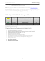
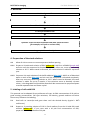
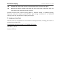
USER MANUAL EdU Click-555 ROTI®kit for Imaging ROTI®kit for Imaging Carl Roth GmbH + Co. KG EdU Click-555 ROTI®kit for Imaging The EdU Click-555 contains chemicals to perform 100 reactions (500 µL each). Introduction and product description: The detection of cell proliferation is of utmost importance for assassing cell health, determining genotoxicity or evaluating anticancer drugs. This is normally performed by adding nucleoside analogs like [3H]thymidine or 5-bromo-2’-deoxyuridine (BrdU) to cells during replication, and their incorporation into DNA is detected or visualized by autoradiography or with an anti-BrdU-antibody respectively. Both methods exhibit several limitations. Working with [3H]thymidine is troublesome because of its radioactivity. Autoradiography is slow and thus not suitable for rapid high-throughput studies. The major disadvantage of BrdU staining is that the double-stranded DNA blocks the the access of the anti-BrdU antibody to BrdU units. Therefore samples have to be subjected to harsh denaturing conditions resulting in degradation of the structure of the specimen. Roth’s EdU Click assays overcome these limitations, providing a superior alternative to BrdU and [3H]thymidine assays for measuring cell proliferation. EdU (5-ethynyl-2’-deoxyuridine) is a nucleoside analog to thymidine and is incorporated into DNA during active DNA synthesis. In contrast to BrdU assays, the EdU Click assays are not antibody based and therefore do not require DNA denaturation for detection of the incorporated nucleoside. Instead, the ROTI®kits for Imaging utilize click chemistry for detection in a variety of dye fluorescent readouts. Furthermore, the streamlined detection protocol reduces both the total number of steps and significantly decreases the total amount of time. The simple click chemistry detection procedure is complete within 30 minutes and is compatible with multiplexing for content and context-rich results. For research use only. Information in this document is subject to change without notice. Carl Roth GmbH + Co. KG assumes no responsibility for any errors that may appear in this document. Carl Roth GmbH + Co. KG disclaims all warranties with respect to this document, expressed or implied, including but not limited to those of merchantability or fitness for a particular purpose. In no event shall Carl Roth GmbH + Co. KG be liable, whether in contract, tort, warranty, or under any statute or on any other basis for special, incidental, indirect, punitive, multiple or consequential damages in connection with or arising from this document, including but not limited to the use thereof. Cautions: EdU: n Danger H340-H360 P202-P280-P308+P313 Catalyst Solution: j g Warning H302-H315-H319-H400-H410 P280-P301+P312a-P302+P352a-P305+P351+P338 1 Carl Roth GmbH + Co. KG ROTI®kit for Imaging Reaction Buffer: g Warning H315-H319 P280-P302+P352a-P305+P351+P338 MSDS: the appropriate MSDS can be downloaded from our website www.carlroth.com. Literature Citation: When describing a procedure for publication using this product, please refer to it as Carl Roth’s ROTI®kit for Imaging (EdU Click-555). 1. Materials provided with the kit and storage conditions Vial-colour yellow red purple orange green blue Amount 5 mg 130 µL 2 x 2 mL 4 x 2 mL 2 x 2 mL 4 x 200 mg Component 5-Ethynyl-deoxyuridine (5-EdU) 5-TAMRA-PEG3-Azide (10 mM) DMSO Reaction buffer (10x) Catalyst solution Buffer additive1 Storage* -20 °C dark, -20 °C RT 2 – 8 °C RT -20 °C *This kit is stable up to 1 year after receipt, when stored as directed. 1 Prepare aliquots to avoid too many freeze and thaw cycles; if the solution starts to develop a brown colour, it has degraded and should be discarded 2. Required Material and Equipment not included in this kit Cells adherently grown on a coverslip Reaction tubes (size depends on the volume of reaction cocktail needed) Phosphate-buffered saline (PBS, pH 7.2 – 7.6) Appropriate cell culture medium Fixation solution (3.7% formaldehyde in PBS) Permeabilization solution (for example, 0.5% Triton® X-100 in PBS) 3% BSA (bovine serum albumine) in PBS (3% BSA in PBS), pH 7.4 18 MΩ purified water 18 x 18-mm coverslips Optional: 6-well microplate 2 ROTI®kit for Imaging Carl Roth GmbH + Co. KG 3. Workflow Seed and grow cells ↓ Optional: sample treatment ↓ Incubate cells with EdU and other live cell stains ↓ Fix and permeabilize cells ↓ Detect EdU ↓ Optional: Treat cells with antibodies and other fixed-cell stains (for example, cell cycle or nuclear stain) ↓ Image aquisition and analysis 4. Preparation of the stock solutions 4.1 Allow all vials to warm to room temperature before opening. 4.1.1 Prepare a 10 mM stock solution of EdU (yellow vial): Add 2 mL of DMSO (purple vial) and mix until the compound is dissolved completely. After use, store any remaining solution at -20 °C. When stored as directed, this stock solution is stable for up to one year. 4.1.2 Prepare a 10x stock solution of the buffer additive (blue vials): Add 2 mL of deionized water to each of the blue vials and mix until the compound is dissolved completely. After use, store any remaining solution at -20°C. When stored as directed, this stock solution is stable for up to 6 months. If the solutions starts to develop a brown colour, it has degraded and should be discarded. We recommend to prepare aliquots to avoid repeated thaw and freeze cycles! 5. Labeling of cells with EdU This protocol can be adapted for any adherent cell type. An EdU concentration of 10 µM is a good starting concentration. Cell type variations, cell density, growth medium and other factors may influence the labeling. 5.1 Seed cells on coverslips and grow them until the desired density (typical ≈ 80% confluence). 5.2 Prepare a 2x working solution of EdU in fresh medium from the 10 mM EdU stock solution (yellow vial). If you start with a 10 µM final concentration of EdU, prepare a 2x working solution of 20 µM. 3 Carl Roth GmbH + Co. KG ROTI®kit for Imaging 5.3 Prewarm the 2x EdU solution and mix it with the same volume of adapted medium from the coverslips to obtain a 1x EdU solution. We do not recommend to replace all of the media with fresh media, because this could affect the rate of cell proliferation. 5.4 Aspirate the rest of medium from the coverslips and add the 1x EdU solution. 5.5 Incubate the cells for the desired pulse lenght under conditions optimal for the cell type. 5.6 Proceed immediately to the cell fixation and permeabilization steps 6.1-6.3. 6. Cell fixation and permeabilization This protocol was developed with a fixation step using 3.7% formaldehyde in PBS, followed by a 0.5% Triton® X-100 permeabilization step, but it is also amenable to other cell fixation/permeabilization reagents. For a better handling and processing, we recommend to transfer the coverslips into a 6-well plate, so that each well contains a single coverslip. 6.1 After incubation, remove the media and add 1 mL 3.7% formaldehyde in PBS (fixation solution) to each well containing the coverslips. Incubate for 15 minutes at room temperature. 6.2 Remove the fixation solution and wash the cells in each well twice with 1 mL of 3% BSA in PBS. 6.3 Remove the wash solution and add 1 mL of 0.5% Triton® X-100 in PBS (permeabilization solution) to each well. Incubate for 20 minutes at room temperature. 7. EdU detection In this protocol, 500 µL of the reaction cocktail per coverslip are used. Also smaller volumes can be used, as long as the reaction components are applied in the same ratios. 7.1 Prepare the reaction cocktail in the same order as described in the following table. If the ingredients are not added in the order listed, the reaction will not proceed optimally or might even fail. Important: Once the reaction cocktail is prepared, use it immediately, at least within the next 15 minutes! Reaction cocktail per coverslip (500 µL): Component Deionized water Reaction buffer (10x) Catalyst solution 5-TAMRA-PEG3-Azide (10 mM) Buffer additive (10x) (prepared in 4.3) Total Volume Vial colour orange green red blue Volume 379 µL 50 µL 20 µL 1 µL 50 µL 500 µL 7.2 Remove the permeabilization solution, then wash the cells in each well twice with 1 mL of 3% BSA in PBS. Remove the wash solution. 7.3 Add 500 µL of reaction cocktail to each well containing a coverslip. Rock the plate gently to distribute the reaction cocktail evenly over the coverslip. 4 ROTI®kit for Imaging Carl Roth GmbH + Co. KG 7.4 Incubate the plate for 30 minutes at room temperature. Protect from light! 7.5 Remove the reaction cocktail, then wash the cells in each well three times with 1 mL of 3% BSA in PBS. Remove the wash solution. Optional: Proceed with nuclear staining (DAPI or Hoechst 33342) or antibody labeling. Important: Keep the samples protected from light during incubations. If no additional staining is desired, proceed with imaging and analysis. 8. Imaging and analysis EdU Click cells are compatible with all methods of slide preparation, including wet mount or prepared mounting media. Excitation and emission maxima for 5-TAMRA-PEG3-Azide: Absorption: 546 nm Emission: 579 nm 5 Carl Roth GmbH + Co. KG ROTI®kit for Imaging Your notes: 6 ROTI®kit for Imaging Carl Roth GmbH + Co. KG Your notes: 7 Carl Roth GmbH + Co. KG ROTI®kit for Imaging Ordering information: (for detailed kit content see Table under 1.) ROTI®kits for Imaging (for 100 reactions): Product number Product Used fluorescent dye Filter 7773.1 EdU Click-488 6-FAM-Azide Green 7775.1 EdU Click-555 5-TAMRA-PEG3-Azide Violet 7776.1 EdU Click-594 5/6-Sulforhodamine 101-PEG3-Azide Orange 7777.1 EdU Click-647 Eterneon-Red 645 Azide (Cyanine 5 Azide analogue) Red To place your order, please contact us: Phone: +49 (0)721/5606-0 Fax: +49 (0)721/5606-149 Email: [email protected] 8 Carl Roth GmbH + Co. KG Phone: +49 (0)721/5606-0 Schoemperlentraße 3-5 Fax: 76185 Karlsruhe, Germany Email: [email protected] +49 (0)721/5606-149