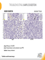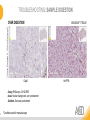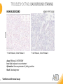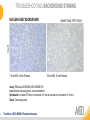
1
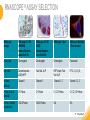
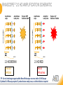
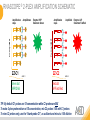
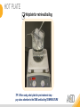
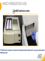
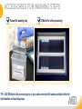
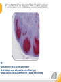
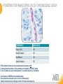
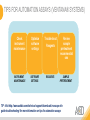
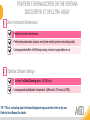
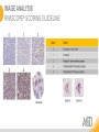
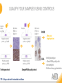
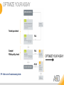
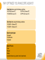
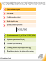
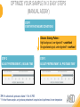
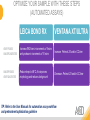
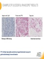
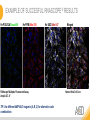
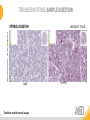
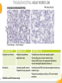
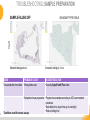
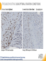
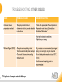
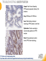
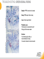
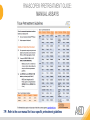
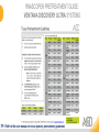
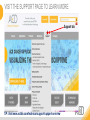
RNASCOPE® TROUBLESHOOTING TIPS Presented by: Jacqueline Akech, Ph.D. June 16th, 2015 Senior Scientist Advanced Cell Diagnostics ©2014 Advanced Cell Diagnostics, Inc. | Confidential and Proprietary | For Research Use Only (RUO), not intended for diagnosis. TOPICS • RNAscope® Recommended Workflow • Tips for RNAscope® Manual and Automation Assays • Troubleshooting Staining Patterns • Q&A 2 RNASCOPE ® WORKFLOW A BREAKTHROUGH PLATFORM UNIQUE Probe Design 3 SIGNAL Amplification + Background Suppression SINGLE Molecule Detection in Single Cells ANY Genome, Gene or Tissue RNASCOPE ® WORKFLOW 4 RNASCOPE ® ASSAY SELECTION RNAscope Assays RNAscope 2.0 HD (BROWN) Ventana Systems Leica Bond Rx RNAscope 2.0 HD (RED) Ventana Systems Leica Bond Rx RNAscope 2-plex RNAscope Multiplex – Fluoroscence Assay type Chromogenic Chromogenic Chromogenic Fluorescent Dye used Diaminobenzene (DAB)-HRP Fast Red -ALP HRP-Green, Fast Red -ALP FITC, Cy3, Cy5, Channel (Manual) Channel 1 Channel 1 Channel 1, 2 Channel 1, 2, 3 Probes channel (Manual) C1 Probes C1 Probes C1, C2 Probes C1, C2, C3 Probes Probes Channel (Automation) VS/LS Probes VS/LS Probes N/A N/A RNASCOPE ® 2.0 HD AMPLIFICATION SCHEMATIC Enzyme: HRP Substrate: DAB Amplification steps Amp6-Red Label probe Pre Amp/AMP1 Amp6-Brown Label probe Pre Amp/AMP1 Amplification steps ZZ mRNA 1 2.0 HD BROWN Brown dot HRP/DAB ZZ mRNA 1 2.0 HD RED Red dot AP/Fast Red TIP : Do not interchange reagents within Brown/Red assays or across similar 2.0 HD Assays By default 2.0 HD assays require C1 probes that are ready to use, no further dilution is required Enzyme: ALP Substrate: FastRed RNASCOPE ® 2-PLEX AMPLIFICATION SCHEMATIC Enzyme: HRP Substrate: Green Amplification steps Amp6-Red Pre Amp/AMP1 Amp6-Brown Pre Amp/AMP1 Amplification steps ZZ-C1 Green dot HRP/DAB mRNA 1 ZZ-C2 mRNA 2 Red dot AP/Fast Red TIP: By default C1 probes are 1X concentration while C2 probes are 50X To make 2-plex probe mixture at 1X concentration, mix C2 probes 1:50 with C1 probes To view C2 probes only, use the “blank-probe-C1”, as a diluent and mix at a 1:50 dilution Enzyme: ALP Substrate: FastRed RNASCOPE ® MULTIPLEX FLUORESCENT SCHEMATIC Amp3-C2 Amp3-C1 Alexa 488 Atto 550 Amp4-Alt A Amp1-C3 Amp1-C1 ZZ-C1 ZZ-C2 495nm/520nm ZZ-C3 mRNA 3 mRNA 2 mRNA 1 Alexa-488 Atto 647 Amp4-Alt A Amp1-C2 Amp4-Alt A Ex/Em Amp3-C3 Atto-550 555nm/575nm Atto-647 645nm/670nm TIP: By default C1 probes are 1X concentration while C2 and C3 probes are 50X To make 3-plex probe mixture at 1X concentration, mix C2 and C3 probes 1:50 with C1 probe If C2 and C3 are all at 50X concentration, use the “blank-probe-C1” as a diluent and mix at a 1:50 dilution RNASCOPE® WORKFLOW: CHROMOGENIC ASSAY RNAscope user workflow Description Deparaffinization Steps H2O2 block (Pretreat 1) Pretreat Time to Completion ~1.5 hours Epitope retrieval (Pretreat 2) Protease (Pretreat 3 or 4) A Hybridize Target probe hybridization Wash B ~2.5 hours Pre-amplifier hybridization Wash Amplify C Amplifier hybridization ~1.5 hours Wash D Label probe hybridization Wash Stain and detect ~1.5 hours Detection Hematoxylin stain Image detection under standard light microscope / scanner TIP : Detection protocols will vary based on the chromogenic assay used Download manuals: http://www.acdbio.com/technical-support/downloads RNASCOPE® WORKFLOW: FLUORESCENT ASSAY RNAscope user workflow Steps Description Fixation Pretreat Dehydration Time to Completion ~1.5 hours Protease digestion (Pretreat 3/4) A Hybridize Target probe hybridization Wash B Pre-amplifier hybridization Wash Amplify C ~3.5 hours Amplifier hybridization Wash D Label probe hybridization Wash Stain and detect ~0.5 hours DAPI Counterstaining Image detection under fluorescent microscope TIP : Pretreatment conditions will vary based on sample type Download manuals: http://www.acdbio.com/technical-support/downloads ONE DAY OR TWO DAY ASSAY? ONE DAY ASSAY TWO DAY ASSAY Sample preparation Sample preparation DAY 1 Sample pretreatment Sample pretreatment DAY 2 RNAscope assay RNAscope assay TIP : Review the User Manuals PART 1 and PART 2 for optional stopping points Refer to the User Manuals for Automation assay workflow TIPS FOR RNASCOPE® MANUAL ASSAYS 12 TIPS FOR MANUAL ASSAYS Follow protocols exactly as described in the user manuals PROTOCOLS Review sample pretreatment recommenda tions SAMPLE PRETREATMENT Always use control probes and slides Review that you are using all required materials USE CONTROLS THE CHECKLIST TIP : Visit http://www.acdbio.com/technical-support/downloads/rnascope-ishguide-troubleshooting/ for more information on tips for manual assays REVIEW THE CHECKLIST: Immedge hydrophobic barrier pen Positive and Negative control probes Hot–Plate for pretreatment/ target retrieval step Superfrost plus slides HybEZ Hybridization system Run RNAscope® control slides Ecomount for 2.0 HD Red & 2-plex chromogenic assay Fresh reagents (ethanol, xylene, 10% NBF) TIP : Visit www. acdbio.com/go for more information on getting started. Checklist is available on the website and in the manual HOT PLATE Hotplate for retrieval/boiling TIP : When using a hot plate for pre-treatment step – pay close attention to the TIME and boiling TEMPERATURE RNASCOPE ® REAGENT KIT CONTENTS OLD NEW Contents of the reagent kit 1. Pretreatment reagents 2. RNAscope detection kit 3. Wash buffer TIP : Warm probes at 40 ºC for 10 minutes before use TIP :Warm 50x wash buffer at 40 ºC for 20 minutes if you notice a precipitation HYBEZ HYBRIDIZATION OVEN HyBEZ hybdrization system TIP: HybEZ oven is required as it provides both temperature and humidity control, necessary to obtain optimal RNAscope results ACCESSORIES FOR WASHING STEPS Tissue Tek washing tray EZ Batch for slide processing TIP : ACD EZ Batch slide processing tray is easy and convenient for loading multiple slides for hybridization and washing steps. FOLLOW WORKFLOW GUIDELINES (MANUAL) Apply all amplification steps in the right order Use “flicking or tapping” technique to remove residual reagent Do not let slides dry out Make sure the hydrophobic barrier remains intact Do not alter the protocol in any way Warm probes and wash buffer at 40°C due to precipitation Maintain adequate humidity in the Humidity Control Chamber Fresh reagents (ethanol, xylene, 10% NBF) TIP : Visit http://www.acdbio.com/technical-support/downloads/rnascope-ishguide-troubleshooting/ for more information on tips for automation assays POINTERS FOR RNASCOPE 2.0 RED ASSAY TIP: •Use Ecomount or PERTEX as the mounting medium •Do not dehydrate sample with alcohol, to avoid a diffused signal •Samples should be dried in a 60 degree oven for 15 minutes before mounting POINTERS FOR RNASCOPE® 2-PLEX CHROMOGENIC ASSAY COMPONENTS MIXING RATIO Probes C2:C1 1:50 Amp 4B: Amp 4A 1:50 Red-B:Red- A 1:60 Green-B: Green-A 1:50 TIP: By default C1 probes are 1X concentration while C2 probes are 50X To make 2-plex probe mixture at 1X concentration, mix C2 probes 1:50 with C1 probes To view C2 probes only, use the “blank-probe-C1”, as a diluent and mix at a 1:50 dilution •Use Ecomount or PERTEX as the mounting medium •Do not dehydrate sample with alcohol, to avoid a diffused signal •Samples should be dried in a 60 degree oven for 15 minutes before mounting POINTERS FOR RNASCOPE® MULTIPLEX FLUORESCENT ASSAY COLOR MODULE OPTIONS Channel 1 (C1) Channel 1 (C2) Channel 1 (C3) AMP 4 Alt A GREEN-Alexa 488 ORANGE-Atto 550 FAR RED-Atto 647 Amp 4Alt B ORANGE-Atto 550 GREEN-Alexa 488 FAR RED-Atto 647 Amp 4 Alt C ORANGE-Atto 550 FAR RED-Atto 647 GREEN-Alexa 488 TIP: By default C1 probes are 1X concentration while C2 and C3 probes are 50X To make 3-plex probe mixture at 1X concentration, mix C2 and C3 probes 1:50 with C1 probe If C2 and C3 are all at 50X concentration, use the “blank-probe-C1” as a diluent and mix at a 1:50 dilution TIPS FOR RNASCOPE® AUTOMATED ASSAYS 23 TIPS FOR AUTOMATION ASSAYS (VENTANA® SYSTEMS) Check instrument maintenance Optimize software settings INSTRUMENT MAINTENANCE SOFTWARE SETTINGS Troubleshoot Reagents REAGENTS Review sample pretreatment recommendati ons SAMPLE PRETREATMENT TIP : Visit http://www.acdbio.com/technical-support/downloads/rnascope-ishguide-troubleshooting/ for more information on tips for automation assays POINTERS FOR RNASCOPE® ON THE VENTANA DISCOVERY® XT OR ULTRA ASSAY 1 Check Instrument Maintenance: Perform instrument maintenance Perform decontamination protocol every three months (prevents microbial growth) Use appropriate buffers for RNAscope assay, remove or purge before a run 2 Optimize Software Settings: *Uncheck the Slide Cleaning option (ULTRA only) Use appropriate hybridization temperature (different for XT versus ULTRA) TIP: *This is a cleaning step in Ventana Equipment may cause the slides to dry out Refer to User Manual for details POINTERS FOR RNASCOPE® (LEICA BOND RX®) Do not shake the contents in the containers as this will form bubbles LS Amp 1, LS Amp 3, 10X LS Wash Buffer, and all target probes require warming up at 40°C for 30 mins LS Brown and LS Red assays utilize Leica Biosystems’ Bond Polymer Refine Detection and Bond Polymer Refine Red Detection kits, respectively Do not alter the staining protocol in any way TIP : Visit http://www.acdbio.com/technical-support/downloads/rnascope-ish-guide-troubleshooting/ for more information on tips for LEICA BOND RX® automation assays QUALIFY YOUR SAMPLES USING CONTROLS 27 IMAGE ANALYSIS RNASCOPE® SCORING GUIDELINE QUALIFY YOUR SAMPLES USING CONTROLS Control slides e.g. Hela Negative control probe, e.g. DapB Positive control probe, e.g. PPIB Technique check QC check PPIB > 2 DapB < 1 Sample QC check PPIB > 2 DapB < 1 PASS Run your target probes Negative control probe, e.g. DapB Positive control probe, e.g. PPIB Sample/RNA quality check TIP : Always start with standard conditions FAIL •Verify technique • Check RNA quality with new samples •Perform Assay optimization OPTIMIZE YOUR ASSAY Technique check FAIL Sample/ RNA quality check FAIL OPTIMIZE YOUR ASSAY PASS TIP : Refer to the Troubleshooting Guide OPTIMIZE YOUR ASSAY 31 WHY OPTIMIZE YOU RNASCOPE ASSAYS? Under-fixed when using the following conditions: •4% PFA/24 hours/4°C 4% PFA ≤ 24 hours /RT •10% NBF/24 hours /4°C 4% PFA <24 hours /4°C Over-fixed when using the following conditions: •10% NBF > 48 hours /RT •10% NBF > 48 hours /4°C Special sample types: •Xenograft •Cultured cells •Cell pellet Special Tissues: •Liver •Muscle •Retina •Lymphoid tissues (e.g. spleen, tonsil, lymph node) FACTORS AFFECTING RNASCOPE® ASSAY PERFORMANCE Fixation conditions are not optimal RNA is degraded Hybridization conditions not optimal Samples drying during assay Special tissues sensitive to pretreatment THE SOLUTIONS Fix samples as recommended. E.g., for FFPE use 10% NBF RT, 16-32 hrs Acquire new samples and assess RNA quality Use the HybEZ hybridization oven only Use Immedge pen and add adequate reagents to avoid drying Start with standard pretreatment, then optimize conditions accordingly NBF: Neutral Buffered Formalin OPTIMIZE YOUR SAMPLE IN 3 EASY STEPS (MANUAL ASSAY) STEP 1 START WITH STANDARD CONDITIONS Observe Staining Pattern High background, over-digested? = underfixed No signal/weak signal, under-digested? = overfixed STEP 2 ADJUST PRETREATMENT 2, BOILING TIME STEP 3 ADJUST PRETREATMENT 3/4, PROTEASE TIME* TIP: For cultured cells, protease is diluted 1:15 in 1X PBS * For fresh frozen samples, only protease pretreatment is required and is performed at room temperature OPTIMIZE YOUR SAMPLE WITH THESE STEPS (AUTOMATED ASSAYS) OVER FIXED/ UNDER DIGESTED UNDER FIXED/ OVER DIGESTED LEICA BOND RX VENTANA XT/ULTRA Increase ER2 time in increments of 5 mins and protease in increments of 10 mins Increase Pretreat 2/3 and/or CC time Reduce temp to 88°C, this improves morphology and reduces background Decrease Pretreat 2/3 and/or CC time TIP: Refer to the User Manuals for automation assay workflow and pretreatment optimization guideline EXAMPLE OF SUCCESFUL RNASCOPE ® RESULTS Negative control, DapB RNAscope 2.0 HD Red Assay Positive control, PPIB Target probe Human breast cancer tissue TIP : Visit http://www.acdbio.com/technical-support/downloads/rnascope-ishguide-troubleshooting/ for more information EXAMPLE OF SUCCESFUL RNASCOPE ® RESULTS Hs POLR2A/Alexa 488 Hs PPIB/Atto 550 Hs UBC/Atto 647 RNAscope Multiplex Fluorescent Assay Amp 4 ALT A* TIP: Use different AMP4 ALT reagents (A, B, C) for alternative color combinations Merged Human Hela Cell Line TROUBLESHOOTING STAINING PATTERNS (CHROMOGENIC MANUAL ASSAYS) 38 39 TROUBLESHOOTING: NO STAINING OBSERVED PROBABLE CAUSE SUGGESTED ACTION Suboptimal fixation Prepare samples according to ACD recommendation Optimize pretreatment conditions •Over fixation •Under fixation Hybridization temperature not optimal Use HybEZ when performing RNAscope HybEZ temperature should be at 40°C Reagents used in the wrong sequence Apply reagents in the correct order Gene of interest no expressed Check positive control for technical accuracy of the assay 40 TROUBLESHOOTING: SAMPLE DIGESTION *8 min Pretreat 2 , 30 min Pretreat 3 OPTIMAL DIGESTION DapB *Conditions used for manual assays XENOGRAFT TISSUE Hs-PPIB 41 TROUBLESHOOTING: SAMPLE DIGESTION UNDER DIGESTION *8 min Pretreat 2 , 15 min Pretreat 3 XENOGRAFT TISSUE DapB Assay: RNAscope 2.0 HD RED Issue: Strong hematoxylin, under pretreatment, weak PPIB Solution: Increase pretreatment *Conditions used for manual assays Hs-PPIB 42 TROUBLESHOOTING: SAMPLE DIGESTION OVER DIGESTION *15 min Pretreat 2 , 30 min Pretreat 3 XENOGRAFT TISSUE DapB Assay: RNAscope 2.0 HD RED Issue: Nuclear background, over pretreatment Solution: Decrease pretreatment *Conditions used for manual assays Hs-PPIB 43 TROUBLESHOOTING: BACKGROUND STAINING HIGH BACKGROUND DapB KIDNEY FFPE TISSUE *15 min Pretreat 2 , 30 min Pretreat 3 *7 min Pretreat 2 , 30 min Pretreat 3 Assay: RNAscope 2.0 HD BROWN Issue: High background, over pretreatment Optimization: Decrease pretreatment 2 (boiling) conditions Result: Clean background *Conditions used for manual assays 44 TROUBLESHOOTING: BACKGROUND STAINING HUMAN TONSIL FFPE TISSUE DapB NUCLEAR HAZY BACKGROUND *15 min ER2, 30 min Protease *20 min ER2, 30 min Protease Assay: RNAscope LS BROWN (LEICA BOND RX) Issue: Nuclear hazy background, under pretreatment Optimization: Increase ER2 time in increments of 5 mins and protease in increments of 10 mins Result: Clean background *Conditions LEICA BOND RX automated assays 45 TROUBLESHOOTING: ASSAY WORKFLOW FFPE HELA PELLET DapB HIGH BACKGROUND/DRYING 15 min Pretreat 2 , 30 min Pretreat 3 BACKGROUND TYPE PROBABLE CAUSE SUGGESTED ACTION Cytoplasmic and nuclear •Samples drying between amplification steps •Completely cover tissue when applying reagents •Process slides one at a time to prevent drying •Ensure HybEZ Oven is at the appropriate temperature •Use the Immedge® hydrophobic barrier pen Extracellular •Incomplete paraffin removal •Suboptimal tissue preparation •Use fresh/unused EtOH and Xylene and agitate slides during incubation steps •Prepare tissue samples according to ACD recommended procedures *Conditions used for manual assays 46 TROUBLESHOOTING: SAMPLE PREPARATION XENOGRAFT FFPE TISSUE DapB SAMPLE FALLING OFF Standard baking protocol Increased baking by 1 hour ISSUE PROBABLE CAUSE SUGGESTED ACTION Tissue detaches from slides •Wrong slides used •Use only SuperFrost® Plus slides •Suboptimal tissue preparation •Prepare tissue samples according to ACD recommended procedures •Bake slides for a longer time (up to overnight) •Reduce boiling time *Conditions used for manual assays TROUBLESHOOTING SUB-OPTIMAL FIXATION CONDITIONS 24 hours fixation/Optimal Sample: FFPE brain sample 3 weeks fixation/Over fixed Synaptophysin Assay: RNAscope 2.0 HD Brown TIP: Sample fixation has a great effect on the success of your assay Solution: Increase pretreatment for better target accessibility 48 TROUBLESHOOTING: OTHER ISSUES ISSUE PROBABLE CAUSE SUGGESTED ACTION Unknown tissue preparation method •Sample provider/clinical •Follow the appropriate Tissue Specimen site/vendor did not provide detailed Preparation and Assay Optimization instructions Guidelines/Technotes* •Start with standard conditions •Optimize your assay Diffused Signal (RED) •Sample not completely dried •Alcohol used to dehydrate sample •Too much Ecomount mounting medium used TIP: Applies to all samples used with RNAscope •Dry sample as recommended (prolonged drying i.e. overnight, may be required •Do not dehydrate samples, dry at 60°C, 15 min •Use Ecomount sparingly and as recommended Positive control, Rn PPIB TROUBLESHOOTING: UNDER FIXATION Sample: Flash Frozen followed by FFPE sample preparation (fixation), Rat intestines Assay: RNAscope 2.0 HD Brown Issue: Weak staining, destroyed morphology, FFPE sample is under fixed Positive control, Rn PPIB Optimization: Fixation according to recommended guidelines for FFPE samples Result: Strong staining for positive control, PPIB, intact morphology 49 TIP : Refer to the Troubleshooting Guide http://www.acdbio.com/technical-support/downloads/rnascope-ish-guide-troubleshooting/ TROUBLESHOOTING: GREEN SIGNAL FADING Sample: FFPE human tonsil sample Hs Kapp/Lambda Assay: RNAscope 2-plex assay Issue: Green signal faded Probable cause: •Hematoxylin or associated low pH •Bluing with Ammonia water Hs Kapp/Lambda Solution: •Use hematoxylin briefly as recommended (30 secs) •Use water instead of ammonia water 50 RNASCOPE® PRETREATMENT GUIDE: MANUAL ASSAYS TIP : Refer to the user manual for tissue specific pretreatment guidelines RNASCOPE® PRETREATMENT GUIDE: VENTANA DISCOVERY ULTRA SYSTEMS TIP : Refer to the user manual for tissue specific pretreatment guidelines TROUBLESHOOTING TIPS MULTIPLEX FLUORESCENT ASSAY 53 TROUBLESHOOTING: SAMPLE DIGESTION Fresh Frozen Mouse Brain Fresh Frozen Mouse Kidney RT protease pretreatment 4 /Optimal TIP: Pretreatment temperature has a great effect on the success of your assay Solution: Perform pretreatment at RT to avoid over digestion of your sample 2-plex Positive Control Probe POLR2A/PPIB 40oC protease pretreatment 4 /Over digested TROUBLESHOOTING: SAMPLE DIGESTION 2-plex Mouse Positive Control Probe Mm POLR2A/PPIB 15 um 20 um Fresh Frozen Mouse Brain 10um Experiment condition: 10% NBF, 15 min Fixation, Pretreatment 4, RT TIP: Sample thickness can signal in your samples Solution: Use recommended sample thickness, 10-20um TROUBLE SHOOTING AUTOFLUORESCENCE Mouse FFPE Kidney Mouse FFPE Intestine Mouse FFPE Colon Mouse FFPE Brain TIP: FFPE sample have inherent autofluorescence Solution: Use appropriate background correction software to reduce autofluorescence MULTIPLEX FLUORESCENT ASSAY 101—PROBLEMS AND SOLUTIONS SOURCE ISSUE PROBLEM SOLUTION Microscopy No/weak signal Nonspecific signal 1. Wrong filter setting/longer emission cut off 2. Wrong exposure 3. Inappropriate imaging enhancing with software 1. Use correct filter settings 2. Do not use using autoexposure at first, verify signal with naked eye 3. Use known image enhancing software e.g. Nuance Sample No/weak signal 1. Compromised RNA quality 2. Sample preparation (high autofluorescence background on the sample 1. Use new sample with good RNA quality 2. Follow the pretreatment guideline recommended 3. Always perform assay with 3-plex positive control and 3plex negative probes to assess RNA quality 4. Always check signal with naked eye under objective lens first MULTIPLEX FLUORESCENT ASSAY 101—TIPS AND TRICKS • Be aware of the suggested filter settings for your microscope • Use the suggested pretreatment condition • Use the sample preparation protocol (PART 1) for your samples for optimal results • Always run a 3-plex positive control and negative control to assess RNA quality and to verify microscope setting are appropriate • Always evaluate the results by eye first before capturing images FREQUENTLY ASKED QUESTIONS 59 FREQUENTLY ASKED QUESTIONS • RNAscope assay compatibility with different tissues RNAscope manual assay can be used with FFPE, fresh-frozen, fixed-frozen and cultured cells. RNAscope automated assays are primarily supported with the FFPE tissue. Please refer to the User Manual Selection Guide: http://www.acdbio.com/technicalsupport/downloads • Key differences between RNAscope ISH assay and IHC No cooling is required during Epitope retrieval, users should directly put the slides in water at room temperature, dehydrate and proceed to Pretreatment 3 step as per the manual Part 1 60 TIP: Visit www.acdbio.com/support for additional FAQs SUMMARY 1. RNAscope ® recommended workflow for - Manual assays - Automated assays 2. Tips for RNAscope manual and automation assays - Check instrument maintenance - Optimize software settings - Optimize your assay 3. Troubleshooting staining patterns - High background, no signal, sample detachment - Optimizing with Pretreatment 2 and 3 optimization (MANUAL) - Adjusting ER2 , protease time and hybridization temperature changes (LEICA) - Offline/online pretreatment optimization (CC and pretreat 2/3) (VENTANA) VISIT THE SUPPORT PAGE TO LEARN MORE Support tab TIP: Visit www.acdbio.com/technical-support/support-overview CONTACT ACD SUPPORT Support via email –[email protected] Support via phone-1-877-376-3636, option 3 Time 8:00am-6:00pm PST Support Resources available on website www.acdbio.com QUESTIONS? PL EA SE C O M PL ET E T H E W EB IN A R SU RVEY, W E VA L U E YO U R F EED B A C K 64 Jacqueline Akech ACD Technical Support Advanced Cell Diagnostics, Inc. 3960 Point Eden Way, Hayward, CA 94545 Advanced Cell Diagnostics ©2013 Advanced Cell Diagnostics, Inc. | Confidential and Proprietary