1
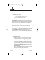
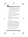
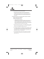
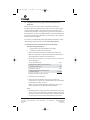
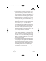
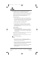
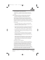
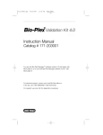
tm045.0505.qxp
7/6/2005
9:39 AM
Page 1
Technical Manual
TNT® Quick Coupled
Transcription/Translation
Systems
INSTRUCTIONS FOR USE OF PRODUCTS L1170, L1171, L2080 AND
L2081.
For your protein purification needs, please see the related
products section of this Technical Manual or visit
www.promega.com for a complete listing of protein purification
products offered by Promega.
PRINTED IN USA.
Revised 5/05
Part# TM045
A F 9 T M0 4 5
0 5 0 5 T M0 4 5
tm045.0505.qxp
7/6/2005
9:39 AM
Page 1
TNT® Quick Coupled
Transcription/Translation Systems
All technical literature is available on the Internet at www.promega.com/tbs
Please visit the web site to verify that you are using the most current version of this
Technical Manual. Please contact Promega Technical Services if you have questions on use
of this system. E-mail [email protected].
I. Description..........................................................................................................2
II. Product Components.........................................................................................5
III. General Considerations....................................................................................6
A. DNA Template Considerations..........................................................................6
B. Creating a Ribonuclease-Free Environment.....................................................8
C. Handling of Lysate...............................................................................................8
IV. Translation Procedure.......................................................................................8
A. General Protocol for TNT® Quick Coupled
Transcription/Translation Reactions Using Plasmid DNA...........................9
B. General Protocol for TNT® T7 Quick Coupled
Transcription/Translation Reactions Using PCR-Generated DNA ...........10
C. Notes.....................................................................................................................11
V. Positive Control Translation Reactions Using Luciferase.......................13
A. Radioactive Luciferase Control Reaction........................................................13
B. Non-Radioactive Luciferase Control Reaction...............................................13
VI. Cotranslational Processing Using Canine Pancreatic
Microsomal Membranes.................................................................................14
A. General Protocol for Translation with Microsomal Membranes ................14
VII. Post-Translational Analysis...........................................................................15
A. Determination of Percent Incorporation of Radioactive Label ...................15
B. Denaturing Gel Analysis of Radioactively Labeled
Translation Products..........................................................................................16
C. Denaturing Gel Analysis of Translation Products Labeled with the
FluoroTect™ GreenLys in vitro Translation Labeling System......................18
D. Denaturing Gel Analysis of Translation Products Labeled with the
Transcend™ Non-Radioactive Translation Detection Systems...................19
VIII. Positive Control Luciferase Assays..............................................................20
A. Using a Luminometer ........................................................................................20
B. Using a Scintillation Counter............................................................................20
IX. Troubleshooting...............................................................................................21
X. References .........................................................................................................23
Promega Corporation · 2800 Woods Hollow Road · Madison, WI 53711-5399 USA
Toll Free in USA 800-356-9526 · Phone 608-274-4330 · Fax 608-277-2516 · www.promega.com
Printed in USA.
Revised 5/05
Part# TM045
Page 1
tm045.0505.qxp
7/6/2005
9:39 AM
Page 2
XI. Appendix ...........................................................................................................25
A. Composition of Buffers and Solutions ............................................................25
B. Luciferase SP6/T7 Control DNAs ...................................................................26
C. Related Products.................................................................................................27
I.
Description
The TNT® Quick Coupled Transcription/Translation Systems(a–e) are convenient
single-tube, coupled transcription/translation reactions for eukaryotic in vitro
translation. The original TNT® Coupled Reticulocyte Lysate Systems(a,b,d,e)
simplified the process and reduced the time required to obtain in vitro
translation results compared with standard rabbit reticulocyte lysate systems (1).
Standard rabbit reticulocyte systems commonly use RNA synthesized in vitro
from SP6, T3 or T7 RNA polymerase (1). The TNT® Quick Coupled
Transcription/Translation System further simplifies the process by combining
the RNA polymerase, nucleotides, salts and Recombinant RNasin® Ribonuclease
Inhibitor(b,c) with the reticulocyte lysate solution to form a single TNT® Quick
Master Mix (Figure 1). For most gene constructs, the TNT® Quick reaction
produces significantly more protein (two- to sixfold) in a 60- to 90-minute
reaction than a standard in vitro rabbit reticulocyte lysate reaction using RNA
templates.
The TNT® Quick Coupled Transcription/Translation System is available in two
configurations for transcription and translation of genes cloned downstream
from either the T7 or SP6 RNA polymerase promoters. To use these systems,
0.2–2.0µg of circular plasmid DNA containing a T7 or SP6 promoter, or a PCR(f)generated fragment containing a T7 promoter, is added to an aliquot of the TNT®
Quick Master Mix and incubated in a 50µl reaction volume for 60–90 minutes at
30°C. The synthesized proteins are then analyzed by SDS-polyacrylamide gel
electrophoresis (SDS-PAGE) and detected. Included with the TNT® Quick System
is a luciferase-encoding control plasmid and Luciferase Assay Reagent(a,e,g),
which can be used in a non-radioactive assay for rapid (<30 seconds) detection of
functionally active luciferase protein. Starting with either circular plasmid DNA
or PCR-generated DNA, in vitro transcription/translation results may be
obtained easily in 5–6 hours.
!
PCR-generated fragments are not recommended for use with the
SP6 promoter. Use the T7 promoter.
Promega Corporation · 2800 Woods Hollow Road · Madison, WI 53711-5399 USA
Toll Free in USA 800-356-9526 · Phone 608-274-4330 · Fax 608-277-2516 · www.promega.com
Part# TM045
Page 2
Printed in USA.
Revised 5/05
tm045.0505.qxp
7/6/2005
9:39 AM
Page 3
TNT® Coupled Reticulocyte
Lysate System
TNT® Rabbit
Reticulocyte Lysate.
TNT® Quick Coupled
Transcription/Translation
System
Add TNT®
Reaction Buffer.
TNT® Quick
Master Mix.
Add TNT®
RNA Polymerase.
Add Amino
Acid Mixture
Minus Methionine.
Add RNasin®
Ribonuclease Inhibitor.
Add label of choice.
Add DNA template and
Nuclease-Free Water.
Separate translation
products by SDS-PAGE.
Detect
1537MB11_2A
Incubate at 30°C for
60-90 minutes.
Figure 1. Comparison of the TNT® Coupled Reticulocyte Lysate System and the
TNT® Quick Coupled Transcription/Translation System protocols.
Promega Corporation · 2800 Woods Hollow Road · Madison, WI 53711-5399 USA
Toll Free in USA 800-356-9526 · Phone 608-274-4330 · Fax 608-277-2516 · www.promega.com
Printed in USA.
Revised 5/05
Part# TM045
Page 3
tm045.0505.qxp
7/6/2005
9:39 AM
Page 4
In addition to verifying the expected molecular weight of a gene construct, the
TNT® Quick System is ideal for screening large numbers of constructs for either
naturally occurring or deliberately engineered mutations. Applications of the
system include:
•
•
•
•
•
•
•
Truncation mutation analysis
[e.g., the Protein Truncation Test
(PTT)] (2)
Drug screening (affecting
translation rates)
Mutation and detection analysis
(i.e., enzyme kinetics)
Protein:protein interactions (using
GST pulldowns)
Immunoprecipitation of protein
complexes
Protein dimerization assays
Ligand-binding region
determination/confirmation/
competition assays
•
•
•
•
•
•
•
In vitro expression cloning (IVEC)
(functional genomics)
Protein structure analysis
Electrophoretic mobility shift
assays (EMSAs) for DNA:protein
interactions
DNA footprinting and protein
cross-linking studies
Protein-RNA binding assays
Post-translational modification
tests
Verification/characterization of
cloned genes
The TNT® Quick Coupled Transcription/Translation Systems are also useful for
detecting protein:protein interactions in vitro. [35S]methionine-labeled proteins
labeled using TNT® Quick Coupled Transcription/Translation System can be
used as probes to detect interactions with suspected protein partners that have
been expressed as GST-(glutathione-S-transferase) or epitope-tagged fusion
proteins (3). [35S]methionine-labeled proteins can be synthesized using coupled
in vitro reactions from either full-length cDNAs or deletion mutants. The fusion
proteins can be bound to an affinity matrix along with the radioactive proteins
with which they interact (4–6). The bound radioactive proteins are then eluted
and analyzed by SDS-PAGE or Western analysis (Figure 2; 6). The fusion tag
approach has been used to study receptor-mediated control of apoptosis (7).
Alternatively, a non-radioactive approach may be used; the protein is labeled
with biotinylated lysine (e.g., Transcend™ Biotinylated tRNA) or is
fluorescently tagged (e.g., FluoroTect™ GreenLys System BODIPY®-FL-labeled
tRNA [Cat.# L5001]) and combined with a GST-tagged protein. The
biotinylated protein is detected by methods similar to those used in Western
blotting (8,9). The fluorescently tagged protein can be detected from within the
gel (10).
For a complete list of references for these and other applications, see reference 6
or visit the Promega Technical Resource Center citations at:
www.promega.com/citations/
Promega Corporation · 2800 Woods Hollow Road · Madison, WI 53711-5399 USA
Toll Free in USA 800-356-9526 · Phone 608-274-4330 · Fax 608-277-2516 · www.promega.com
Part# TM045
Page 4
Printed in USA.
Revised 5/05
tm045.0505.qxp
!
7/6/2005
9:39 AM
Gene 1
Page 5
GST Gene 1
Express in E. coli
TNT® System
35
Purify on Affinity Column
GST Protein 2
S Protein 1
W
E
M
W
E
M
W-Wash
M-Marker
Autoradiography Western
2598MA03_9A
E-Eluate
Figure 2. The study of protein:protein interactions using the TNT® Systems (6).
This schematic shows translation of one protein with radioactive [35S]methionine in a
TNT® System reaction. Large amounts of the suspected partner protein are expressed
and purified. A fusion tag (most commonly GST) is incorporated into this second
protein to facilitate purification and subsequent capture steps. After the GST fusion
protein is immobilized on sepharose (GST pulldowns), it is mixed with the protein
produced in the TNT® reaction. The sepharose is washed to remove unbound
protein, and the remaining bound proteins are eluted and analyzed on a gel. This
technique allows measurement of the protein:protein interactions for both proteins
and is often used to verify the in vivo results obtained from yeast two-hybrid
experiments. Promega offers the MagneGST™ Pull-Down System (Cat.# V8870) for
GST pull-down experiments.
II.
Product Components
Product
Cat.#
TNT® T7 Quick Coupled Transcription/Translation System
L1170
TNT® SP6 Quick Coupled Transcription/Translation System
L2080
For Laboratory Use. Each system contains sufficient reagents to perform approximately
40 × 50µl translation reactions. Includes:
•
•
•
•
•
•
•
1.6ml
5µg
100µl
50µl
250µl
1.25ml
1
TNT® Quick Master Mix (8 × 200µl)
SP6 or T7 Luciferase Control DNA (0.5µg/µl)(a)
T7 TNT® PCR Enhancer (L1170 only)
Methionine, 1mM
Luciferase Assay Reagent
Nuclease-Free Water
Protocol
Promega Corporation · 2800 Woods Hollow Road · Madison, WI 53711-5399 USA
Toll Free in USA 800-356-9526 · Phone 608-274-4330 · Fax 608-277-2516 · www.promega.com
Printed in USA.
Revised 5/05
Part# TM045
Page 5
tm045.0505.qxp
7/6/2005
9:39 AM
Page 6
Product
Cat.#
TNT® T7 Quick Coupled Transcription/Translation System, Trial Size
L1171
TNT® SP6 Quick Coupled Transcription/Translation System, Trial Size
L2081
For Laboratory Use. Each system contains sufficient reagents to perform approximately
5 × 50µl translation reactions. Includes:
•
•
•
•
•
200µl
5µg
100µl
50µl
1
TNT® Quick Master Mix
SP6 or T7 Luciferase Control DNA (0.5µg/µl)
T7 TNT® PCR Enhancer (L1171 only)
Methionine, 1mM
Protocol
Storage and Stability: Store all components at –70°C. Product components are
sensitive to CO2 (avoid prolonged exposure), frequent temperature fluctuations
and multiple freeze-thaw cycles, which can adversely affect stability, activity
and performance. Luciferase Assay Reagent (LAR) is stable for at least
12 months if stored and handled properly.
Note: See Note 5, Section IV.C, for details on how to refreeze the lysate.
Note that the systems are shipped in foil packaging because the system is
sensitive to carbon dioxide released from dry ice. If storing the system in a
freezer containing dry ice, keep system components sealed in foil packaging for
best results. DO NOT store the unfoiled lysate in the presence of dry ice.
Prolonged exposure to dry ice causes significant loss of activity. The expiration
date for the TNT® Quick Master Mix is listed on the product vial. Do not
freeze-thaw the Master Mix more than two times.
III. General Considerations
III.A. DNA Template Considerations
DNA Expression Elements
1. In addition to circular plasmid DNA, PCR-generated DNA templates can
be transcribed/translated using the T7 System. For maximal expression
from such templates, we recommend that approximately 11bp be present
upstream of the T7 RNA polymerase promoter for efficient promoter
binding. A stop codon (usually UAA) is important for truncated gene
products in order to prevent ribosomes from stalling at the ends of RNAs
without stop codons. This can be done through appropriate primer design
(11). The best transcription/translation results are obtained when the
fragment contains the T7 RNA polymerase promoter. We do not
recommend using linear DNA with the SP6 System because of reduced
transcription efficiencies.
Note: For coupled transcription/translation from PCR-generated templates,
Promega offers TNT® T7 Quick for PCR DNA (Cat.# L5540).
Promega Corporation · 2800 Woods Hollow Road · Madison, WI 53711-5399 USA
Toll Free in USA 800-356-9526 · Phone 608-274-4330 · Fax 608-277-2516 · www.promega.com
Part# TM045
Page 6
Printed in USA.
Revised 5/05
tm045.0505.qxp
7/6/2005
9:39 AM
Page 7
2. While Rabbit Reticulocyte Lysate-based systems are less sensitive to
5´-untranslated region (UTR) secondary structure than other systems, it is
still important to avoid strong hairpin secondary structure in the 5´-UTR
region, because this can impair translation efficiency (12).
3. We have observed enhanced translation of proteins when using DNA
constructs containing a poly(A) sequence downstream of the gene of
interest. Poly(A) sequences are important for mRNA stability and can play
a role in translation initiation in Rabbit Reticulocyte Lysate (13). For
example, we have observed a two- to fivefold increase in the production of
luciferase when the gene is cloned into the pSP64 Poly(A) Vector (Cat.#
P1241).
Plasmid DNA
1. Residual ethanol should be removed from DNA preparations before they
are added to the TNT® Quick Master Mix.
2. Linearized templates produced by restriction enzyme digestion should be
cleaned up either by using the Wizard® PCR Preps DNA Purification
System or by phenol:chloroform extraction, followed by ethanol
precipitation, before use in the TNT® Quick reaction.
3. Plasmid DNA can be purified using the Wizard® Plus or Wizard® Plus SV
Minipreps DNA Purification System or the PureYield™ Plasmid Midiprep
System. DNA prepared by the standard alkaline lysis method described by
Sambrook, Fritsch and Maniatis (14) is also sufficiently clean for use in the
TNT® Quick Coupled Transcription/Translation System. For most
constructs, optimal results are obtained when 1µg of plasmid DNA
template is used. However, we have used 0.2–2.0µg of DNA template and
obtained satisfactory levels of translation. The use of more than 1µg of
plasmid does not necessarily increase the amount of protein produced.
4. If linearizing plasmid DNA for use with the T7 System, avoid the use of
restriction enzymes that yield 3´-overhangs (Pst I, Kpn I, Sac I, Sac II, BstX I,
Nsi I, Apa I and Aat II), as aberrant transcription products can be produced
(15). If no alternative enzyme is available, the 3´-overhang can be removed
by adding T4 DNA polymerase.
Note: If you are using a linearized plasmid as a template, include 1µl of the
T7 TNT® PCR Enhancer in each 50µl reaction.
5. Check the sequence of the DNA template for the presence of additional
upstream start codons. During translation, the ribosome is thought to scan
from the 5´ end of the RNA and begin translation at the first AUG
encountered. Thus, any AUGs within the transcribed portion of the vector
or untranslated sequence of the insert may cause translation initiation to
occur prior to the desired start codon and result in a shift in the reading
frame or production of a larger protein than expected.
Promega Corporation · 2800 Woods Hollow Road · Madison, WI 53711-5399 USA
Toll Free in USA 800-356-9526 · Phone 608-274-4330 · Fax 608-277-2516 · www.promega.com
Printed in USA.
Revised 5/05
Part# TM045
Page 7
tm045.0505.qxp
7/6/2005
9:39 AM
Page 8
PCR-Generated DNA Templates
1. Because PCR DNA templates are usually much smaller than plasmid
templates, the amount of DNA necessary for optimal expression is often
less than for inserts cloned into plasmid vectors (e.g., for a 500bp PCR
product, use 100–800ng for each 50µl TNT® Quick reaction).
Note: For coupled transcription/translation from PCR-generated templates,
Promega offers TNT® T7 Quick for PCR DNA (Cat.# L5540).
2. PCR products (5–7µl) can be used directly from the amplification reaction.
Note: If you are using a PCR-generated template, include 1µl of the
T7 TNT® PCR Enhancer in each 50µl reaction.
III.B. Creating a Ribonuclease-Free Environment
To reduce the chance of RNase contamination, gloves should be worn when
setting up experiments, and microcentrifuge tubes and pipette tips should be
RNase-free. It is not necessary to add Recombinant RNasin® Ribonuclease
Inhibitor to the TNT® Quick reactions to prevent degradation of RNA, because
it is already present in the TNT® Quick Master Mix.
III.C. Handling of Lysate
Except for the actual transcription/translation incubation, all handling of the
TNT® Quick Master Mix should be done at 4°C. Any unused Master Mix
should be refrozen as soon as possible after thawing to minimize loss of
translational activity (see Note 5, Section IV.C). Do not freeze-thaw the Master
Mix more than two times.
IV. Translation Procedure
The following is a general guideline for setting up a transcription/translation
reaction. Also provided are examples of standard reactions using
[35S]methionine (radioactive), Transcend™ Non-Radioactive Detection System
(colorimetric or chemiluminescent) or FluoroTect™ GreenLys Systems
(fluorescent). Using the Transcend™ Systems, biotinylated lysine residues are
incorporated into nascent proteins during translation. This biotinylated lysine is
added to the transcription/translation reaction as a precharged ε-labeled,
biotinylated lysine-tRNA complex (Transcend™ tRNA) rather than a free
amino acid. For more information on the Transcend™ Systems, request
Technical Bulletin #TB182. The FluoroTect™ System uses a charged lysine
tRNA labeled with the fluorophore, BODIPY®-FL, to incorporate fluorescently
labeled lysine residues into the in vitro translation product. For more
information on the FluoroTect™ System, request Technical Bulletin #TB285.
Note: Technical Manuals and Bulletins are available online at:
www.promega.com/tbs/ or by request from Technical Services.
Promega Corporation · 2800 Woods Hollow Road · Madison, WI 53711-5399 USA
Toll Free in USA 800-356-9526 · Phone 608-274-4330 · Fax 608-277-2516 · www.promega.com
Part# TM045
Page 8
Printed in USA.
Revised 5/05
tm045.0505.qxp
7/6/2005
9:39 AM
Page 9
IV.A. General Protocol for TNT® Quick Coupled Transcription/Translation
Reactions Using Plasmid DNA
Materials to Be Supplied by the User
•
Nuclease-Free Water (Cat.# P1193)
•
Radiolabeled amino acid (for radioactive detection; Note 4, Section IV.C) or
Transcend™ tRNA (Cat.# L5061) or Transcend™ Colorimetric (Cat.#
L5070) or Chemiluminescent (Cat.# L5080) Translation Detection System
(for non-radioactive detection) or FluoroTect™ GreenLys in vitro Translation
Labeling System (for fluorescent detection; Cat.# L5001).
1. Remove the reagents from storage at –70°C. Rapidly thaw the TNT® Quick
Master Mix by hand-warming and place on ice. The other components can
be thawed at room temperature and then stored on ice.
2. Following the example below, assemble the reaction components in a 0.5ml
or 1.5ml microcentrifuge tube. After adding of all the components, gently
mix by pipetting. If necessary, centrifuge briefly to return the reaction to
the bottom of the tube. For additional information on performing a TNT®
Quick reaction, see Notes 1–9 in Section IV.C.
3. We recommend including a control reaction containing no added DNA.
This reaction allows measurement of any background incorporation of
labeled amino acids.
Example of a TNT® Quick Reaction Using Plasmid DNA
Components
Standard
Reaction Using
Transcend™
tRNA
Standard
Reaction Using
FluoroTect™
GreenLys tRNA
40µl
40µl
40µl
–
1µl
1µl
2µl
–
–
2µl
2µl
2µl
–
1–2µl
–
–
–
1–2µl
50µl
50µl
50µl
Standard
Reaction Using
[35S]methionine
TNT® Quick Master Mix
(see Note 3, Section IV.C)
Methionine, 1mM
(mix gently prior to use)
[35S]methionine
(1,000Ci/mmol at 10mCi/ml)
(see Note 4, Section IV.C)
plasmid DNA template(s)
(0.5µg/µl)
(see Note 6, Section IV.C)
Transcend™ Biotin-Lysyl-tRNA
(see Note 9, Section IV.C)
FluoroTect™ GreenLys tRNA
(see Note 9, Section IV.C)
Nuclease-Free Water
to a final volume of
Note: Small-scale reactions may be performed by reducing the recommended
volumes proportionally.
Promega Corporation · 2800 Woods Hollow Road · Madison, WI 53711-5399 USA
Toll Free in USA 800-356-9526 · Phone 608-274-4330 · Fax 608-277-2516 · www.promega.com
Printed in USA.
Revised 5/05
Part# TM045
Page 9
tm045.0505.qxp
7/6/2005
9:39 AM
Page 10
4. Incubate the reaction at 30°C for 60–90 minutes.
5. Analyze the results of translation. Procedures for determination of
radiolabel incorporation (Section VII.A) and SDS-PAGE analysis of
translation products (Section VII.B) are provided. If using FluoroTect™
GreenLys tRNA, see Section VII.C; for Transcend™ tRNA reactions, see
Section VII.D.
IV.B. General Protocol for TNT® T7 Quick Coupled Transcription/ Translation
Reactions Using PCR-Generated DNA
Materials to Be Supplied by the User
•
Nuclease-Free Water (Cat.# P1193)
•
Radiolabeled amino acid (for radioactive detection; Note 4, Section IV.C) or
Transcend™ tRNA (Cat.# L5061) or Transcend™ Colorimetric (Cat.#
L5070) or Chemiluminescent (Cat.# L5080) Translation Detection System
(for non-radioactive detection) or FluoroTect™ GreenLys in vitro Translation
Labeling System (for fluorescent detection; Cat.# L5001).
1. Remove the reagents from storage at –70°C. Rapidly thaw the TNT® Quick
Master Mix by hand-warming and place on ice. The other components can
be thawed at room temperature and then stored on ice.
2. Following the example below, assemble the reaction components in a 0.5ml
or 1.5ml microcentrifuge tube. After addition of all the components, gently
mix by pipetting. If necessary, centrifuge briefly to return the reaction to
the bottom of the tube. For additional information on performing a TNT®
Quick reaction, see Notes 1–9 in Section IV.C.
3. We recommend including a control reaction containing no added DNA.
This reaction allows measurement of any background incorporation of
labeled amino acids.
Promega Corporation · 2800 Woods Hollow Road · Madison, WI 53711-5399 USA
Toll Free in USA 800-356-9526 · Phone 608-274-4330 · Fax 608-277-2516 · www.promega.com
Part# TM045
Page 10
Printed in USA.
Revised 5/05
tm045.0505.qxp
7/6/2005
Components
9:39 AM
Page 11
Standard
Reaction Using
Transcend™
tRNA
Standard
Reaction Using
FluoroTect™
GreenLys tRNA
40µl
40µl
40µl
–
1µl
1µl
2µl
–
–
2.5–5µl
2.5–5µl
2.5–5µl
1µl
1µl
1µl
–
1–2µl
–
–
–
1–2µl
50µl
50µl
50µl
Standard
Reaction Using
[35S]methionine
TNT® T7 Quick Master Mix
(see Note 3, Section IV.C)
Methionine, 1mM
(mix gently prior to use)
[35S]methionine
(1,000Ci/mmol at 10mCi/ml)
(see Note 4, Section IV.C)
PCR-generated DNA template(s)
(see Note 1, Section IV.C)
T7 TNT® PCR Enhancer
(see Note 2, Section IV.C)
Transcend™ Biotin-Lysyl-tRNA
(see Note 9, Section IV.C)
FluoroTect™ GreenLys tRNA
(see Note 9, Section IV.C)
Nuclease-Free Water
to a final volume of
Note: Small-scale reactions may be performed by reducing the
recommended volumes proportionally.
4. Incubate the reaction at 30°C for 60–90 minutes.
5. Analyze the results of translation. Procedures for determination of
radiolabel incorporation (Section VII.A) and SDS-PAGE analysis of
translation products (Section VII.B) are provided. If using FluoroTect™
GreenLys tRNA, see Section VII.C; for Transcend™ tRNA reactions, see
Section VII.D.
IV.C.Notes
1. PCR-generated templates can be used directly from the amplification
reaction. We recommend using 2.5–5µl from the amplification reaction, but
up to 7µl can be used in a 50µl reaction. For PCR-generated DNA that has
been purified following amplification, we recommend using 100–800ng of
the purified product for each reaction.
2. We recommend using 1µl of the T7 TNT® PCR Enhancer in a 50µl reaction
to increase transcription/translation when using PCR-generated DNA,
linear plasmid or viral enhanced plasmids.
Promega Corporation · 2800 Woods Hollow Road · Madison, WI 53711-5399 USA
Toll Free in USA 800-356-9526 · Phone 608-274-4330 · Fax 608-277-2516 · www.promega.com
Printed in USA.
Revised 5/05
Part# TM045
Page 11
tm045.0505.qxp
7/6/2005
9:39 AM
Page 12
3. The TNT® Quick Master Mix is designed to give the highest expression for
most expression constructs. However, we have observed that certain gene
constructs may differ in the Mg2+ and K+ concentrations required for
optimal expression in the coupled reaction. For example, some viral leaders
will increase translation efficiency and fidelity if additional magnesium
acetate and potassium chloride are added to the TNT® Quick reaction. If
using a construct with a viral leader, we suggest adding 1–2µl of the T7
TNT® PCR Enhancer.
4. We recommend using a grade of [35S]methionine, such as Amersham
Biosciences Redivue® L-[35S]methionine (Amersham Biosciences Cat.#
AG1094), which does not cause the background labeling of the rabbit
reticulocyte lysate 42kDa protein. Background labeling of the 42kDa
protein can occur using other grades of label (16). In addition, a stabilizer
has been added to the Redivue® [35S]methionine to increase the stability of
this product over conventional radiolabeled amino acids, so that the release
of volatile gases is reduced substantially. This [35S]methionine may be
stored at 4°C without dispensing into aliquots. Other types of 35S-labeled
amino acids may be oxidized easily to translation-inhibiting sulfoxides and
should be stored in aliquots at –70°C in buffer containing DTT.
Between 10–40µCi (1–4µl) of [35S]methionine can be added to the TNT®
Quick reactions, depending upon the balance between labeling efficiency
and cost. For gene constructs that express well and contain several
methionines, the 10µCi level (1µl) is sufficient for adequate detection.
5. Except for the actual transcription/translation incubation, all handling of
the TNT® Quick System components should be done at 4°C or on ice.
Optimum results are obtained when any unused Master Mix is quickfrozen with liquid nitrogen as soon as possible after thawing to minimize
loss of translational activity.
6. For most plasmid constructs, optimal results are obtained when 1µg of
plasmid DNA template is used. We recommend using 0.2–2.0µg of plasmid
DNA in TNT® Quick reactions. The use of more than 1µg of plasmid does
not necessarily increase the amount of protein produced.
7. Avoid adding calcium to the transcription/translation reaction. Calcium
may reactivate the micrococcal nuclease used to destroy endogenous RNA
in the Master Mix and result in degradation of DNA or RNA templates.
8. The TNT® Quick Master Mix contains roughly 100–200mg/ml of
endogenous protein.
9. The level of added Transcend™ tRNA and FluoroTect™ GreenLys tRNA
can be increased (1–4µl) to allow more sensitive detection of proteins that
contain few lysines or are poorly expressed.
Promega Corporation · 2800 Woods Hollow Road · Madison, WI 53711-5399 USA
Toll Free in USA 800-356-9526 · Phone 608-274-4330 · Fax 608-277-2516 · www.promega.com
Part# TM045
Page 12
Printed in USA.
Revised 5/05
tm045.0505.qxp
V.
7/6/2005
9:39 AM
Page 13
Positive Control Translation Reactions Using Luciferase
The assay for firefly luciferase activity is extremely sensitive, rapid and easy to
perform. It is a good control for in vitro translations because only full-length
luciferase is active. Additionally, luciferase is a monomeric protein (61kDa) that
does not require post-translational processing or modification for enzymatic
activity. The Luciferase Assay System(a,e,g) is a substantial improvement over
conventional methods in both sensitivity and simplicity (17).
The control reaction can be performed with or without the addition of
radiolabeled amino acids.
V.A. Radioactive Luciferase Control Reaction
1. The following example contains [35S]methionine:
TNT® Quick Master Mix (see Note 3, Section IV.C)
[35S]methionine (1,000Ci/mmol at 10mCi/ml)
(see Note 4, Section IV.C)
Appropriate Luciferase Control DNA (0.5µg/µl)
(see Section XI.B)
Nuclease-Free Water to a final volume of
40µl
2µl
2µl
50µl
2. Incubate the reaction at 30°C for 60–90 minutes (see Note 3, Section IV.C).
3. Analyze the results of translation by measuring direct incorporation of
radiolabel (Section VII.A) and/or gel analysis of translation products
(Section VII.B).
4. The Luciferase Control reactions can be stored at –20°C for up to 2 months
or at –70°C for up to 6 months with little loss of luciferase activity.
V.B. Non-Radioactive Luciferase Control Reaction
1. The following example contains Methionine:
TNT® Quick Master Mix (see Note 3, Section IV.C)
40µl
Methionine, 1mM
1µl
Appropriate Luciferase Control DNA (0.5µg/µl)
(see Section XI.B)
2µl
Nuclease-Free Water to a final volume of
50µl
2. Incubate the translation reaction at 30°C for 60–90 minutes.
3. Test for the synthesis of functional luciferase using the standard luciferase
assay (see Section VIII.A).
4. The Luciferase Control reactions can be stored at –20°C for up to 2 months
or at –70°C for up to 6 months with little loss of luciferase activity.
Promega Corporation · 2800 Woods Hollow Road · Madison, WI 53711-5399 USA
Toll Free in USA 800-356-9526 · Phone 608-274-4330 · Fax 608-277-2516 · www.promega.com
Printed in USA.
Revised 5/05
Part# TM045
Page 13
tm045.0505.qxp
7/6/2005
9:39 AM
Page 14
VI. Cotranslational Processing Using Canine Pancreatic Microsomal
Membranes
Microsomal vesicles are used to study cotranslational and initial posttranslational processing of proteins. Processing events such as signal peptide
cleavage, membrane insertion, translocation and core glycosylation can be
examined by the translation of the appropriate gene in vitro in the presence of
these membranes. To ensure consistent performance with minimal background,
Canine Pancreatic Microsomal Membranes (Cat.# Y4041) have been isolated so
that they are free from mRNA.
For assistance in troubleshooting Microsomal Membrane translation reactions,
contact Promega Technical Services. E-mail: [email protected]
VI.A.General Protocol for Translation with Microsomal Membranes
Materials to Be Supplied by the User
•
Canine Pancreatic Microsomal Membranes (Cat.# Y4041)
•
[35S]methionine (1,000Ci/mmol at 10mCi/ml)
1. Remove the reagents from the freezer and allow them to thaw on ice.
Note: The storage buffer for Canine Pancreatic Microsomal Membranes is
50mM triethanolamine, 2mM DTT and 250mM sucrose.
2. Mix the following components on ice, in the order given, in a sterile 1.5ml
microcentrifuge tube:
T7 TNT® Quick Master Mix
20µl
[35S]methionine
(1,000Ci/mmol at 10mCi/ml)
(see Note 4, Section IV.C)
plasmid DNA, 0.5µg/µl
Canine Pancreatic Microsomal Membranes
(see Note 1, below)
Nuclease-Free Water to a final volume of
2.0µl
0.5µl
0.3–1.8µl
25µl
3. Incubate at 30°C for 60–90 minutes.
4. Analyze the results of translation and processing. Procedures for
incorporation assays (Section VII.A) and SDS-PAGE analysis of translation
products (Section VII.B) are provided.
Note: TNT® Quick Coupled Transcription/Translation Systems are not
tested for performance with Canine Microsomal Membranes.
Notes:
1. We do not recommend using Canine Microsomal Membranes when using
SP6 TNT® Quick Coupled Transcription/Translation Systems, because SP6
polymerase is sensitive to salts. Transcription may be inhibited as much as
70% by the presence of Canine Microsomal Membranes in the reaction.
Promega Corporation · 2800 Woods Hollow Road · Madison, WI 53711-5399 USA
Toll Free in USA 800-356-9526 · Phone 608-274-4330 · Fax 608-277-2516 · www.promega.com
Part# TM045
Page 14
Printed in USA.
Revised 5/05
tm045.0505.qxp
7/6/2005
9:39 AM
Page 15
2. The amount of Canine Microsomal Membranes used in the reaction may
need to be titrated. While these reaction conditions will be suitable for most
applications, the efficiency of processing using membranes may vary. Thus,
reaction parameters may need to be altered to suit individual requirements.
In general, increasing the amount of membranes in the reaction increases
the proportion of polypeptides processed but reduces the total amount of
polypeptides synthesized.
3. For reactions using the TNT® Quick CoupledTranscription/Translation
System, the Canine Microsomal Membranes will inhibit transcription. We
do not recommend exceeding 1.8µl of Canine Microsomal Membranes.
Transcription/Translation may be inhibited by as much as 50% with 0.6µl
of Canine Microsomal Membranes.
4. The amount of protein produced in TNT® Quick reactions using Canine
Pancreatic Microsomal Membranes will be less than the amount produced
in TNT® Quick reactions alone. Depending on the construct used, protein
synthesis efficiency can be expected to drop between 10–50% in the
presence of Microsomal Membranes.
5. In some cases, it is difficult to determine if efficient processing or
glycosylation has occurred by gel analysis alone. Other assays, such as
various protection assays (18), may be required to determine if processing
events have taken place.
VII. Post-Translational Analysis
Materials to Be Supplied by the User
(Solution compositions are provided in Section XI.A.)
•
1M NaOH/2% H2O2
•
Whatman® 3MM filter paper
•
30% acrylamide solution
•
25% TCA/2% casamino
•
separating gel 4X buffer
acids (Difco® brand, Vitamin
•
stacking gel 4X buffer
Assay Grade)
•
SDS sample buffer
•
5% TCA
•
SDS polyacrylamide gels
•
Whatman® GF/A
•
optional: precast
glass fiber filter
polyacrylamide gels
(Whatman® Cat.# 1820 021)
•
acetone
VII.A. Determination of Percent Incorporation of Radioactive Label
1. After the 50µl translation reaction is complete, remove 2µl from the reaction
and add it to 98µl of 1M NaOH/2% H2O2.
2. Vortex briefly and incubate at 37°C for 10 minutes.
3. At the end of the incubation, add 900µl of ice-cold 25% TCA/2% casamino
acids to precipitate the translation product. Incubate on ice for 30 minutes.
Promega Corporation · 2800 Woods Hollow Road · Madison, WI 53711-5399 USA
Toll Free in USA 800-356-9526 · Phone 608-274-4330 · Fax 608-277-2516 · www.promega.com
Printed in USA.
Revised 5/05
Part# TM045
Page 15
tm045.0505.qxp
7/6/2005
9:39 AM
Page 16
4. Wet a Whatman® GF/A glass fiber filter with a small amount of ice-cold
5% TCA. Collect the precipitated translation product by vacuum filtering
250µl of the TCA reaction mix. Rinse the filter 3 times with 1–3ml of icecold 5% TCA. Rinse once with 1–3ml of acetone. Allow the filter to dry at
room temperature or under a heat lamp for at least 10 minutes.
5. For determination of 35S incorporation, put the filter in the appropriate
scintillation cocktail, invert to mix and count in a liquid scintillation counter.
6. To determine total counts present in the reaction, spot a 5µl aliquot of the
TCA reaction mix directly onto a filter. Dry the filter for 10 minutes. Count
in a liquid scintillation counter as in Step 5.
7. To determine background counts, remove 2µl from a 50µl translation
reaction containing no DNA and proceed as described in Steps 1–5.
8. Perform the following calculation to determine percent incorporation:
cpm of washed filter (Step 5)
× 100 = percent incorporation
cpm of unwashed filter (Step 6) × 50
9. Perform the following calculation to determine the fold stimulation over
background:
cpm of washed filter (Step 5)
= fold stimulation
cpm of “no DNA control reaction” filter (Step 7)
VII.B. Denaturing Gel Analysis of Radioactively Labeled Translation Products
Precast polyacrylamide gels are available from a number of manufacturers. For
protein analysis, NOVEX® and Bio-Rad Laboratories, Inc., offer a variety of
precast mini-gels, which are compatible with their vertical electrophoresis and
blotter systems. These companies offer Tris-Glycine, Tricine and Bis-Tris gels
for resolution of proteins under different conditions and over a broad
spectrum of protein sizes. The NOVEX® 4–20% Tris-Glycine gradient gels
(Invitrogen Cat.# EC6025BOX or EC60355BOX) and the Bio-Rad Ready Gel
4–20% Tris-Glycine Gel, 10-well (Bio-Rad Cat.# 161-0903) are convenient for
resolving proteins over a wide range of molecular weights. In addition to
convenience and safety, precast gels provide consistent results.
1. Once the 50µl translation reaction is complete (or at any desired timepoint),
remove a 1–5µl aliquot and add it to 20µl of SDS sample buffer. The
remainder of the reaction may be stored at –20°C, or at –70°C for long-term
storage.
2. Cap the tube and heat at 100°C for 2 minutes to denature the proteins. This
may cause protein aggregation. Incubation at a lower temperature (e.g.,
20 minutes at 60°C, 10 minutes at 70°C or 3–4 minutes at 80–85°C) may be
more appropriate.
3. A small aliquot (5–10µl) of the denatured sample can then be loaded onto an
SDS-polyacrylamide gel or stored at –20°C. It is not necessary to separate
labeled polypeptides from free amino acids by acetone precipitation.
Promega Corporation · 2800 Woods Hollow Road · Madison, WI 53711-5399 USA
Toll Free in USA 800-356-9526 · Phone 608-274-4330 · Fax 608-277-2516 · www.promega.com
Part# TM045
Page 16
Printed in USA.
Revised 5/05
tm045.0505.qxp
7/6/2005
9:39 AM
Page 17
4. Typically, electrophoresis is carried out at a constant current of 15mA in
the stacking gel and 30mA in the separating gel (or 30mA for a gradient
gel). Electrophoresis is usually performed until the bromophenol blue dye
has run off the bottom of the gel. Disposal of unincorporated label may be
easier if the gel is stopped while the dye front remains in the gel, as the dye
front also contains the unincorporated labeled amino acids. If transferring
the gel to a membrane filter for Western blotting, proceed to Step 7.
5. Place the polyacrylamide gel in a plastic box and cover the gel with fixing
solution (as prepared in Section XI.A) for 30 minutes. Agitate slowly on an
orbital shaker. Pour off the fixing solution. Proceed to Step 6 (gel drying
prior to film exposure).
Optional: Labeled protein bands in gels may be visualized by
autoradiography or fluorography. Fluorography dramatically increases the
sensitivity of detection of 35S-, 14C- and 3H-labeled proteins and is
recommended for the analysis of in vitro translation products. The
increased detection sensitivity of fluorography is obtained by infusing an
organic scintillant into the gel. The scintillant converts the emitted energy
of the isotope to visible light and increases the proportion of energy that
may be detected by X-ray film. Commercial reagents, such as Amplify®
Reagent (Amersham Biosciences), can be used for fluorographic
enhancement of signal. Alternatively, the fixed gel can be exposed to a
phosphorimaging screen. These systems provide greater sensitivity, greater
speed and the ability to quantitate the radioactive bands.
6. Dry the gel before exposure to film as follows: Soak the gel in 10% glycerol
for 5 minutes to prevent the gel from cracking during drying. Place the gel
on a sheet of Whatman® 3MM filter paper, cover with plastic wrap and dry
at 80°C for 30–90 minutes under a vacuum using a conventional gel dryer;
dry completely. The gel also may be dried overnight using the Gel Drying
Kit (Cat.# V7120). To decrease the likelihood of cracking gradient gels, dry
them with the wells pointing down. Expose the gel on Kodak X-OMAT®
AR film for 1–6 hours at –70°C (with fluorography) or 6–15 hours at room
temperature (with autoradiography).
7. For Western blot analysis of proteins, transfer (immobilize) the protein from
the gel onto nitrocellulose or PVDF membrane (19,20). Usually Western blots
are made by electrophoretic transfer of proteins from SDS-polyacrylamide
gels. Detailed procedures for electrophoretic blotting usually are included
with commercial devices and can be found in references 19, 21, 22 and 23. A
general discussion of Western blotting with PVDF membranes is found in
reference 24. PVDF membranes must be prewet in methanol or ethanol
before equilibrating in transfer buffer. The blot may then be subjected to
immunodetection analysis. For more information, refer to the Promega
Protocols and Applications Guide, Third Edition (25).
Promega Corporation · 2800 Woods Hollow Road · Madison, WI 53711-5399 USA
Toll Free in USA 800-356-9526 · Phone 608-274-4330 · Fax 608-277-2516 · www.promega.com
Printed in USA.
Revised 5/05
Part# TM045
Page 17
tm045.0505.qxp
7/6/2005
9:39 AM
Page 18
VII.C. Denaturing Gel Analysis of Translation Products Labeled with the
FluoroTect™ GreenLys in vitro Translation Labeling System
The fluorescent translation product should be resolved by SDS-PAGE and then
visualized by placing the gel on a laser-based fluorescence scanning device.
Note: The use of gel systems other than Tris-Glycine may cause different
migration patterns for the expressed and background bands.
Denaturing Gel Analysis
1. Once the translation reaction is complete (or at any desired time point),
remove a 5µl aliquot and add it to 20µl of 1X SDS gel-loading buffer. Store
the remainder of the translation reaction at –20°C. The FluoroTect™ tRNA
fluorophore is sensitive to extreme heating. If heating to denature the
proteins, do not exceed 70°C for more than 2–3 minutes.
2. Load the sample from Step 1 on an SDS-PAGE gel.
3. Peform electrophoresis using standard conditions for your apparatus.
Typically, electrophoresis is carried out at a constant current of 20mA.
Electrophoresis usually is performed until the bromophenol blue dye has
run to the bottom of the gel.
Fluorescent Detection
Materials to Be Supplied by the User
•
Fluorescent Imaging Instrument (i.e., FluorImager® SI or FluorImager® 595
[Molecular Dynamics], both with a 499 argon laser; the Typhoon® 8600
[Molecular Dynamics], with a 532nm excitation, or the FMBIO® II [Hitachi],
with a 505 channel)
Note: The Storm® instrument (Molecular Dynamics) is not recommended for use
with the FluoroTect™ System due to reduced sensitivity.
After electrophoresis is completed, immediately place the gel in water, then
complete fluorescent scanning.
!
Use gloves when handling the gels.
Notes:
1. Fixing polyacrylamide gels does not interfere with the detection of
FluoroTect™ GreenLys-labeled in vitro translation products, although the
signal intensity may be somewhat decreased.
2. Drying fixed polyacrylamide gels in cellophane does not interfere with the
detection of FluoroTect™ GreenLys-labeled in vitro translation products,
although signal intensity may be somewhat decreased.
3. Fixing and/or drying gels may decrease the signal intensity of prestained
molecular weight markers, making them difficult to detect with
fluorescent scanners.
Promega Corporation · 2800 Woods Hollow Road · Madison, WI 53711-5399 USA
Toll Free in USA 800-356-9526 · Phone 608-274-4330 · Fax 608-277-2516 · www.promega.com
Part# TM045
Page 18
Printed in USA.
Revised 5/05
tm045.0505.qxp
7/6/2005
9:39 AM
Page 19
Immunoprecipitation and Western Blot Analysis
Anti-BODIPY®-FL is available from Molecular Probes (Invitrogen Cat.#
A-5770) for immunoprecipitation and Western blot analysis of translation
products.
VII.D. Denaturing Gel Analysis of Translation Products Labeled with the
Transcend™ Non-Radioactive Translation Detection Systems
Biotinylated protein standards (Bio-Rad Cat.# 161-0319) can be used to
determine the apparent molecular weight of the translated biotinylated
protein. Alternatively, fluorescently labeled size standards can be observed
after transfer and marked with a pencil under UV irradiation. The positions of
unlabeled size standards also can be determined by staining the blot after
transfer (see Transcend™ Non-Radioactive Translation Detection Systems Technical
Bulletin #TB182).
1. Once the 50µl translation reaction is complete (or at any desired time
point), remove a 1µl aliquot and add it to 15µl of SDS sample buffer. The
remainder of the reaction may be stored at –20°C.
2. Close the tube and heat at 90–100°C for 2 minutes to denature the proteins.
Note: In some cases, high molecular weight complexes are formed at
100°C, and denaturation may need to be performed at lower temperatures
(e.g., 20 minutes at 60°C, 10 minutes at 70°C or 3–4 minutes at 80–85°C).
3. Load the denatured sample on an SDS-polyacrylamide gel. (Protocols for
SDS polyacrylamide gel electrophoresis may be found in the Protocols and
Applications Guide [25]).
4. Perform electrophoresis using standard conditions for your apparatus.
Typically, electrophoresis is carried out at a constant current of 20mA.
Electrophoresis usually is performed until the bromophenol blue dye has
run off the bottom of the gel.
Note: If a gene product is weakly expressed or contains few lysines, up to
2µl of the translation reaction (Reticulocyte Lysate) can be loaded on an
SDS gel without the loss of resolution observed with autoradiography.
However, loading more of the translation reaction can result in high
background on the blot.
Electroblotting of Proteins to Membrane
For colorimetric detection, see Section V.C of the Transcend™ Non-Radioactive
Translation Detection Systems Technical Bulletin #TB182. The translation products
can be blotted from the SDS-polyacrylamide gel to (in decreasing order of
preference) PVDF, nitrocellulose or another membrane using any standard
apparatus and protocol, including semi-dry systems. Detailed procedures for
electrophoretic blotting are usually included with commercial devices. We
routinely transfer at a constant voltage of 100V for 60 minutes using a minigelsize electroblotting unit or 15 minutes using a semi-dry system. PVDF membrane
must be pre-wet in methanol before it is equilibrated in transfer buffer.
Promega Corporation · 2800 Woods Hollow Road · Madison, WI 53711-5399 USA
Toll Free in USA 800-356-9526 · Phone 608-274-4330 · Fax 608-277-2516 · www.promega.com
Printed in USA.
Revised 5/05
Part# TM045
Page 19
tm045.0505.qxp
7/6/2005
9:39 AM
Page 20
Instructions for chemiluminescent detection of products are found in Section
V.D of the Transcend™ Non-Radioactive Translation Detection Systems Technical
Bulletin #TB182.
VIII. Positive Control Luciferase Assays
Light intensity is a measure of the rate of catalysis by luciferase and is therefore
dependent upon temperature. The optimum temperature for luciferase activity is
approximately room temperature (20–25°C). It is important that the Luciferase
Assay Reagent be fully equilibrated to room temperature before beginning
measurements. To ensure temperature equilibration, place a thawed aliquot of the
Luciferase Assay Reagent in a sealed tube into a water bath maintained at ambient
temperature, and equilibrate for at least 30 minutes. The sample to be assayed
should also be at ambient temperature.
Either a luminometer or a scintillation counter can be used for quantitation. (There
is usually insufficient light output for qualitative visual detection.) A luminometer
can measure as little as 10–20 moles (0.001pg) of luciferase, whereas a scintillation
counter typically has a less sensitive detection limit. However, the limits of
sensitivity may vary depending upon the particular instrument used. The assay
should be linear in some portion of the detection range of the instrument. Please
consult your instrument operator’s manual for general operating instructions.
VIII.A. Using a Luminometer
1. Dispense 50µl of the Luciferase Assay Reagent into luminometer tubes, one
tube per sample.
2. Program the luminometer to perform a 2-second measurement delay
followed by a 10-second measurement read for luciferase activity. The
read time may be shortened if sufficient light is produced.
3. Add 2.5µl of cell lysate to a luminometer tube containing the Luciferase
Assay Reagent. Mix by pipetting 2–3 times or vortex briefly.
4. Place the tube in the luminometer and initiate reading.
5. If the luminometer is not connected to a printer or computer, record the
reading.
VIII.B. Using a Scintillation Counter
Ideally, the coincidence circuit of the scintillation counter should be turned off.
Usually, this is achieved through an option of the programming menu or by a
switch within the instrument. Consult the user’s manual or the manufacturer
of the scintillation counter. If the circuit cannot be turned off, a linear
relationship between luciferase concentration and cpm still can be produced
by calculating the square root of measured counts per minute (cpm) minus
background cpm (i.e., [sample – background]1/2). To measure background
cpm, use water or Luciferase Assay Reagent as a blank.
Promega Corporation · 2800 Woods Hollow Road · Madison, WI 53711-5399 USA
Toll Free in USA 800-356-9526 · Phone 608-274-4330 · Fax 608-277-2516 · www.promega.com
Part# TM045
Page 20
Printed in USA.
Revised 5/05
tm045.0505.qxp
7/6/2005
9:39 AM
Page 21
Use the same protocol as luciferase assays using a luminometer (Section
VII.A). The sample may be placed directly in the scintillation vial if it
completely covers the bottom of the vial (clear or translucent vials are
acceptable). Do not add scintillant, because it will inactivate luciferase.
Alternatively, place the sample in a microcentrifuge tube, and then place the
tube in the scintillation vial. To ensure consistency when working with
multiple samples, place each microcentrifuge tube at the same relative position
within the scintillation vial.
For consistency in measuring luciferase activity, use the scintillation counter in
manual mode. Initiate each sample reaction immediately before measurement,
and read the samples one at a time. Because the enzymatic reaction produces
light at all wavelengths, read the samples with all channels open (open
window). To reduce background counts, it may be necessary to wait
10–30 seconds before counting. Read individual samples for 1–5 minutes.
IX. Troubleshooting
For questions not addressed here, please contact your local Promega Branch Office or Distributor.
Contact information available at: www.promega.com. E-mail: [email protected]
Symptoms
Causes and Comments
The control reaction
produces no luciferase
Loss of reaction component(s) activity. The
lysate should not be used after more than two
freeze-thaw cycles. Do not use reagents after the
expiration date.
Ethanol or salt present in the reaction may
inhibit translation.
Low translation efficiency
Certain gene constructs may require different
Mg2+ and K+ concentrations for optimal
expression. Add 1–3µl of the T7 TNT® PCR
Enhancer.
Calcium is present in the translation reaction.
Avoid adding calcium to the translation
reaction. Calcium may reactivate the
micrococcal nuclease used used to destroy
endogenous mRNA in the lysate and result in
degradation of the DNA or mRNA template.
Ethanol present in the translation reaction.
Residual ethanol should be removed from
template DNA preparations and amino acids
before they are added to the translation
reaction.
Incubation of the reaction at 37°C causes
decreased protein synthesis. Incubate the
reaction at 30°C, the optimal temperature.
Promega Corporation · 2800 Woods Hollow Road · Madison, WI 53711-5399 USA
Toll Free in USA 800-356-9526 · Phone 608-274-4330 · Fax 608-277-2516 · www.promega.com
Printed in USA.
Revised 5/05
Part# TM045
Page 21
tm045.0505.qxp
7/6/2005
9:39 AM
Page 22
IX. Troubleshooting (continued)
Symptoms
Causes and Comments
Unexpected bands present
at higher molecular weights
or bands stuck in stacking gel
Denaturing temperature too high. Denature
sample at a lower temperature (e.g., 60–80°C)
for 10–15 minutes.
Unexpected bands
present on the gel
Proteolysis of translation product. Add protease
inhibitors, such as α2-macroglobulin, leupeptin
or chymostatin (0.5–1µg/ml).
More than one peptide is translated from the
template. Leaky scanning for translation
initiation can result in translation initiating at
internal methionines. Optimizing the Mg2+ or
K+ concentration can increase fidelity (26).
The [35S]methionine used is not translational
grade or beyond its expiration date. There are
reports of a 42kDa band with some grades of
[35S]methionine (15). We recommend
Amersham Biosciences Redivue®
L-[35S]methionine (Amersham Biosciences Cat.#
AG1094) to avoid this 42kDa band.
Globin may appear on the autoradiogram or
stained gel. It appears as a broad band
migrating at 10–15kDa.
Aminoacyl tRNAs may produce background
bands (~25kDa). Add RNase A to the lysate
reaction (after completion) to a final
concentration of 0.2mg/ml. Incubate for
5 minutes at 30°C.
Oxidized β-mercaptoethanol is present or not
enough SDS in the loading buffer. Use a loading
buffer that contains 2% SDS and 100mM DTT.
Unexpected bands present when
isolating polyhistidine-tagged protein
A nickel-based resin is used to purify
polyhistidine tagged proteins. Hemoglobin
present in the rabbit reticulocyte lysate will
bind to the nickel and co-elute with the
polyhistidine-tagged protein. Use the MagZ™
Protein Purification System (Cat.# V8830) or
an alternate purification tag to isolate the
protein from the TNT® lysate and avoid this
problem.
Promega Corporation · 2800 Woods Hollow Road · Madison, WI 53711-5399 USA
Toll Free in USA 800-356-9526 · Phone 608-274-4330 · Fax 608-277-2516 · www.promega.com
Part# TM045
Page 22
Printed in USA.
Revised 5/05
tm045.0505.qxp
7/6/2005
9:39 AM
Page 23
IX. Troubleshooting (continued)
Symptoms
Causes and Comments
Smearing on
the gel
Gel not clean. Gel must be washed before placing onto film. Once gel electrophoresis is
complete, soak the gel in either a standard
Coomassie® destaining solution (50% methanol,
7.5% glacial acetic acid) or in water for
15–30 minutes prior to drying.
Too much protein loaded on the gel. Check the
amount of samples loaded on the gel and the
amount of loading buffer. Too much protein
loaded can cause smearing.
Acrylamide concentration in the gel is too low.
Acrylamide concentration can be increased
to 12%.
Sample contains ethanol, which can cause gel
smearing.
X.
References
1. Pelham, H.R.B. and Jackson, R.J. (1976) An efficient mRNA-dependent translation
system from reticulocyte lysates. Eur. J. Biochem. 67, 247–56.
2. Bibliography of References Using the TNT ® Coupled Transcription/Translation Systems
#BL001 (1996) Promega Corporation.
3. Chinnaiyan, A.M. et al. (1995) FADD, a novel death domain-containing protein,
interacts with the death domain of Fas and initiates apoptosis. Cell 81, 505–12.
4. Cowell, I. and Hurst, H. (1996) Protein-protein interaction between the transcriptional
repressor E4BP4 and the TBP-binding protein Dr1. Nucl. Acids Res. 24, 3607–13.
5. Sharp, T.V., Witzel, J.E. and Jagus, R. (1997) Homologous regions of the alpha subunit
of eukaryotic translational initiation factor 2 (eIF2alpha) and the vaccinia virus K3L
gene product interact with the same domain within the dsRNA-activated protein
kinase (PKR). Eur. J. Biochem. 250, 85–91.
6. Jagus, R. and Beckler, G.S. (1998) Overview of eukaryotic in vitro translation and
expression systems. Current Protocols in Cell Biology Bonifacirro et al., eds. John Wiley
& Sons, Inc., 11.1.1–11.1.13.
7. Cleveland, D.L. and Ihle, J.H. (1995) Contenders in FasL/TNF death signaling. Cell
81, 479–82.
8. Pei, L. (1999) Pituitary tumor-transforming gene protein associates with ribosomal
protein S10 and a novel human homologue of DnaJ in testicular cells. J. Biol. Chem.
274, 3151–8.
9. Chien, W. and Pei, L. (2000) A novel binding factor facilitates nuclear translocation
and transcriptional activation function of the pituitary tumor-transforming gene
product. J. Biol. Chem. 275, 19422–7.
Promega Corporation · 2800 Woods Hollow Road · Madison, WI 53711-5399 USA
Toll Free in USA 800-356-9526 · Phone 608-274-4330 · Fax 608-277-2516 · www.promega.com
Printed in USA.
Revised 5/05
Part# TM045
Page 23
tm045.0505.qxp
7/6/2005
9:39 AM
Page 24
10. FluoroTect™ GreenLys in vitro Translation Labeling System Technical Bulletin, #TB285,
Promega Corporation.
11. Beckler, G. et al. (2000) A new TNT® System for enhanced expression of PCR DNA.
Promega Notes 74, 10–13.
12. Frances, V., Morle, F. and Godet, J. (1992) Identification of two critical base pairings
in 5´ untranslated regions affecting translation efficiency of synthetic uncapped
globin mRNAs. Biochim. Biophys. Acta. 1130, 29–37.
13. Jackson, R.J. and Standart, N. (1990) Do the poly(A) tail and 3´ untranslated region
control mRNA translation? Cell 62, 15–24.
14. Sambrook, J., Fritsch, E.F. and Maniatis, T. (1989) Molecular Cloning: A Laboratory
Manual. Cold Spring Harbor Laboratory, Cold Spring Harbor, NY.
15. Schenborn, E.T. and Mierendorf, R.C., (1985) A novel transcription property of SP6
and T7 RNA polymerases: Dependence on template structure. Nucl. Acids Res. 13,
6223–36.
16. Jackson, R.J. and Hunt, T. (1983) Preparation and use of nuclease-treated rabbit
reticulocyte lysates for the translation of eukaryotic messenger RNA. Meth. Enzymol.
96, 50–74.
17. Wood, K.V. (1991) Recent advances and prospects for use of beetle luciferases as
genetic reporters. In: Bioluminescence and Chemiluminescence: Current Status, Stanley,
P.E., and Kricka, J., eds., John Wiley and Sons, Chichester, N.Y.
18. Andrews, D. (1987) Assaying protein translocation across the endoplasmic reticulum
membrane. Promega Notes 11, 1–4.
19. Towbin, H. et al. (1979) Electrophoretic transfer of proteins from polyacrylamide gels
to nitrocellulose sheets: Procedure and some applications. Proc. Natl. Acad. Sci. USA
76, 4350–4.
20. Burnette, W.N. (1981) “Western blotting”: Electrophoretic transfer of proteins from
sodium dodecyl sulfate--polyacrylamide gels to unmodified nitrocellulose and
radiographic detection with antibody and radioiodinated protein A. Anal. Biochem.
112, 195–203.
21. Bittner, M. et al. (1980) Electrophoretic transfer of proteins and nucleic acids from slab
gels to diazobenzyloxymethyl cellulose or nitrocellulose sheets. Anal. Biochem. 102,
459–71.
22. Towbin, H. and Gordon, J. (1984) Immunoblotting and dot immunobinding—current
status and outlook. J. Immunol. Meth. 72, 313–40.
23. Bers, G. and Garfin, D. (1985) Protein and nucleic acid blotting and immunobiochemical detection. BioTechniques 3, 276–88.
24. Hicks, D. et al. (1986) Immobilon™ PVDF Transfer Membrane: A new membrane
substrate for Western blotting of proteins. BioTechniques 4, 272.
25. Protocols and Applications Guide, Third Edition (1996) Promega Corporation.
26. Hurst, R. et al. (1996) The TNT® T7 Quick Coupled Transcription/Translation System.
Promega Notes 58, 8–11.
27. Kozak, M. (1986) Point mutations define a sequence flanking the AUG initiator codon
that modulates translation by eukaryotic ribosomes. Cell 44, 283–92.
Promega Corporation · 2800 Woods Hollow Road · Madison, WI 53711-5399 USA
Toll Free in USA 800-356-9526 · Phone 608-274-4330 · Fax 608-277-2516 · www.promega.com
Part# TM045
Page 24
Printed in USA.
Revised 5/05
tm045.0505.qxp
7/6/2005
9:39 AM
Page 25
XI. Appendix
XI.A. Composition of Buffers and Solutions
acrylamide solution, 30% (37.5:1)
30g acrylamide
0.8g bisacrylamide
Add water to a final volume of
100ml. Store at 4°C.
fixing solution
50% methanol
10% glacial acetic acid
40% water
1X SDS gel-loading buffer
50mM
100mM
2%
0.1%
10%
Tris-HCl (pH 6.8)
dithiothreitol
SDS
bromophenol blue
glycerol
1X SDS gel-loading buffer lacking
dithiothreitol can be stored at room
temperature. Dithiothreitol should
be added from a 1M stock just
before the buffer is used.
SDS polyacrylamide running 10X
buffer
30g Tris base
144g glycine
100ml 10% SDS
Add deionized water to a final
volume of 1 liter. Store at room
temperature.
separating gel 4X buffer
18.17g Tris base
4ml 10% SDS
Bring the volume to approximately
80ml with deionized water. Adjust
to pH 8.8 with 12N HCl and add
deionized water to a final volume of
100ml. Store at room temperature.
stacking gel 4X buffer
6.06g Tris base
4ml 10% SDS
Bring the volume to approximately
80ml with deionized water. Adjust
to pH 6.8 with 12N HCl and add
deionized water to a final volume of
100ml. Store at room temperature.
Promega Corporation · 2800 Woods Hollow Road · Madison, WI 53711-5399 USA
Toll Free in USA 800-356-9526 · Phone 608-274-4330 · Fax 608-277-2516 · www.promega.com
Printed in USA.
Revised 5/05
Part# TM045
Page 25
tm045.0505.qxp
7/6/2005
9:39 AM
Page 26
XI.B. Luciferase SP6/T7 Control DNAs
The Luciferase SP6/T7 Control DNAs are used as functional controls in the
TNT® Quick Coupled Transcription/Translation System. The Control DNAs
contain the gene for luciferase under transcriptional control of a phage RNA
polymerase promoter. The constructs carry a 30bp poly[d(A)/d(T)] tail
following the luciferase gene. The maps of the Luciferase SP6 Control DNA
and T7 Control DNA are shown in Figures 3 and 4, respectively. Please note
that these vectors are intended for use as control luciferase expression vectors
only. They are not intended for use as cloning vectors.
SP6 Promoter
SP6 Initiation (1)
Hind III (8)
Not I (21)
BamH I (41)
luc
Xmn I
(3651)
Amp r
Luciferase SP6
Control DNA
(4747bp)
ori
Xmn I
(1804)
Sac I
(1764)
1917VA04_6A
(dA:dT)30
Figure 3. Luciferase SP6 Control DNA circle map and sequence reference points.
Additional description: Ampr, β-lactamase gene (resistant to ampicillin); ori, origin
of plasmid replication.
Sequence reference points:
SP6 RNA polymerase initiation
GLPrimer2
Luciferase gene
Poly(A) (dA)30
pUC/M13 reverse primer (17mer)
pUC/M13 reverse primer (22mer)
β-lactamase gene (Ampr)
SP6 RNA polymerase promoter primer
SP6 RNA polymerase promoter
1
49–71
48–1700
1767–1796
1833–1817
1838–1817
3838–2978
4731–1
4731–3
Promega Corporation · 2800 Woods Hollow Road · Madison, WI 53711-5399 USA
Toll Free in USA 800-356-9526 · Phone 608-274-4330 · Fax 608-277-2516 · www.promega.com
Part# TM045
Page 26
Printed in USA.
Revised 5/05
tm045.0505.qxp
7/6/2005
9:39 AM
Page 27
Sac I (1767)
(dA:dT)30
luc
Luciferase T7
Control DNA
(4331bp)
Xmn I
(2632)
ori
BamH I (44)
Not I (22)
Hind III (11)
T7 Initiation (1)
T7 Promoter
1916VA04_6A
Amp r
Figure 4. Luciferase T7 Control DNA circle map and sequence reference points.
Additional description: Ampr, β-lactamase gene (resistant to ampicillin); ori, origin
of plasmid replication.
Sequence reference points:
T7 RNA polymerase initiation
GLPrimer2
Luciferase gene
Poly(A) (dA)30
β-lactamase gene (Ampr)
T7 RNA polymerase promoter
T7 RNA polymerase promoter primer
1
52–74
51–1700
1770–1799
2444–3304
4315–3
4315–3
XI.C. Related Products
The in vitro synthesis of proteins is a popular method in biological research.
Among other applications, translation systems are used to rapidly characterize
plasmid clones, study structural mutations and examine translational signals.
Two basic approaches to in vitro protein synthesis are available: 1) systems
programmed with RNA (translation systems), or 2) systems programmed with
DNA (coupled transcription/translation systems). Several general
considerations to assist you in selecting the appropriate Promega product(s)
are discussed in this section.
Promega Corporation · 2800 Woods Hollow Road · Madison, WI 53711-5399 USA
Toll Free in USA 800-356-9526 · Phone 608-274-4330 · Fax 608-277-2516 · www.promega.com
Printed in USA.
Revised 5/05
Part# TM045
Page 27
tm045.0505.qxp
7/6/2005
9:39 AM
Page 28
Translation Systems
A number of cell-free translation systems have been developed for the
translation of mRNA isolated from tissue or generated in vitro. Promega offers
several Rabbit Reticulocyte Lysate and Wheat Germ Extract Systems. All are
reliable, convenient and easy-to-use systems to initiate translation and produce
full-size polypeptide products. Rabbit Reticulocyte Lysate is appropriate for
the translation of larger mRNA species and generally is recommended when
microsomal membranes are to be added for cotranslational processing of
translation products. Flexi® Rabbit Reticulocyte Lysate is recommended where
optimization of translation of particular RNAs through adjustments to salt and
DTT concentrations is required. Wheat Germ Extract is recommended for
translation of RNA preparations containing low concentrations of doublestranded RNA (dsRNA) or oxidized thiols, which are inhibitory to reticulocyte
lysate.
Coupled Transcription/Translation Systems
DNA sequences cloned in plasmid vectors also may be expressed directly
using either TNT® Coupled Reticulocyte Lysate Systems, Wheat Germ Extract
Systems or E. coli S30 Extract Systems. The TNT® Systems are used to direct
eukaryotic translation, whereas the S30 Systems are under prokaryotic
translational controls. The TNT® Systems require plasmid constructs containing
a phage RNA polymerase promoter (SP6, T3 or T7) for the initiation of
transcription, but translation in this system is under eukaryotic controls.
Optimal translation will occur if the AUG initiation codon is in a “Kozak
consensus” context (A/GCCAUGG) (27) in the absence of inhibiting secondary
structure. The template DNA to be expressed in the S30 Systems must contain
E. coli promoter sequences or a phage T7 promoter sequence and prokaryotic
ribosome binding sites (GGAGG) for translation. The TNT® and E. coli S30
Systems can use either circular or linear DNA templates.
TNT® Coupled Reticulocyte Lysate Systems
Product
TNT® SP6 Coupled Reticulocyte Lysate System
TNT® T7 Coupled Reticulocyte Lysate System
TNT® T3 Coupled Reticulocyte Lysate System
TNT® T7/T3 Coupled Reticulocyte Lysate System
TNT® T7/SP6 Coupled Reticulocyte Lysate System
TNT® T7 Quick for PCR DNA
TNT® SP6 Coupled Reticulocyte Lysate System,
Trial Size
TNT® T7 Coupled Reticulocyte Lysate System,
Trial Size
Size
40 reactions
40 reactions
40 reactions
40 reactions
40 reactions
40 reactions
Cat.#
L4600
L4610
L4950
L5010
L5020
L5540
8 reactions
L4601
8 reactions
L4611
For Laboratory Use.
Promega Corporation · 2800 Woods Hollow Road · Madison, WI 53711-5399 USA
Toll Free in USA 800-356-9526 · Phone 608-274-4330 · Fax 608-277-2516 · www.promega.com
Part# TM045
Page 28
Printed in USA.
Revised 5/05
tm045.0505.qxp
7/6/2005
9:39 AM
Page 29
TNT® Coupled Wheat Germ Extract Systems
Product
TNT® T3 Coupled Wheat Germ Extract System
TNT® SP6 Coupled Wheat Germ Extract System
TNT® T7 Coupled Wheat Germ Extract System
TNT® T7/SP6 Coupled Wheat Germ
Extract System
TNT® T7/T3 Coupled Wheat Germ Extract System
Size
40 reactions
40 reactions
40 reactions
Cat.#
L4120
L4130
L4140
40 reactions
40 reactions
L5030
L5040
Size
5 × 200µl
1ml
Cat.#
L4960
L4151
Size
5 × 200µl
Cat.#
L4540
Size
5 × 200µl
40 × 50µl reactions
10 × 50µl reactions
Cat.#
L4380
L3250
L3251
For Laboratory Use.
Rabbit Reticulocyte Lysate
Product
Rabbit Reticulocyte Lysate, Nuclease-Treated*
Rabbit Reticulocyte Lysate, Untreated
*For Laboratory Use.
Bulk Rabbit Reticulocyte Lysate is available from Promega.
Flexi® Rabbit Reticulocyte Lysate System
Product
Flexi® Rabbit Reticulocyte Lysate System
Bulk Flexi® Rabbit Reticulocyte Lysate is available from Promega.
Wheat Germ Extract
Product
Wheat Germ Extract
Wheat Germ Extract Plus
Rabbit Reticulocyte Lysate/Wheat Germ Extract Combination System
Product
Rabbit Reticulocyte/Wheat Germ
Extract Combination System
Size
Cat.#
12 reactions
L4330
Size
30 reactions
30 reactions
30 reactions
Cat.#
L1030
L1020
L1130
E. coli S30 Extract Systems
Product
E. coli S30 Extract System for Linear Templates
E. coli S30 Extract System for Circular DNA
E. coli T7 S30 Extract System for Circular DNA
Promega Corporation · 2800 Woods Hollow Road · Madison, WI 53711-5399 USA
Toll Free in USA 800-356-9526 · Phone 608-274-4330 · Fax 608-277-2516 · www.promega.com
Printed in USA.
Revised 5/05
Part# TM045
Page 29
tm045.0505.qxp
7/6/2005
9:39 AM
Page 30
Transcend™ Non-Radioactive Translation Detection Systems
Product
Transcend™ Colorimetric Non-Radioactive
Translation Detection System
Transcend™ Chemiluminescent Non-Radioactive
Translation Detection System
Transcend™ Biotinylated tRNA
Size
Cat.#
30 reactions
L5070
30 reactions
30µl
L5080
L5061
For Laboratory Use.
FluoroTect™ GreenLys in vitro Translation Labeling System
Product
FluoroTect™ GreenLys in vitro Translation
Labeling System
Size
Cat.#
40 reactions
L5001
Size
50µl
Cat.#
Y4041
Size
30 purifications
40 reactions
200 reactions
50ml
1 × 96
5 × 96
Cat.#
V8830
V8600
V8603
V8821
V3680
V3681
Size
80 reactions
Cat.#
V8870
Size
25 preps
100 preps
Cat.#
A2492
A2495
For Laboratory Use.
Canine Pancreatic Microsomal Membranes
Product
Canine Pancreatic Microsomal Membranes
Protein Purification
Product
MagZ™ Protein Purification System
MagneGST™ Protein Purification System
HisLink™ Protein Purification Resin
HisLink™ 96 Protein Purification System
Protein:Protein Interactions
Product
MagneGST™ Pull-Down System
Plasmid Purification
Product
PureYield™ Plasmid Midiprep System
Promega Corporation · 2800 Woods Hollow Road · Madison, WI 53711-5399 USA
Toll Free in USA 800-356-9526 · Phone 608-274-4330 · Fax 608-277-2516 · www.promega.com
Part# TM045
Page 30
Printed in USA.
Revised 5/05
tm045.0505.qxp
7/6/2005
10:14 AM
Page 31
(a)The method of recombinant expression of Coleoptera luciferase is covered by U.S. Pat. Nos. 5,583,024, 5,674,713 and
5,700,673. A license (from Promega for research reagent products and from The Regents of the University of California for all
other fields) is needed for any commercial sale of nucleic acid contained within or derived from this product.
(b)U.S. Pat. Nos. 4,966,964, 5,019,556 and 5,266,687, Australian Pat. Nos. 616881 and 641261 and other pending and issued
patents, which claim vectors encoding a portion of human placental ribonuclease inhibitor, are exclusively licensed to
Promega Corporation.
(c)U.S. Pat. No. 5,552,302, European Pat. No. 0 422 217, Australian Pat. No. 646803 and Japanese Pat. Nos. 3009458 and 3366596
have been issued to Promega Corporation for the methods and compositions for production of human recombinant placental
ribonuclease inhibitor.
(d)U.S. Pat. Nos. 5,324,637 and 5,492,817, European Pat. No. 0 566 714 B1, Australian Pat. No. 660329 and Japanese Pat. No.
2904583 have been issued to Promega Corporation for coupled transcription/translation systems that use RNA polymerases
and eukaryotic lysates.
(e)U.S. Pat. Nos. 5,283,179, 5,641,641, 5,650,289 and 5,814,471, Australian Pat. No. 649289, European Pat. No. 0 553 234 and
Japanese Pat. No. 3171595 have been issued to Promega Corporation for a beetle luciferase assay method, which affords
greater light output with improved kinetics as compared to the conventional assay.
(f)The PCR process is covered by patents issued and applicable in certain countries*. Promega does not encourage or support
the unauthorized or unlicensed use of the PCR process.
*In Europe, effective March 28, 2006, European Pat. Nos. 201,184 and 200,362, will expire. In the U.S., the patents covering the
foundational PCR process expired on March 29, 2005.
(g)Certain applications of this product may require licenses from others.
© 1996–2005 Promega Corporation. All Rights Reserved.
Flexi, RNasin, TNT and Wizard are registered trademarks of Promega Corporation. FluoroTect, MagneGST, MagZ, PureYield
and Transcend are trademarks of Promega Corporation.
Amplify, FluorImager, Redivue, Storm and Typhoon are registered trademarks of Amersham Biosciences Ltd. BODIPY is
a registered trademark of Molecular Probes, Inc. Coomassie is a registered trademark of Imperial Chemical Industries, Ltd.
Difco is a registered trademark of Difco Inc. FMBIO is a registered trademark of Hitachi Software Engineering Company, Ltd.
Immobilon is a trademark of Millipore Corporation. NOVEX is a registered trademark of Novel Experimental Technology.
Whatman is a registered trademark of Whatman Paper Company, Ltd. X-OMAT is a registered trademark of Eastman
Kodak Co.
Products may be covered by pending or issued patents or may have certain limitations. Please visit our Web site for more
information.
All prices and specifications are subject to change without prior notice.
Product claims are subject to change. Please contact Promega Technical Services or access the Promega online catalog for the
most up-to-date information on Promega products.
Promega Corporation · 2800 Woods Hollow Road · Madison, WI 53711-5399 USA
Toll Free in USA 800-356-9526 · Phone 608-274-4330 · Fax 608-277-2516 · www.promega.com
Printed in USA.
Revised 5/05
Part# TM045
Page 31