1
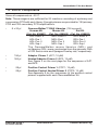
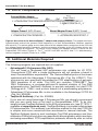
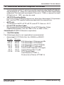
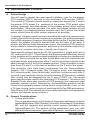
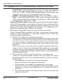
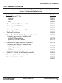
GenomeWalker™ Kits User Manual Cat Nos. 638901 638902 638903 PT1116-1 (PR47603) Published 23 August 2004 GenomeWalker™ Kits User Manual Table of Contents I. Introduction 3 II. List of Components 7 III. Additional Materials Required 8 IV. GenomeWalker Protocol A. Primer Design B. General Considerations C. Procedure for PCR-based DNA Walking in GenomeWalker Libraries 10 10 10 V. Expected Results and Troubleshooting Guide 16 12 VI. Suggestions for Characterizing GenomeWalker Products 18 VII. References 22 VIII. Related Products 23 Appendix A:Sequences of the Positive Control Primers 24 Appendix B: Design of the GenomeWalker Adaptor 25 Appendix C: Parameters for the GeneAmp Systems 2400 & 9600 27 List of Figures Figure 1. Flow chart of the GenomeWalker protocol Figure 2. Map of the human tissue-type plasminogen activator (tPA) locus and results of primary and secondary GenomeWalker PCR using tPA primers Figure 3. Positive control results with the Mouse and Rat GenomeWalker Kits Figure 4. Structure of the GenomeWalker adaptor and adaptor primers Figure 5. Simple restriction mapping of GenomeWalker PCR products from the human tPA locus Figure 6. The suppression PCR effect Clontech Laboratories, Inc. www.clontech.com 4 5 6 8 18 26 Protocol No. PT1116-1 Version No. PR47603 GenomeWalker™ Kits User Manual I. Introduction GenomeWalker™ Kits provide researchers with ready access to a novel method for walking upstream (i.e., towards promoters) or downstream in genomic DNA from a known sequence such as a cDNA (Siebert et al., 1995a, 1995b). Each kit contains four “libraries” of uncloned, adaptor-ligated genomic DNA fragments. (These are not libraries in the conventional sense; that is, the DNA fragments are not ligated into a vector which is then propagated in E. coli. However, like conventional libraries, GenomeWalker Libraries are a pool of specially prepared DNA fragments from which researchers can identify, isolate, and clone specific pieces of DNA.) Construction of GenomeWalker Libraries begins with isolation of very clean genomic DNA that has a very high average molecular weight. The starting DNA must be of considerably higher quality than the minimum suitable for Southern blotting or conventional PCR. Four separate aliquots are then thoroughly digested with four different restriction enzymes that recognize a 6base site, leaving blunt ends. Following digestion, each pool of DNA fragments is ligated to the GenomeWalker Adaptor (see Appendix B). The GenomeWalker protocol takes just two days and consists of two PCR amplifications per library (Figure 1). The first or “primary” PCR amplification uses the outer adaptor primer (AP1) provided in the kit and an outer, gene-specific primer (GSP1) provided by the researcher. The primary PCR mixture is then diluted and used as a template for a secondary or “nested” PCR amplification using the nested adaptor primer (AP2) and a nested gene-specific primer (GSP2). This generally produces a single, major PCR product from at least three of the four libraries (and often in all four). Each of the DNA fragments—which begin in known sequence at the 5’ end of GSP2 and extend into the unknown adjacent genomic DNA—can then be cloned and further analyzed. Figure 2 shows sample results of primary and secondary GenomeWalker PCR. Amplification of each of the GenomeWalker human libraries with the adaptor primers and primers derived from exon 1 of the human tissue-type plasminogen activator (tPA) gene generated single major products of the size expected based on the map of the tPA locus. Each kit also includes positive control primers PCP1 and PCP2, which generate a single major product from each library. Thermostable DNA polymerase(s) are not included in the kit (see next paragraph). Long-distance PCR with the Advantage® 2 PCR Kit GenomeWalker reactions should be performed with a 50X polymerase mix containing a combination of DNA polymerases suitable for long-distance PCR (LD PCR; Barnes, 1994; Cheng et al., 1994). In LD PCR, a combination of two thermostable DNA polymerases is used to increase the range and accuracy of PCR amplification. Most of the extension is carried out by a primary polymerase, while a secondary polymerase provides the critical 3’→5' exonuclease or "editing" function that corrects misincorporated nucleotides. Protocol No. PT1116-1 www.clontech.com Clontech Laboratories, Inc. Version No. PR47603 GenomeWalker™ Kits User Manual I. Introduction continued GenomeWalker™ "Libraries" Amplify gene of interest from all four libraries Genomic DNA fragment No binding site for AP1 Binding site is created on template of interest by extension of GSP1 GSP2 GSP1 5'– AP1 AP2 N N –5' GenomeWalker Adaptor AP1 GSP1 Primary PCR with Advantage™ 2 Polymerase Mix (contains TaqStart™ Antibody) AP2 GSP2 Secondary or "nested" PCR with Advantage™ 2 Polymerase Mix Examine products on an agarose/EtBr gel M 3.0 2.0 1.6 1.0 0.5 1 G 2 3 4 M Amplify gene of interest from all four libraries Genomic DNA fragment No binding site for AP1 5'– Binding site is created on template of interest by extension of GSP1 P P2 GSP2 GSP – 5' BD GenomeWalker Adaptor P GSP Primary PCR with BD Advantage™ 2 Polymerase Mix (contains BD TaqStart™ Antibody) P2 GSP2 Secondary or "nested" PCR with BD Advantage™ 2 Polymerase Mix Examine products on an agarose/EtBr gel M 1 2 3 4 M 3.0 2.0 1.6 1.0 0.5 • Clone & characterize major PCR products • Test for promoter activity by cloning into reporter vector • Clone & characterize major PCR products • Test for promoter activity by cloning into reporter vector Figure 1. Flow chart of the GenomeWalker™ protocol. The gel shows typical results generated by nested PCR with the GenomeWalker human libraries and gene-specific primers. Primary and secondary (nested) PCR was performed using Advantage 2 Polymerase Mix and the cycling parameters described in the protocol. Lane 1: EcoR V Library. Lane 2: Dra I Library. Lane 3: Pvu II Library. Lane 4: Ssp I Library. Lane M: DNA size markers. The absence of a major product in one of the libraries is not unusual. In our experience, there is no major band in one or more lanes in approximately half of the GenomeWalker experiments. As explained in the Expected Results and Troubleshooting Guide (Section V), this is usually because the distance between the primer and the upstream restriction site is greater than the capability of the system. N: Amine group that blocks extension of the 3’ end of the adaptor-ligated genomic fragments. AP: Adaptor primers. GSP: Gene-specific primers. Clontech Laboratories, Inc. www.clontech.com Protocol No. PT1116-1 Version No. PR47603 GenomeWalker™ Kits User Manual I. Introduction continued Map of tPA locus and expected PCR products tPA2 tPA1 Presumed promoter Ssp I EcoR V Pvu II Dra I Exon I 1.8 kb EcoR V Library 0.9 kb Dra I Library 1.5 kb Pvu II Library 3.9 kb Ssp I Library Gel of secondary PCR reaction products: Gel of primary PCR reaction products: M 1 2 3 4 M 4.0 3.0 2.0 1.6 4.0 3.0 2.0 1.6 1.0 1.0 0.5 0.5 1 2 3 4 Figure 2. Map of the human tissue-type plasminogen activator (tPA) locus (Friezner-Degen et al., 1986) and results of primary and secondary GenomeWalker™ PCR using tPA primers. Primary and secondary (nested) PCR was performed using Advantage 2 Polymerase Mix and the cycling parameters described in the protocol. The tPA primers used in this experiment are the positive control primers PCP1 and PCP2 provided with the GenomeWalker Human Kit. Lane 1: EcoR V Library. Lane 2: Dra I Library. Lane 3: Pvu II Library. Lane 4: Ssp I Library. Lane M: 1 kb DNA ladder. A 1.3 kb band is often observed in HDL-Ssp 1. Protocol No. PT1116-1 www.clontech.com Version No. PR47603 Clontech Laboratories, Inc. GenomeWalker™ Kits User Manual I. Introduction continued In the GenomeWalker protocol, the use of LD PCR extends the range of possible PCR products to about 6 kb. (The precise reason for the upper limit on GenomeWalker products is not clear. It may be due to the loss of the suppression PCR effect [see Appendix B]). As discussed in Section III, we recommend our Advantage® 2 Polymerase Mix (Cat. No. 639201). Advantage 2 Polymerase Mix is available separately and in the Advantage® 2 PCR Kit (Cat. No. 639206). Applications The primary application of GenomeWalker Kits is the rapid cloning of the promoters (and other upstream regulatory elements) in genes for which only cDNA sequence was previously available. In addition to obtaining promoters, GenomeWalker DNA walking can also be used to map intron/exon junctions and to walk bidirectionally from any sequence-tagged site (STS) or expressed sequence tag (EST). Although individual steps are limited to about 6 kb, multiple steps can be strung together to create longer walks. Consequently, this method is useful for filling in gaps in genome maps, particularly when the missing clones have been difficult to obtain by conventional library screening methods. In all applications, the PCR products are generally pure enough to allow restriction mapping without cloning. A discussion of cloning GenomeWalker PCR products and testing them for promoter activity is included at the end of this manual. Mouse Positive Control Results M 4.0 3.0 2.0 1.6 1.0 0.5 1 2 3 Rat Positive Control Results 4 M 1 2 3 4 4.0 3.0 2.0 1.6 1.0 0.5 Figure 3. Positive control results obtained with the Mouse and Rat GenomeWalker™ Kits (see Figure 2 for the human positive control results). Primary and secondary (nested) PCR was performed using Advantage 2 Polymerase Mix and the cycling parameters described in the protocol. Lane 1: EcoR V Library. Lane 2: Dra I Library. Lane 3: Pvu II Library. Lane 4: Ssp I Library. Lane M:1kb DNA ladder. Clontech Laboratories, Inc. www.clontech.com Protocol No. PT1116-1 Version No. PR47603 GenomeWalker™ Kits User Manual II. List of Components Store all components at –20°C. Note: These reagents are sufficient for 20 reactions consisting of a primary and a secondary PCR with each library. Enough primers are provided for 150 primary PCR and 300 secondary PCR amplifications. • 5 x 20 µl GenomeWalker™ DNA Libraries (100 ng each) Human Kit Mouse Kit Rat Kit (Cat. No. 638901) (Cat. No. 638902) (Cat. No. 638903) HDL-EcoR V MDL-EcoR V RDL-EcoR V HDL-Dra I MDL-Dra I RDL-Dra I HDL-Pvu II MDL-Pvu II RDL-Pvu II HDL-Ssp I MDL-Ssp I RDL-Ssp I The GenomeWalker mouse libraries (MDL) and rat libraries (RDL) were constructed from the genomic DNA of ICR Swiss mice and Sprague-Dawley rats, respectively. • 150 µl Adaptor Primer 1 (AP1; 10 µM) • 300 µl Nested Adaptor Primer 2 (AP2; 10 µM) See Figure 4 on the next page for the sequences of AP1 and AP2. • 25 µl Positive Control Primer 1 (PCP1; 10 µM) • 25 µl Positive Control Nested Primer 2 (PCP2; 10 µM) See Appendix A for the sequences of the positive control primers supplied with each GenomeWalker Kit. Protocol No. PT1116-1 www.clontech.com Clontech Laboratories, Inc. Version No. PR47603 GenomeWalker™ Kits User Manual II. List of Components continued Srf I Sal I Sma I/Xma I Mlu I 5'–GTAATACGACTCACTATAGGGCACGCGTGGTCGACGGCCCGGGCTGGT–3' 3'–H2N-CCCGACCA-PO4–5' GenomeWalker Adaptor Adaptor Primer 1 (AP1; 22-mer) Nested Adaptor Primer 2 (AP2; 19-mer) 5'–GTAATACGACTCACTATAGGGC–3' 5'–ACTATAGGGCACGCGTGGT–3' Figure 4. Structure of the GenomeWalker™ adaptor and adaptor primers. The adaptor has been ligated to both ends of the genomic DNA fragments in all four GenomeWalker Libraries supplied with each kit. The amine group on the lower strand of the adaptor blocks extension of the 3’ end of the adaptor-ligated genomic fragments, and thus prevents formation of an AP1 binding site on the general population of fragments. The design of the adaptor and adaptor primers is critical for the suppression PCR effect (Figure 6). The Tm’s of AP1 and AP2 are 59°C and 71°C, determined by nearest neighbor analysis (Freier et al., 1986). III. Additional Materials Required The following reagents are required but not supplied: • Advantage® 2 Polymerase Mix (50X) You will need a Taq-based 50X polymerase mix suitable for LD PCR. Conventional PCR with a single polymerase will not produce a band in most GenomeWalker experiments. The GenomeWalker protocol has been optimized with the Advantage 2 Polymerase Mix (Cat. No. 639201). This enzyme mix was specifically developed for PCR amplifications of genomic DNA templates of all sizes. This 50X mix contains TITANIUM™ Taq DNA Polymerase—a nuclease-deficient N-terminal deletion of Taq DNA polymerase plus TaqStart® Antibody to provide automatic hot start PCR (Kellogg et al., 1994)—and a minor amount of a proofreading polymerase. Advantage 2 Polymerase Mix is also available in the Advantage 2 PCR Kit (Cat. No. 639206). • TaqStart® Antibody (Cat. No. 639250) If you are not using Advantage 2 Polymerase Mix, we strongly recommend that you use some form of hot start in GenomeWalker PCR. To do this, simply include TaqStart Antibody in the 50X polymerase mix (see PT1576-1, available at www.clontech.com). TaqStart Antibody is included in Advantage 2 Polymerase Mix. This antibody is an effective method for hot start PCR that is simpler and more convenient than wax-based or manual methods. The TaqStart Antibody binds to and inactivates Taq DNA polymerase and thus eliminates DNA synthesis from nonspecifically bound primers while reactions are being assembled. PCR amplification proceeds efficiently after an initial Clontech Laboratories, Inc. www.clontech.com Protocol No. PT1116-1 Version No. PR47603 GenomeWalker™ Kits User Manual III. Additional Materials Required continued 1 min incubation at 94°C, which irreversibly inactivates the TaqStart Antibody. See Kellogg et al. (1994) for a discussion of hot start PCR with inactivating antibodies. Hot start with wax beads (Chou et al., 1992) or manual hot start (D’Aquila et al., 1991) can also be used . • 10X PCR Reaction Buffer If you are using a DNA polymerase mix other than Advantage 2 Polymerase Mix, use the PCR reaction buffer provided with the enzyme mix. • dNTP mix 10 mM each of dATP, dCTP, dGTP, and dTTP. Store at –20°C. • 0.5-ml PCR reaction tubes We recommend using GeneAmp 0.5-ml PCR Reaction Tubes. (Applied Biosystems, Cat. No. N801-0737 or N801-0180.) •Deionized H O (Milli-Q filtered or equivalent) • 1 kb DNA ladder 2 The following product is not required but recommended: • Advantage® 2 PCR Kit (Cat. No. 639206 or 639207) 30 rxns 100 rxns 30 µl 100 µl 50X Advantage 2 Polymerase Mix 200 µl 600 µl 10X Advantage 2 PCR Buffer 200 µl 600 µl 10X Advantage 2 SA PCR Buffer 50 µl 120 µl 50X dNTP Mix (10 mM each) 30 µl 100 µl Control DNA Template (100 ng/µl) 30 µl 100 µl Control Primer Mix (10 µM) 2.5 ml 5.0 ml PCR-Grade Water User Manual (PT3281-1) Protocol-at-a-Glance (PT3281-2) Protocol No. PT1116-1 www.clontech.com Version No. PR47603 Clontech Laboratories, Inc. GenomeWalker™ Kits User Manual IV. GenomeWalker Protocol A. Primer Design You will need to design two gene-specific primers—one for the primary PCR reaction (GSP1) and one for the secondary PCR reaction (GSP2). The nested PCR primer should anneal to sequences beyond the 3’ end of the primary PCR primer (i.e., upstream of the primary PCR primer when walking upstream and downstream of the primary primer when walking downstream). Whenever possible, the outer and nested primers should not overlap; if overlapping primers must be used, the 3’ end of the nested primer should have as much unique sequence as possible. In general, the gene-specific primers should be derived from sequences as close to the end of the known sequence as possible. For walking upstream from cDNA sequence, the primer should be as close to the 5’ end as possible. Ideally, the primers should be derived from the first exon of the gene. If primers are derived from downstream exons, the resulting PCR products are less likely to contain the promoter, particularly if the intervening intron(s) and exon(s) comprise more than a few kb (see Figure 2). Gene-specific primers should be 25–28 nucleotides in length and have a GC-content of 40–60%. This will ensure that the primers will effectively anneal to the template at the recommended annealing and extension temperature of 67°C. Primers should not be able to fold back and form intramolecular hydrogen bonds, and sequences at the 3’ end of your primers should not be able to anneal to the 3’ end of the adaptor primers. There should be no more than three G’s and C’s in the last six positions at the 3’ end of the primer. Five restriction sites have been incorporated into the GenomeWalker Adaptor—Sal I (cohesive ends), Mlu I (cohesive ends), and overlapping Srf I (cohesive ends), Sma I (blunt ends), and Xma I (cohesive ends) sites. If you wish to use restriction sites to clone the resulting PCR products, suitable sites should also be designed into the 5’ end of GSP2 (i.e., the nested gene-specific primer used for the secondary PCR reaction.) The sites in the Adaptor Primer allow easy insertion of PCR products into commonly used promoter reporter vectors. Alternatively, GenomeWalker products can be cloned into a general purpose cloning vector using restriction sites, or into a TA-type cloning vector using the A overhang left byTaq DNA polymerase. (See Section VI.B.3 for a discussion of our various promoter-cloning reporter vectors and reporter assay systems.) B. General Considerations 1. Cycling parameters The cycling parameters in this protocol have been optimized using the Applied Biosystems DNA Thermal Cycler 480, Advantage 2 Polymerase Mix, and the reagents and positive control primers provided in the GenomeWalker Kit. The optimal cycling parameters may vary with different polymerase mixes, gene-specific primers, and thermal cyclers. Clontech Laboratories, Inc. www.clontech.com 10 Protocol No. PT1116-1 Version No. PR47603 GenomeWalker™ Kits User Manual IV. GenomeWalker Protocol continued Recommended cycling parameters for the Applied Biosystems GeneAmp PCR Systems 2400 and 9600 are provided in Appendix C. Please refer to the Troubleshooting Guide (Section V) for suggestions on optimizing PCR conditions. 2. Use some form of hot start PCR It is advantageous to use some form of hot start, and the protocol assumes that TaqStart Antibody has been included in the 50X polymerase mix (see Section III, Additional Materials). Hot start can also be performed using wax beads (Chou et al., 1992) or manually (D’Aquila et al., 1991). If you use a manual or wax-based hot start, you will need to adapt the protocol to these particular methods. 3. Touchdown PCR The PCR cycling parameters in Steps IV.C.8 and IV.C.16 are for “touchdown” PCR (Don et al., 1991; Roux, 1995; Hecker and Roux, 1996). Touchdown PCR involves using an annealing/extension temperature that is several degrees higher than the Tm of the primers during the initial PCR cycles. Although primer annealing (and amplification) is less efficient at this higher temperature, it is also much more specific. The higher temperature also enhances the suppression PCR effect with AP1 (see Appendix B). This allows a critical amount of gene-specific product to accumulate. The annealing/extension temperature is then reduced to slightly below the primer Tm for the remaining PCR cycles, permitting efficient, exponential amplification of the gene-specific template. As noted above, we recommend using primers with Tm’s greater than 68°C to allow you to use the touchdown cycling programs in the protocol. 4. Use of the positive controls In each experiment, we suggest that you include a positive control in which you amplify one of the libraries using the positive control PCP primers. This will confirm that your DNA polymerase mix is functional and thermal cycling parameters are compatible with this protocol. To ensure that you do not run out of any one library prematurely, use a different library as the positive control for each experiment. Note: You may wish to perform an initial experiment using all four libraries with the positive control primers prior to using the kit with their gene-specific primers. Protocol No. PT1116-1 www.clontech.com Version No. PR47603 Clontech Laboratories, Inc. 11 GenomeWalker™ Kits User Manual IV. GenomeWalker Protocol continued 5. Amplify all four libraries with each set of GSPs To maximize your chances of success in finding a promoter or taking the largest possible step in a genomic walk, we recommend that you amplify all four libraries with each new gene-specific primer. 6. Use the recommended amounts of enzymes The enzyme amounts have been carefully optimized for the GenomeWalker amplification protocol and reagents. C. Procedure for PCR-based DNA Walking in GenomeWalker Libraries GenomeWalker PCR has been optimized with our Advantage 2 Polymerase Mix, which includes TaqStart Antibody for automatic hot start PCR. 1. Label the 0.5-ml PCR tubes. At Clontech, we use the following system (GSP1 and GSP2 indicate your gene-specific primers): table i. suggested labeling plan DNA 1° PCR 2° PCR Library (DL) Tube No. Primers Tube No. Primers DL-EcoR V 1A GSP1 & AP1A 1B GSP2 & AP2B DL-Dra I 2A “ 2B “ DL-Pvu II 3A “ 3B “ DL-Ssp I 4A “ 4B “ Negative control None 5A “ 5B “ Positive control DL-EcoR VC 6A PCP1 & AP1 6B PCP2 & AP2 A B C Primers contained in primary master mix. Primers contained in secondary master mix Any of the libraries can be used for the positive control. To prevent running out of any one library prematurely, use a different library as the positive control for each experiment. Clontech Laboratories, Inc. www.clontech.com 12 Protocol No. PT1116-1 Version No. PR47603 GenomeWalker™ Kits User Manual IV. GenomeWalker Protocol continued 2. Prepare the primary PCR master mix by combining the following reagents in an 0.5-ml tube: 6 rxns per rxn 240 µl 40 µl H2O 30 µl 5 µl 10X Advantage 2 PCR Buffer 6 µl 1 µl dNTP (10 mM each) 6 µl 1 µl AP1 (10 µM) 6 µl 1 µl GSP1 (10 µM)† 6 µl 1 µl Advantage 2 Polymerase Mix (50X) 294 µl 49 µl Total volume † GSP1 is your outer gene-specific primer. Mix well by vortexing (without introducing bubbles) and briefly spin the tube in a microcentrifuge. 3. Add 49 µl of the primary PCR master mix to the appropriately labeled tubes and to the negative control. Do not add master mix to the positive control (see Step 6). 4. Add 1 µl of each DNA library (i.e., HDL-EcoR V, etc.) to the appropriately labeled tubes, including the positive control. Do not add any library DNA to the negative control. 5. Add 1 µl of H2O to the negative control. 6. Prepare your positive control by combining the following reagents in a 0.5-ml tube: 40 µl H2O 5 µl 10X Advantage 2 PCR Buffer 1 µl dNTP (10 mM each) 1 µl AP1 (10 µM) 1 µl PCP1 (10 µM) 1 µl DL-EcoR V (or DL-Dra I, etc.) 1 µl Advantage 2 Polymerase Mix (50X) 50 µl Total volume 7. Overlay the contents of each test tube with one drop of mineral oil and place caps firmly on each tube. Protocol No. PT1116-1 www.clontech.com Version No. PR47603 Clontech Laboratories, Inc. 13 GenomeWalker™ Kits User Manual IV. GenomeWalker Protocol continued 8. Commence cycling in a DNA Thermal Cycler 480 (Applied Biosystems) using the following two-step cycle parameters: • 7 cycles: 94°C 72°C 25 sec 4 min • 32 cycles: 94°C 67°C 25 sec 4 min • 67°C for an additional 4 min after the final cycle. Notes: Do not use a three-step cycling program (e.g., 95°C melting, 60°C annealing, 68°C extension). See Appendix C for cycling parameters for GeneAmp PCR Systems 2400 and 9600. 9. Analyze 5 µl of the primary PCR products on a 1.5% agarose/EtBr gel, along with DNA size markers such as a 1-kb ladder. If you do not see any product, perform five additional cycles. Expected results of primary PCR: You should observe multiple fragments, ranging in size from about 500 bp to 5 kb, in all four lanes. There may be some smearing in some lanes. See Figure 2 in the Introduction (Section I) for a sample gel showing products of primary PCR. If you obtain any bands or smearing with your gene-specific primer, continue with secondary PCR as described in Steps 10–17 (even if your products are weaker than the positive control or the bands in Figure 2). If you do not observe any product with your gene-specific primers, consult the Troubleshooting Guide. 10. Using a clean 0.5-ml tube for each sample, dilute 1 µl of each primary PCR (including positive and negative controls) into 49 µl of sterile H2O. 11. Prepare a secondary PCR master mix by combining the following reagents in an 0.5-ml tube: 6 rxns per rxn 240 µl 40 µl H2O 30 µl 5 µl 10X Advantage 2 PCR Buffer 6 µl 1 µl dNTP (10 mM each) 6 µl 1 µl AP2 (10 µM) 6 µl 1 µl GSP2 (10 µM)† 6 µl 1 µl Advantage 2 Polymerase Mix (50X) 294 µl 49 µl Total volume † GSP2 is your nested gene-specific primer. Mix well by vortexing (without introducing bubbles) and briefly spin the tube in a microcentrifuge. Keep on ice until ready to use. Clontech Laboratories, Inc. www.clontech.com 14 Protocol No. PT1116-1 Version No. PR47603 GenomeWalker™ Kits User Manual IV. GenomeWalker Protocol continued 12. Add 49 µl of the secondary PCR master mix to the appropriately labeled tubes and to the negative control. Do not add master mix to the positive control. 13. Prepare your secondary positive control by combining the following reagents in an 0.5-ml tube: 40 µl H2O 5 µl 10X Advantage 2 PCR Buffer 1 µl dNTP (10 mM each) 1 µl AP2 (10 µM) 1 µl PCP2 (10 µM) 1 µl Advantage 2 Polymerase Mix (50X) 49 µl Total volume 14. Add 1 µl of each diluted primary PCR product to the appropriate tube. Be sure to include both the positive and negative controls. 15. Overlay the contents of each test tube with one drop of mineral oil, and place caps firmly on each tube. 16. Commence cycling in a DNA Thermal Cycler 480 (PE Biosystems) using the following two-step cycle parameters: • 5 cycles: 94°C 72°C 25 sec 4 min • 18–22 cycles: 94°C 25 sec 67°C 4 min • 67°C for an additional 4 min after the final cycle. Note: Do not use a three-step cycling program (e.g., 95°C melting, 60°C annealing, 68°C extension). See Appendix C for cycling parameters for the GeneAmp PCR Systems 2400 and 9600. 17. Analyze 5 µl of the secondary PCR products on a 1.2% agarose/EtBr gel, along with DNA size markers such as a 1 kb DNA ladder or λ/Hind III digest. As discussed in the Troubleshooting Section (Section V.A.2), more than one band may occasionally be observed after secondary PCR. Store the unused portion of the secondary PCR samples at 4°C until you have confirmed that the procedure has been successful. At that point, proceed with analyzing and cloning the fragments of interest (e.g., putative promoter fragments), as described in Section VI. Protocol No. PT1116-1 www.clontech.com Version No. PR47603 Clontech Laboratories, Inc. 15 GenomeWalker™ Kits User Manual V. Expected Results and Troubleshooting Guide A. Expected Results 1. Primary PCR Figure 2 (Section I) shows a typical result after primary GenomeWalker PCR. In general, primary PCR should produce multiple fragments, ranging in size from about 500 bp to 5 kb, in all four lanes. There may be some smearing in some lanes. In general, you should continue with secondary PCR if you obtain any bands or smearing with your genespecific primer. 2. Secondary PCR a. Positive control PCP primers Positive control secondary PCR should produce major bands of the following sizes: GenomeWalker DNA Libraries EcoR V Dra I Pvu II Ssp I Human (HDLs) 1.8 kb 0.9 kb 1.5 kb 3.9 kb* Mouse (MDLs) 1.5 kb 1.3 kb 0.6 kb 3.2 kb Rat (RDLs) 1.5 kb 1.2 kb 0.7 kb 1.0 kb * a 1.3 kb band is often observed in HDL-Ssp I. See the Introduction for gels showing the expected results following the secondary PCR amplification with the human (Figure 2) and mouse and rat (Figure 3) positive control primers. b. Experimental PCR primers. In approximately half the cases, single major bands will be observed with each of the four libraries. On rare occasions, more than one band may be observed. The exact size of the major bands will depend on the positions of restriction sites in your gene. Typically, secondary PCR products will range from 0.2 to 6 kb. Fragments generated from nested gene-specific primers that are less than 0.4 kb from one of the restriction sites represented in the GenomeWalker libraries may appear as a low molecular weight smear on a 1.2% agarose/EtBr gel. If this is the case with one or more of the GenomeWalker libraries, run this particular PCR product(s) on a 2% agarose/EtBr gel. In our experience, no product is observed in one or more of the libraries in approximately half the cases. This is usually because the distance from the primer to the restriction site is greater than the capability of the system (~6 kb). This limit reflects the diminished suppression PCR effect as template size increases. Targets greater than ~6 kb often become indistinguishable in a smear of high molecular weight material. Such smearing may also occur in lanes that do contain major bands, but should not affect the major bands. The absence of a major band in one or more of the libraries does not mean that products obtained with other libraries are not correct. Clontech Laboratories, Inc. www.clontech.com 16 Protocol No. PT1116-1 Version No. PR47603 GenomeWalker™ Kits User Manual V. Expected Results and Troubleshooting Guide continued B. Troubleshooting Guide 1. No products with the positive control primers (even after increasing the number of primary cycles from 32 to 37) a. Reduce all annealing/extension temperatures by 2°C (i.e., 72°C to 70°C and 67°C to 65°C) b. Reduce the length of the incubation at 94°C. c. Check your 50X polymerase mix by PCR using two specific primers and a 1–10 kb template that works in your hands. 2. Expected products observed with positive control primer, but no product observed with your gene-specific primers a. Check the design of your primers. If the positive control PCP primers produce the expected PCR products, but your gene-specific primers do not produce major PCR products with any of the libraries, there is probably a problem with your primers. If your primer sequence was derived from cDNA sequence information, the primary or secondary PCR primer may cross an exon/intron junction. If this is the case, it will be necessary to redesign one or both gene-specific primers. Remember that all primers should be able to anneal efficiently at 67°C (i.e., have a Tm ≥ 67°C). If you are sure your primers do not cross intron/exon boundaries, recheck the sequence of your primers. In some instances, primers will fail to produce any products due to a mistake in primer design or synthesis. b. Your target template may have a high GC content. Such templates are difficult to amplify. Repeat your experiment using a final concentration of 5% DMSO in your primary and secondary PCR. For each PCR, add 2.5 µl of DMSO (and only 37.5 µl of H2O) to the Master Mix (Step IV.C.2 & 11). Add the DMSO to the Master Mix last. Note: You will need to perform more cycles in the presence of DMSO. For the primary PCR, perform 36 cycles instead of 32; for the secondary PCR, perform 24 cycles. If this fails, repeat again using a final concentration of 6% DMSO and 3% glycerol in your primary and secondary PCR. If neither DMSO concentration solves the problem, try increasing the temperature to 99°C for 5 seconds at the beginning of the first cycle. 3. Smears or multiple major bands observed in positive control and genespecific primers If the positive control primers give the expected pattern but your gene-specific primers still generate a smear after secondary PCR, try repeating your experiment using 5% DMSO as discussed above. However, in most cases, it will be necessary to redesign your gene-specific primers if you get smears after the secondary PCR. Protocol No. PT1116-1 www.clontech.com Version No. PR47603 Clontech Laboratories, Inc. 17 GenomeWalker™ Kits User Manual VI. Suggestions for Characterizing GenomeWalker Products A. Restriction Mapping of GenomeWalker PCR Products GenomeWalker PCR products are generally clean enough to allow simple restriction mapping without cloning. An example of such an experiment is shown in Figure 5. Pvu II tPA tPA1 BamH I Ssp I EcoR V Pvu II Dra I Exon I 1.8 kb EcoR V Library 0.9 kb Dra I Library 1.5 kb Pvu II Library 3.9 kb Ssp I Library 4.0 3.0 2.0 1.6 1.0 0.5 oR V aI Dr M M Ec Ec oR Dr V aI Pv u Ss II pI M Pv u Ss II pI Ec oR Dr V aI Pv u Ss II pI Restriction digests: BamH I Pvu II PCR products: M 4.0 3.0 2.0 1.6 1.0 0.5 Figure 5. Simple restriction mapping of GenomeWalker™ PCR products from the human tPA locus. The map shows the positions of the relevant restriction sites in the genomic DNA and in the predicted PCR products. The gel on the left shows the products of GenomeWalker PCR. The gel on the right shows the pattern of restriction fragments generated by digestions of each PCR product with either BamH I or Pvu II. Lane M: DNA size markers. Clontech Laboratories, Inc. www.clontech.com 18 Protocol No. PT1116-1 Version No. PR47603 GenomeWalker™ Kits User Manual VI. Suggestions for Characterizing Products continued B. Cloning GenomeWalker Products and Testing for Promoter Activity 1. Cloning GenomeWalker products Once you have obtained major bands using your gene-specific primer, you will usually want to clone the fragments into a general purpose cloning vector using restriction sites, or into a TA-type cloning vector using the A overhang left by the DNA polymerase. In some cases, you may wish to clone directly into a promoter reporter vector (See Section B.3 below). If your secondary PCR produces a single, major band with little background and no minor bands, you may be able to clone the fragment directly. If the product of your secondary reaction has significant background, you will need to gel-purify the desired band. There are several options for gel-purifying DNA fragments. We recommend either the NucleoSpin® Extract Kit (Cat. No. 635960 or 635961) or the NucleoTrap® Gel Extraction Kit (Cat. No. 636018) for gel-purifying PCR products. Note on TAE vs. TBE gels: We recommend that you use Tris-AcetateEDTA (TAE) buffer instead of Tris-Borate-EDTA (TBE) buffer in your agarose gels when purifying DNA fragments for cloning. In our experience, DNA purified from TBE gels is more difficult to clone than DNA purified from TAE gels. Note on EtBr and UV damage to DNA: Minimize the exposure of your DNA to UV light. 2. Sequencing and scanning for regulatory elements Prior to testing GenomeWalker products for promoter activity, most researchers will want to sequence at least part of their clones and look for common regulatory sequence motifs such as promoters, enhancers, etc. 3. Testing for promoter activity GenomeWalker products can be cloned into a promoter reporter vector to test for the presence of a promoter. Cloning in both orientations will provide a positive and negative control. Suitable promoter-cloning vectors from Biosciences Clontech include the following: • pSEAP2-Basic is sold separately (Cat. No. 631715) and as a component in the chemiluminescent Great EscAPe™ SEAP Reporter System 3 (Cat. No. 631706; Yang et al, 1994). This kit also includes pSEAP2-Control and reagents necessary for 100 chemiluminescent assays. The reporter molecule in the Great EscAPe system is a secreted form of alkaline phosphatase (SEAP), which can be conveniently measured directly in the culture medium using a sensitive chemiluminescent assay. Protocol No. PT1116-1 www.clontech.com Version No. PR47603 Clontech Laboratories, Inc. 19 GenomeWalker™ Kits User Manual VI. Suggestions for Characterizing Products continued • pβgal-Basic is sold separately (Cat. No. 631707) and as a component in the Luminescent β-gal Reporter System 3 (Cat. No. 631713; Sinai et al., 1994). This kit also includes pβgal-Control Vector and reagents necessary for 100 chemiluminescent assays. • pEGFP-1 Promoter Reporter Vector (Cat. No. 632319) uses a bright, codon-optimized variant of the green fluorescent protein (GFP) to monitor promoter activity (Cormack et al., 1996; April 1996 Clontechniques; Kitts et al., 1995). Note on ATG start codon: If your gene-specific primer was downstream of the ATG start codon in your gene of interest, then you may wish to eliminate the ATG from your promoter reporter construct(s). This may prevent a possible false negative result due to the expression of a bicistronic message (See Section 4.b.v below). 4. Explanation of possible results of tests for promoter activity Some GenomeWalker products will have no promoter activity when cloned in both orientations in a promoter reporter vector. There are several possible explanations. a. None of the fragments contain the promoter. Your primer may be several kb from the promoter and/or there may be intervening restriction sites between the primer and the promoter. This may also be an indication that the primer does not fall into the first exon (or within a downstream exon that is within 6 kb of the promoter). If this is the case, you may need to obtain sequence data from closer to the 5’ end of the transcript. Alternatively, you can “walk another step” by sequencing the distal end of the GenomeWalker product(s), designing a new gene-specific primer, and repeating the amplification protocol. b. The promoter is present, but the reporter is not expressed. There are several possible reasons why you might not detect promoter activity even if your promoter-reporter construct contains the promoter. i. The fragment is cloned in the wrong orientation. Reclone and test in the opposite orientation. ii. The promoter is too weak to be detected in your assay. If this is the case, it may be possible to add an enhancer to your construct or reclone your fragment(s) in a vector that has an enhancer. iii. The promoter needs to be induced (and you do not have the means to induce it). Clontech Laboratories, Inc. www.clontech.com 20 Protocol No. PT1116-1 Version No. PR47603 GenomeWalker™ Kits User Manual VI. Suggestions for Characterizing Products continued Again, recloning into a vector that has a strong enhancer may allow you to detect promoter activity. iv. The promoter is tissue- or stage-specific. Again, recloning into a vector that has a strong enhancer may allow you to detect promoter activity. Alternatively, it may be possible to demonstrate the presence of a promoter by testing the construct in another host cell or in the whole organism. v. Reporter construct makes a bicistronic message. The cloned fragment contains the ATG and some portion of the open reading frame from the gene of interest. This results in a bicistronic message in which two ORFs may compete for translation; the downstream ORF (i.e., the reporter) may not be efficiently translated. If you suspect this to be the case, test for promoter activity at the RNA level by performing RT-PCR. (Reporter expression can be assayed by Northern blot; however, RT-PCR is much faster and more sensitive if suitable primers are available.) vi. The cloned fragment(s) contains a strong negative enhancer. There are numerous instances of so-called “negative enhancers” that prevent transcription of a functional promoter. If you suspect this to be the case, try recloning in the presence of a known strong enhancer, or testing subclones in which upstream sequences have been deleted. 5. Deletion analysis of promoters After finding fragments that have promoter activity, many researchers will want to perform a deletion analysis to define the minimal promoter. Any standard nested-deletion method is compatible with this system. C. Other Applications of GenomeWalker Other possible applications of the GenomeWalker DNA walking method include the following: • Mapping intron/exon boundaries. • Walking short distances upstream or downstream in genomic DNA from known sequences (e.g., expressed sequence tags [EST] or other sequence tagged sites [STS]). Although individual steps are limited to ~6 kb, multiple steps can be strung together to create longer walks. • Walking from 5’ or 3’ ends generated by RACE using the SMART™ RACE cDNA Amplification Kit (Cat. No. 634914). You can also clone full-length cDNAs and the surrounding genomic sequences without ever screening a library using both our SMART RACE cDNA Amplification Kit and the GenomeWalker Kit. Protocol No. PT1116-1 www.clontech.com Clontech Laboratories, Inc. Version No. PR4760321 GenomeWalker™ Kits User Manual VII. References Barnes, W. M. (1994) PCR amplification of up to 35-kb DNA with high fidelity and high yield from λ bacteriophage templates. Proc. Natl. Acad. Sci. USA 91:2216–2220. Cheng, S., Fockler, C., Barnes, W. M. & Higuchi, R. (1994) Effective amplification of long targets from cloned inserts and human genomic DNA. Proc. Natl. Acad. Sci. USA 91:5695–5699. Chou, Q., Russell, M., Birch, D., Raymond, J. & Bloch, W. (1992) Prevention of pre-PCR mispriming and primer dimerization improves low-copy-number amplifications. Nucleic Acids Res. 20:1717–1723. Cormack, B. P., Valdivia, R. & Falkow, S. (1996) FACS-optimized variants of the green fluorescent protein. Gene 173:33–38. D’Aquila, R. T., Bechtel, L. J., Videler, J. A., Eron, J. J., Gorczyca, & Kaplan, J. C. (1991) Maximizing sensitivity and specificity by preamplification heating. Nucleic Acids Res. 19:3749. Don, R. H., Cox, P. T., Wainwright, B. J., Baker, K. & Mattick, J. S. (1991) ‘Touchdown’ PCR to circumvent spurious priming during gene amplification. Nucleic Acids Res. 19:4008. Freier, S. M., Kierzek, R., Jaeg er, J. A., Sugimoto, N., Caruthers, M. H., Neilson, T., and Tumer, D. H. (1986) Improved free-energy parameters for predictions of RNA duplex stability. Proc. Natl. Acad. Sci. USA 83:9373–9377 Friezner-Degen, S. J., Rajput, B. & Reich, E. (1986) Structure of the human tissue-type plasminogen activator gene. J. Biol. Chem. 261:6972–6985. Hecker, K.H. & Roux, K. H. (1996) High and low annealing temperatures increase both specificity and yield in touchdown and stepdown PCR. BioTechniques 20:478–485. Kellogg, D. E., Rybalkin, I., Chen, S., Mukhamedova, N., Vlasik, T., Siebert, P. & Chenchik, A. (1994) TaqStart Antibody: Hotstart PCR facilitated by a neutralizing monoclonal antibody directed against Taq DNA polymerase. BioTechniques 16:1134–1137. Kitts, P. Adams, M., Kondepudi, A., Gallagher, D. & Kain, S. (January 1995) Green fluorescent protein: A novel reporter for monitoring gene expression in living cells and organisms. Clontechniques X(1):1–3. Nelson, K., Brannan, J. & Kretz, K. (1995) The fidelity of TaqPlus DNA Polymerase in PCR. Strategies in Mol. Biol. 8:24–25. Roux, K. H. (1995) Optimization and troubleshooting in PCR. PCR Methods & Applications 4:5185– 5194. Sambrook, J. & Russell, D.W. (2001) Molecular Cloning: A Laboratory Manual, (Cold Spring Harbor Laboratory Press, Cold Spring Harbor, NY). Siebert, P. D., Chenchik, A., Kellogg, D. E., Lukyanov, K. A. & Lukyanov, S. A. (1995a) An improved method for walking in uncloned genomic DNA. Nucleic Acids Res. 23:1087–1088. Siebert, P. D., Chen, S. & Kellogg, D. E. (April 1995b) The Human GenomeWalker DNA Walking Kit: A new PCR method for walking in uncloned genomic DNA. Clontechniques X(II):1–3. Sinai, P., Kondepudi, A., Yang, T., Adams, M., Kitts, P. & Kain, S. (October 1994) The Luminescent β-Gal chemiluminescent assay for β-galactosidase: Application to the analysis of cis-regulatory elements. Clontechniques IX(4):1–4. Yang, T., Kondepudi, A., Adams, M., Kitts, P. & Kain, S. (July 1994) Quantitative detection of specific gene regulation with the Great EscAPe secreted alkaline phosphatase Genetic Reporter System. Clontechniques IX(3):1–5. Clontech Laboratories, Inc. www.clontech.com 22 Protocol No. PT1116-1 Version No. PR47603 GenomeWalker™ Kits User Manual VIII. Related Products For a complete listing of all Biosciences Clontech products, please visit www.clontech.com Products • GenomeWalker™ Kits Human Mouse Rat Cat. No. • GenomeWalker™ Universal Kit 638904 • Advantage® 2 PCR Kit 639206 639207 • Advantage® 2 Polymerase Mix 639201 • TaqStart® Antibody 639250 • Great EscAPe™ SEAP2 Reporter System 3 (Includes two vectors, listed below) pSEAP2-Basic Vector pSEAP2-Control Vector 631706 Luminescent β-gal Reporter System 3 (Includes two vectors, listed below) pβgal-Basic Vector pβgal-Control Vector 631713 • pEGFP-1 Promoter Reporter Vector 632319 • SMART™ RACE cDNA Amplification Kit 634914 • NucleoTrap® Gel Extraction Kit 636018 • NucleoSpin® Extract Kit 635960 635961 • 638901 638902 638903 631715 631717 631707 631709 Protocol No. PT1116-1 www.clontech.com Clontech Laboratories, Inc. Version No. PR4760323 GenomeWalker™ Kits User Manual Appendix A: Sequence of the Positive Control Primers • The positive control primers in the GenomeWalker Human Kit (Cat. No. 638901) are derived from exon 1 of the tissue-type plasminogen activator (tPA) cDNA. PCP1 Primer (tPA1): 5’-AGA AAC CCG ACC TAC CAC GGC TTG CTC CTT-3’ PCP2 Primer (tPA2): 5’-CCC TTT CCT CGC AGA GGT TTT CTC TCC AGC-3’ • The positive control primers in the GenomeWalker Mouse Kit (Cat. No. 638902) are derived from intron 6 of the interleukin 1β (IL1β) gene. PCP1 Primer (IL1β2): 5’-TCC GTG TGC ATG TTG CAT GTA TGA CAG AAA GG-3’ PCP2 Primer (IL1β1): 5’-TAC CAC GGT AGA CAT ATT CTC AGG GCT GCT GG-3’ • The positive control primers in the GenomeWalker Rat Kit (Cat. No. 638903) are derived from exon 5 of the interleukin 6 (IL6) gene. PCP1 Primer (IL61): 5’-CCA CAG TGA GGA ATG TCC ACA AAC TGA TAT GC-3’ PCP2 Primer (IL62): 5’-ACT AGG TTT GCC GAG TAG ACC TCA TAG TGA CC-3’ Clontech Laboratories, Inc. www.clontech.com 24 Protocol No. PT1116-1 Version No. PR47603 GenomeWalker™ Kits User Manual Appendix B: Design of the GenomeWalker Adaptor The GenomeWalker Adaptor has three design features that are critical to the success of GenomeWalker DNA walking. These features, which can be seen schematically in Figure 1 (in the Introduction), are as follows: 1) The use of a 5’-extended adaptor that has no binding site for the AP1 primer used in primary PCR. An AP1 binding site can only be generated by extension of the gene-specific primer. 2) Blocking of the exposed 3’ end of the adaptor with an amine group to prevent extension of the 3’ end (which would create an AP1 binding site). 3) The use of an adaptor primer that is shorter than the adaptor itself (“suppression PCR”). As shown in Figure 6, the suppression PCR effect prevents amplification of templates where the 3’ end has been extended to create an AP1 binding site. Though rare, such extension does occur, presumably due to incomplete amine modification or incomplete adaptor ligation. Given the exponential nature of PCR amplification, such events would lead to nonspecific amplification and unacceptable backgrounds in the absence of suppression PCR. Each of these features helps eliminate nonspecific amplification among the general population of DNA fragments. In combination with touchdown PCR and nested PCR, these innovations allow amplification of a specific target from a very complex mixture of DNA fragments—all of which have the same terminal structure—using a single set of gene-specific primers. Of the three features, suppression PCR is the most critical (unpublished data). Protocol No. PT1116-1 www.clontech.com Clontech Laboratories, Inc. Version No. PR4760325 GenomeWalker™ Kits User Manual Appendix B: continued • On rare occasions, the 3' end of the GenomeWalker Adaptor is extended, creating a template with the full adaptor sequence on both ends. OR • Melt at 95°C Suppression PCR • Anneal at 68°C AP1 • DNA synthesis No primer binding: panhandle structure suppresses PCR Even when the adaptor is extended, very little full-length amplification occurs. Figure 6. The suppression PCR effect. In rare cases, the 3’ end of the GenomeWalker Adaptor gets extended. (Though rare, such extension does occur, presumably due to incomplete amine modification during oligonucleotide synthesis or incomplete adaptor ligation.) This creates a molecule that has the full-length adaptor sequence on both ends and can serve as a template for end-to-end amplification. Without suppression PCR, these rare events would lead to unacceptable backgrounds due to the exponential nature of PCR amplification. However, in suppression PCR, the adaptor primer is much shorter than the adaptor itself. Thus, during subsequent thermal cycling, nearly all the DNA strands will form the “panhandle” structure shown above, which cannot be extended. At the appropriate annealing/ extension temperature, this intramolecular annealing event is strongly favored over (and more stable than) the intermolecular annealing of the much shorter adaptor primer to the adaptor. The suppression PCR effect will be reduced or lost if you use an annealing temperature lower than 60–65°C. The upper limit of the suppression PCR effect is about 6 kb. Clontech Laboratories, Inc. www.clontech.com 26 Protocol No. PT1116-1 Version No. PR47603 GenomeWalker™ Kits User Manual Appendix C: Parameters for GeneAmp Systems 2400 & 9600 As noted elsewhere in this manual, cycling parameters may have to be optimized for different thermal cyclers. For example, the cycling parameters in this protocol (which were developed on an Applied Biosystems DNA Thermal Cycler 480) do not work with the Applied Biosystems GeneAmp PCR Systems 2400 and 9600. Both the 2400 and 9600 systems use much shorter cycling parameters and smaller thin-walled tubes (0.2-ml vs. 0.5-ml). These systems also eliminate the need to overlay the reaction with mineral oil. The following parameters for primary and secondary GenomeWalker PCR give good results with the standard 50-µl positive control reaction (with no mineral oil overlay) and the 2400 and 9600 thermal cyclers. 1. Primary PCR (Step IV.C.8) • 7 cycles: 94°C 2 sec 72°C 3 min • 32 cycles: 94°C 2 sec 67°C 3 min • 67°C for an additional 4 min. 2. Secondary PCR (Step IV.C.16) • 5 cycles: 94°C 2 sec 72°C 3 min • 20 cycles: 94°C 2 sec 67°C 3 min • 67°C for an additional 4 min. Notes: Length of denaturation time: We have observed that differences of only a few seconds in the denaturation time at 94°C can dramatically affect results with the 2400 and 9600 systems. For example, positive control products larger than 2–3 kb were not detectable when the incubation time is increased from 2 to 5 sec. The extremely short incubation time at 94°C may be necessary to preserve the integrity of the larger genomic DNA templates required for LD PCR in the GenomeWalker protocol. Reaction volume: Although the 2400 and 9600 systems allow you to reduce the reaction volumes in many applications, we have not optimized the GenomeWalker protocol for lower reaction volumes. Protocol No. PT1116-1 www.clontech.com Clontech Laboratories, Inc. Version No. PR4760327 GenomeWalker™ Kits User Manual Notes Clontech Laboratories, Inc. www.clontech.com 28 Protocol No. PT1116-1 Version No. PR47603 GenomeWalker™ Kits User Manual Notes Protocol No. PT1116-1 www.clontech.com Clontech Laboratories, Inc. Version No. PR4760329 GenomeWalker™ Kits User Manual Notes Clontech Laboratories, Inc. www.clontech.com 30 Protocol No. PT1116-1 Version No. PR47603 GenomeWalker™ Kits User Manual Notes Notice to Purchaser This product is intended to be used for research purposes only. It is not to be used for drug or diagnostic purposes, nor is it intended for human use. Clontech products may not be resold, modified for resale, or used to manufacture commercial products without written approval of Clontech Laboratories, Inc. Suppression PCR is covered by U.S. Patent No. 5,565,340. NucleoTrap® and NucleoSpin® are registered trademarks of Macherey-Nagel, GmbH and Co. A license under U.S. Patent Nos. 4,683,202, 4,683,195, and 4,965,188 and U.S. Patents Nos. 5,407,800, 5,322,770, and 5,310,652 or their foreign counterparts, owned by Roche Molecular Systems, Inc, and F. Hoffmann-La Roche Ltd, for use in research and development, has an up-front fee component and a running-royalty component. The purchase price of this product includes limited, nontransferable rights under the running-royalty component to use only this amount of the product to practice the Polymerase Chain Reaction (PCR) and related processes described in said patents where the processes are covered by patents solely for the research and development activities of the purchaser when this product is used in conjunction with a thermal cycler whose use is covered by the up-front fee component. Rights to the up-front fee component must be obtained by the enduser in order to have a complete license to use this product in the PCR process where the process is covered by patents. These rights under the up-front fee component may be purchased from Applied Biosystems or obtained by purchasing an Authorized Thermal Cycler. No right to perform or offer commercial services of any kind using PCR, where the process is covered by patents,including without limitation reporting the results of purchaser’s activities for a fee or other commercial consideration, is hereby granted by implication or estoppel. Further information on purchasing licenses to practice the PCR Process where the process is covered by patents may be obtained by contacting the Director of Licensing at Applied Biosystems, 850 Lincoln Centre Drive, Foster City, California94404 or the Licensing Department at Roche Molecular Systems, Inc., 1145 Atlantic Avenue, Alameda, California 94501. GeneAmp® and AmpliTaq® are registered trademarks of Roche Molecular Systems, Inc., licensed to the Applied Biosystems Corporation. Clontech, Clontech logo and all other trademarks are the property of Clontech Laboratories, Inc. Clontech is a Takara Bio Company. ©2005 Protocol No. PT1116-1 www.clontech.com Version No. PR47603 Clontech Laboratories, Inc. 31