1
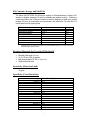
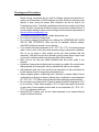
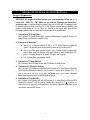
PRODUCT INFORMATION AND MANUAL Enzyme-linked immunosorbent assay for quantitative detection of mouse GM-CSF For Research Use Only Not for diagnostic or therapeutic procedures. 96 Wells Mouse GM-CSF Product # 453510 944 Nandino Blvd. Lexington KY 40511-1205 USA 859/254-1221 or 800/477-8201 USA/CANADA Fax: 859/255-5532 | Email: [email protected] | Web: www.neogen.com/LifeSciences Technical assistance is available Monday-Friday between 8:00 a.m. and 6:00 p.m. EST. COPYRIGHT All rights reserved worldwide. No part of this publication may be reproduced, transmitted, transcribed, or stored in any information-retrieval system, or translated into any human or computer language in any form or by any means (manual, electronic, mechanical, magnetic, optical, chemical, or otherwise) without expressed written permission. WARRANTY Neogen Corporation makes no warranty of any kind, either expressed or implied, except that the material from which its products are made are of standard quality. If any materials are defective, Neogen Corporation will provide a replacement product. Buyer assumes all risk and liability resulting from the use of this product and any of the predictive models. There is no warranty of merchantability of this product, or of the fitness of the product for any purpose. Neogen Corporation shall not be liable for any damages, including special or consequential damage, or expense arising directly or indirectly from the use of this product. TABLE OF CONTENTS GENERAL INFORMATION .......................................................................................... 2 Product Description ............................................................................................................. 2 Procedure Overview ............................................................................................................. 2 Kit Contents, Storage and Shelf Life ............................................................................ 3 Required Materials Not Provided With the Kit ....................................................... 3 Sensitivity (Detection Limit) ............................................................................................. 3 Specificity (Cross-Reactivity) .......................................................................................... 3 Warnings and Precautions ................................................................................................ 4 MOUSE GM-CSF ELISA TEST KIT PROTOCOL ..................................................... 5 Reagent Preparation ............................................................................................................ 5 ELISA Testing Protocol....................................................................................................... 6 Mouse GM-CSF Concentration Calculations ......................................................... 7 TROUBLESHOOTING ................................................................................................... 8 No Color Development or No Signals with Standards ........................................ 8 Low Optical Density (OD) Readings ........................................................................... 8 High Background or High Optical Density (OD) Readings ............................ 8 High Intra-Plate or Inter-Plate Variance .................................................................. 9 One or More of the Standard Curve Points Are Out of Range ........................ 9 1 GENERAL INFORMATION GENERAL INFORMATION Product Description The Mouse GM-CSF ELISA Test Kit is designed for quantitative determination of the concentration of mouse GM-CSF in serum, plasma, and cell culture supernatant. Mouse granulocyte/macrophage-colony stimulating factor (GM-CSF) is a 14 kDa protein that is believed to contain 124 amino acid residues based on its cDNA sequence. Evidence from several laboratories demonstrates that it is primarily produced by activated T cells and macrophages in mice. It is also produced by other cell types including endothelium and fibroblasts when stimulated by TNF, IL-1, IL-2, and IFN-γ. GM-CSF stimulates growth of macrophages, granulocytes, and dendritic cells and it plays a pivotal role in the immune response in mice. There are membrane-bound and soluble forms of GM-CSF. Soluble GM-CSF is found in a complex associated with the extracellular matrix. Both forms function in the biological processes mentioned above. Recombinant GM-CSF synthesized in E. coli is nonglycosylated and is also biologically active. The specifications of the Mouse GM-CSF ELISA Test Kit are as follows: Format: 96-well strip plate Assay range: 10-640 pg/mL Sensitivity: 10 pg/mL Total Assay Time: 3 hours and 45 minutes Sample Size: 100 L/well Sample types: Serum, Plasma and Cell Culture Supernatant Wavelength for plate reading: 450 nm Procedure Overview The method is a solid phase sandwich ELISA (Enzyme-Linked Immunosorbent Assay). It utilizes a monoclonal antibody (capture antibody) specific for mouse GM-CSF coated on a 96-well plate. Standards and samples are added to the wells, and any mouse GM-CSF present binds to the immobilized antibody. The wells are washed and biotinylated polyclonal anti-mouse GM-CSF antibody (detection antibody) is added. After a second wash, avidin-horseradish peroxidase (avidin-HRP) is added, producing an antibody-antigen-antibody sandwich. The wells are again washed and a substrate solution is added, which produces a blue color in direct proportion to the amount of mouse GM-CSF present in the initial sample. The stop buffer is then added to terminate the reaction. This results in a color change from blue to yellow. The wells are then read at 450 nm. 2 Kit Contents, Storage and Shelf Life The Mouse GM-CSF ELISA Test Kit has the capacity for 96 determinations or testing of 40 samples in duplicate (assuming 16 wells for standards and negative controls). Return any unused microwells to the foil bag and reseal them with the desiccant provided in the original package. Store the kit components as recommended in the table below. The shelf life is 12 months when the kit is properly stored. Kit Contents Amount Storage Capture Ab-coated Microtiter Plate Mouse GM-CSF Standard 500X Detection Antibody 500X Avidin-HRP 5X Assay Diluent** 20X Wash Solution** Stop Buffer** TMB Substrate** 1 x 96-well plate (8 wells x 12 strips) 3 X 20 L 40 L 40 L 15 mL 28 mL 20 mL 12 mL 2-8C -20C -20C -20C 2-8C 2-8C 2-8C 2-8C ** Components with the same part numbers are interchangeable among kits as long as they are used before their expiration dates. Required Materials Not Provided With the Kit Microtiter plate reader (450 nm) 10, 20, 100 and 1000 L pipettes Multi-channel pipette: 50-300 L (Optional) Distilled deionized water Sensitivity (Detection Limit) 10 pg/mL Specificity (Cross-Reactivity) Analytes Mouse GM-CSF: Human IL-1: Human IL-2: Human IL-4: Human TGF-: Human IL-5: Mouse IL-1α: Mouse IL-2: Mouse IL-4: Mouse TGF: Mouse IL-6: Mouse IL-15 Mouse IFN-γ: Mouse IL-17: Rat IL-6: Rat TNF-α: Cross-Reactivity (%) 100 <0.01 <0.01 <0.01 <0.01 <0.01 <0.01 <0.01 <0.01 <0.01 <0.01 <0.01 <0.01 <0.01 <0.01 <0.01 3 Warnings and Precautions Neogen strongly recommends that you read the following warnings and precautions to ensure your full awareness of ELISA techniques and other details you should pay close attention to when running the assays. More information can also be found in the Troubleshooting section. Periodically, optimizations and revisions are made to the kit and manual. Therefore, it is important to follow the version of the protocol included with the kit. If you need further assistance, please contact a Neogen technical services representative at [email protected]. The standard contains mouse GM-CSF. Handle with particular care. Do not use the kit past the expiration date. Do not intermix reagents from different kits or different lots. ANTIBODIES AND PLATES ARE KIT- AND LOT-SPECIFIC. Make sure that the standards, detection antibody, avidin-HRP, and diluent are mixed in correct volumes. Try to maintain a laboratory temperature of (20 – 25°C / 68 – 77°F). Avoid running assays under or near air vents, as this may cause excessive cooling, heating and/or evaporation. Also, do not run assays in direct sunlight, as this may cause excessive heat and evaporation. Cold bench tops should be avoided by placing several layers of paper towel or some other insulation material under the assay plates during incubation. Make sure you are using only distilled deionized water since water quality is very important. Incubations of assay plates should be timed as precisely as possible. Be consistent when adding standards to the assay plate. Add your standards first and then your samples. Add standards to plate only in the order from low concentration to high concentration as this will minimize the risk of compromising the standard curve. Always refrigerate plates in sealed bags with a desiccant to maintain stability. Prevent condensation from forming on plates by allowing them to equilibrate to room temperature (20 – 25C / 68 – 77F) before opening (plates provided in packaging contain dessicant). Be sure samples are properly stored. In general, samples should be refrigerated at 2-4°C for no more than 1-2 days. Freeze samples to -20°C or colder if they need to be stored for a longer period. Frozen samples can be thawed at room temperature (20 – 25C / 68 – 77F) or in a refrigerator before use. Mouse GM-CSF in plasma, sera, and cell culture supernatant can be measured directly using this kit without extraction. 4 MOUSE GM-CSF TEST KIT PROTOCOL MOUSEELISA GM-CSF ELISA TEST KIT PROTOCOL Reagent Preparation IMPORTANT: All reagents should be brought up to room temperature before use (1 – 2 hours at 20 – 25C / 68 – 77F). Make sure you read the “Warnings and Precautions” section on page 3. Solutions should be prepared just prior to ELISA test. All reagents should be mixed by gently inverting or swirling prior to use. Prepare volumes that are needed for the number of wells being run. Do not return the reagents to the original stock tubes/bottles. Disposable reservoirs are recommended to minimize the risk of contamination. 1. Preparation of 1X Assay Diluent Mix 1 volume of 5X Assay Diluent with 4 volumes of distilled water. In general, 50 mL of 1X Assay Diluent is sufficient for one whole plate. 2. Preparation of Standards a. Add 15 L of Standard solution to 200 L of 1X Assay Diluent to prepare 64 ng/mL. This standard stock can be used for a week when it is stored at 4C. b. Add 10 L of 64 ng/mL standard stock to 1mL of Assay Diluent to prepare 640 pg/mL. Make serial 2X dilutions in the same diluent to make standards of 320 pg/mL, 160 pg/mL, 80 pg/mL, 40 pg/mL, 20 pg/mL, and 10 pg/mL. c. Use 1X Assay Diluent as negative control. 3. Preparation of 1X Wash Solution Mix 1 volume of the 20X Wash Solution with 19 volumes of distilled water. 4. Preparation of 1X Detection Antibody Mix 1 volume of 500X Detection Antibody with 499 volumes of 1X Assay Diluent. When the 500X Detection Antibody is thawed, it can be used for 2 weeks when stored at 4C. If you do not plan to use it for a long time, you should store it at a freezer. Repeated freeze-thaw cycles may result in reduced antibody activity. 5. Preparation of 1X Avidin-HRP Mix 1 volume of 500X Avidin-HRP with 499 volumes of 1X Assay Diluent. When the 500X Avidin-HRP is thawed, it can be used for 2 weeks when stored at 4C. If you do not plan to use it for a long time, you should store it at a freezer. Repeated freeze-thaw cycles may result in reduced HRP activity. 5 ELISA Testing Protocol Label the individual strips that will be used and prepare working solutions of reagents as shown in the following example. Adjust the total amount as needed for number of wells that will be used. Component Volume per Well 1X Detection Antibody 1X Avidin-HRP 1X Wash Solution Stop Buffer TMB Substrate 100 L 100 L 3.0 mL 100 L 100 L 24 Wells 2.4 mL 2.4 mL 72 mL 2.4 mL 2.4 mL 1. Add 100 L of 1X Assay Diluent to duplicate wells to serve as negative controls. Add 100 L of each dilution of Mouse GM-CSF Standards in duplicate into different wells ( Add standards to plate only in the order from low concentration to high concentration.). 2. Add 100 L of each sample in duplicate into different sample wells. 3. Incubate for 2 hours at room temperature (20 – 25C / 68 – 77F) ( Avoid direct sunlight and cold bench tops during the incubation. Covering the plate with aluminum foil while incubating is recommended). 4. Aspirate all fluid from each well after the incubation and wash wells 3 times with 250 L of 1X Wash Solution per wash. It is not necessary to agitate the plate during the wash steps. Allow Wash Solution to remain in wells for 1-2 min during each wash step. After last wash, invert the plate and gently tap the plate on paper towels to remove residual fluid. ( Perform the next step immediately after plate washings. Do not allow the plate to air dry between working steps). 5. Add 100 L of 1X Detection Antibody into each well and incubate the plate for 1 hour at room temperature. 6. Aspirate and wash the plate 3 times with 250 L of 1X Wash Solution per wash. After the last wash, invert the plate and gently tap the plate on paper towels ( Perform the next step immediately after the third wash. Do not allow the plate to air dry between steps). 7. Add 100 L of 1X Avidin-HRP into each well. Incubate the plate for 30 minutes at room temperature (20 – 25C / 68 – 77F). 8. Wash the plate 3 times with 250 L of 1X Wash Solution per wash. After the last wash, invert the plate and gently tap the plate dry on paper towels ( Perform the next step immediately after the third wash. Do not allow the plate to air dry between steps.) 9. Add 100 L of TMB Substrate and incubate for 15 minutes at room temperature (20 – 25C / 68 – 77F). Start timing the reaction immediately after adding the substrate to the last well. Mix the solution by gently rocking the plate manually during the first minute of incubation. ( Do not put any substrate back into the original container which could lead to contamination. Any substrate solution exhibiting coloration is indicative of deterioration and should be discarded. Covering the plate with aluminum foil while incubating is recommended). 10. Add 100 L of Stop Buffer in the order of adding TMB substrate to stop the enzyme reaction. 11. Read the plate as soon as possible following the addition of Stop Buffer. Read plate on a 6 plate reader at 450 nm wavelength ( Before reading, wipe the bottom of the plate with a lint-free tissue to remove any moisture or fingerprints that could interfere with the reading.) Mouse GM-CSF Concentration Calculations A standard curve can be constructed by plotting the adjusted average absorbance for each reference standard against its concentration in pg/mL on a logarithmic scale as shown in the figure below. The adjusted average absorbance is obtained by subtracting the average of the negative control absorbance from the average of each observed absorbance. Mouse GM-CSF Standard Curve 1.0 Log10(OD 450) To determine the corresponding concentration of mouse GM-CSF in pg/mL in samples, plot the adjusted absorbance value for each sample on the standard curve. The figure to the right shows a typical mouse GM-CSF standard curve. 0.5 0.0 -0.5 -1.0 -1.5 -2.0 1.0 1.5 2.0 2.5 3.0 Log10(Conce ntration) (pg/mL) 7 TROUBLESHOOTING TROUBLESHOOTING No Color Development or No Signals with Standards Possible Causes Reagents were used in the wrong order or a step was skipped. Wrong antibodies were used. Either Detection Antibody or Avidin-HRP was prepared incorrectly or has deteriorated. TMB Substrate has deteriorated. Recommended Action Follow the protocol carefully and repeat the assay. Make sure that the antibodies used are the ones that came with the kit. All antibodies are kit- and lot-specific. Make sure that the Detection Antibody, Avidin-HRP and diluent are mixed in correct volumes. Use a new set of TMB substrate. Note, if TMB substrate shows any color before use, it should not be used for the assay. Low Optical Density (OD) Readings Possible Causes Reagents were expired or mixed with a different lot number. Wash solution was prepared incorrectly. Too many wash cycles were used. Incubation times were too short. Lab temperature was too low. Reagents and plates were too cold. Reader was at wrong wavelength, or reader was malfunctioning. Excessive kit stress has occurred. Assay plates were compromised. Recommended Action Verify the expiration dates and lot numbers. Verify that the solution was prepared as described in the protocol. Make sure to use the number of washes per the protocol instruction. Follow protocol and ensure accurate incubation time. Maintain the lab room temperature within (20 – 25°C / 68 – 77°F). Do not run assays under air conditioning vents or near cold windows. Make sure that the plates and reagents are brought up to room temperature. Make sure that the wavelength is set to 450 nm and read the plate again. Verify reader calibration and lamp alignment. Check records to see how many times the kit has cycled from the refrigerator. Check to see if the kit was left at extreme temperatures for too long. Always refrigerate plates in sealed bags with a desiccant to maintain stability. Prevent condensation from forming on plates by allowing them to equilibrate to room temperature (20 – 25C / 68 – 77F) while in the packaging. High Background or High Optical Density (OD) Readings Possible Causes Recommended Action Poor quality water was used in wash solution. Substrate solution has deteriorated. If water quality is questionable, try substituting an alternate source of distilled deionized water to prepare the wash solution. Make sure that the substrate is colorless prior to addition to the plate. Use the number of washes per the protocol instruction. Make sure that 250 L of wash solution is dispensed per well per wash. If you use a multichannel pipette or robotic liquid handling system, verify its performance; have the system repaired if any ports drip, dispense or aspirate poorly. There was insufficient washing or poor liquid handling technique. Reader was malfunctioning or not blanked properly. This is a high possibility if the OD readings were high and the color was light. Verify the reader’s performance using a calibration plate and check the lamp alignment. Verify the blanking procedure, if applicable, and reblank. Lab temperature was too high. Maintain the room temperature within (20 – 25°C / 68 – 77°F). Avoid running assays near heat sources or in direct sunlight. Reagents were intermixed, contaminated or prepared incorrectly. Ensure that the correct reagents were used, that working solutions were prepared correctly and that contamination has not occurred. 8 High Intra-Plate or Inter-Plate Variance Possible Causes Inconsistent time was taken when adding standards, reagents or samples within and/or between plates. Multichannel pipette was not functioning properly. There was inconsistent washing or poor liquid handling technique. Inconsistent incubation times occurred from plate to plate. Pipette was inaccurate. Kit plates, reagents, standards and samples were at different temperatures. Reagents used were intermixed from different kit lots, or the kits were of different expiration dates. Recommended Action Make sure that all materials are set up and ready to use. Use a multichannel pipette to add reagents to multiple wells whenever possible. Do not interrupt procedure while adding standards, reagents and samples. Verify pipette calibration and check that tips are on tight. Be sure all channels of the pipette draw and dispense equal volumes. Use the number of washes per the protocol instruction. Make sure that 250 L of wash solution is dispensed per well per wash. If you use a multichannel pipette or robotic liquid handling system, verify its performance; have the system repaired if any ports drip, dispense or aspirate poorly. Time each plate separately to ensure consistent incubation times. Check the pipette calibration. Verify that pipette tips are on tight before use and that all channels draw and dispense equal volumes. Make sure to allow sufficient time for kit plates, reagents, standards and samples to come to room temperature (20 – 25C / 68 – 77F). Larger volumes will require longer equilibration time. If using a water bath to hasten equilibration, make sure that it is maintained at room temperature; do not use a warm water bath to warm reagents, samples and kit standards. Carefully label each user-prepared reagent to make sure that the reagents are not intermixed. Kits with different expiration dates might generate different range of OD readings, however, the relative absorbance values will typically be comparable. In general, a value of less than 1.0 reading for the highest standard may indicate deterioration of reagents. One or More of the Standard Curve Points Are Out of Range Possible Causes Standards were added in wrong order or recorded in wrong position. Standards were contaminated or intermixed with other standards. There was inconsistent washing or poor liquid handling technique. Inconsistent time was taken to add standards and reagents to plate. Multichannel pipette was not functioning properly. Recommended Action Make sure that the standards are applied and recorded correctly. Prepare a new set of standards. Always add standards to plate in the order from low concentration to high concentration. Use the number of washes per the protocol instruction. Make sure that 250 L of wash solution is dispensed per well per wash. If you use a multichannel pipette or robotic liquid handling system, verify its performance; have the system repaired if any ports drip, dispense or aspirate poorly. Make sure all materials are set up and ready to use. Add standards to plate only in the order from low concentration to high concentration at undisrupted constant pace. Use a multichannel pipette to add reagents to multiple wells simultaneously to increase consistency. Verify pipette calibration and check that tips are on tight. Be sure all channels of the pipette draw and dispense equal volumes. ©Neogen Corporation, 2012. Neogen® is a registered trademark of Neogen Corp., Lansing, MI. D453510-3/12/12 9