1
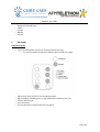
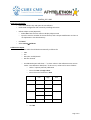
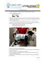
XLMTM_D.2.2.002 Please quote this SOP in your Methods. Respiratory assessment of a canine model of XLMTM using noninvasive respiratory impedence plethysmography SOP (ID) Number XLMTM_D.2.2.002 Version 1 Issued March 26th, 2014 Last reviewed March 26th, 2014 Author Melissa A Goddard University of Washington, Seattle WA Working group members Martin K Childers, David L Mack SOP responsible Melissa A Goddard Official reviewer Martin K Childers University of Washington, Seattle WA Page 1 of 8 XLMTM_D.2.2.002 TABLE OF CONTENTS OBJECTIVE.................................................................................................................................. 3 SCOPE AND APPLICABILITY ........................................................................................................ 3 CAUTIONS .................................................................................................................................. 3 MATERIALS ................................................................................................................................ 3 METHODS .................................................................................................................................. 4 EVALUATION AND INTERPRETATION OF RESULTS .................................................................... 8 REFERENCES .............................................................................................................................. 8 Page 2 of 8 XLMTM_D.2.2.002 1. OBJECTIVE Anesthetised dogs are fitted with wired bands and connected to a calibrated pneumotachometer at the time of assessment. After a baseline ventilatory pattern is established and measured, the respiratory stimulant doxapram hydrochloride is given. Ventilatory parameters are assessed from measures of airflow and by impedence plethysmography as a function of time in response to doxapram. 2. SCOPE AND APPLICABILITY Respiratory impedence plethysmography (RIP) and pneumotachography have been used successfully in larger animal models to measure respiration. Similar non-invasive systems have been applied clinically in patients with neuromuscular disorders to assess the respiratory dysfunction that is often present in these diseases. 3. CAUTIONS The dog should not be shifted or moved during measurement as can cause noise or disruptions in the RIP signal 4. MATERIALS EKG pads EMKA underjacket and outer jacket of an appropriate size (typically the underjacket is one size smaller; all come in small, medium and large)(EMKA Technologies) Alcohol Shaver Wired band material (EMKA Technologies) Scissors Abdominal, thoracic and EKG leads (EMKA Technologies) Pneumotachometer (EMKA Technologies) Calibration unit and t/r box (EMKA Technologies) Antennae (EMKA Technologies) Small ‘screw driver’ (included with calibration unit) (EMKA Technologies) Laptop, with iox2 software 2.8.0.13 (Windows XP Professional, service pack 2 or 3) Telemetry pack (EMKA Technologies) 1L super syringe (EMKA Technologies) Tubing AA Duracell Procell batteries Electrode gel (optional) Page 3 of 8 XLMTM_D.2.2.002 5. Software key (Iox) (EMKA Technologies) Doxapram hydrochloride Saline Syringe Needle SD card METHODS Equipment Set Up Ensure that equipment is properly connected and functioning. T/r box connections should be as follows when viewed at the back: Lights on the front of the T/r unit should glow green Signal strength should be green or high yellow when telemetry box is on Connect the antenna Turn on the unit Connect SD card to T/l box and insert into laptop Page 4 of 8 XLMTM_D.2.2.002 Load saved config file Insert the software key and open the Iox Software Load saved configuration file created by installing technician Define subject names (Optional) Select Run from the main menu to display subject name Input the name or number of the test animal, with a unique modification so there is no duplication in the data directory Click Run! Select TuningCalibrate Calibrate the inputs Calibration values should load automatically as follows for EKG Act RIP (Thor and Ab) bands RIP sum channel Uncalibrated inputs will show “--” as their values in the calibration main screen under “Last calibration date/time.” If this occurs, values can be set as follows: Select “2 points (manual) calibration Enter the Low and High values Enter the unit of measure in Unit Low High Unit EKG 0 1 mV Act 0 1 G -5V -1.45V 0 1000 0 1V 0 1000 RIP RIP Sum m/s m/s Click Ok Page 5 of 8 XLMTM_D.2.2.002 Calibrate RF Calc, the pneumotach signal Select Tuning Calibrate from the run menu. Select “integrate calibration” using the dial Enter the Low value (0) and the High value (1000) Enter the unit of measure in Unit (ml/sec or l/sec) Set the calibrating unit/amplifier to DC using the switch Set the gain to 1 Adjust the coarse adjustment (lowest screw) using the provided ‘screw driver’ until the signal is close to 0V (no flow) Adjust the gain in a stepwise fashion, dialing back until the signal returns to 0 using the fine adjustment dial Set the integration period to the number of seconds sufficient to inject the volume. 2s or 3s is ideal for accurate calibration, although longer periods can be used initially. Attach the 1L syringe to the pneumotachometer using the tubing. When connected correctly, pushing air in should cause a negative deflection to be measured Flip the metal transducer if this is not observed and reconnect the pneumotach Inject the volume and immediately click the High button Ensure that the signal is within range and that injection time does not exceed the integration period. (Area under the curve should be between the 3s.) If not, dial up the gain until the scale fits. Switch the calibration unit back to AC and deselect “remove AC offset” on the program Click Ok Save this new configuration file under a new name (which includes the date of creation) Preparation of Dogs and Jacket Fitting Shave the anaesthetised dog as cleanly as possible in the appropriate areas : under the left ‘armpit’ of the forelegs under the right ‘armpit’ of the forelegs On the left side of the groin Size the dog for the thoracic and abdominal bands in such a way that they fit snuggly but still allow two fingers under the band Cut using the scissors Page 6 of 8 XLMTM_D.2.2.002 Wipe the area with alcohol to remove any stray hairs and fix the EKG pads firmly to the dog. Electrode gel can also be used to improve adhesion and conductivity. Connect the EKG leads as follows: Green at the groin White on the right Black on the left Feed the band material through the loops of the inner jacket and place the inner jacket on the dog, so that the bands face outwards. The metal part of the bands should also be visible (ie, facing outwards) Check to ensure that the thoracic and abdominal leads are in an appropriate ‘open’ position, feed the end of the band material into each and then close. Ends should lock firmly, but should not need to be forced as this may cause damage The blue measures the thoracic band The yellow measures the abdominal Dog should appear as follows: Place the outer jacket on the dog (Optional) Connect thoracic, abdominal and EKG leads to the telemetry pack. Connecting the EKG lead turns the unit on. Ensure that a signal is being obtained on the computer Place the telemetry unit in the pouch of the outer jacket before continuing with any planned experiments (Optional) A catheter should also be placed in the fore or hindlimb for the injection of doxapram hydrochloride. Page 7 of 8 XLMTM_D.2.2.002 Run the experiment and start streaming and storage To start streaming, select Start/Stop Storage/Streaming, Click start streaming Check the Save beats box Click Ok Or click the pre-programed (F1) key To stop streaming press F2 To start storage, select Start/Stop Storage/Streaming, Click start storage Select the site or press Select All sites Click Ok Or click the pre-programed (F3) key To stop storage press F4 Doxapram hydrocloride administration 6. Create a label in Iox by pressing the F12 button and entering the amount of doxapram. Administer a bolus injection of 1.0mg/kg of doxapram hydrochloride, followed rapidly by a saline flush Press Enter at the same time the dose is administered to label Collect a minimum of 20 clear breaths once respiration has reached its peak EVALUATION AND INTERPRETATION OF RESULTS Iox and ecgGAUTO (version 2.8.2.6) (EMKA Technologies) calculates various respiratory parameters from the measured curves such as respiratory rate, tidal volume, peak inspiratory and expiratory flows, and thoracic and abdominal volume. The software can also be used to generate reports of these values, including flow-volume or phase curves. 7. REFERENCES IOX2 user manual. EMKA technologies. V2.4. rev 2. 2010 Goddard MA, mitchell EL, Smith BK, Childers MK. Establishing clinical end points of respiratory function in large animals for clinical translation. Phys Med Rehabil Clin N Am. 2012;23(1):75-94, xi. Page 8 of 8