1
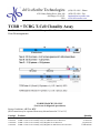
InVivoScribe Technologies 6330 Nancy Ridge Drive, Suite 106 San Diego, CA 92121 USA (858) 623-8105 – Phone (858) 623-8109 – Fax [email protected] www.invivoscribe.com TCRB + TCRG T-Cell Clonality Assay For Identification of Clonal T Cell Receptor Beta Chain and Gamma Chain Gene Rearrangements FOR RESEARCH USE ONLY (Not for use in diagnostic procedures) Storage Conditions: -65ºC to -85ºC (DNA controls may be separated from assay kits and stored at 2°C to 8°C) Catalog# Products 1-200-0010 TCRB + TCRG T-Cell Clonality Assay for Gel Detection 1-200-0011 1-200-0020 1-200-0021 TCRB + TCRG T-Cell Clonality Assay for ABI Fluorescence Detection TCRB + TCRG T-Cell Clonality Assay MegaKit for Gel Detection TCRB + TCRG T-Cell Clonality Assay MegaKit for ABI Fluorescence Detection Quantity 33 Reactions 33 Reactions 330 Reactions 330 Reactions Page 2 of 20 Table of Contents 1. NOTICE .............................................................................................................................................................................. 3 2. PRINCIPLE ........................................................................................................................................................................ 3 3. ASSAY USES ...................................................................................................................................................................... 5 4. SPECIMEN REQUIREMENTS ....................................................................................................................................... 5 5. KIT CONTENTS ................................................................................................................................................................ 6 STATEMENT OF WARNINGS ............................................................................................................................................... 6 6. STORAGE CONDITIONS ................................................................................................................................................ 7 7. REAGENTS REQUIRED BUT NOT INCLUDED ......................................................................................................... 7 PCR AMPLIFICATION ................................................................................................................................................................. 7 ABI FLUORESCENCE DETECTION ............................................................................................................................................... 7 8. RECOMMENDED POSITIVE CONTROLS .................................................................................................................. 7 9. PROCEDURE NOTES ...................................................................................................................................................... 8 10. REAGENT PREPARATION ............................................................................................................................................ 8 11. SAMPLE PREPARATION ............................................................................................................................................... 8 12. AMPLIFICATION ............................................................................................................................................................. 9 13. DETECTION ...................................................................................................................................................................... 9 TEMPLATE AMPLIFICATION CONTROL ....................................................................................................................................... 9 GEL DETECTION – HETERODUPLEX ANALYSIS........................................................................................................................... 9 ABI FLUORESCENCE DETECTION WITH ABI 310, 3100 AND 3130XL INSTRUMENTS ................................................................ 10 14. INTERPRETATION AND REPORTING ..................................................................................................................... 10 EXPECTED SIZE OF AMPLIFIED PRODUCTS ............................................................................................................................... 11 SAMPLE INTERPRETATION ........................................................................................................................................................ 12 15. LIMITATIONS OF PROCEDURE ................................................................................................................................ 12 16. REFERENCES ................................................................................................................................................................. 12 17. APPENDIX ....................................................................................................................................................................... 13 REAGENTS AND SPECIAL SUPPLIES .......................................................................................................................................... 13 Ficoll Separation ................................................................................................................................................................ 13 Gel Electrophoresis ............................................................................................................................................................ 13 Differential Fluorescence Detection ................................................................................................................................... 13 18. TROUBLE SHOOTING GUIDE .................................................................................................................................... 13 19. SAMPLE DATA ............................................................................................................................................................... 14 GEL DETECTION ....................................................................................................................................................................... 14 ABI FLUORESCENCE DETECTION ............................................................................................................................................. 16 20. SINGLE PAGE FLOW CHART..................................................................................................................................... 20 GEL DETECTION – HETERODUPLEX ANALYSIS......................................................................................................................... 20 ABI FLUORESCENCE DETECTION WITH ABI 310, 3100 AND 3130XL INSTRUMENTS ................................................................ 20 TCRB + TCRG T-Cell Clonality Assay FOR RESEARCH USE ONLY; not for use in diagnostic procedures 120000XXv1.01 Page 3 of 20 Thank you for purchasing our TCRB + TCRG T-Cell Clonality Assay. We appreciate your business. We are happy to assist you in the validation of this assay, and will provide ongoing technical assistance to keep the assays performing efficiently in your laboratory. Technical assistance is most rapidly obtained using our Internet site: http://www.invivoscribe.com or by sending an email inquiry to: [email protected]. Questions received during business hours usually receive a response within an hour. Alternatively, you can call for technical assistance and for information on our testing kits at (858) 623-8105 between the hours of 8:00 AM and 5:00 PM Pacific Standard Time. 1. Notice This product and the methods employed are covered by United States Letters Patent Numbered 5,296,351 and 5,418,134; Australian Patent Number 626,601 and Japanese Patent Number 2,781,438, all of which are licensed exclusively to InVivoScribe Technologies (“IVS”). Purchase of this product includes a limited sublicense for non-commercial practice of this technology for use within (or with respect to data or product that are transmitted to) the United States, Japan or Australia only when the purchaser is registered with IVS as an exclusively non-commercial user of IVS products. No sublicense is granted simply by purchase of these products. Non-commercial practice of the technology means sample testing done for teaching and basic research. Non-Commercial practice excludes testing if any of the following apply: (a) test results, products or information derived from the tests are used for or in support of patient care, or are transferred to a healthcare professional involved in patient care; (b) test results are clinically utilized to determine cause of death; (c) compensation, in any form or manner, is received for performing the tests. To request a form for registration as an exclusively non-commercial product user, to discuss terms for a potential sublicense for broader practice of these methods, or for any questions concerning the scope or content of the non-commercial sublicense please contact our legal department by email at [email protected], or by telephone at (858) 623-8105. These methods also require nucleic acid amplification methods such as Polymerase Chain Reaction (PCR), which is covered by patents owned by Hoffmann-LaRoche, Inc. and F. Hoffmann-LaRoche Ltd. No license under these patents to use the PCR process is conveyed expressly or by implication to the purchaser by the purchase of these products. The assays described herein are not approved by any regulatory agency for clinical use. These assays are not for diagnostic or therapeutic use. This product is sold FOR RESEARCH USE ONLY; not for use in diagnostic procedures. 2. Principle NOTICE: InVivoScribe Technologies’ Gene Rearrangement and Translocation Assays represent a new approach to PCR-based clonality testing. These standardized assays were carefully optimized testing positive and negative control samples using multiplex master mixes. Assay development was followed by extensive validation testing more than 400 clinical samples using Revised European/American Lymphoma (REAL) Classification. Testing was done at more than thirty prominent independent testing centers throughout Europe in a collaborative study known as the BIOMED-2 Concerted Action. Results from this BIOMED-2 study appear in a leading peer-reviewed journal, Leukemia. 2003 Dec; 17(12):2257-2317 (Nature Publishing Group). BACKGROUND: TCRB Clonality Testing The human TCR beta gene locus on chromosome 7 (7q35) includes 64-67 V genes belonging to 32 subgroups, 2 D segments, and 13 J segments, spread over 620 kilobases. The diversity of this locus has complicated PCRbased testing, and extended dependence on Southern blot analysis in many testing centers. However, this TCRB + TCRG T-Cell Clonality Assay FOR RESEARCH USE ONLY; not for use in diagnostic procedures 120000XXv1.01 Page 4 of 20 standardized multiplex PCR assay detects the vast majority of clonal TCR beta gene rearrangements using only 3 multiplex master mixes. The assay provides rapid TCR clonality assessment in 4-6 hours, reducing the number of Southern blot tests performed in the laboratory. The detection rate of clonal TCR beta gene rearrangements using this assay is unprecedentedly high. The performance characteristics of this assay have been independently determined by a European collaborative study involving 32 diagnostic PCR laboratories (BIOMED-2 Concerted Action) testing hundreds of clinical samples defined according to the WHO classification. Three multiplex master mixes target conserved regions within the variable (V), diversity (D), and the joining (J) regions that flank the unique hypervariable antigen-binding region 3 (CDR3). TCRB Tube A contains 23 Vb primers + 6 Jb1 primers + 3 Jb2 primers. TCRB Tube B contains 23 Vb + 4 Jb2 primers. TCRB Tube C contains 2 Db + 13 Jb primers. PCR products can be analyzed by differential fluorescence detection using capillary electrophoresis or gene sequencing instruments, by heteroduplex analysis, or using standard gel electrophoresis with ethidium staining. Clonality is indicated if any one of the master mixes generates clonal band(s). TCRG Clonality Testing The human TCR gamma gene locus on chromosome 7 (7q14) includes 14 V genes belonging to 4 subgroups (6 are functional; 3 Open Reading Frames and 5 pseudogenes), 5 J segments, and 2 C genes spread over 200 kilobases. The diversity of this locus has complicated PCR-based testing, and extended dependence on Southern blot analysis in many testing centers. However, this standardized multiplex PCR assay detects the vast majority of clonal TCR gamma gene rearrangements using only 2 multiplex master mixes. The assay provides rapid TCR clonality assessment in 4-6 hours, reducing the number of Southern blot tests performed in the laboratory. The detection rate of clonal TCR gamma gene rearrangements using this assay is unprecedentedly high. The performance characteristics of this assay have been independently determined by a European collaborative study involving 32 diagnostic PCR laboratories (BIOMED-2 Concerted Action) testing hundreds of clinical samples defined according to the WHO classification. Two multiplex master mixes target conserved regions within the variable (V) and the joining (J) regions that flank the unique hypervariable antigen-binding region 3 (CDR3). TCRG Tube A contains primers that target the V gamma 1-8 + V gamma 10 genes and all J gamma exon segments. TCRG Tube B contains primers that target the V gamma 9 + V gamma 11 genes and all J gamma exon segments. PCR products can be analyzed by differential fluorescence detection using capillary electrophoresis or gene sequencing instruments, by heteroduplex analysis, or using standard gel electrophoresis with ethidium staining. Clonality is indicated if any one of the master mixes generates clonal band(s). Polymerase chain reaction (PCR) assays are routinely used for the identification of clonal B- & T-cell populations. These tests amplify the DNA between primers that target the conserved framework (FR) and joining (J) regions (Tubes A & B), or the diversity and joining regions (Tube C). These conserved regions lie on either side of an area within the V-J region where programmed genetic rearrangements occur during maturation of all B and T lymphocytes. The antigen receptor genes that undergo rearrangement are the immunoglobulin heavy chain & light chains genes in B-cells, and the T cell receptor genes in T-cells. Each Band T-cell has a single productive V-J rearrangement that is unique in both length and sequence. Therefore, when this region is amplified using DNA primers that flank this region, a clonal population of cells yields one or two prominent amplified products (amplicons) within the expected size range. Two products are produced in cases when the initial rearrangement was non-productive and was followed by rearrangement of the other homologous chromosome. In contrast, DNA from a normal or polyclonal (many clones) population produces a bell-shaped curve of amplicon products (or Gaussian distribution) that reflect the heterogeneous population of V-J region rearrangements. TCRB + TCRG T-Cell Clonality Assay FOR RESEARCH USE ONLY; not for use in diagnostic procedures 120000XXv1.01 Page 5 of 20 Since the antigen receptor genes are polymorphic (consisting of a heterogeneous population of related DNA sequences), it is difficult to employ a single set of DNA primer sequences to target all of the conserved flanking regions around the V-J rearrangement. N-region diversity and somatic mutation further scramble the DNA sequences in these regions. Therefore multiplex master mixes, which target several FR regions, are required to identify the majority of clonal rearrangements. As indicated, clonal rearrangements are identified as prominent, single-sized products within the smear of different-sized amplicon products that form a Gaussian distribution around a statistically favored, average-sized rearrangement. Gel electrophoresis is commonly used to resolve the different-sized amplicon products and ethidium bromide or other DNA intercalating dyes to stain and detect these products. A powerful alternative method is use of differential fluorescence detection with primers conjugated with fluorescent dyes that correspond to different targeted regions. Reaction products from several different master mixes can be pooled, fractionated using capillary electrophoresis, and detected simultaneously. This detection system results in unsurpassed sensitivity, resolution, differential product detection, and quantification. In addition, the laboratory can eliminate the use of agarose and polyacrylamide gels, as well as the use of carcinogens such as ethidium bromide. Further, differential detection allows accurate, reproducible and objective interpretation of primer-specific products and automatic archiving of data. The limit of detection of this assay has been determined to be approximately 5 clonal cells in 100 hundred normal cells, and inter-assay and intra-assay reproducibility in size determination using capillary electrophoresis is approximately 1-2 basepairs. This reproducibility and sensitivity allows monitoring and tracking of individual tumors during research or methods development. The automatic archiving of specimen data allows comparison of data collected at different times. This test kit includes 6 master mixes. TCRB Tubes A and B target framework regions within the variable region, and the joining region of the TCR beta chain locus. TCRB Tube C targets the diversity and joining regions. TCRG Tubes A and B target framework regions within the variable region, and the joining region of the TCR gamma chain locus. The last master mix, the Specimen Control Size Ladder, targets multiple genes and generates a series of amplicons of 100, 200, 300, 400, and 600 base pairs to ensure that the quality and quantity of input DNA is adequate to yield a valid result. A single thermocycler program and similar detection methodologies are used with all of the BIOMED tests. Many of our customers have remarked that this improves consistency and facilitates cross training on a broad range of different assays. These robust InVivoScribe assays can be used to test DNA extracted from virtually any source. 3. Assay Uses T Cell Receptor Beta Chain and Gamma Chain Gene Rearrangement Assays are useful for: Identifying clonal T-cell populations highly suggestive of T-cell malignancies Lineage determination of leukemias and lymphomas Monitoring and evaluation of disease recurrence Detection and assessment of residual disease Evaluation of new research and methods in malignancy studies 4. Specimen Requirements This assay tests genomic DNA 1. 5cc of peripheral blood, bone marrow biopsy, or bone marrow aspirate anti-coagulated with heparin or EDTA. Ship at ambient temperature; OR 2. Minimum 5mm cube of tissue shipped frozen; or at room temperature or on ice in RPMI 1640; OR 3. 2μg of genomic DNA; OR 4. Formalin-fixed paraffin embedded tissue or slides. TCRB + TCRG T-Cell Clonality Assay FOR RESEARCH USE ONLY; not for use in diagnostic procedures 120000XXv1.01 Page 6 of 20 5. Kit Contents Controls and Standards IVS-0009 Clonal Control DNA IVS-0004 Clonal Control DNA IVS-0021 Clonal Control DNA IVS-0000 Polyclonal Control DNA Master Mixes IVS Catalog # 4-088-0490 4-088-0190 4-088-1210 4-092-0010 IVS Catalog # Concentration 100μl @ 200μg/ml 100μl @ 200μg/ml 100μl @ 200μg/ml 100μl @ 200μg/ml Target Multiple Vβ + Jβ1 + Jβ2 2-205-001X Multiple Vβ + Jβ2 2-205-002X 2-205-003X Multiple Dβ + Jβ 2-207-003X Vg1-8 + Vg10 + Jg Regions 2-207-004X Vg9 + Vg11 + Jg Regions 2-096-002X Multiple Genes Note: X = Detection format code Note: MegaKits contain 10 units of each master mix and 5 units of each Controls and Standards TCRB Tube A TCRB Tube B TCRB Tube C TCRG Tube A TCRG Tube B Specimen Control Size Ladder STATEMENT OF WARNINGS The assay kit has been optimized to be used as a system. Do not substitute other manufacturer’s reagents. Dilution, reducing amplification reaction volumes, or other deviation in this protocol may affect the performance of this test and/or nullify any limited sublicense that comes with the purchase of this testing kit. Close adherence to the protocol will assure optimal performance and reproducibility. It is recommended that glass distilled de-ionized molecular biology grade water be used with the preparation of specimen DNA. This can be purchased from several manufacturers. In addition, laboratory personnel are reminded to wear appropriate personal protective equipment and follow good laboratory practices and universal precautions when working with specimens. Specimens should be handled in approved biological safety containment facilities and opened only in certified biological safety cabinets. Please see Section 9 for further details. TCRB + TCRG T-Cell Clonality Assay FOR RESEARCH USE ONLY; not for use in diagnostic procedures 120000XXv1.01 Page 7 of 20 6. Storage Conditions PCR master mixes are sensitive to freeze/thaw cycles. Therefore, for any duration other than immediate use, our master mixes and assay kits should be stored at -65°C to -85°C. The reason for this is quite straightforward: Due to the high salt concentrations in our master mixes, the effective freezing and thawing temperature of the master mixes is approximately –10°C. The temperature in a standard laboratory –20°C freezer can easily reach –10°C or warmer during the day when these freezers are opened on a regular basis. At these temperatures, PCR master mixes may go through multiple freeze/thaw cycles, resulting in precipitation of the primers. Accordingly, to minimize the exposure of your master mixes to freeze/thaw cycles, IVS recommends that master mixes be stored at -65°C to -85°C. Please note that our DNA standards are best stored at 2°C to 8°C. However, these standards can be stored at any lower temperature as long as they are vortexed after thawing and before use to ensure that they are resuspended completely. If you have any questions, please contact our technical staff. We are happy to help you determine your optimal storage needs. 7. Reagents Required But Not Included PCR Amplification AmpliTaq Gold DNA Polymerase (RECOMMENDED) AmpliTaq DNA Polymerase (Applied Biosystems, Cat# N808-0241) (Applied Biosystems, Cat# N808-0161) ABI Fluorescence Detection HI-DI Formamide with ROX size standards - ABI 310 HI-DI Formamide with ROX size standards - ABI 3100 (IVS, Cat# 6-098-0051) (IVS, Cat# 6-098-0061) 8. Recommended Positive Controls Master Mix Target TCRB Tube A Vβ + Jβ1/2 Color Green TCRB Tube B Vβ + Jβ2 TCRB Tube C Dβ + Jβ1/2 Blue TCRG Tube A TCRG Tube B Specimen Control Size Ladder Note: Green Vg1-8, Vg10 + Jg 1.3/2.3 Green Vg9 and Vg11 + Jg 1.3/2.3 Green Multiple Genes Blue Control DNA Catalog # Product Size in Basepairs Valid Size Range IVS-0009 Clonal Control DNA Valid Size Range IVS-0004 Clonal Control DNA Valid Size Range IVS-0009 Clonal Control DNA Valid Size Range IVS-0021 Clonal Control DNA Valid Size Rang IVS-0021 Clonal Control DNA Valid Size Range IVS-0000 Polyclonal Control DNA --4-088-0490 --4-088-0190 --4-088-0490 --4-088-1210 --4-088-1210 --4-092-0010 240-285 264 240-285 253 170-210 (Dβ2), 285-325 (Dβ1) 309 145-255 211 80-220 167 84, 96, 200, 300, 400, 600 84, 96, 200, 300, 400, 600 The amplicon sizes listed above were determined using an ABI 3100 platform. Amplicon sizes seen on your specific CE instrument may differ 1-4bp from those listed above depending on the platform of detection (ABI) and the version of the analysis software used. Once identified, the amplicon size as determined on your specific platform will be consistent from run to run. This reproducibility is extremely useful when tracking MRD. TCRB + TCRG T-Cell Clonality Assay FOR RESEARCH USE ONLY; not for use in diagnostic procedures 120000XXv1.01 Page 8 of 20 9. Procedure Notes Autoclaving does not eliminate DNA contamination. Work flow in the PCR laboratory should always be in a one way direction between separate work areas; beginning in Master Mix Preparation, moving to the Specimen Preparation, then to the Amplification, and finally to Detection. 1. Do not bring amplified DNA into the areas designated for master mix or specimen preparation. 2. Due to the analytical sensitivity of this test, extreme care should be taken to avoid the contamination of reagents or amplification mixtures with samples, controls or amplified materials. All reagents should be closely monitored for signs of contamination (e.g., negative controls giving positive signals). Discard reagents suspected of contamination. 3. All pipettes, pipet tips, and any equipment used in a particular area should be dedicated to and kept to that area of the laboratory. 4. PCR trays, bases, and retainers must to be decontaminated in 10% bleach and rinsed with distilled water two separate times before returning them to the starting areas. 5. Sterile, disposable plastic ware should be used whenever possible to avoid RNase or cross-contamination. 10. Reagent Preparation All unknown samples should be tested using the template Specimen Control Size Ladder. This is to ensure that no inhibitors of amplification are present, and there is DNA of sufficient quality and quantity to generate a valid result. All samples should be tested in singlicate. Positive, negative and no template controls should be tested for each of the master mixes. 1. Using gloved hands, remove the master mixes from the freezer. Allow the tubes to thaw; then gently vortex to mix. 2. In containment hood or dead air box remove an appropriate aliquot to clean, sterile microfuge tube (one tube for each of the master mixes). Aliquot volumes should be 45μl for each sample + 135μl (3 x 45µl) for the positive, negative and no template controls. We recommend adding an additional 20μl to correct for pipetting errors. 3. Add the appropriate amount of AmpliTaq Gold polymerase (0.45μl of AmpliTaq Gold @ 5U/μl per 50μl total PCR reaction volume for TCRB Tubes A & B, 0.25μl of AmpliTaq Gold @ 5U/μl per 50μl total PCR reaction volume for TCRB Tube C, TCRB Tubes A & B and the Specimen Control Size Ladder) to each of the master mixes and gently mix by inverting several times or gently vortexing. The master mixes are now ready for distribution to reaction tubes or plate, and addition of sample. 11. Sample Preparation Using any method of DNA extraction, extract the genomic DNA from unknown samples. Resuspend DNA to final concentration of 100μg - 400μg per ml in TE (10 mM Tris-HCl, 1mM EDTA, pH 8.0) or distilled water. This is a robust assay system. A wide range of DNA concentrations will generate a valid result. Therefore, quantifying and adjusting DNA concentrations is generally not necessary. Testing sample DNAs with the Control Size Ladder will ensure that DNA of sufficient quality and quantity was present to yield a valid result. TCRB + TCRG T-Cell Clonality Assay FOR RESEARCH USE ONLY; not for use in diagnostic procedures 120000XXv1.01 Page 9 of 20 12. Amplification 1. Aliquot 45μl of the master mix/enzyme solutions into individual PCR wells or tubes. 2. Add 5μl of sample or control DNA to the individual tubes or wells containing the respective master mix reactions. Pipette up and down several times to mix. Amplify the reactions using the following PCR program We recommend the MJ Research PTC-100, PTC-200 or the PE 2600, 9600, or 9700 thermocyclers, using the following PCR parameters for the amplifications: Note: We recommend using the calculated option for temperature measurement with the PTC instruments. Standard Program for AmpliTaq Gold Step 1: 95°C for 7 minutes Step 2: 95°C for 45 seconds Step 3: 60°C for 45 seconds Step 4: 72°C for 90 seconds Step 5: Go to step 2; 34 more times Step 6: 72°C for 10 minutes Step 7: 15°C forever Remove the amplification plate from the thermocycler 13. Detection Not all detection formats are available for all assays Template Amplification Control The Specimen Control Size Ladder master mix primers may be labeled with a fluorescent dye (6-FAM). This label is detected as BLUE using the differential fluorescence software. The amplicons produced with this master mix are at ~100, 200, 300, 400, and 600 basepairs. Please note that the ~100bp band is comprised of a 84bp and 96bp bands. Both of these bands co-migrate on a gel. The products of this master mix should be run separately. Gel Detection – Heteroduplex Analysis 1. Denature 20μl of PCR products at 94ºC for 5 minutes. 2. Re-anneal PCR products at 4ºC for 60 minutes. 3. Assemble electrophoresis unit using a 6% non-denaturing polyacrylamide TBE gel (made with 1X TBE, Invitrogen Cat# EC62652Box) and 0.5X TBE running buffer (Invitrogen 5X TBE Cat# LC6675). 4. Add 5μl of ice-cold non-denaturing bromophenol blue loading buffer to samples 5. Load 20μl of mixture into wells of the gel. 6. Run gel at 110V for 2-3 hours or 40-50V overnight. Voltage and electrophoresis time depend on the PCR amplicon size, acrylamide gel thickness, and type of PCR equipment. Voltage and run time can be adapted accordingly. 7. Gels are stained in 0.5μg/ml EtBr (in water or 0.5X TBE Buffer) for 5-10 minutes. 8. Gels are destained 2X in water for 5-10 minutes. 9. UV illumination is used for visualization. 10. Gel is photographed and data are interpreted. TCRB + TCRG T-Cell Clonality Assay FOR RESEARCH USE ONLY; not for use in diagnostic procedures 120000XXv1.01 Page 10 of 20 ABI Fluorescence Detection with ABI 310, 3100 and 3130xl instruments 1. In a new microcentrifuge tube, mix an appropriate amount (for a total of 10μl per PCR reaction) of Hi-Di Formamide with ROX Size Standardsa. Vortex well. 2. In a new 96-well PCR plate, add 10μl of Hi-Di Formamide with ROX size standards to individual wells for each PCR reaction. 3. Transfer 1μl of each PCR reaction to the wells containing Hi-Di Formamide with ROX size standards. Add only one sample per well. Pipette up and down to mix. 4. Cap or cover the PCR plate or tubes. 5. Heat denature the samples at 95ºC for 2 minutes then snap chill on ice for 5 minutes. 6. Prepare a sample sheet and injection list for the samples. 7. Run the samples on an ABI capillary electrophoresis instrument according to the user manualb. 8. Data are automatically displayed as size and color specific peaks. Review profile and controls, report results. Note a: Please see Applied Biosystems’ accompanying product insert for mixing Hi-Di Formamide with ROX size standards for different ABI instruments. Alternatively, pre-mixed aliquots may be purchased directly from InVivoScribe Technologies. Note b: As the samples are run on the machine, they are fractionated, detected and analyzed by the instrument. Runs are 20-24 minutes in duration. The ABI capillary electrophoresis instruments routinely handle 2 runs per hour (for the 1-, 4-, and 16-capillary instruments this is equal to 48, 192, and 768 samples per day, respectively), and automatically analyze and store data for review or comparison with other test results. 14. Interpretation and Reporting Note: This assay is for research use only. Although positive results are highly suggestive of malignancy, these assays are designed for Research Use Only and, if used in a clinical setting, should only be used in support of diagnosis. Positive and negative results should be interpreted in the context of all clinical information and laboratory test results. PCR based testing does not identify 100% of clonal cell populations; therefore, repeat testing by Southern blot may be advisable to rule out clonality. The size range for each of the master mixes has been determined testing positive control samples. For accurate and meaningful interpretation it is important to ignore peaks that occur outside of the proscribed/valid size range for each of the master mixes. Peaks that are outside of the range cannot be assumed to be valid. Note: “Color” indicates the color of products generated with the master mix when using differential fluorescence detection format (e.g., ABI instruments). TCRB + TCRG T-Cell Clonality Assay FOR RESEARCH USE ONLY; not for use in diagnostic procedures 120000XXv1.01 Page 11 of 20 Expected Size of Amplified Products Master Mix Color Mix TCRB Blue Vβ + Jβ (Jβ2.X) Tube A + Green Control DNA Cat# Product Size in basepairs Valid Size Range --240-285 IVS-0000 Polyclonal Control DNA 4-092-0010 240-285, 2711 IVS-0009 Clonal Control DNA 4-088-0490 264 IVS-0004 Clonal Control DNA 4-088-0190 295 (Jβ1.X) Vg1-8 + Jg 1.1/2.1 Vg1-8 + Jg 1.3/2.3 Vg10 + Jg 1.1/2.1 Vg10 + Jg 1.3/2.3 Blue Green Blue Green Vg1-8 + Jg 1.3/2.3 Vg1-8 + Jg 1.3/2.3 Green Green IVS-0009 Clonal Control DNA IVS-0021 Clonal Control DNA Vβ + Jβ2 TCRB Tube C Dβ + Jβ Blue (Jβ2.X) Blue (Jβ2.X) + Green (Jβ1.X) TCRG Tube A Valid Size Range --240-285 IVS-0000 Polyclonal Control DNA 4-092-0010 240-285, 2212 IVS-0009 Clonal Control DNA 4-088-0490 No Product IVS-0004 Clonal Control DNA 4-088-0190 253 Valid Size Range --170-210 (Db2), 285-325 (Db1) IVS-0000 Polyclonal Control DNA 4-092-0010 1282, 170-210, 285-325, 3372 IVS-0009 Clonal Control DNA 4-088-0490 309 IVS-0004 Clonal Control DNA 4-088-0190 295 Valid Size Range --145-255 IVS-0000 Polyclonal Control DNA 4-092-0010 230-255 IVS-0000 Polyclonal Control DNA 4-092-0010 195-230 IVS-0000 Polyclonal Control DNA 4-092-0010 175-195 IVS-0000 Polyclonal Control DNA 4-092-0010 145-175 TCRB Tube B Valid Size Range Vg9 + Jg 1.1/2.1 Vg9 + Jg 1.3/2.3 Vg11 + Jg 1.1/2.1 Vg11 + Jg 1.3/2.3 Blue Green Blue Green IVS-0000 Polyclonal Control DNA IVS-0000 Polyclonal Control DNA IVS-0000 Polyclonal Control DNA IVS-0000 Polyclonal Control DNA Vg11 + Jg 1.3/2.3 Vg9 + Jg 1.3/2.3 Specimen Multiple Genes Control Size Ladder Green Green Blue IVS-0009 Clonal Control DNA IVS-0021 Clonal Control DNA Any Human DNA TCRG Tube B Note: Note 1: Note 2: Note3: Note4: Note5: Note6: 4-088-0490 4-088-1210 --4-092-0010 4-092-0010 4-092-0010 4-092-0010 212 211 80-220 195-220 160-1954 110-1405 80-1103 4-088-0490 1153 4-088-1210 1436, 167 --84, 96, 200, 300, 400, 600 The amplicon sizes listed above were determined using an ABI 3100 platform. Amplicon sizes seen on your specific CE instrument may differ 1-4bp from those listed above depending on the platform of detection (ABI) and the version of the analysis software used. Once identified, the amplicon size as determined on your specific platform will be consistent from run to run. This reproducibility is extremely useful when tracking MRD. The 271bp band (mainly visible with GeneScan analysis) is particularly seen in samples with low numbers of contaminating lymphoid cells. Under sub-optimal conditions aspecific products of 128, 221, and 337bps can be detected in Tubes B and C. If present, these bands will normally be faint. This may be seen as a weak amplicon. Amplicon product is often not seen in this size range. Amplicon product is often not seen. This is an extremely restricted repertoire. Amplicon product is often not seen in this size range. Results can be reported as “Positive” or “Negative” for “Detection of clonal T cell receptor beta chain or gamma chain gene rearrangement consistent with the presence of a clonal cell population” TCRB + TCRG T-Cell Clonality Assay FOR RESEARCH USE ONLY; not for use in diagnostic procedures 120000XXv1.01 Page 12 of 20 1. Samples that fail to amplify following repeat testing should be reported as “A result cannot be reported on this specimen because there was DNA of insufficient quantity or quality for analysis”. 2. It is acceptable to call a sample “Positive” when a product is generated in the valid size range yet the positive control for that master mix fails. 3. Samples that test negative should be repeated if the positive control reaction failed. 4. All assay controls must be examined prior to interpretation of sample results. If the controls do not yield the correct results, the assay is not valid and the samples should not be interpreted. The following describes the analysis of each of the controls, and the decisions necessary based upon the results. 1. Negative Control: (Polyclonal control, water or no template blank). If the negative control is: Positive: Possible contamination of all PCR amplification reactions. Do not continue with the interpretation of results. Prepare fresh master mix and repeat amplification. Negative: Continue with the analysis. 2. Positive Control: Positive: Negative: (This can also be an extraction control if positive control material is taken through extraction processes). If the positive control is: Continue with analysis. Repeat assay unless specimen tests positive. 3. Specimen Control Size Ladder: (This is run on unknown samples only). If the amplification control is: Positive: ~100, 200, 300, 400, and 600 basepair products are seen. Because smaller PCR fragments are preferentially amplified, it is not unusual for the 600 basepair fragment to have a diminished signal or to be missing entirely. Continue with analysis. Negative: Repeat assay unless specimen tests positive. Sample Interpretation Following the acceptance of the controls, the clinical samples are interpreted as follows: One or two prominent bands within the valid size range for TCRB Tubes A, B or C is reported as: “Detection of clonal T cell receptor beta chain gene rearrangement consistent with the presence of a clonal cell population.” One or two prominent bands within the valid size range for TCRG Tubes A or B is reported as: “Detection of clonal T cell receptor gamma chain gene rearrangement consistent with the presence of a clonal cell population.” 15. Limitations of Procedure The assay is subject to interference by degradation of DNA or inhibition of PCR due to heparin or other agents. The assay cannot reliably detect less than 1 positive cell per 100 normal cells. 16. References 1. 2. 3. Miller, JE, Wilson, SS, Jaye, DJ, Kronenberg, M. An automated semiquantitative B and T cell clonality assay. Mol. Diag. 1999, 4(2):101-117. van Dongen, JJM et al. Design and standardization of PCR primers and protocols for detection of clonal immunoglobulin and T-cell receptor gene recombinations in suspect lymphoproliferations: Report of the BIOMED-2 Concerted Action BMH4-CT98-3936. Leukemia. 2003, 17(12):2257-2317. van Krieken JH, Langerak AW, Macintyre EA, Kneba M, Hodges E, Sanz RG, Morgan GJ, Parreira A, Molina TJ, Cabeçadas J, Gaulard P, Jasani B, Garcia JF, Ott M, Hannsmann ML, Berger F, Hummel M, Davi F, Brüggemann M, Lavender FL, Schuuring E, Evans PA, White H, Salles G, Groenen PJ, Gameiro P, Pott Ch, van Dongen JJM. Improved reliability of lymphoma diagnostics via PCR-based clonality testing: report of the BIOMED-2 Concerted Action BHM4-CT98-3936. Leukemia. 2007; 21(2):201-6. TCRB + TCRG T-Cell Clonality Assay FOR RESEARCH USE ONLY; not for use in diagnostic procedures 120000XXv1.01 Page 13 of 20 17. Appendix Reagents and Special Supplies Ficoll Separation Ficoll-Hypaque or Ficoll-Paque 1X PBS diluted from 10X PBS RPMI 1640 DMSO Hybri-Max Fetal Bovine Serum (Pharmacia, Cat# 17-0840-02) (Gibco/BRL, Cat# 70011-044) (Gibco/BRL, Cat# 11875-093) (Sigma, Cat# D2650) (Hyclone, Cat# SH30071.03) Gel Electrophoresis UltraPure™ 10 mg/ml Ethidium Bromide 10X BlueJuice™ Gel Loading Buffer Ready-LoadTM 100 bp Ladder Novex® TBE gels (6%, 12 well) Novex® TBE Running Buffer (5X) Novex® Hi-Density TBE Sample Buffer (5X) (Invitrogen, (Invitrogen, (Invitrogen, (Invitrogen, (Invitrogen, (Invitrogen, Differential Fluorescence Detection HI-DI Formamide with ROX size standards - ABI 310 HI-DI Formamide with ROX size standards - ABI 3100 HI-Deionized Formamide HI-Deionized Formamide GS ROX 50-400HD Size Standard (IVS, Cat# 6-098-0051) (IVS, Cat# 6-098-0061) (IVS, Cat# 6-098-0041) (Applied Biosystems, Cat# 4311320) (Applied Biosystems, Cat# 402985) Cat# 15585-011) Cat# 10816-015) Cat# 10380-012) Cat# EC62652Box) Cat# LC6675) Cat# LC6678) 18. Trouble Shooting Guide Our laboratories are located in San Diego, California. Technical assistance is most rapidly obtained using our Internet site: http://www.invivoscribe.com or by sending an email inquiry to: [email protected]. Alternatively, you can call (858) 623-8105 for technical assistance and information on our testing kits between the hours of 8:00 AM and 5:00 PM Pacific Standard Time. Questions received during business hours usually receive a response within an hour. TCRB + TCRG T-Cell Clonality Assay FOR RESEARCH USE ONLY; not for use in diagnostic procedures 120000XXv1.01 Page 14 of 20 19. Sample Data Gel Detection The data shown below were generated using the master mixes indicated. Amplified products were run on a 6% polyacrylamide gel. For each TCRB and TCRG master mix (Figures 1-5): Lane 1 displays data generated testing an alternative 100% clonal control DNA. Lane 2 displays data generated testing the recommended 100% clonal control DNA. Lane 3 displays data generated testing a 10% dilution of the recommended clonal control DNA. Lane 4 displays data generated testing IVS-0000 Polyclonal Control DNA. Figure 1 Figure 2 Figure 3 Figure 4 TCRB + TCRG T-Cell Clonality Assay FOR RESEARCH USE ONLY; not for use in diagnostic procedures 120000XXv1.01 Page 15 of 20 Figure 5 The data shown below were generated using the master mix indicated. Amplified products were run on a 2% agarose gel. For the Specimen Control Size Ladder master mix: Lane 1 displays a 100 basepair DNA ladder. Lane 2 displays a 50 basepair DNA ladder. Lanes 3 and 4 display data generated testing two different 100% clonal control DNAs. Figure 6 TCRB + TCRG T-Cell Clonality Assay FOR RESEARCH USE ONLY; not for use in diagnostic procedures 120000XXv1.01 Page 16 of 20 ABI Fluorescence Detection The data shown below were generated using the master mixes indicated. Amplified products were run on an ABI 3100 instrument. For each TCRB and TCRG master mix (Figures 7-11): Panel 1 displays data generated testing an alternative 100% clonal control DNA. Panel 2 displays data generated testing the recommended 100% clonal control DNA. Panel 3 displays data generated testing a 10% dilution of the recommended clonal control DNA. Panel 4 displays data generated testing IVS-0000 Polyclonal Control DNA. Figure 7 TCRB + TCRG T-Cell Clonality Assay FOR RESEARCH USE ONLY; not for use in diagnostic procedures 120000XXv1.01 Page 17 of 20 Figure 8 Figure 9 TCRB + TCRG T-Cell Clonality Assay FOR RESEARCH USE ONLY; not for use in diagnostic procedures 120000XXv1.01 Page 18 of 20 Figure 10 Figure 11 TCRB + TCRG T-Cell Clonality Assay FOR RESEARCH USE ONLY; not for use in diagnostic procedures 120000XXv1.01 Page 19 of 20 For the Specimen Control Size Ladder master mix: Panel 1 displays data generated testing a negative water control. Panel 2 displays data generated testing the recommended positive control, IVS-0000 Polyclonal Control DNA. Panels 3 and 4 display data generated testing two different 100% clonal control DNAs. Figure 12 TCRB + TCRG T-Cell Clonality Assay FOR RESEARCH USE ONLY; not for use in diagnostic procedures 120000XXv1.01 Page 20 of 20 20. Single Page Flow Chart 1. 2. 3. 4. 5. Using gloved hands, remove the master mixes from the freezer. Allow the tubes to thaw; then gently vortex to mix. In a containment hood or dead air box remove an appropriate aliquot to clean, sterile microfuge tube (one tube for each of the master mixes). Aliquot volumes should be 45μl for each sample + 135μl for the positive, negative and no template controls. We recommend adding an additional 20μl to correct for pipetting errors. Add the appropriate amount of AmpliTaq Gold polymerase (0.45μl of AmpliTaq Gold @ 5U/μl per 50μl total PCR reaction volume for TCRB Tubes A & B, 0.25μl of AmpliTaq Gold @ 5U/μl per 50μl total PCR reaction volume for TCRB Tube C, TCRB Tubes A & B and the Specimen Control Size Ladder) to each of the master mixes and gently mix by inverting several times or gently vortexing. Aliquot 45μl of master mix to individual wells of a PCR plate. Add 5μl of DNA from the unknown and control samples to individual tubes or wells containing the respective master mix reactions, and pipette up and down several times to mix. Amplify target DNA using the universal thermocycler program. Gel Detection – Heteroduplex Analysis 6. Denature 20μl of PCR products at 94ºC for 5 minutes. 7. Re-anneal PCR products at 4ºC for 60 minutes. 8. Assemble electrophoresis unit using a 6% non-denaturing polyacrylamide TBE gel (made with 1X TBE, Invitrogen Cat# EC62652Box) and 0.5X TBE running buffer (Invitrogen 5X TBE Cat# LC6675). 9. Add 5μl of ice-cold non-denaturing bromophenol blue loading buffer to samples 10. Load 20μl of mixture into wells of the gel. 11. Run gel at 110V for 2-3 hours or 40-50V overnight. Voltage and electrophoresis time depend on the PCR amplicon size, acrylamide gel thickness, and type of PCR equipment. Voltage and run time can be adapted accordingly. 12. Gels are stained in 0.5μg/ml EtBr (in water or 0.5X TBE Buffer) for 5-10 minutes. 13. Gels are destained 2X in water for 5-10 minutes. 14. UV illumination is used for visualization. 15. Gel is photographed and data are interpreted. ABI Fluorescence Detection with ABI 310, 3100 and 3130xl instruments 6. In a new microcentrifuge tube, mix an appropriate amount (for a total of 10μl per PCR reaction) of Hi-Di Formamide with ROX Size Standards. Vortex well. 7. In a new 96-well PCR plate, add 10μl of Hi-Di Formamide with ROX size standards to individual wells for each PCR reaction. 8. Transfer 1μl of each PCR reaction to the wells containing Hi-Di Formamide with ROX size standards. Add only one sample per well. Pipette up and down to mix. 9. Cap or cover the PCR plate or tubes. 10. Heat denature the samples at 95ºC for 2 minutes then snap chill on ice for 5 minutes. 11. Prepare a sample sheet and injection list for the samples. 12. Run the samples on an ABI capillary electrophoresis instrument according to the user manual. 13. Data are automatically displayed as size and color specific peaks. Review profile and controls, report results. TCRB + TCRG T-Cell Clonality Assay FOR RESEARCH USE ONLY; not for use in diagnostic procedures 120000XXv1.01