1
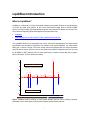
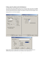
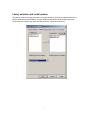
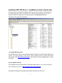
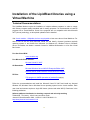
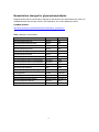
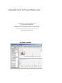
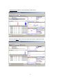
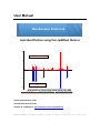
User Manual
Metabolomics Fiehn Lab
Lipid identification using the LipidBlast libraries
673
100
Experimental MS/MS spectrum
Experimental MS/MS spectrum
50
476
496
715
549
0
426
511 527
451
613
585 604
634
686
658
549
50
715
476 494
In-silico MS/MS spectrum
In-silico MS/MS spectrum
100
673
440 460
480 500 520
732.555 [Da].dta
540 560 580 600
620 640 660
Head to Tail MF=438 RMF=901
680 700 720
740
PC 32:1; [M+H]+; GPCho(16:1(9Z
Tobias Kind and Oliver Fiehn
Fiehnlab November 2012 v46
Contact: Dr. Tobias Kind - http://fiehnlab.ucdavis.edu/staff/kind/
General Disclaimer: The following information is provided for research purposes only and may contain errors.
1
LipidBlast Introduction
What is LipidBlast?
LipidBlast is a collection of in-silico electrospray tandem mass spectral libraries for the identification
of neutral and polar lipid species. In the current development stage around 120,000 MS/MS
spectra, covering multiple glycerophospholipid classes and possible ESI adducts are included. For
more resources regarding lipids and analytical techniques please visit:
•
LipidMaps
•
Lipid Analysis with GC-MS, LC-MS, FT-MS - Metabolomics Fiehn Lab
The LipidBlast libraries were developed from known theoretical fragmentations and experimental
fragmentation and abundance information from tandem mass spectra (MS/MS). The experimental
basis was a Thermo Finnigan LTQ linear iontrap instrument equipped with an Advion Nanomate
nanoESI robotic infusion system. However the libraries are platform independent and can be used
for all MS/MS or MSE platforms from all mass spectrometry vendors including ABI Sciex, Agilent,
Bruker, Shimadzu, Thermo, Waters and others.
673
100
Experimental MS/MS spectrum
Experimental MS/MS spectrum
50
476
496
715
549
0
426
511 527
451
613
585 604
634
658
686
549
50
715
476 494
In-silico MS/MS spectrum
In-silico MS/MS spectrum
100
673
440 460 480 500 520 540 560 580 600 620 640 660 680 700 720 740
732.555 [Da].dta
Head to Tail MF=438 RMF=901
PC 32:1; [M+H]+; GPCho(16:1(9Z
Figure: LipidBlast libraries showing an experimental MS/MS spectrum and a matching computer
generated in-silico mass spectrum with similar fragment peaks and abundances.
2
How can the LipidBlast libraries be used?
The LipidBlast can be used for identification of polar lipids using high resolution and low
resolution tandem mass spectrometer data. In any case the precursor mass is needed from the MS1
spectrum and the datadependent MS2 spectra must be extracted from the experimental runs.
Therefore the LipidBlast libraries can be used for identification of lipids from:
A) Infusion mass spectrometric experiments with data dependent MS/MS scans
B) LC-MS experiments with data dependent MS/MS scans
C) LC-MS experiments with TOF MSE experiments
The LipidBlast libraries are traditional mass spectral libraries. One difference to other in-silico
libraries is that accurate mass information is included for all precursor and product ions, as well as
fragment names and modeled abundances. The following mass spectral search modes can be
applied:
A) Traditional mass spectral library search
B) MS/MS database search
The LipidBlast libraries can be used with traditional library search procedures such as PBM search
or dot product (NIST MS) search. Better performance and better hit rates and higher identification
rates are obtained if the accurate precursor mass is included (for pre-filtering possible lipid species)
and a subsequent MS/MS peak matching is performed. The NIST MS Search algorithm is capable
of finding a single mass spectrum in a library of one million spectra in only milliseconds.
How can I extract MS/MS spectra?
If your platform software has MS/MS search capabilities, just import the provided ASCII MSP
spectra into your vendor software. If your platform has no MS/MS search capability (which includes
precursor search and product ion search step, not to be confused with normal mass spectral library
search) you can extract MS/MS spectra, export them as MFG files (MASCOT format) and import
them directly into NIST MS Search or NIST MSPepSearch. These approaches are used in the
proteomics community since many years, so talking to a fellow proteomics researcher will solve that
problem.
A) For extraction of MGF directly from raw files, download ProteoWizard
http://proteowizard.sourceforge.net/
B) Other software can be found at the Sashimi tools
(http://sourceforge.net/projects/sashimi/files/
C) Trans Proteomic Pipeline software page
(http://tools.proteomecenter.org/wiki/index.php?title=Formats:mzXML).
3
How can I perform quantification of lipid species?
The LipidBlast library is a mass spectral library for identification of lipids. In order to quantify lipids
other commonly available vendor software tools must be used. This includes the use of internal or
external standards (reference compounds), the use of proper calibration and peak integration
methods and programs for alignment of LC-MS or direct infusion MS data.
For a collection of programs see:
http://fiehnlab.ucdavis.edu/staff/kind/Metabolomics/LipidAnalysis/
For alignment programs see peak alignment of LC, GC, MS, NMR data
http://fiehnlab.ucdavis.edu/staff/kind/Metabolomics/Peak_Alignment/
Or see our poster using the Genedata Refiner MS program: An automated workflow for rapid
alignment and identification of lipid biomarkers obtained from chip-based direct infusion
nanoelectrospray tandem mass spectrometry
http://fiehnlab.ucdavis.edu/staff/kind/Metabolomics/LipidAnalysis/lipids-biomarker-workflow.pdf
4
Which lipid classes are covered in the LipidBlast libraries?
Currently the glycerophospholipids are available in NIST MS MSP format. Other libraries
(monoacylglycerols, diacylglycerols and triacylglycerols) and galactolipids (MGDG, DGDG, SQDG)
are also available. Additional libraries can be developed on request or by using the provided data
templates. The creation of in-silico libraries depends on the correct molecular species enumeration
and existence of molecular structures and secondly on a correct nomenclature to help during the
discovery process. Secondly experimental spectra from reference standards must be obtained for
all possible classes all possible side chain modifications. That includes symmetric sn1 and sn2 side
chains, different degrees of unsaturation and different head groups.
Table: LipidBlast lipids and current adduct libraries including lyso-forms for some species
Short
Class
Positive mode
Negative mode
Glycerophosphocholines
PC
M+H; M+Na
low abundant
Glycerophosphoethanolamines
PE
M+H; M+Na
M-H
Glycerophosphoserines
PS
M+H
M-H
Glycerophosphoglycerols
PG
low abundant
M-H
Glycerophosphoinositols
PI
low abundant
M-H
Glycerophosphates
PA
M+NH4; M+Na; M+2Na
M-H
Sphingomyelins
SM
M+H; M+Na
low abundant
Diacylglycerols
DAG
M+NH4; M+H; M+Na
-
Triacylglycerols
TAG
M+NH4; M+H; M+Na
-
Monogalactosyldiacylglycerols
MGDG
M+NH4; M+Na
M-H
Digalactosyldiacylglycerols
DGDG
M+NH4; M+Na
M-H
Sulfoquinovosyldiacylglycerols
SQDG
M-H
Gangliosides
[glycan]-Cer
M-H
Ceramide-1-phosphates
Sulfatides
CerP
ST
Gangliosides
Diacylated phosphatidylinositol
monomannoside
Diacylated phosphatidylinositol
dimannoside
Triacylated phosphatidylinositol
dimannoside
Tetraacylated
phosphatidylinositol
dimannoside
[glycan]-Cer
M-H
Ac2PIM1
M-H
Ac2PIM2
M-H
Ac3PIM2
M-H
Ac4PIM2
M-H
Sulfatides
ST
M-H
M+H
M-H
M-H
Some of the adducts may not occur in the literature, because ions may be suppressed in mixtures,
but ions were observed from authentic standards. Also common adducts such as [M+Li]+ may not
5
be included because experimental spectra were not acquired and in-silico spectra were not
modeled yet.
How large is each single adduct library? The size depends on the lipid species but is usually
around 5,500 in-silico MS/MS spectra. In case of sphingomyelins the library size is smaller. So at
the current development stage around 212,516 MS/MS spectra from 119,200 structures are
included. In case of TAGs the library size was reduced to the 2640 most common compounds
(TG(12:0/12:0/16:0) to TG(22:6/22:6/22:6)) due to the large number of combinations.
6
NIST MS Search program for easy identification
The NIST MS Search GUI is a freely available program developed by the Mass Spectral Data group
at NIST. The procedure of lipid identification, can be easily performed by loading the result *.mgf file
into the NIST MS GUI search. By clicking the green GO button or each single experimental MS/MS
spectrum a library search is performed. Hit scores are provided in the left result window. See
http://chemdata.nist.gov/ and the NIST help file (Ver20Man.pdf) for further information.
Experimental MS/MS list
Experimental MS/MS list
exp. MS/MS
exp. MS/MS
exp. MS/MS
exp. MS/MS
in-silico MS/MS
in-silico MS/MS
Library hit scores
Library hit scores
in-silico MS/MS
in-silico MS/MS
Figure: NIST MS search GUI with the LipidBlast libraries loaded
Automation of library search is obtained by starting
nistms.exe with the /par=4 option from the DOS
command prompt which writes all results into
NISTLOG.txt in the same directory. The option
nistms.exe /par=8 stops the automated result logging.
The automation options are set in the library options
dialog. The number of hits is the most important
selection.
7
Library search options and m/z tolerances
The NIST MS Search option MS/MS search has to be selected as seen in the picture. An MS/MS
search precursor mass tolerance of 0.001 Da or in case of accurate mass data and 0.4 m/z units for
unit mass spectrometers should be selected. The product ion mass can be less stringent, due to
hydrogen shifts, 0.4 to 0.8 Da is acceptable.
Figure: MS/MS search and appropriate m/z windows must be selected. MS/MS options are
selected using reverse search and low score thresholds, all other options are off.
8
Library selection and install options
The libraries must be activated and visible in the right window. If no library is selected make sure to
edit the install directory (NISTMS.ini) or simply delete the NISTMS.ini file and restart nistms.exe.
The program will create a new INI file and will ask for the current library directory.
9
Installation NIST MS Search + LipidBlast procedure step-by-step
The install assumes a fully patched Windows OS (Vista, XP, Server). Extract the directories from
the zip file and place it under C:\ for instant use, or install into any subdirectory of your choice
(requires selection of library path). The folder should be C:\LipidBlastBeta-MSSearch\
Start nistms.exe to work with the program.
(1) The NIST MS Search GUI
The NIST MS Search GUI is a freely available program developed by the Mass Spectral Data group
at NIST. The current distribution of the ZIP file contains a working version of the NIST MS Search
GUI program. See http://chemdata.nist.gov/ and the NIST help file (Ver20Man.pdf) for further
information.
(2) The LipidBlast libraries
The LipidBlast libraries are developed by Tobias Kind, Kwang-Hyeon Liu, Do Yup Lee and Oliver
Fiehn; Fiehnlab June 2010 v31 http://fiehnlab.ucdavis.edu/staff/kind/
(1) and (2) are independent developments and *not* associated with each other.
10
Using LipidBlast in high-throughput mode
LipidBlast can be used to annotate lipids from large MS/MS files with the NIST MSPepSearch. The
search speed is several hundred spectra per second and thousands of spectra can be annotated
at-once. (1) Import the MGF file on the top window (2) Select an output drive (4) Select the
LipidBlast directories from the prior GUI installation such as C:\LipidBlastBeta-MSSearch\
Download here: http://peptide.nist.gov/software/ms_pep_search_gui/MSPepSearch.html
Define precursor and product ion accuracy, select number of output hits, do *not* load into memory
(current bug that prevents libraries searched) and press run. An *.tsv file with library search scores
will be created which can be imported into EXCEL or OpenOffice Calc.
11
Detailed technical information
12
Installation of the LipidBlast libraries using a
Virtual Machine
Technical Recommendations
The LipidBlast libraries require the installation of multiple software programs. In order to comply
with industry computer safety standards and for testing purposes it is recommended to install all
programs into a Virtual Machine. Some of the programs are not compatible with older Microsoft
.NET (dot net) technology, so all required updates must be installed.
Visit Virtual machine - Wikipedia, the free encyclopedia to learn more about Virtual Machines. For
ease of use we use the freely Microsoft Virtual PC and install a licensed (purchase required)
operating system or test license from Microsoft for Windows XP, Windows Vista or Windows
Server. Universities can obtain a research license for VMWare Workstation or a free Sun Virtual
Box license.
Free Sun Virtual BOX:
www.virtualbox.org
Free Microsoft Virtual PC 2007:
Microsoft Virtual PC 2007
OS Downloads:
Microsoft Windows Server 2003 R2 Enterprise Edition VHD
Windows Server 2008 Evaluation Virtual Hard Drive Images
ZIP files:
7Zip program for UNZIP operations of packed files (zip/gz)
Follow the recommendations and install the Microsoft Virtual PC 2007 and install any licensed
Windows XP, Windows Vista or Windows Server operating system into the virtual machine. Each
new virtual environment requires a single OS license (unless used under WIN7). Read more in the
following publication:
Software platform virtualization in chemistry research and university teaching
Tobias Kind , Tim Leamy , Julie A Leary and Oliver Fiehn
Journal of Cheminformatics 2009, 1:18doi:10.1186/1758-2946-1-18
http://www.jcheminf.com/content/1/1/18
13
Useful software
The LipidBlast libraries can be utilized with multiple software packages and workflows. We suggest
using the freely available and latest version of the NIST MS Search GUI (compiled in May 2010).
For technical questions please refer to the PDF Handbook (Ver20Man.pdf) - NIST Mass Spectral
Search Program (Version 2.0f) provided by the NIST Mass Spectrometry Data Center
NIST MS Search GUI at http://chemdata.nist.gov/
NIST MSPepSearch for high-throughput annotation
•
Software for batch interpretation of tandem mass spectra. Especially useful for thousands
of tandem mass spectra, evaluations can be around 1500 spectra / second
http://peptide.nist.gov/software/ms_pep_search_gui/MSPepSearch.html
Sashimi file conversion utilities
•
Tools for conversion of MS/MS spectra to mzXML and subsequent *.dta and *.mgf
conversion can be found at http://proteowizard.sourceforge.net/ and
http://sashimi.sourceforge.net/
MS-Clustering
•
The clustering tool is required to improve signal to noise ratio from infusion mass spectra.
Similar MS/MS spectra are clustered according to selected precursor accuracy and a single
consensus spectrum is created. Mscluster.exe accepts several formats, among them dta,
mgf, mzXML. For LC-MS data a retention time tolerance match may be required or true
mass spectral deconvolution, because peak data at different retention times should not be
clustered. MSCluster can be downloaded from http://peptide.ucsd.edu/ or
http://proteomics.ucsd.edu/Software/MSClustering.html
14
Thermo extract_msn.exe for dta file extraction
•
This tool is part of Thermo Xcalibur 2.0 and used for extraction of *.dta files from data
dependent scans and for the creation of *.mgf files (combined *.dta files). A license must be
obtained from Thermo Fisher Inc.
DeconMSn - optional tool for dta and mgf extraction
•
DeconMSn is a tool for dta and mgf file extraction from PNNL. It requires the installation of
the Thermo XCalibur software and multiple Microsoft dot NET upgrades. DeconMSn can be
downloaded at http://omics.pnl.gov/
DTASuperCharge - optional tool for dta and mgf extraction
•
DTASuperCharge is a GUI driven tool for MGF file creation and can be obtained from the
following server http://msquant.sourceforge.net/
mgfprecursor.exe – tool for precursor ion annotation
•
After the clustering algorithm spectra contain only cluster IDs. For the NIST MS Peptide
Search it is more convenient to have attached m/z precursor values. That also allows better
evaluation during automation issues. The tool requires the MS Virtual C++ V2008
redistributable package installed as well as all Microsoft dot NET updates. The tool is
provided as source in C for Visual C++ and also as executable.
create-clustererd-mgf.bat – automatic processing of Thermo *.raw files
•
This Windows batch files automates the whole process of a) creating dta files b) clustering
similar spectra c) rewriting the clustered m/z precursor ions e) creating the mgf file which
can be imported into NIST MS Peptide search. This batch file uses DeconMSn.exe as
MGF file creator
create-cluster-mfg.bat – automatic processing of Thermo *.raw files
•
This Windows batch files automates the whole process of a) creating dta files b) clustering
similar spectra c) rewriting the clustered m/z precursor ions e) creating the mgf file which
can be imported into NIST MS Peptide search. This batch file uses extract_msn.exe as
DTA file creator and MGF files must be created independently or by MSCluster.
Visual C++ 2008 Redistributable Package (x86)
•
Required for C++ applications can be downloaded from Microsoft
vcredist_x86.exe
15
Nomenclature changes for glycerophospholipids
Please be aware that the nomenclature changed so now the short term abbreviations are used in all
LipidBlast libraries with the long version in the explanation. For current updates see online.
LipidMaps Updates:
http://www.lipidmaps.org/data/classification/classification_updates.html
Table: Changes in nomenclature
Class
Previous
New
Glycerophosphocholines
GPCho
PC
Glycerophosphoethanolamines
GPEtn
PE
Glycerophosphoserines
GPSer
PS
Glycerophosphoglycerols
GPGro
PG
Glycerophosphoglycerophosphates
GPGroP
PGP
Glycerophosphoinositols
GPIns
PI
Glycerophosphoinositol monophosphates
GPInsP
PIP
Glycerophosphoinositol bis-phosphates
GPInsP2
PIP2
Glycerophosphoinositol tris-phosphates
GPInsP3
PIP3
Glycerophosphates
GPA
PA
Glyceropyrophosphates
GPP
PPA
Glycerophosphoglycerophosphoglycerols
CL
CL
CDP-glycerols
GCDP
CDP-DG
Glycosylglycerophospholipids
[glycan]-GP
[glycan]-GP
Glycerophosphoinositolglycans
[glycan]-GPIns
[glycan]-PI
Glycerophosphonocholines
GPnCho
PnC
Glycerophosphonoethanolamines
GPnEtn
PnE
16
How was the LipidBlast library created?
The fragmentations were obtained from the literature and LipidMaps data and validated using
external reference compounds standards. Those reference compounds were investigated using
different ionization voltages and using multiple solvents.
Figure: The LipidBlast libraries were developed from EXCEL templates containing all highly
probable fragmentations.
In principle every possible electrospray (ESI) or atmospheric pressure chemical ionization (APCI)
adduct can be modeled. Currently [M+H]+, [M+Na]+, [M+NH4]+, [M-H]- for both positive and negative
ESI are covered. Special iontrap fragmentations not mentioned in the literature before are also
covered. Intensities can be modeled according to iontrap or QTOF data and different CID ionization
potentials. The current libraries are developed using 25V CID as reference for peak abundance
modeling.
Figure: Around 2000 lines of EXCEL VBA code were used for MSP export.
17
Automated generation of clustered MGF files for Thermo XCalibur
The install assumes a clean totally patched 32-bit Windows OS (Vista, XP, Server). The current
procedure describes the extraction of MS/MS data as dta files. Please refer to the Internets for
more information about DTA, MGF, PKL files. Those formats are very common in the proteomics
community and there exist many web resources. The clustered MGF files are important because
they will be imported into the NIST MS Search program. MGF files usually contain multiple MS/MS
scans. The current procedure describes the installation for Thermo Finnigan XCalibur *.RAW
format. For other vendors please refer to web or vendor information.
1) Install XCalibur 2.0 with all service packs.
2) Create a directory C:\MSMS\
Create a directory C:\MSMS\work\
Create a directory C:\MSMS\MSCluster
Copy create-cluster-mfg.bat into the work directory
Copy mgfprecursor.exe into the work directory; The files UnifiedFile.dll; Fregistry.dll and
Fileio.dll are part of XCalibur and are needed in the directory unless no PATH is set
Copy extract_msn.exe from the XCalibur install into the C:\MSMS\work\ directory
Copy PCs_Pos_ID_CE45_01.raw as example file into the C:\MSMS\work\ directory
When using DeconMSn.exe –XMGF the MGF file is directly created by DeconMSn. In this
case please use create-clustererd-mgf.bat. Otherwise the DTA file have to be bundled into
an DTA file independently. In case of problems open the BATCH file (Copy create-clustermfg.bat) which contains a documented structure of all calls which will be executed.
3) Download any Windows compatible Perl distribution (AtiveState Perl) and install it. Set it
PATH so PERL scripts can be executed in every directory.
4) Copy merge.pl to the work directory C:\MSMS\work\
If DeconMSn.exe –XMGF is used please adjust all parameters in the BATCH file for MGF
file creation including the isolation width and m/z tolerance
5) Start mgfprecursor.exe – if an error message occurs install the
Visual C++ 2008 Redistributable Package (x86) vcredist_x86.exe
Microsoft Core XML Services (MSXML) 6.0 msxml6.msi
Microsoft .NET Framework Version 2.0 Redistributable Package (x86) dotnetfx.exe
(For compiling the source make sure to statically link the MFC in the compiler options)
6) Download MSCluster (MSCluster.20080609-1.zip) extract the ZIP into the folder
C:\MSMS\MSCluster
Create the directory C:\MSMS\MSCluster\in\
Create the directory C:\MSMS\MSCluster\out\
Create the directory C:\MSMS\MSCluster\tmp\
18
7) Start the file create-cluster-mfg.bat
That will process the example file PCs_Pos_ID_CE45_01.raw and will create the combined
MGF file merge-ok.mgf which will be used in the NIST MS Search GUI.
The MGF file is created in the folder is directly used for library search.
Comment: The mscluster.exe accepts multiple formats dta, mgf, mzXML. Two example batchfiles
are provided for msn_extract
Important: The old *.raw file in the folder C:\MSMS\work\ needs to be deleted so only the current
*.raw file will be processed. Multiple *.raw files can be processed together.
Congratulations! You created a combined and clustered MGF file with multiple MS/MS scans.
19
Automated transfer for Thermo XCalibur users
Special section for Thermo XCalibur users
for LC-MS and Infusion MS data
MS/MS spectra are automatically transferred to NIST MS GUI
XCalibur currently does not transfer accurate product ion masses, only accurate precursor masses
(that works in principle with every software via the locator
file autoimp.msd and an invoked start of nistms.exe)
Start XCalibur, goto MS/MS
20
Right click with mouse in Xcalibur
NIST MS starts automatically
Double click spectrum (1) – positive ID with
21
Single search examples for ABI, Agilent, Bruker, Thermo, Waters
Example 1) Name: Waters QTOF Premier; [M-H]-; PE(17:0/17:0);
Example 2) Name: Thermo LTQ; PC 34:1; [M+H]+ ; PC 34:1
22
Example 3) Name: SHIMADZU LCMS-IT-TOF; [M-H]-; SQDG(34:3);
Example 4) Name: Bruker Esquire ion trap mass spectrometer ESI MS/MS, [M-H]MGDG 38:9;
23
Example 5) Name: Agilent Ion Trap SL; [M+Na]+; PC (18:1/16:0);
Example 6) ABI Name: 4000 Q-Trap mass spectrometer; M+NH4; TAG(16:0/18:1/18:2);
24
How does the text based format look like?
The library is natively stored in NIST MSP text format. That allows an easy conversion to all
possible machine specific or program specific formats.
Name: PC 36:4; [M+H]+; GPCho(20:4(7E,10E,13E,16E)/16:0)
MW: 782.56995
PRECURSORMZ: 782.56995
Comment: Parent=782.56995 Mz_exact=782.56995 ; PC 36:4; [M+H]+;
GPCho(20:4(7E,10E,13E,16E)/16:0); C44H80NO8P
Num Peaks: 7
764.55939 400 "[M+H]-H2O (-18)"
723.49645 999 "[M+H]-C3H9N (-59)"
599.50391 400 "[M+H]-C5H14NO4P (-183)"
544.34043 600 "[M+H]-sn2"
526.32987 600 "[M+H]-sn2-H2O"
496.34043 600 "[M+H]-sn1"
478.32987 600 "[M+H]-sn1-H2O"
Due to the standardized format the library is versatile and universal applicable. Fragments are
annotated as accurate masses and specific fragments are named.
experimental MS/MS
In silico MS/MS
Name: PC 32:1; [M+H]+; GPCho(16:1(9Z)/16:0)
MW: 732.55433
PRECURSORMZ: 732.55433
Num Peaks: 7
714.54377 400 "[M+H]-H2O (-18)"
673.48083 999 "[M+H]-C3H9N (-59)"
549.48829 400 "[M+H]-C5H14NO4P (-183)"
496.34045 600 "[M+H]-sn1"
478.32989 600 "[M+H]-sn1-H2O"
494.32481 600 "[M+H]-sn2"
476.31425 600 "[M+H]-sn2-H2O"
K7823L #408 RT: 4.68 AV: 1 NL: 3.18E1
T: ITMS + c NSI d Full ms2 [email protected] [190.00-745.00]
476.48
100
95
90
85
80
75
496.38
70
Name: PC 32:1; [M+H]+; GPCho(17:1(9Z)/15:0)
MW: 732.55433
PRECURSORMZ: 732.55433
Num Peaks: 7
714.54377 400 "[M+H]-H2O (-18)"
673.48083 999 "[M+H]-C3H9N (-59)"
549.48829 400 "[M+H]-C5H14NO4P (-183)"
482.32481 600 "[M+H]-sn1"
464.31425 600 "[M+H]-sn1-H2O"
508.34045 600 "[M+H]-sn2"
490.32989 600 "[M+H]-sn2-H2O"
713.61
65
Relative Abundance
Name: PC 32:1; [M+H]+; GPCho(17:0/15:1(9Z))
MW: 732.55433
PRECURSORMZ: 732.55433
Num Peaks: 7
714.54377 400 "[M+H]-H2O (-18)"
673.48083 999 "[M+H]-C3H9N (-59)"
549.48829 400 "[M+H]-C5H14NO4P (-183)"
480.30917 600 "[M+H]-sn1"
462.29861 600 "[M+H]-sn1-H2O"
510.35609 600 "[M+H]-sn2"
492.34553 600 "[M+H]-sn2-H2O"
60
55
673.49
50
45
40
35
714.94
30
25
672.07
20
591.51
15
5
695.39
544.39
10
534.25
455.38
526.60
549 585.03
724.61
630.28 670.40
623.82
658.19
0
450
500
550
600
m/z
650
700
Figure: The in-silico fragment (left) match the experimental MS/MS spectra (right). The peak
abundances in experimental MS/MS spectra may fluctuate or in case of overlapping compounds
may differ in some cases from the in-silico model.
25
I want to integrate LipidBlast with my own software program
The LipidBlast libraries available under the Creative Commons by attribution (CC-BY) license.
-------------------------------------------------------------------LipidBlast - In silico created MS/MS libraries for lipid profiling
License (CC-by) Tobias Kind; Metabolomics Fiehn Lab (2009)
http://fiehnlab.ucdavis.edu/staff/kind/
-------------------------------------------------------------------http://creativecommons.org/licenses/by/3.0/
CC-BY means you are free:
to Share - to copy, distribute and transmit the work
to Remix - to adapt the work
Under the following conditions:
Attribution - You must attribute the work in the manner specified
by the author or licensor (but not in any way that suggests that
they endorse you or your use of the work).
--------------------------------------------------------------------
There are three easy options:
1) Direct Integration.
Load the MSP files LipidBlastBeta-neg.msp and LipidBlastBeta-pos.msp via an internal parser into
your vendor specific mass spectral search program. Make sure to implement an accurate precursor
window search and an additional MS/MS matching (dot product). Add the CC-BY license.
2) Automatic transfer to NIST MS Search GUI
Your program transfers the mass spectral file into NIST MS Search GUI. It uses and locator file
(autoimp.msd). The accurate mass file with precursor and product ions is transferred to the NIST
MS Search GUI and a function call invokes the automatic start of the NIST MS Search GUI. Please
refer to http://chemdata.nist.gov/ for DLL integration and all technical question.
3) Use of MGF (compiled *.dta) file
This method is used by Hundreds of proteomics groups and supported by all major mass
spectrometry vendors. NIST MS GUI can read *.MGF files. Please use Google to find out more.
http://www.google.com/#num=100&hl=en&safe=active&q=MGF+and+DTA+files
26