Download - Diagenode
Transcript
ExoIP kits
Cat. No. C
28010001
C28010002
C28010003
C28010004
C28010005
(ExoIP Composite kit)
(ExoIP CD9 kit)
(ExoIP CD63 kit)
(ExoIP CD81 kit)
(ExoIP EpCAM kit)
Version 1 I 10.15
PAGE 2
DIAGENODE EXOIP USER MANUAL
Contacts
DIAGENODE HEADQUARTERS
Diagenode s.a. BELGIUM | EUROPE
LIEGE SCIENCE PARK
Rue Bois Saint-Jean, 3
4102 Seraing - Belgium
Tel: +32 4 364 20 50
Fax: +32 4 364 20 51
[email protected]
[email protected]
Diagenode Inc. USA | NORTH AMERICA
400 Morris Avenue, Suite #101
Denville, NJ 07834
Tel: +1 862 209-4680
Fax: +1 862 209-4681
[email protected]
[email protected]
For a complete listing of Diagenode’s international distributors visit:
http://www.diagenode.com/company/distributors.php
For rest of the world, please contact Diagenode sa.
Diagenode website: www.diagenode.com
Innovating Epigenetic Solutions
PAGE 3
Content
Introduction. . . . . . . . . . . . . . . . . . . . . . . . . . . . . . . . . . . . . . . . . . . . . . . . . . . . . . . . . . . . . . . . . . . . . . . . . . . . . . . . . . . . . . . . . . . . . . . . . . . . . . . . . . . . . . . . . . . . . . . . . . . . . . . . . . . . . . . . . . . . . . . . . . . . . 4
Product components. . . . . . . . . . . . . . . . . . . . . . . . . . . . . . . . . . . . . . . . . . . . . . . . . . . . . . . . . . . . . . . . . . . . . . . . . . . . . . . . . . . . . . . . . . . . . . . . . . . . . . . . . . . . . . . . . . . . . . . . . . . . . . . . . . . . . . . 4
Storage. . . . . . . . . . . . . . . . . . . . . . . . . . . . . . . . . . . . . . . . . . . . . . . . . . . . . . . . . . . . . . . . . . . . . . . . . . . . . . . . . . . . . . . . . . . . . . . . . . . . . . . . . . . . . . . . . . . . . . . . . . . . . . . . . . . . . . . . . . . . . . . . . . . . . . . . . . . . 5
Required equipments . . . . . . . . . . . . . . . . . . . . . . . . . . . . . . . . . . . . . . . . . . . . . . . . . . . . . . . . . . . . . . . . . . . . . . . . . . . . . . . . . . . . . . . . . . . . . . . . . . . . . . . . . . . . . . . . . . . . . . . . . . . . . . . . . . . . . . 5
Recommended protocols . . . . . . . . . . . . . . . . . . . . . . . . . . . . . . . . . . . . . . . . . . . . . . . . . . . . . . . . . . . . . . . . . . . . . . . . . . . . . . . . . . . . . . . . . . . . . . . . . . . . . . . . . . . . . . . . . . . . . . . . . . . . . . . . 5
Protocols . . . . . . . . . . . . . . . . . . . . . . . . . . . . . . . . . . . . . . . . . . . . . . . . . . . . . . . . . . . . . . . . . . . . . . . . . . . . . . . . . . . . . . . . . . . . . . . . . . . . . . . . . . . . . . . . . . . . . . . . . . . . . . . . . . . . . . . . . . . . . . . . . . . . . . . . . 6
1: Cell culture guidance . . . . . . . . . . . . . . . . . . . . . . . . . . . . . . . . . . . . . . . . . . . . . . . . . . . . . . . . . . . . . . . . . . . . . . . . . . . . . . . . . . . . . . . . . . . . . . . . . . . . . . . . . . . . . . . . . . . . . . . . . . . . . 6
2. Pre-enrichment/concentration . . . . . . . . . . . . . . . . . . . . . . . . . . . . . . . . . . . . . . . . . . . . . . . . . . . . . . . . . . . . . . . . . . . . . . . . . . . . . . . . . . . . . . . . . . . . . . . . . . . . . . . . . . . . . . . 6
3. Immunocapture protocol . . . . . . . . . . . . . . . . . . . . . . . . . . . . . . . . . . . . . . . . . . . . . . . . . . . . . . . . . . . . . . . . . . . . . . . . . . . . . . . . . . . . . . . . . . . . . . . . . . . . . . . . . . . . . . . . . . . . . . . . 7
4. Exosome Elution . . . . . . . . . . . . . . . . . . . . . . . . . . . . . . . . . . . . . . . . . . . . . . . . . . . . . . . . . . . . . . . . . . . . . . . . . . . . . . . . . . . . . . . . . . . . . . . . . . . . . . . . . . . . . . . . . . . . . . . . . . . . . . . . . . 10
www.diagenode.com |
PAGE 4
DIAGENODE EXOIP USER MANUAL
Introduction
ExoIP kits are the appropriate solution to isolate and enrich exosomes from diverse biological fluid samples and avoid
contamination from vesicules or aggregates.
Exosomes are extracellular small vesicules of 30-100 nm constitutively released under certain stimuli in biological fluids
including blood, urine, saliva, breast milk, culture medium of cell cultures, etc. Exosomes contain various molecular
constituents of their cell of origin, including proteins and RNA. Circulant exosomes can potentially act as cargo messenger
and transfer molecules from one cell to another via membrane vesicle trafficking. Exosomes are suitable for a variety of
downstream applications including RNA analysis, protein analysis, etc.
ExoIP kits consist of immunoprecipitation method using magnetic capture beads coupled with antibodies that recognize
exosome surface antigens for an optimal pure and high quality of exosome isolation.
5 exosome isolation kits were developed to optimize serum, plasma and cell culture supernatant exosome isolation:
• ExoIP CD9 kit
• ExoIP CD63 kit
• ExoIP CD81 kit
• ExoIP EpCAM kit
• ExoIP Composite Kit
Advantages of Diagenode’s ExoIP kits:
• Highly pure exosome population obtained after isolation: ideal for RNA and non-coding RNA analysis (microRNA)
protein analysis, Western blot, ELISA, flow cytometry, and more!
• Ready to use, high-quality: includes antibody-conjugated beads and optimized BSA-free buffers
• Sample diversity: serum, plasma, and cell culture supernatant
• Highly validated with multiple methods: Western blot, electron microscopy, RT-qPCR, dynamic light scattering,
and flow cytometry
Innovating Epigenetic Solutions
PAGE 5
Product components
• Magnetic capture beads of 3µm diameter coupled with anti-CD63 antibodies that recognize exosome surface
CD63 antigens.
• Treatment buffer that decreases the non-specific binding on the magnetic bead
• Washing/dilution buffer
• Exosome elution buffer to recover after immunocapture fully intact and functional exosomes
ExoIP kit components
Amount
Magnetic capture beads
2 ml
Treatment buffer
30 ml
Washing/dilution buffer
60 ml
Elution buffer
1.5 ml
ExoIP kits
Magnetic capture beads
ExoIP CD9
CD9 capture beads
ExoIP CD63
CD63 capture beads
ExoIP CD81
CD81 capture beads
ExoIP EpCam
EpCAM capture beads
ExoIP Composite
Mixture of CD9, CD63, CD81 and EpCAM capture beads
Storage
Store à 2-8°C, DO NOT FREEZE.
Required equipments
• Magnetic tube stand.
• Vortexer and sample shaker.
• Buffer for suspending bead-exosome complex.
• Gel filtration device or ultrafiltration for buffer exchange [optional]
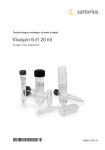
• Vivaspin 20 (Sartorius); PES; 50k Da
Recommended protocols
Sample
Treatment Buffer *
Capture Beads **
Total
100 µl
100 µl
Western blotting 100 µl
200 µl
250 µl
250 µl
Flow cytometry 50 µl
500 µl
500 µl
500 µl
qPCR for microRNA 500 µl
1000 µl
1000 µl
1000 µl
(remove dispersion liquid before use)
2000 µl
* We recommend to substitute treatment buffer with washing/dilution buffer for low-protein sample.
** This is a recommended volumes for a standard usage. It should be adapted according target abundance and application.
www.diagenode.com |
PAGE 6
DIAGENODE EXOIP USER MANUAL
Protocols
1. Cell culture guidance
The two cell lines have been used as models for the characterization of the exosomes: HeLa & HEK293T. The cell culture
was conducted into traditional cell culture flasks. The possibility of switching to a bioreactor culture is of great advantage
since it can possibly greatly reduce the main concern of the workflow that is the low exosome recovery in the final
preparation. In this case, we will recommend using the bioreactors from Integra.
The cell culture is actually the critical point and cannot be resume by a standardization.
Here is some guidance about how cell culture can drastically change the exosome secretion:
• The exosome secretion depends directly of the cell type considered.
(For example, it is generally known that endothelial, dendritic and immune cells are cell types abundantly
secreting exosomes)
• The medium chosen for the cell culture may influence the exosome secretion of the cells since exosomes are a
way of intercellular communications. This communication may change if the growing environment is changing
itself
• The time of secretion obviously play a role in the quantity of exosomes that will be recovered when harvesting the
conditioned culture media. Usually, the research papers are describing a secretion time varying from 24h to 72h.
The user will have to determine the optimal secretion time for his cell culture since not all the cells can handle a
long secretion time of 72 hours.
• For the secretion period during the cell culture, FBS depleted extracellular vesicules (EV’s) has to be used in
the culture medium. Either culture medium without any serum at all or culture medium complemented with
serum but depleted from EV’s. The depletion of the serum from extracellular vesicles is usually performed by
ultracentrifugation.
The following depletion protocol was used to eliminate EV’s out of the Fetal Bovine Serum (FBS):
• Ultracentrifuge: Optima L-90K (Beckman Coulter); Rotor: SW32
• Centrifugation 18 hours (overnight) at +4°C with a speed of 110.000g
• The serum has first to be diluted in culture medium in order to decrease its inherent viscosity that may hamper
the exosomes depletion by centrifugation → pelleting efficiency. A dilution factor of 2.4 was used for our depletion
protocol (50 ml of serum in 120 ml complete medium).
• After the depletion by ultracentrifugation, the resulting serum diluted in medium is sterilized through a 0.2 µm
filter.
2. Pre-enrichment/concentration
The aim of the concentration step is to increase the exosome quantity in the sample of interest. This is particularly
important for cell culture supernatant that may not contain quantity of exosomes since some cell lines may be poor at
extracellular vesicles secretion.
For other kind of samples such as serum or plasma, this step can be avoided since it may already contain enough
exosomes for the subsequent immunocapture and the following downstream analyses.
Two important factors have to be considered before performing the concentration:
1. Do my cells secrete a lot of extracellular vesicles? If not, pre-enrichment/concentration will be done.
2. Do I need a lot of exosomes for the downstream analyses that I plan to perform?
Several pre-enrichment/concentration techniques can be considered such as:
• Ultracentrifugation
• Size exclusion chromatography (SEC)
• Precipitation with volume-excluding polymer (such as PEG)
Innovating Epigenetic Solutions
PAGE 7
All of these techniques are suitable for the pre-enrichment of the exosome fraction in the sample and can be used
before the selective immunocapture but based on our quality criteria, ultrafiltration seems to be the best compromise
to concentrate exosomes.
Ultrafiltration (UF) can yield to highly concentrated exosome preparations with a great efficiency in comparison to other
well-known techniques to only can enrich a sub-fraction of the exosome population or even damage the integrity of the
vesicles.
To perform a concentration, we recommend the Vivaspin columns from Sartorius. Amicon columns from Millipore are
also a suitable device but have been less extensively used during the design of those kits.
Using the right UF column is of the upmost importance! That’s why it’s important to respect the following advices:
• Membrane chemistry : Polyethersulfone (PES) or cellulose acetate (CA)
• Membrane molecular weight cut-off (MWCO) : 50k Da or 100k Da but not lower or higher since lower cut-off will
result in an increase of retained soluble proteins that may hamper the further immunocapture and higher can
lead to an exosome loss.
• Wash and hydrate the membrane before loading the sample
• Use centrifugation based devices and not pressure driven units
Here is the protocol used by the research team during the design of the kits:
• Wash the column with 20 ml PBS(1X) at 3000g for 20 minutes at room temperature
• Saturate the membrane and the housing with 20 ml of 1% BSA solution diluted in PBS (1X). The saturation is
performed overnight at room temperature and has for aim to decrease the effect of non-specific binding of the
proteins and thus the exosomes to the column.
• After the overnight passivation, wash the membrane manually by pipetting with 1 or 2 ml of PBS (1X)
• Wash the membrane with 20 ml PBS (1X) with a 20’ centrifugation at 3000g at room temperature.
• The sample can be loaded on the membrane. This sample is called conditioned culture medium (CCM) because
it contains now exosomes ready to be processed. Before loading it on the ultrafiltration membrane, a few steps
have to be done:
1. Centrifuge the CCM at 3000g for 30’ at room temperature in order to pellet the floating cells and large debris.
2. Filter the resulting supernatant through a 0.2 µm cut-off in order to deplete the filtrate from large vesicles or
aggregates. Pressure is again one important factor to take into consideration since vesicles even larger than
200 nm can pass through the filter or break down if the pressure applied is too important.
The concentration factor depends on the sample volume input but it is preferable not to reach a high fold, above 50X, to
avoid exosome aggregation, vesicle damage or loss. The final concentrate volume for one sample to be processed for
the immunocapture needs to be less than 1 ml.
For those reasons, a 20X concentration fold was chosen.
To reach this concentration, a speed of 3000g during 30’ at room temperature can be applied. It is not advised to increase
the centrifugation speed to reach earlier the desired fold since it can cause vesicles aggregation or vesicles trapping
inside the pores of the membrane
• Then, the concentrate can be recovered manually by pipetting and stored for the day at +4°C before processing it
for immunocapture. It is not advised to keep the concentrated conditioned culture medium for more than several
hours at +4°C since it can lead to exosome degradation. For the same reason, it is better not to freeze the sample
before processing it to immunocapture.
3. Immunocapture protocol
Advices
Use fresh samples prior immunocapture since freezing may damage the surface integrity of the vesicles and thus
decrease the binding efficiency
Always prefer a long incubation period at +4°C. Optimal binding efficiency can be reached between 21 to 24 hours
incubation.
www.diagenode.com |
PAGE 8
DIAGENODE EXOIP USER MANUAL
It is important to use 2 ml tube for proper mixing conditions during the incubation period
Protocole 1. Cell and Debris Pre-Clearance Procedure
1. Prepare the appropriate size tubes for your sample.
2. Dispense the sample into tube.
3. Centrifuge the tubes at 300 x g at 4°C for 10 minutes.
4. Transfer supernatant to a new tube.
5. Centrifuge the tubes at 2000 x g at 4°C for 20 minutes.
6. Transfer supernatant to a new tube.
7. Centrifuge the tubes at 10,000 x g at 4°C for 30 minutes.
8. (Optional) Filter the final supernatant with a 0.22 μm filter unit.
9. The sample is now ready for immediate use with ExoIP kit or storage at -80°C.
Protocole 2. Exosome Capture Procedure for Western blotting
(Example: 100 μL beads, 1 mL sample. We recommended to adjust capture beads amount according to your target
abundance)
1. Set 2 mL microfuge tubes on a magnetic tube stand.
2. Suspend ExoIP magnetic capture beads well using a vortexer and put 100 μL of the suspension into each microfuge
tubes per sample.
3. Place the tube on the magnetic tube stand for about a minute and remove the supernatant.
4. Add 1000 μL of treatment buffer for serum and plasma, or washing/dilution buffer for cell culture supernatant.
5. Add 1000 μL of sample that has been cleared of cells and debris. (See above protocol 1. Cell and Debris PreClearance Procedure)
6. Incubate the sample for 20 minutes - 24 hours at room temperature or 4°C with gentle mixing. (Note: the optimal
reaction time and temperature may depend on the target abundance and stability.)
7. Briefly spin the tube to remove beads from the top of the tube.
8. Place the tube on the magnetic tube stand for about a minute and remove the supernatant.
9. Wash the beads 2 times with 500 µL washing/dilution buffer. Mix the beads briefly but thoroughly. After 2 times
washing, resuspend with 500 µL washing/dilution buffer and transfer whole this buffer containing ExoIP magnetic
capture beads to a fresh tube.
10. Place the tube on the magnetic tube stand for about a minute and remove the supernatant carefully.
Move to Protocole 3. Preparation Protocol for SDS-PAGE or 2.4 section. Exosome Elution Procedure.
Option: In order to elute exosome proteins for Western blotting, you may add the appropriate lysis buffer directly to the
beads according to your preferred method/kit. Place the tube on the magnetic tube stand for about a minute and collect
the eluted sample.
Protocole 3. Preparation Protocol for SDS-PAGE
Continued from protocol 2
1. Add 20 µL of 1x sample buffer to the magnetic beads and mix well.
2. Incubate 95°C for 5 minutes.
3. Vortex and spin down.
4. Place the tube on magnetic tube stand about a minute.
5. Apply the total supernatant to a lane of SDS-PAGE.
Innovating Epigenetic Solutions
PAGE 9
Protocole 4. Exosome Capture Procedure for flow cytometry
(Example: 50 μL beads, 300 µL sample. We recommended to titrate the magnetic beads amount according to your target
abundance.)
1. Set 2 mL microfuge tubes on a magnetic tube stand.
2. Suspend ExoIP magnetic capture beads well using a vortexer and put 50 μL of the suspension into each microfuge
tubes per sample.
3. Place the tube on the magnetic tube stand for about a minute and remove the supernatant.
4. Add 300 μL of treatment buffer for serum and plasma, or washing/dilution buffer for cell culture supernatant.
5. Add 300 μL of sample that has been cleared of cells and debris. (Note 1: please see above protocol 1. Cell and
Debris Pre-Clearance Procedure, Note 2: Customer may use as little as 100 μL sample from according to target
abundance.)
6. Incubate the sample for 24 hours at room temperature or 4°C with gentle mixing. (Note: the optimal reaction time
and temperature vary with the target abundance.)
7. Briefly spin the tube to remove beads from the top of the tube.
8. Place the tube on the magnetic tube stand for about a minute and remove the supernatant.
9. Wash the beads 2 times with 250 µL washing/dilution buffer and resuspend in 300 µL PBS or your preferred buffer.
Protocole 5. Preparation of exosome-bead complexes for flow cytometer Analysis
Continued from protocol 4
1. Add optimized volume of fluorescent antibody of a target or isotype control to 100 μL of prepared exosome-beads
complex. Mix gently by rotator for 1 hour. (Note 1: titration of antibody conditions are important for optimal signal.)
2. Place the tube on the magnetic tube stand for about a minute and remove the supernatant.
3. Wash the beads 1 time with 300 µL PBS or your preferred buffer.
4. Add 300 μL of PBS or your preferred buffer for flow cytometry.
5. Perform flow cytometer analysis.
Protocole 6. Exosome Capture Procedure for quantitative PCR
We recommend 1 mL sample volume. Samples less than 1 mL may deteriorate Ct value. Recommended Capture Beads
amount for qPCR sample preparation. You may titrate the magnetic beads amount according to your target abundance.
1. Set 2 mL microfuge tubes on a magnetic tube stand.
2. Suspend ExoIP magnetic capture beads well using a vortexer and put 500 μL of the suspension into each microfuge
tube per sample.
3. Place the tube on the magnetic tube stand for about a minute and remove the supernatant.
4. Add 1000 μL of treatment buffer for serum and plasma, or washing/dilution buffer for cell culture supernatant.
5. Add 1000 μL of sample that has been cleared of cells and debris. (Note 1: please see above protocol 1. Cell and
Debris Pre-Clearance Procedure), Note 2: Customer may use as little as 100 μL sample from according to target
abundance.)
6. Incubate the sample for 24 hours at room temperature with gentle mixing. (Note: the optimal reaction time and
temperature vary with the target abundance.)
7. Briefly spin the tube to remove beads from the top of the tube.
8. Place the tube on the magnetic tube stand for about a minute and remove the supernatant.
9. Wash the beads 2 times with 500 µL washing/dilution buffer. Mix the beads briefly but thoroughly. Re-suspend
with 500 µL and transfer the final washed beads to a fresh tube.
www.diagenode.com |
PAGE 10
DIAGENODE EXOIP USER MANUAL
10. Place the tube on the magnetic tube stand for about a minute and remove the supernatant carefully.
11. Exosomes bound with ExoIP magnetic capture beads are ready for nucleic acids isolation.
4. Exosome Elution
After the immunocapture step, the samples can be recovered by a quick spin. A pre-centrifugation is preferred before
the magnetic capture of the beads since it will greatly reduce the risks of losing beads in the supernatant and reduce the
time to attract all the beads dispersed in the aqueous medium to the magnet.
A quick high speed centrifugation (9000g during 30’’ at room temperature) seems to be a good way to pellet all the beads
to the bottom of the tube prior magnetic capture.
Two washing steps with the wash/dilution buffer are good to clean-up the preparation after the immunocapture. It is
not necessary to perform more washing steps and this can be a source of potential beads loss and thus exosomes loss.
When the beads pellet is properly washed, the exosomes are then ready to be processed either for the elution or for the
lysis:
Exosome Elution Procedure
Continued from Protocol 2. Exosome Capture Procedure for Western blotting
1. Add 25-50 μL of exosome elution buffer to resuspend the beads. Mix gently by pipetting (do not use a vortexer).
2. Incubate the beads without mixing for 3 minutes at room temperature whitout shaking
3. Place the tube on the magnetic tube stand for about a minute and transfer the supernatant to a fresh tube.
4. Immediately proceed to exchange the exosome elution buffer to washing/dilution buffer or your preferred buffer,
such as PBS etc. The recommended buffer exchange is a diafiltration protocol using Amicon 0.5 3kDa columns.
Vivaspin 0.5 3k Da are also suitable! but less used during our experiments
a. Load the eluted exosomes properly diluted with PBS 1X to a final volume of 500 µl on the Amicon column
b. Operate centrifugation cycles at 14000g at room temperature until a dilution factor of 50X is reached for the
elution buffer
25 µl elution buffer (one sample) + 475 µl PBS 1X : 20X dilution
Centrifuge 10’ 14000g RT → concentrate = 114 µl
114 µl + 386 µl PBS 1X → dilution factor of 4.3X → 20*4.3 = 86X for the initial amount of elution buffer → above
50X dilution : exosomes are safe !
Centrifuge one last time to reach to desired volume of concentrate
c. Recover manually by pipetting the concentrate containing the exosomes
Note: Exosome elution buffer contains a denaturing agent. The exosome elution buffer may interfere with
downstream applications unless diluted to 20-100 times or exchanged with washing/dilution buffer or your
preferred buffer. Washing/dilution buffer contains a synthetic polymer to reduce adsorption of exosomes on tubes
and buffer exchange devices. However, the polymer may disturb certain downstream analysis of the exosomes.
Exosome direct lysis on the beads Procedure
For one sample:
1. Add 10-15 µl of RIPA 1X + protease inhibitors to the beads pellet.
2. Incubate 15’ at +4°C.
3. Recover the supernatant containing the lysed exosomes.
4. Process it directly for downstream analysis or store it at -80°C until further use.
Innovating Epigenetic Solutions
PAGE 11
FOR RESEARCH USE ONLY.
Not intended for any animal or human therapeutic or diagnostic use.
© 2014 Diagenode SA. All rights reserved. No part of this publication may be reproduced, transmitted, transcribed,
stored in retrieval systems, or translated into any language or computer language, in any form or by any means:
electronic, mechanical, magnetic, optical, chemical, manual, or otherwise, without prior written permission from
Diagenode SA (hereinafter, "Diagenode”) . The information in this guide is subject to change without notice. Diagenode
and/or its affiliates reserve the right to change products and services at any time to incorporate the latest technological
developments. Although this guide has been prepared with every precaution to ensure accuracy, Diagenode and/or its
affiliates assume no liability for any errors or omissions, nor for any damages resulting from the application or use of this
information. Diagenode welcomes customer input on corrections and suggestions for improvement.
NOTICE TO PURCHASER
LIMITED LICENSE
The information provided herein is owned by Diagenode and/or its affiliates. Subject to the terms and conditions that govern
your use of such products and information, Diagenode and/or its affiliates grant you a nonexclusive, nontransferable, nonsublicensable license to use such products and information only in accordance with the manuals and written instructions
provided by Diagenode and/or its affiliates. You understand and agree that except as expressly set forth in the terms and
conditions governing your use of such products, that no right or license to any patent or other intellectual property owned
or licensable by Diagenode and/or its affiliates is conveyed or implied by providing these products. In particular, no right
or license is conveyed or implied to use these products in combination with any product not provided or licensed to you
by Diagenode and/or its affiliates for such use.
Limited Use Label License: Research Use Only The purchase of this product conveys to the purchaser the limited, nontransferable right to use the product only to perform internal research for the sole benefit of the purchaser. No right
to resell this product or any of its components is conveyed expressly, by implication, or by estoppel. This product is for
internal research purposes only and is not for use in commercial applications of any kind, including, without limitation,
quality control and commercial services such as reporting the results of purchaser's activities for a fee or other form of
consideration. For information on obtaining additional rights, please contact [email protected].
TRADEMARKS
The trademarks mentioned herein are the property of Diagenode or their respective owners. Bioruptor is a registered
trademark of Diagenode SA.
Bioanalyzer is a trademark of Agilent Technologies, Inc. Agencourt and AMPure are registered trademarks of Beckman
Coulter, Inc. Microcon is a registered trademark of Millipore Inc. Illumina is a registered trademark of Illumina Inc. Ion
Torrent and Personal Genome Machine are trademarks of Life Technologies Corporation. Qubit is a registered trademark
of Life Technologies Corporation.
POWERED BY JSR
www.diagenode.com |
DIAGENODE
HEADQUARTERS
DIAGENODE S.A.
BELGIUM | EUROPE
DIAGENODE INC.
USA | NORTH AMERICA
LIEGE SCIENCE PARK
Rue Bois Saint-Jean, 3
4102 Seraing - Belgium
Tel: +32 4 364 20 50
Fax: +32 4 364 20 51
[email protected]
[email protected]
400 Morris Avenue, Suite #101
Denville, NJ 07834
Tel: +1 862 209-4680
Fax: +1 862 209-4681
[email protected]
[email protected]
FOR A COMPLETE LISTING OF
DIAGENODE’S INTERNATIONAL
DISTRIBUTORS VISIT:
http://www.diagenode.com/company/
distributors.php
For rest of the world, please contact
Diagenode sa.
www.diagenode.com
© 2015 Diagenode, Inc. All rights reserved. The content of this document cannot be reproduced without prior permission of the authors. Bioruptor & IP-Star are registered trademarks of Diagenode
MA-ExoIP-V1-10_15


