1
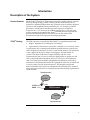
pBAD/TOPO® ThioFusion™ Expression Kit Five-minute cloning of Taq polymerase-amplified PCR products for soluble, regulated expression and purification in E. coli Catalog no. K370-01 Revision date : 21 July 2009 Manual part no. 25-0257 MAN0000095 Corporate Headquarters Invitrogen Corporation 1600 Faraday Avenue Carlsbad, CA 92008 T: 1 760 603 7200 F: 1 760 602 6500 E: [email protected] For country-specific contact information visit our web site at www.invitrogen.com User Manual ii Contents Kit Contents and Storage........................................................................................................................... iv Introduction ................................................................................................................... 1 Description of the System............................................................................................................................1 Experiment Outline......................................................................................................................................3 Methods ......................................................................................................................... 4 Producing PCR Products.............................................................................................................................6 TOPO® Cloning Reaction and Transformation ........................................................................................7 Optimizing the TOPO® Cloning Reaction...............................................................................................12 Expressing the PCR Product .....................................................................................................................13 Analyzing Samples.....................................................................................................................................16 Purifying Recombinant Protein ................................................................................................................18 Appendix...................................................................................................................... 20 Recipes .........................................................................................................................................................20 Purifying the PCR Products ......................................................................................................................22 Adding 3´ A-Overhangs Post-Amplification..........................................................................................24 pBAD/TOPO® ThioFusion™ Control Reactions.....................................................................................25 Troubleshooting..........................................................................................................................................27 Map and Features of pBAD/Thio-TOPO® ..............................................................................................28 Map of pBAD/Thio....................................................................................................................................30 Regulation by Arabinose ...........................................................................................................................31 Accessory Products ....................................................................................................................................32 Technical Support.......................................................................................................................................34 Purchaser Notification ...............................................................................................................................35 References....................................................................................................................................................37 iii Kit Contents and Storage Shipping and Storage The pBAD/TOPO® ThioFusion™ Expression Kit is shipped on dry ice. Each kit contains a box with pBAD/Thio TOPO® TA Cloning® reagents (Box 1), a box with One Shot® TOP10 Chemically Competent E. coli (Box 2), and a stab of LMG194. Store Box 1 at –20°C and Box 2 at –80°C. Store the LMG194 stab at 4°C. pBAD/Thio TOPO® TA Cloning® Reagents pBAD/Thio TOPO® TA Cloning® reagents (Box 1) are listed below. Note that you must supply the Taq polymerase. Store Box 1 at –20°C. Item Concentration ® Amount pBAD/Thio-TOPO vector, linearized 10 ng/μL plasmid DNA in: 50% glycerol 50 mM Tris-HCl, pH 7.4 (at 25°C) 1 mM EDTA 2 mM DTT 0.1% Triton X-100 100 μg/mL BSA 30 M phenol red 20 reactions 10X PCR Buffer 100 mM Tris-HCl, pH 8.3 (at 42°C) 500 mM KCl 25 mM MgCl2 0.01% gelatin 100 μL dNTP Mix 12.5 mM dATP 12.5 mM dCTP 12.5 mM dGTP 12.5 mM dTTP neutralized at pH 8.0 in water 10 μL Salt Solution 1.2 M NaCl 0.06 M MgCl2 50 μL 20% L-Arabinose 20% in sterile water 1 mL Trx Forward Sequencing Primer 0.1 μg/μL in TE Buffer, pH 8 20 μL pBAD Reverse Sequencing Primer 0.1 μg/μL in TE Buffer, pH 8 20 μL Control PCR Primers 0.1 μg/μL in TE Buffer, pH 8 10 μL Control PCR Template 0.05 μg/μL in TE Buffer, pH 8 10 μL Continued on next page iv Kit Contents and Storage, Continued pBAD/Thio TOPO® TA Cloning® Reagents, continued Item Sequences of the Primers Concentration Sterile Water -- 1 mL Expression Control Plasmid (pBAD/Thio, supercoiled) 500 ng/μL in TE buffer, pH 8 10 μL The table below provides the sequences of the Trx Forward and pBAD Reverse sequencing primers. Two micrograms of each primer are supplied. Primer One Shot® Reagents Sequence pMoles Supplied Trx Forward 5´-TTCCTCGACGCTAACCTG-3´ 371 pBAD Reverse 5´-GATTTAATCTGTATCAGG-3´ 363 The table below describes the items included in the One Shot® TOP10 Chemically Competent E. coli kit. Store at –80°C. Item Genotype of TOP10 Amount Composition Amount 21 50 μL TOP10 cells -- SOC Medium (may be stored at room temperature or 4°C) 2% Tryptone 0.5% Yeast Extract 10 mM NaCl 2.5 mM KCl 10 mM MgCl2 10 mM MgSO4 20 mM glucose 6 mL pUC19 Control DNA 10 pg/μL in 5 mM TrisHCl, 0.5 mM EDTA, pH 8.0 50 μL TOP10: Use this strain for general cloning and expression of PCR products in pBAD/Thio-TOPO®. This strain cannot be used for single-strand rescue of DNA. F- mcrA (mrr-hsdRMS-mcrBC) 80lacZM15 lac74 recA1 araD139 (araleu)7697 galU galK rpsL (StrR) endA1 nupG Continued on next page v Kit Contents and Storage, Continued Genotype of LMG194 F- lacX74 gal E thi rpsL phoA (Pvu II) ara714 leu::Tn10. Storing LMG194 Stab The LMG194 E. coli cells supplied as a stab with the kit are guaranteed until the expiration date marked on tube when stored at 4°C. Note: This strain is deleted for araBADC, and is streptomycin and tetracycline resistant. Upon receipt, we recommend that you prepare a set of glycerol master stocks within two weeks of receiving the kit. To prepare 5–10 glycerol master stocks for long-term storage: 1. Streak a small portion of the LMG194 cells that you have received as a stab on an LB plate containing the appropriate antibiotics. 2. Invert the plate and incubate at 37°C overnight. 3. Isolate a single colony and inoculate into 5–10 mL of LB medium containing the appropriate antibiotics. 4. Grow the culture to stationary phase (OD600 = 1–2). 5. Mix 0.8 mL of culture with 0.2 mL of sterile glycerol and transfer to a cryovial. Store at –80°C. Use one master stock to create working stocks for regular use. vi Introduction Description of the System Product Features pBAD/TOPO® ThioFusion™ Expression Kit provides a highly efficient, 5-minute, one-step cloning strategy ("TOPO® Cloning") for the direct insertion of Taq polymerase-amplified PCR products into a plasmid vector for soluble, regulated expression and simplified protein purification in E. coli. No ligase, post-PCR procedures, or PCR primers containing specific sequences are required. Expression in E. coli is driven by the araBAD promoter (PBAD). The AraC gene product encoded on the pBAD/Thio-TOPO® plasmid positively regulates this promoter. Recombinant proteins are expressed as fusions to His-Patch thioredoxin for high-level expression and simple purification. TOPO® Cloning The PCR expression vector (pBAD/Thio-TOPO®) is supplied linearized with: Single 3´ thymidine (T) overhangs for TA Cloning® Topoisomerase I bound to the vector (this is referred to as “activated” vector) Taq polymerase has a nontemplate-dependent terminal transferase activity that adds a single deoxyadenosine (A) to the 3´ ends of PCR products. The linearized vector supplied in this kit has single, overhanging 3´ deoxythymidine (T) residues. This allows PCR inserts to ligate efficiently with the vector. Topoisomerase I from Vaccinia virus binds to duplex DNA at specific sites and cleaves the phosphodiester backbone after 5-CCCTT in one strand (Shuman, 1991). The energy from the broken phosphodiester backbone is conserved by formation of a covalent bond between the 3 phosphate of the cleaved strand and a tyrosyl residue (Tyr-274) of topoisomerase I. The phospho-tyrosyl bond between the DNA and enzyme can subsequently be attacked by the 5 hydroxyl of the original cleaved strand, reversing the reaction and releasing topoisomerase (Shuman, 1994). Topoisomerase Tyr-274 O CCCTT GGGA P OH A PCR Product HO Tyr-274 O A AGGG TTCCC P Topoisomerase Continued on next page 1 Description of the System, Continued Regulation of Expression by Arabinose In the presence of arabinose, expression from PBAD is induced while only very low levels of transcription are observed from PBAD in the absence of arabinose (Lee, 1980; Lee et al., 1987). Uninduced levels are repressed even further by growth in the presence of glucose (0.1% to 0.2%). Glucose reduces the levels of 3´, 5´-cyclic AMP, lowering expression from the catabolite-repressed PBAD promoter (Miyada et al., 1984). By varying the concentration of arabinose, protein expression levels can be optimized to ensure maximum expression of protein. In addition, the tight regulation of PBAD by AraC is useful for expression of potentially toxic or essential genes (Carson et al., 1991; Dalbey and Wickner, 1985; Guzman et al., 1992; Kuhn and Wickner, 1985; Russell et al., 1989; San Millan et al., 1989). For more information on the mechanism of expression and repression of the ara regulon, see page 30 or refer to Schleif, 1992. Thioredoxin The 11.7 kDa thioredoxin protein is found in yeast, plants, and mammals, as well as in bacteria. It was originally isolated from E. coli as a hydrogen donor for ribonuclease reductase (for a review, see Holmgren, 1985 ). The gene has been completely sequenced (Wallace and Kushner, 1984). The protein has been crystallized and its three-dimensional structure determined (Katti et al., 1990). When overexpressed in E. coli, thioredoxin is able to accumulate to approximately 40% of the total cellular protein and still remains soluble. Thioredoxin is used to increase translation efficiency, and in some cases, solubility, of eukaryotic proteins expressed in E. coli. Murine interleukin-2, human interleukin-3, murine interleukin-4, murine interleukin-5, human macrophage-colony stimulating factor, murine steel factor, murine leukemia inhibitory factor and human bone morphogenetic protein-2 are some of the proteins that have been produced as soluble C-terminal fusions to the thioredoxin protein in E. coli (LaVallie et al., 1993). His-Patch Thioredoxin To create a metal binding domain in the thioredoxin protein, the glutamate residue at position 32 and the glutamine residue at position 64 were mutated to create histidine residues. When His-Patch thioredoxin folds, the histidines at positions 32 and 64 interact with a native histidine at position 8 to form a "patch". This histidine patch was shown to have high affinity for divalent cations (Lu et al., 1996). His-Patch thioredoxin (HP-thioredoxin) proteins can therefore be purified on metal-chelating resins (e.g., ProBond™) See page 33 for ordering information. 2 Experiment Outline Experiment Flowchart The flow chart below describes the general steps needed to amplify, TOPO® Clone, and express your protein of interest. Design Primers for PCR Produce PCR product TOPO® Cloning Reaction: Mix together PCR product and pBAD/Thio-TOPO® vector Incubate 5 minutes at room temperature Transform into TOP10 E. coli cells Select and analyze colonies Select a positive transformant and induce expression with arabinose 3 Methods Designing PCR Primers Introduction It is important to properly design your PCR primers to ensure that you obtain the recombinant protein you need for your studies. Use the information below and the diagram on the next page to design your PCR primers. Considerations pBAD/Thio-TOPO® is designed with some specific features to facilitate expression. They are: The initiation ATG is correctly spaced from the optimized ribosome binding site to ensure optimal translation HP-thioredoxin acts as a translation leader to facilitate high-level expression and in some cases, solubility Note: You can remove HP-thioredoxin after protein purification using enterokinase (i.e., EKMax™, see page 19). Primer Design Suggestions for primer design are provided in the table below. Remember that your PCR product will have single 3´ adenine overhangs. If you wish to.... clone in frame with thioredoxin Then... the forward PCR primer must be designed to ensure that your protein is in frame with the N-terminal leader peptide. include the V5 epitope and polyhistidine region the reverse PCR primer must be designed to remove the native stop codon in the gene of interest and preserve the reading frame through the C-terminal tag. NOT include the V5 epitope and polyhistidine region include the native sequence containing the stop codon in the reverse primer or make sure the stop codon is upstream from the reverse PCR primer binding site. remove the N-terminal leader (for expressing truly native protein) the forward PCR primer can be designed to include a unique Nco I site which contains the first ATG of the protein. Example: 5´-ACC ATG G.... The vector can be digested with Nco I after cloning and religated, assuming there are no internal Nco I sites in your PCR product. Note: Removing the N-terminal leader generally decreases expression levels. When synthesizing PCR primers, do not add 5’ phosphates to the primers, because 5’ phosphates prevent the synthesized PCR product from ligating into the pBAD-TOPO® vector. Continued on next page 4 Designing PCR Primers, Continued TOPO® Cloning Site The diagram below is supplied to help you design appropriate PCR primers to correctly clone and express your PCR product. Restriction sites are labeled to indicate the actual cleavage site. The complete sequence of the vector is available for downloading at www.invitrogen.com or from Technical Support (page 34). O2 Region 1 AAGAAACCAA TTGTCCATAT TGCATCAGAC ATTGCCGTCA CTGCGTCTTT TACTGGCTCT TCTCGCTAAC CAAACCGGTA 81 ACCCCGCTTA TTAAAAGCAT TCTGTAACAA AGCGGGACCA AAGCCATGAC AAAAACGCGT AACAAAAGTG TCTATAATCA pBAD Forward priming site O1 Region CAP binding site 161 CGGCAGAAAA GTCCACATTG ATTATTTGCA CGGCGTCACA CTTTGCTATG CCATAGCATT TTTATCCATA AGATTAGCGG -35 I2 and I1 Region -10 241 ATCCTACCTG ACGCTTTTTA TCGCAACTCT CTACTGTTTC TCCATACCCG TTTTTTTGGG CTAGAAATAA TTTTGTTTAA 321 CTTTAAGAAG GAGATATACA TACCC ATG GGA TCT GAT AAA ATT ATT CAT CTG ACT GAT GAT TCT TTT GAT Met Gly Ser Asp Lys Ile Ile His Leu Thr Asp Asp Ser Phe Asp RBS 391 Nco I HP-Thioredoxin translational start site HP-Thioredoxin ORF (indicated by italicized amino acids) ACT GAT GTA CTT AAG GCA GAT GGT GCA ATC CTG GTT GAT TTC TGG GCA CAC TGG TGC GGT CCG TGC Thr Asp Val Leu Lys Ala Asp Gly Ala Ile Leu Val Asp Phe Trp Ala His Trp Cys Gly Pro Cys 457 AAA ATG ATC GCT CCG ATT CTG GAT GAA ATC GCT GAC GAA TAT CAG GGC AAA CTG ACC GTT GCA AAA Lys Met Ile Ala Pro Ile Leu Asp Glu Ile Ala Asp Glu Tyr Gln Gly Lys Leu Thr Val Ala Lys 523 CTG AAC ATC GAT CAC AAC CCG GGC ACT GCG CCG AAA TAT GGC ATC CGT GGT ATC CCG ACT CTG CTG Leu Asn Ile Asp His Asn Pro Gly Thr Ala Pro Lys Tyr Gly Ile Arg Gly Ile Pro Thr Leu Leu 589 CTG TTC AAA AAC GGT GAA GTG GCG GCA ACC AAA GTG GGT GCA CTG TCT AAA GGT CAG TTG AAA GAG Leu Phe Lys Asn Gly Glu Val Ala Ala Thr Lys Val Gly Ala Leu Ser Lys Gly Gln Leu Lys Glu 655 TTC CTC GAC GCT AAC CTG GCC GGC TCT GGA TCC GGT GAT GAC GAT GAC AAG CTC GAG Phe Leu Asp Ala Asn Leu Ala Gly Ser Gly Ser Gly Asp Asp Asp Asp Lys Leu BseR I V5 epitope GGC GAG CTT GAA GGT AAG CCT ATC CCT AAC CCT CTC CTC GGT CTC GAT TCT ACG Gly Glu Leu Glu Gly Lys Pro Ile Pro Asn Pro Leu Leu Gly Leu Asp Ser Thr Trx Forward priming site 721 Polyhistidine region NgoM I Nae I Enterokinase recognition site Pme I Enterokinase cleavage site GCC CTT CGG GA A Ala Leu PCR Product A AG TTC Lys CGT ACC GGT CAT Arg Thr Gly His pBAD Reverse 787 CAT CAC CAT CAC CAT TGA GTTTAAACG GTCTCCAGCT TGGCTGTTTT GGCGGATGAG AGAAGATTTT CAGCCTGATA His His His His His *** priming site 861 CAGATTAAAT CAGAACGCAG AAGCGGTCTG ATAAAACAGA ATTTGCCTGG CGGCAGTAGC GCGGTGGTCC CACCTGACCC 941 CATGCCGAAC TCAGAAGTGA AACGCCGTAG CGCCGATGGT AGTGTGGGGT CTCCCCATGC GAGAGTAGGG AACTGCCAGG 1021 CATCAAATAA AACGAAAGGC TCAGTCGAAA GACTGGGCCT TTCGTTTTAT CTGTTGTTTG TCGGTGAACG CTCTCCTGAG rrnB T1 and T2 transcriptional terminator 5 Producing PCR Products Introduction Once you have decided on a PCR strategy and have synthesized the primers you are ready to produce your PCR product. Materials Supplied by the User Taq polymerase Note: For improved specificity and higher yields, we recommend using Platinum® Taq DNA Polymerase available from Invitrogen (see page 32 for ordering information) to generate your PCR product. Thermocycler DNA template and primers to produce your PCR product Note: dNTPs (adjusted to pH 8) are provided in the kit. Polymerase Mixtures If you wish to use a mixture containing Taq polymerase and a proofreading polymerase, Taq must be used in excess of a 10:1 ratio to ensure the presence of 3´ A-overhangs on the PCR product. If you use polymerase mixtures that do not have enough Taq polymerase or a proofreading polymerase only, you can add 3 A-overhangs using the method on page 24. Producing PCR Products 1. Set up the following 50 μL PCR reaction. Use less DNA if you are using plasmid DNA as a template and more DNA if you are using genomic DNA as a template. Use the cycling parameters suitable for your primers and template. Be sure to include a 7 to 30 minute extension at 72°C after the last cycle to ensure that all PCR products are full length and 3´ adenylated. DNA Template 10–100 ng 10X PCR Buffer 5 μL 50 mM dNTPs Primers Sterile water Taq Polymerase (1 unit/μL) Total Volume 2. 0.5 μL 100–200 ng each add to a final volume of 49 μL 1 μL 50 μL Check the PCR product by agarose gel electrophoresis. You should see a single, discrete band. If you do not see a single band, refer to the Note below. If you do not obtain a single, discrete band from your PCR, gel-purify your fragment before TOPO® Cloning (see page 22). Take special care to avoid sources of nuclease contamination and long exposure to UV light. Alternatively, optimize your PCR to eliminate multiple bands and smearing (Innis et al., 1990). The PCR Optimizer™ Kit from Invitrogen can help you optimize your PCR (see page 32 for ordering information). 6 TOPO® Cloning Reaction and Transformation Introduction TOPO® Cloning technology allows you to produce your PCR products, ligate them into pBAD/Thio-TOPO®, and transform the recombinant vector into E. coli in one day. It is important to have everything ready to use to ensure you obtain the best possible results. If this is the first time you have TOPO® Cloned, perform the control reactions on pages 25–26 in parallel with your samples. Experiments at Invitrogen demonstrate that inclusion of salt (200 mM NaCl, 10 mM MgCl2) in the TOPO® Cloning reaction increases the number of transformants 2- to 3-fold. We have also observed that in the presence of salt, incubation times of greater than 5 minutes can also increase the number of transformants. This is in contrast to earlier experiments without salt where the number of transformants decreases as the incubation time > 5 minutes. Including salt in the TOPO® Cloning reaction allows for longer incubation times, because it prevents topoisomerase I from rebinding and potentially nicking the DNA after ligating the PCR product and dissociating from the DNA. The result is more intact molecules leading to higher transformation efficiencies. Important We recommend adding salt to the TOPO® Cloning reaction. A stock salt solution is provided in the kit for this purpose. Note that the amount of salt added to the TOPO® Cloning reaction varies depending on whether you plan to transform chemically competent cells (provided) or electrocompetent cells (see below). For this reason, two different TOPO® Cloning reaction protocols are provided to help you obtain the best possible results. Chemically Competent E. coli For TOPO® Cloning and transformation into chemically competent E. coli, adding NaCl and MgCl2 to a final concentration of 200 mM NaCl, 10 mM MgCl2 in the TOPO® Cloning reaction increases the number of colonies over time. A Salt Solution (1.2 M NaCl; 0.06 M MgCl2) is provided to adjust the TOPO® Cloning reaction to the recommended concentration of NaCl and MgCl2. Electrocompetent E. coli For TOPO® Cloning and transformation of electrocompetent E. coli, salt must also be included in the TOPO® Cloning reaction, but the amount of salt must be reduced to 50 mM NaCl, 2.5 mM MgCl2 to prevent arcing. The Salt Solution is diluted 4-fold to prepare a 300 mM NaCl, 15 mM MgCl2 solution for convenient addition to the TOPO® Cloning reaction (see next page). Materials Supplied by the User 42°C water bath (or electroporator with cuvettes, optional) LB plates containing 50–100 μg/mL ampicillin (two for each transformation) 37°C shaking and non-shaking incubator Continued on next page 7 TOPO® Cloning Reaction and Transformation, Continued Preparing for Transformation Setting Up the TOPO® Cloning Reaction For each transformation, you need one vial of competent cells and two selective plates. Equilibrate a water bath to 42°C (for chemical transformation) or set up your electroporator if you are using electrocompetent E. coli. For electroporation, dilute a small portion of the Salt Solution 4-fold to prepare Dilute Salt Solution (e.g., add 5 μL of the Salt Solution to 15 μL of sterile water) Warm the vial of SOC medium from Box 2 to room temperature. Warm selective plates at 37°C for 30 minutes. For each transformation, thaw 1 vial of One Shot® cells on ice. The table below describes how to set up your TOPO® Cloning reaction (6 μL) for eventual transformation into chemically competent TOP10 One Shot® E. coli (provided) or electrocompetent E. coli. Refer to page 11 for additional information on optimizing the TOPO® Cloning reaction. An Insert:vector molar ratio of 1:1 gives the optimal efficiency in TOPO® Cloning reaction. Note: The red color of the TOPO® vector solution is normal and is used to visualize the solution. Reagent Chemically Competent E. coli Electrocompetent E. coli Fresh PCR product 0.5 to 4 μL 0.5 to 4 μL Salt Solution 1 μL -- Dilute Salt Solution -- 1 μL Sterile Water add to a total volume of 5 μL add to a total volume of 5 μL TOPO® vector 1 μL 1 μL Final Volume 6 μL 6 μL * Store all reagents at –20C when finished. Salt solutions and water can be stored at room temperature or 4C. Performing the TOPO® Cloning Reaction 1. Mix reaction gently and incubate for 5 minutes at room temperature. Note: For most applications, incubation for 5 minutes yields plenty of colonies for analysis. Depending on your needs, you can vary the length of the TOPO® Cloning reaction from 30 seconds to 30 minutes. For routine subcloning of PCR products, 30 seconds is sufficient. For large PCR products (> 1 kb) or if you are TOPO® Cloning a pool of PCR products, increasing the reaction time yields more colonies. 2. Place the reaction on ice and proceed to the One Shot® Chemical Transformation or Transformation by Electroporation (next page). Note: You may store the TOPO® Cloning reaction at –20C overnight. Continued on next page 8 TOPO® Cloning Reaction and Transformation, Continued One Shot® TOP10 Chemical Transformation 1. 2. Add 2 μL of the TOPO® Cloning reaction from Step 2 previous page into a vial of One Shot® Chemically Competent E. coli and mix gently. Do not mix by pipetting up and down. Incubate on ice for 5 to 30 minutes. Note: Longer incubations on ice do not seem to have any affect on transformation efficiency. The length of the incubation is at the user’s discretion (see page 12). Transformation by Electroporation 3. Heat-shock the cells for 30 seconds at 42°C without shaking. 4. Immediately transfer the tubes to ice. 5. Add 250 μL of room temperature SOC medium. 6. Cap the tube tightly and shake horizontally (200 rpm) at 37°C for 1 hour. 7. Spread 25–200 μL from each transformation on a prewarmed selective plate and incubate overnight at 37°C. We recommend that you plate two different volumes to ensure that at least one plate will have well-spaced colonies. 8. An efficient TOPO® Cloning reaction will produce hundreds of colonies. Pick ~10 colonies for analysis (see Analyzing Positive Clones, next page). 1. Add 2 μL of the TOPO® Cloning reaction into a 0.1 cm cuvette containing 50 l of electrocompetent E. coli and mix gently. Do not mix by pipetting up and down. Avoid formation of bubbles. Electroporate your samples using the protocol for your electroporator. 2. Note: If you have problems with arcing, see below. 3. Immediately add 250 μL of room temperature SOC medium. 4. Transfer the solution to a 15 mL snap-cap tube and shake for at least 1 hour at 37°C to allow expression of the antibiotic resistance gene. 5. Spread 10–50 μL from each transformation on a prewarmed selective plate and incubate overnight at 37°C. To ensure even spreading of small volumes, add 20 μL of SOC. We recommend that you plate two different volumes to ensure that at least one plate will have well-spaced colonies. 6. An efficient TOPO® Cloning reaction will produce hundreds of colonies. Pick ~10 colonies for analysis (see Analyzing Positive Clones, next page). Adding the Dilute Salt Solution in the TOPO® Cloning Reaction brings the final concentration of NaCl and MgCl2 in the TOPO® Cloning reaction to 50 mM and 2.5 mM, respectively. To prevent arcing of your samples during electroporation, the volume of cells should be between 50 and 80 μL for 0.1 cm cuvettes or 100 to 200 μL for 0.2 cm cuvettes. If you experience arcing during transformation, try one of the following: Reduce the voltage normally used to charge your electroporator by 10% Reduce the pulse length by reducing the load resistance to 100 ohms Ethanol-precipitate the TOPO® Cloning reaction and resuspend in water prior to electroporation Continued on next page 9 TOPO® Cloning Reaction and Transformation, Continued Analyzing Positive 1. Clones Alternative Method of Analysis Important Culture 10 colonies overnight in LB or SOB medium with 50–100 μg/mL ampicillin. 2. Isolate plasmid DNA using your method of choice. If you need ultra-pure plasmid DNA for automated or manual sequencing, we recommend using Invitrogen’s PureLink™ HQ Mini Plasmid Purification or PureLink™ HiPure Plasmid Miniprep kits (see page 32 for ordering information). Refer to www.invitrogen.com or contact Technical Support for more information on a large selection of plasmid purification columns. 3. Because the PCR product will clone in either direction, analyze for orientation as well as insertion by restriction analysis or by sequencing. The Trx Forward and pBAD Reverse sequencing primers are included to sequence your insert. Refer to the diagram on page 5 for primer binding sites. You may directly analyze positive transformants using PCR. You may use the Trx Forward and pBAD Reverse sequencing primers as PCR primers. We recommend performing restriction analysis in parallel to confirm that PCR gives you the correct result. Artifacts can be obtained because of mispriming or contaminating template. The following protocol is provided for your convenience. Other protocols are suitable. 1. Prepare a PCR cocktail consisting of PCR buffer, dNTPs, primers, and Taq polymerase. Use a 20 μL reaction volume. Multiply by the number of colonies to be analyzed (e.g., 10). 2. Pick 10 colonies and resuspend them individually in 20 μL of the PCR cocktail. 3. Incubate the reaction for 10 minutes at 94°C to lyse the cells and inactivate nucleases. 4. Amplify for 20 to 30 cycles (94°C for 1 minute, 55°C for 1 minute, and 72°C for 1 minute). 5. For the final extension, incubate at 72°C for 10 minutes. Hold at 4°C. 6. Analyze by agarose gel electrophoresis. If you have problems obtaining transformants or the correct insert, see pages 25–26. Control reactions are described using reagents supplied in the kit. Continued on next page 10 TOPO® Cloning Reaction and Transformation, Continued Long-Term Storage After you have identified the correct clone, purify the colony and make a glycerol stock for long term storage. We recommend that you also store a stock of plasmid DNA at –20°C. 1. Streak the original colony out for single colony on LB plates containing 50–100 μg/mL ampicillin. 2. Isolate a single colony and inoculate into 1–2 mL of LB containing 50–100 μg/mL ampicillin. 3. Grow overnight until culture is saturated. 4. Mix 0.85 mL of culture with 0.15 mL of sterile glycerol and transfer to a cryovial. 5. Store glycerol stock at –80°C, and a stock of plasmid DNA at –20°C. 11 Optimizing the TOPO® Cloning Reaction Introduction The information below will help you optimize the TOPO® Cloning reaction for your particular needs. Faster Subcloning The high efficiency of TOPO® Cloning technology allows you to streamLine the cloning process. If you routinely clone PCR products and wish to speed up the process, consider the following: More Transformants Incubate the TOPO® Cloning reaction for only 30 seconds instead of 5 minutes. You may not obtain the highest number of colonies, but with the high cloning efficiency of TOPO® Cloning, most of the transformants will contain your insert. After adding 2 μL of the TOPO® Cloning reaction to chemically competent cells, incubate on ice for only 5 minutes. Increasing the incubation time to 30 minutes does not significantly improve transformation efficiency. If you are TOPO® Cloning large PCR products, toxic genes, or cloning a pool of PCR products, you may need more transformants to obtain the clones you want. To increase the number of colonies, incubate the salt-supplemented TOPO® Cloning reaction for 20 to 30 minutes instead of 5 minutes. Increasing the incubation time of the salt-supplemented TOPO® Cloning reaction allows more molecules to ligate, increasing the transformation efficiency. Addition of salt appears to prevent topoisomerase from rebinding and nicking the DNA after it has ligated the PCR product and dissociated from the DNA. Cloning Dilute PCR Products 12 To clone dilute PCR products, you may: Increase the amount of the PCR product Incubate the TOPO® Cloning reaction for 20 to 30 minutes Concentrate the PCR product Expressing the PCR Product Introduction Since each recombinant protein has different characteristics that may affect optimal expression, it is helpful to vary the arabinose concentration and/or run a time course of expression to optimize the expression of your particular protein. pBAD/Thio is included for use as a positive expression control. TOP10 cells may be used as a general host for expression. LMG194 Strain The E. coli strain LMG194 (Guzman et al., 1995) is included in the kit to allow additional repression for low basal level expression of toxic genes. This strain is capable of growth on minimal medium (RM medium) which allows repression of PBAD by glucose. After you have determined that you have the correct construct, transform it into LMG194 prior to performing expression experiments. Follow the guidelines below for using LMG194: Induce the pBAD promoter when cells are growing in LB or RM-Glucose. If you are growing your construct under maximal repression, i.e., with D-glucose in RM media, then you must spin down the culture and resuspend it in RM containing 0.2% glycerol and Arabinose (i.e., substitute glycerol for the glucose in the media recipe on page 21). pBAD/Thio Vector The positive control vector, pBAD/Thio, is included in the kit as an expression control. Details of this vector are provided on page 28. Transform the vector (10 ng) into TOP10 cells using the procedure on page 9. Basic Strategy Once you have some clones that you wish to characterize, we recommend the following strategy to determine the optimal expression level. 1. Pilot Expression. Vary the amount of arabinose over a 10,000-fold range (0.00002% to 0.2%) to determine the approximate amount of arabinose needed for maximum expression of your protein. See next page for protocol. 2. To optimize expression of your protein, try arabinose concentrations spanning the amount determined in Step 1, or you may perform a time course. Note: If your protein is insoluble, analyze the supernatant and the pellet of lysed cells for expression of soluble protein (page 16). Expressing your protein with the N-terminal HP-thioredoxin peptide and the C-terminal tag increases the size of your protein by 13 kDa and 3 kDa, respectively. Be sure to account for any additional amino acids between the tag and your protein. Continued on next page 13 Expressing the PCR Product, Continued Materials Required Pilot Expression SOB or LB containing 50–100 μg/mL ampicillin (see page 19 for recipe) 37°C shaking incubator 20% L-arabinose (provided). Additional L-arabinose is available from Sigma (Cat. no. A3256). In addition to testing your transformants, we recommend that you include the pBAD/Thio vector as a positive control and cells without vector as a negative control. 1. For each transformant or control, inoculate 2 mL of SOB or LB containing 50–100 μg/mL ampicillin with a single recombinant E. coli colony. Note: If you are using LMG194 as a host, use RM medium containing glucose and 100 μg/mL ampicillin for overnight growth (see page 21 for a recipe), and then substitute glycerol for glucose in medium at Step 3 below (see Using LMG194, previous page). 2. Grow overnight at 37°C with shaking (225–250 rpm) to OD600 = 1–2. 3. The next day, label five tubes 1 through 5 and add 10 mL of SOB or LB containing 50–100 μg/mL ampicillin. 4. Inoculate each tube with 0.1 mL of the overnight culture. 5. Grow the cultures at 37°C with vigorous shaking to an OD600 = ~0.5 (the cells should be in mid-log phase). 6. While the cells are growing, prepare four 10-fold serial dilutions of 20% arabinose with sterile water using aseptic technique (e.g., 2%, 0.2%, 0.02%, and 0.002%). 7. Remove a 1 mL aliquot of cells from each tube, centrifuge at maximum speed in a microcentrifuge for 30 seconds, and aspirate the supernatant. 8. Freeze the cell pellet at –20°C. This is the zero time point sample. (protocol continued on next page) Continued on next page 14 Expressing the PCR Product, Continued Pilot Expression, continued 9. Use the stock solutions prepared in Step 6 and add arabinose to the five 9 mL cultures as follows. Note: For the positive and negative controls, it is not necessary to test all concentrations of arabinose. Use only the highest concentration of arabinose. Tube Stock Solution Volume (mL) Final Concentration 1 0.002% 0.09 0.00002% 2 0.02% 0.09 0.0002% 3 0.2% 0.09 0.002% 4 2% 0.09 0.02% 5 20% 0.09 0.2% 10. Grow at 37°C with shaking for 4 hours. 11. Take 1 mL samples at 4 hours and treat as in Step 7 and 8. You will have a total of 10 samples for each transformant and two samples for each control. Expressing Toxic Proteins To ensure low levels of expression, you may find it useful to utilize 0.2% glucose to repress the araBAD promoter further. Follow the steps below to express your protein. 1. Transform your construct into LMG194. LMG194 can be grown in RM medium that enables repression of PBAD by glucose or glycerol. 2. Follow the Pilot Expression protocol, using RM medium containing 0.2% glycerol to grow the cells (i.e., substitute glycerol for glucose in the media recipe on page 21). 3. Be sure to monitor the OD600 as the cells will grow more slowly in RM medium. 4. Induce with various concentrations of arabinose as described in the Pilot Expression. 5. Monitor OD600 over time to be sure cells are growing. 15 Analyzing Samples Materials Needed Preparing Samples Preparing Samples for Soluble/Insoluble Protein Polyacrylamide Gel Electrophoresis Reagents and apparatus for SDS-PAGE gel 1X and 2X SDS-PAGE sample buffer Boiling water bath Lysis Buffer (see page 21 for recipe) Liquid nitrogen, optional Before starting, prepare SDS-PAGE gels or use one of the pre-cast polyacrylamide gels available from Invitrogen (see next page) to analyze the collected samples. 1. When all the samples have been collected from the pilot expression, resuspend each cell pellet in 100 μL of 1X SDS-PAGE sample buffer. 2. Boil 5 minutes and centrifuge briefly. 3. Load 5–10 μL of each sample on an SDS-PAGE gel and electrophorese. Save your samples by storing them at –20°C. 1. Thaw and resuspend each pellet in 500 μL of Lysis Buffer. 2. Freeze sample in dry ice or liquid nitrogen and then thaw at 42°C. Repeat 2 to 3 times. Note: To facilitate lysis, you may add lysozyme to the sample or sonicate the cells. 3. Centrifuge samples at maximum speed in a microcentrifuge for 1 minute at 4°C to pellet insoluble proteins. Transfer supernatant to a fresh tube and store on ice. 4. Mix together equivalent amounts of supernatant and 2X SDS-PAGE sample buffer and boil for 5 minutes. 5. Add 500 μL of 1X SDS-PAGE sample buffer to the pellets from Step 3 and boil 5 minutes. 6. Load 10 μL of the supernatant sample and 5 μL of the pellet sample onto an SDS-PAGE gel and electrophorese. To facilitate separation and visualization of your recombinant fusion protein by polyacrylamide gel electrophoresis, a wide range of pre-cast NuPAGE® and Novex® Tris-Glycine polyacrylamide gels and electrophoresis apparatus are available from Invitrogen. The NuPAGE® Gel System avoids the protein modifications associated with LaemmLi-type SDS-PAGE, ensuring optimal separation for protein analysis. In addition, Invitrogen also carries a large selection of molecular weight protein standards and staining kits. For more information about the appropriate gels, standards, and stains to use to visualize your recombinant protein, refer to www.invitrogen.com or contact Technical Support (page 34). Continued on next page 16 Analyzing Samples, Continued Analyzing Samples Low Expression 1. Stain the gel and look for a band of increasing intensity in the expected size range for the recombinant protein. 2. Use the positive control (pBAD/Thio) to confirm that growth and induction was done properly. The positive control should yield a 16 kDa protein when induced with 0.02% arabinose. 3. Determine the approximate arabinose concentration for maximum expression. If you don't see any expression on a gel, re-run your samples on an SDS-PAGE gel and perform a western blot. Use antibody to your protein or any of the antibodies listed on page 33. If you still don't see expression of your protein, sequence your construct and make sure it is in frame with the N-terminal and/or C-terminal peptide. Optimizing Expression After you have detected expression of your protein of interest, perform some experiments to further optimize expression. Use the Pilot Expression protocol on pages 14–15, but vary the arabinose concentration over a smaller range. For example, if you obtained the best expression at 0.002% arabinose in the medium, try 0.0004%, 0.0008%, 0.001%, 0.004%, and 0.008%. You may also perform a time course of induction to determine if varying the time increases expression. Take time points every hour, over a 5 to 6 hour period. If your protein is insoluble, analyze the supernatant and pellet of lysed cells when you vary the arabinose concentration (see Preparing Samples for Soluble/Insoluble Protein, previous page). Store your cell lysates at –20°C. 17 Purifying Recombinant Protein Introduction After you have expressed your recombinant fusion protein, you are ready to purify your fusion protein using a metal-chelating resin such as ProBond™. ProBond™ ProBond™ is a nickel-charged Sepharose® resin that can be used for affinity purification of fusion proteins containing the HP-thioredoxin leader peptide and/or a 6His tag. Proteins bound to the resin may be eluted with either low pH buffer or competition with imidazole or histidine. To scale up your pilot expression for purification, see below. To purify your fusion protein using ProBond™, refer to the ProBond™ Purification manual. To purify your fusion protein using another metal-chelating resin, refer to the manufacturer’s instructions. Note that denaturing conditions will destroy the Ni2+ binding site created by the histidine ‘patch’ in HP-thioredoxin. Additional Purification Steps Scaling Up Expression for Purification on ProBond™ There may be cases when your specific HP-thioredoxin fusion protein may not be completely purified by metal affinity chromatography. Other protein purification techniques may be utilized in conjunction with ProBond™ to purify the fusion protein (Deutscher, 1990). The capacity of ProBond™ is about 1 mg of protein per mL. Depending on the expression level of your recombinant fusion protein, you may need to adjust the culture volume to bind the maximum amount of recombinant fusion protein. For a prepacked 2 mL ProBond™ column, start with 50 mL of bacterial culture. To grow and induce a 50 mL bacterial culture: 1. Inoculate 2 mL of SOB or LB containing 50–100 μg/mL ampicillin with a single recombinant E. coli colony. 2. Grow overnight at 37°C with shaking (225–250 rpm) to OD600 = 1–2. 3. The next day, inoculate 50 mL of SOB or LB containing 50–100 μg/mL ampicillin with 1 mL of the overnight culture. 4. Grow the culture at 37°C with vigorous shaking to an OD600 = ~0.5 (the cells should be in mid-log phase). 5. Add the optimal amount of arabinose to induce expression. 6. Grow at 37°C with shaking until the optimal time point is reached. Harvest the cells by centrifugation (3,000 g for 10 minutes at 4°C). 7. At this point, you may proceed directly to purification (ProBond™ Purification System manual) or store the cells at –80°C for future use. Continued on next page 18 Purifying Recombinant Protein, Continued Removing the N-terminal Leader The enterokinase recognition site in the HP-thioredoxin leader may be utilized to remove the leader sequence from your protein after purification. Note that after digestion with enterokinase, there will be three vector-encoded amino acids (Leu-Ala-Leu) remaining at the N-terminus of the protein (see page 5). To digest your fusion protein with enterokinase, follow the manufacturer's recommendations. A recombinant preparation of the catalytic subunit of bovine enterokinase (EKMax™) is available from Invitrogen. Instructions for digestion are included with the product. To remove EKMax™ from the digest, you may use EK-Away™ Resin, also available from Invitrogen (see page 32 for ordering information). 19 Appendix Recipes LB (Luria-Bertani) Medium and LB Agar Plates 1.0% Tryptone 0.5% Yeast Extract 1.0% NaCl pH 7.0 1. For 1 liter, dissolve 10 g tryptone, 5 g yeast extract, and 10 g NaCl in 950 mL deionized water. 2. Adjust the pH of the solution to 7.0 with NaOH and volume to 1 liter. 3. Autoclave on liquid cycle for 20 minutes at 15 psi. Allow solution to cool to 55°C and add antibiotic if needed (50–100 g/mL ampicillin). 4. Store at room temperature or at 4°C. LB agar plates 1. Prepare LB medium as above, but add 15 g/L agar before autoclaving. X-Gal Stock Solution 2. Autoclave on liquid cycle for 20 minutes at 15 psi. 3. After autoclaving, cool to ~55°C, add antibiotic (50–100 μg/mL of ampicillin), and pour into 10 cm plates. 4. Let harden, then invert and store at 4°C, in the dark. 5. To add X-gal to the plate, warm the plate to 37°C. Pipette 40 μL of the 40 mg/mL stock solution (see below) onto the plate, spread evenly, and let dry 15 minutes. Protect plates from light. 1. For a 40 mg/mL stock solution, dissolve 400 mg X-Gal in 10 mL dimethylformamide. 2. Protect from light by storing in a brown bottle at –20°C. SOB Medium (with 2% Tryptone Ampicillin) 0.5% Yeast Extract 0.05% NaCl 2.5 mM KCl 10 mM MgCl2 1. Dissolve 20 g tryptone, 5 g yeast extract, and 0.5 g NaCl in 950 mL deionized water. 2. Make a 250 mM KCl solution by dissolving 1.86 g of KCl in 100 mL of deionized water. Add 10 mL of this stock KCl solution to the solution in Step 1. 3. Adjust pH to 7.5 with 5 M NaOH and add deionized water to 1 liter. 4. Autoclave this solution, cool to ~55°C, and add 10 mL of sterile 1 M MgCl2. You may also add ampicillin to 50–100 μg/mL. 5. Store at 4°C. Medium is stable for only 1–2 weeks. Continued on next page 20 Recipes, Continued RM Medium + Glucose 1X M9 Salts (see recipe below for 10X M9 Salts) 2% Casamino Acids 0.2% glucose 1 mM MgCl2 50–100 μg/mL ampicillin 1. For 1 liter of RM medium, mix 20 g Casamino Acids and 890 mL deionized water. 2. Autoclave 20 minutes on liquid cycle. 3. Cool the autoclaved solution, and add the following sterile solutions: 10X M9 Salts 100 mL 1 M MgCl2 1 mL 20% glucose 10 mL 100 mg/mL ampicillin 0.5 to 1 mL 4. Mix well and store at 4°C for 1 month. 10X M9 Salts 1. 2. 3. Lysis Buffer Dissolve the following reagents in 900 mL water and adjust the pH to 7.4 with 10 M NaOH. Na2HPO4 60 g KH2PO4 30 g NaCl 5g NH4Cl 10 g Add water to 1 liter and autoclave for 20 minutes on the liquid cycle. Store at room temperature. 50 mM potassium phosphate, pH 7.8 400 mM NaCl 100 mM KCl 10% glycerol 0.5% Triton X-100 10 mM imidazole 1. Prepare 1 M stock solutions of KH2PO4 and K2HPO4. 2. For 100 mL, dissolve the following reagents in 90 mL of deionized water: 1 M KH2PO4 0.3 mL 1 M K2HPO4 4.7 mL NaCl 2.3 g KCl 0.75 g Glycerol 10 mL Triton X-100 0.5 mL Imidazole 68 mg 3. Mix thoroughly and adjust pH to 7.8 with HCl. Bring the volume to 100 mL. 4. Store at 4°C. 21 Purifying the PCR Products Introduction Smearing, multiple banding, primer-dimer artifacts, or large PCR products (>3 kb) may necessitate gel purification. If you intend to purify your PCR product, be extremely careful to remove all sources of nuclease contamination. There are many protocols to isolate DNA fragments or remove oligonucleotides. Refer to Current Protocols in Molecular Biology, Unit 2.6 (Ausubel et al., 1994) for the most common protocols. Two simple protocols are described below. Using the PureLink™ Quick Gel Extraction Kit The PureLink™ Quick Gel Extraction Kit allows you to rapidly purify PCR products from regular agarose gels (see page 32 for ordering information). 1. Equilibrate a water bath or heat block to 50°C. 2. Cut the area of the gel containing the desired DNA fragment using a clean, sharp blade. Minimize the amount of surrounding agarose excised with the fragment. Weigh the gel slice. 3. Add Gel Solubilization Buffer (GS1) supplied in the kit as follows: For <2% agarose gels, place up to 400 mg gel into a sterile, 1.5-mL polypropylene tube. Divide gel slices exceeding 400 mg among additional tubes. Add 30 μL Gel Solubilization Buffer (GS1) for every 10 mg of gel. For >2% agarose gels, use sterile 5-mL polypropylene tubes and add 60 μL Gel Solubilization Buffer (GS1) for every 10 mg of gel. 4. Incubate the tube at 50°C for 15 minutes. Mix every 3 minutes to ensure gel dissolution. After gel slice appears dissolved, incubate for an additional 5 minutes. 5. Preheat an aliquot of TE Buffer (TE) to 65–70°C 6. Place a Quick Gel Extraction Column into a Wash Tube. Pipette the mixture from Step 4, above onto the column. Use 1 column per 400 mg agarose. 7. Centrifuge at >12,000 g for 1 minute. Discard the flow-through. Place the column back into the Wash Tube. 8. Optional: Add 500 μL Gel Solubilization Buffer (GS1) to the column. Incubate at room temperature for 1 minute. Centrifuge at >12,000 g for 1 minute. Discard the flow-through. Place the column back into the Wash Tube. 9. Add 700 μL Wash Buffer (W9) with ethanol (add 96–100% ethanol to the Wash Buffer according to instructions on the label of the bottle) to the column and incubate at room temperature for 5 minutes. Centrifuge at >12,000 g for 1 minute. Discard flow-through. 10. Centrifuge the column at >12,000 g for 1 minute to remove any residual buffer. Place the column into a 1.5 mL Recovery Tube. 11. Add 50 μL warm (65–70C) TE Buffer (TE) to the center of the cartridge. Incubate at room temperature for 1 minute. 12. Centrifuge at >12,000 g for 2 minutes. The Recovery Tube contains the purified DNA. Store DNA at –20C. Discard the column. 13. Use 4 μL of the purified DNA for the TOPO® Cloning reaction. Continued on next page 22 Purifying the PCR Products, Continued Low-Melt Agarose Method Note that gel purification will dilute your PCR product. Use only chemically competent cells for transformation. 1. Electrophorese as much as possible of your PCR reaction on a low-melt agarose gel (0.8 to 1.2%) in TAE buffer. 2. Visualize the band of interest and excise the band. 3. Place the gel slice in a microcentrifuge tube and incubate the tube at 65°C until the gel slice melts. 4. Place the tube at 37°C to keep the agarose melted. 5. Use 4 μL of the melted agarose containing your PCR product in the TOPO® Cloning reaction (page 7). 6. Incubate the TOPO® Cloning reaction at 37°C for 5 to 10 minutes. This is to keep the agarose melted. 7. Transform 2 to 4 μL directly into TOP10 One Shot® cells using the method on page 9. Cloning efficiency may decrease with purification of the PCR product. To produce a single band, optimize your PCR conditions. 23 Adding 3´ A-Overhangs Post-Amplification Introduction Direct cloning of DNA amplified by Vent® or Pfu polymerases into TOPO® Cloning vectors is often difficult because of very low cloning efficiencies. These low efficiencies are caused by the lack of the terminal transferase activity that adds the 3´ A-overhangs necessary for TOPO® Cloning. Invitrogen has developed a simple method to clone these blunt-ended fragments. Before Starting Taq polymerase A heat block equilibrated to 72°C Phenol-chloroform (optional) 3 M sodium acetate (optional) 100% ethanol (optional) 80% ethanol (optional) TE buffer (optional) Procedure This is just one method for adding 3´ adenines. Other protocols are also suitable. 1. After amplification with Vent® or Pfu polymerase, place vials on ice and add 0.7–1 unit of Taq polymerase per tube. Mix well. It is not necessary to change the buffer. 2. Incubate at 72°C for 8-10 minutes (do not cycle). 3. Place the vials on ice. The DNA amplification product is now ready for ligation into pBAD/Thio-TOPO®. Note: If you plan to store your sample(s) overnight before proceeding with TOPO® Cloning, you may want to extract your sample(s) with phenol-chloroform to remove the polymerases. After phenol-chloroform extraction, precipitate the DNA with ethanol and resuspend the DNA in TE buffer to the starting volume of the amplification reaction. You may also gel-purify your PCR product after amplification with Vent® or Pfu (see previous page for protocol). After purification, add Taq polymerase buffer, dATP, and 0.5 unit of Taq polymerase and incubate 10–15 minutes at 72°C. Use 4 μL in the TOPO® Cloning reaction. 24 pBAD/TOPO® ThioFusion™ Control Reactions Introduction If you have trouble obtaining transformants or vector containing insert, perform the following control reactions to help troubleshoot your experiment. Performing the control reactions involves producing a control PCR product containing the lac promoter and the LacZ fragment using the reagents included in the kit. Successful TOPO® Cloning of the control PCR product will yield blue colonies on LB agar plates containing antibiotic and X-gal. Before Starting Prepare the following reagents before performing the control reaction: Producing Control PCR Product 40 mg/mL X-gal in dimethylformamide LB plates containing 50–100 μg/mL ampicillin and X-gal (see page 20) 1. To produce the 500 bp control PCR product containing the lac promoter and LacZ, set up the following 50 μL PCR: Control DNA Template (50 ng) 1 μL 10X PCR Buffer 5 μL 50 mM dNTPs 0.5 μL Control PCR Primers (0.1 μg/μL) Sterile Water 41.5 μL Taq Polymerase (1 unit/μL) 1 μL Total Volume 2. 3. 1 μL 50 μL Amplify using the following cycling parameters: Step Time Temperature Cycles Initial Denaturation 2 minutes 94°C 1X Denaturation 1 minute 94°C Annealing 1 minute 55°C Extension 1 minute 72°C Final Extension 7 minutes 72°C 25X 1X Remove 10 μL from the reaction and analyze by agarose gel electrophoresis. A discrete 500 bp band should be visible. Proceed to the Control TOPO® Cloning Reactions, next page. Continued on next page 25 pBAD/TOPO® ThioFusion™ Control Reactions, Continued Control TOPO® Cloning Reactions Using the control PCR product produced on the previous page and the pBAD/Thio-TOPO® vector, set up two 6 μL TOPO® Cloning reactions as described below. 1. Set up control TOPO® Cloning reactions: Reagent "Vector Only" "Vector + PCR Insert" -- 1 μL Salt Solution or Dilute Salt Solution 1 μL 1 μL Sterile Water 4 μL 3 μL 1 μL 1 μL Control PCR Product ® pBAD/Thio-TOPO Analyzing Results 2. Incubate at room temperature for 5 minutes and place on ice. 3. Transform 2 μL of each reaction into separate vials of TOP10 One Shot® cells (page 9). 4. Spread 10–50 μL of each transformation mix onto LB plates containing 50–100 μg/mL ampicillin and X-Gal (see page 20). Plate two different volumes to ensure that at least one plate has well-spaced colonies. To plate small volumes, add 20 μL of SOC to allow even spreading. 5. Incubate overnight at 37°C. The vector + PCR insert reaction should produce hundreds of colonies, and greater than 90% of the colonies should be blue. The “vector only” plate should yield very few colonies (<10% of the vector + PCR insert plate), and these should be all white. Transformation Control pUC19 plasmid is included as a control to check the transformation efficiency of One Shot® competent cells. Transform One Shot® competent cells with 10 pg of plasmid per 50 μL of cells using the protocol on page 10. Plate 10 μL of the transformation mixture plus 20 μL of SOC to help ensure even spreading on LB plates containing 100 μg/mL ampicillin. Transformation efficiency should be ~1 109 cfu/μg DNA. Continued on next page 26 Troubleshooting Factors Affecting Cloning Efficiency Lower cloning efficiencies may be a result from different variables. Most of these are easily correctable, but if you are cloning large inserts, you may not obtain the expected 90% (or more) cloning efficiency. Variable Solution pH > 9 in PCR amplification reaction Check the pH of the PCR amplification reaction and adjust with 1 M Tris-HCl, pH 8. Incomplete extension during PCR Be sure to include a final extension step of 7 to 30 minutes during PCR. Longer PCR products will need a longer extension time. Cloning large inserts (>3 kb) Increase amount of insert, or gel-purify as described on page 22. Excess (or overly dilute) PCR product Reduce (or concentrate) the amount of PCR product. You may add up to 4 μL of your PCR to the TOPO® Cloning reaction (page 8). Cloning blunt-ended fragments Add 3´ A-overhangs by incubating with Taq polymerase (page 24). PCR cloning artifacts ("false positives") TOPO® Cloning is very efficient for small fragments (< 100 bp) present in certain PCR reactions. Gel-purify your PCR product (page 22) or optimize your PCR. If your template DNA carries an ampicillin marker, carryover into the TOPO® Cloning reaction from the PCR may lead to false positives. Linearize the template DNA prior to PCR to eliminate carryover. PCR product does not contain sufficient Taq polymerase is less efficient at adding a nontemplate 3´ A 3´ A-overhangs even though you used next to another A. Taq is most efficient at adding a nontemplate 3´ A next to a C. You may have to redesign your Taq polymerase primers so that they contain a 5´ G instead of a 5´ T (Brownstein et al., 1996). 27 Map and Features of pBAD/Thio-TOPO® The map below shows the features of pBAD/Thio-TOPO®. The complete sequence of the vector is available for downloading at www.invitrogen.com or from Technical Support (page 34). A HP-thioredoxin EK site PCR Product T A Pme I Nco I pBAD/Thio-TOPO® Map 6xHis Stop V5 epitope T Am AD 1 pBAD/ThioTOPO® ar a 4454 bp C Comments for pBAD/Thio-TOPO® 4454 nucleotides lin cil pi PB Arabinose O2 operator region: bases 4-19 Arabinose O1 operator region: bases 161-182 CAP binding site: bases 203-216 Arabinose I1 and I2 region: bases 213-251 Arabinose minimal promoter: bases 248-276 Ribosome binding site: bases 329-334 His-Patch Thioredoxin ORF: bases 346-674 Trx Forward priming site: bases 655-672 Enterokinase recognition site: bases 691-705 TOPO® Cloning site: bases 714-715 V5 epitope: bases 730-771 Polyhistidine region: bases 781-801 pBAD Reverse priming site: bases 854-871 rrnB transcriptional termination region: bases 904-1061 Ampicillin resistance gene (ORF): bases 1341-2201 pUC origin: bases 2346-3019 AraC ORF: bases 3550-4428 (opposite strand) o pUC ri Continued on next page 28 Map and Features of pBAD/Thio-TOPO®, Continued Features of pBAD/Thio-TOPO® The important elements of pBAD/Thio-TOPO® (4,454 bp) are described in the following table. All features have been functionally tested. For more information on the regulation of gene expression by arabinose, see page 31. Feature Benefit araBAD promoter (PBAD) Provides tight, dose-dependent regulation of heterologous gene expression (Guzman et al., 1995). O2 region Binding site of AraC that represses transcription from PBAD. O1 region Binding site of AraC that represses transcription of the araC promoter (PC) (transcribed on the opposite strand). CAP binding site Site where CAP (cAMP binding protein) binds to activate transcription from PBAD and PC. I2 and I1 regions Binding sites of AraC that activate transcription from PBAD. –10 and –35 regions Binding sites of RNA polymerase for transcription from PBAD. Optimized ribosome binding site Increases efficiency of recombinant fusion protein expression. HP-thioredoxin Provides a highly efficient fusion partner for translation of the fusion protein. TOPO® Cloning site Allows quick insertion of your PCR product for expression. C-terminal V5 epitope tag (Gly-Lys-Pro-Ile-Pro-Asn-Pro-Leu-Leu-GlyLeu-Asp-Ser-Thr) Allows detection of the fusion protein by the Anti-V5 Antibody or the Anti-V5-HRP Antibody (Southern et al., 1991) C-terminal polyhistidine region (optional) Permits purification of recombinant fusion protein on metalchelating resins (i.e. ProBond™). In addition, it allows detection of the recombinant protein with the Anti-His(C-term) Antibody or the Anti-His(C-term)-HRP Antibody (Lindner et al., 1997) rrnB transcription termination region Strong transcription termination region. Ampicillin resistance gene (β-lactamase) Allows selection of the plasmid in E. coli. pUC origin Replication and growth in E. coli. araC gene Encodes the regulatory protein for tight regulation of the PBAD promoter (Lee, 1980; Schleif, 1992). 29 Map of pBAD/Thio pBAD/Thio is a 4,436 bp control vector expressing a 16 kDa HP-thioredoxin fusion protein. The whole fusion gene may be excised with Nco I and Pme I. Map of Control Vector The figure below summarizes the features of the pBAD/Thio vector. The complete nucleotide sequence for pBAD/Thio is available for downloading at www.invitrogen.com or by contacting Technical Support (see page 34). Nco I Pme I Description HP-thioredoxin EK site V5 epitope Am AD lin cil pi PB 1 6xHis Stop pBAD/Thio 4436 bp ara C Comments for pBAD/Thio 4436 nucleotides Arabinose O2 operator region: bases 4-19 Arabinose O1 operator region: bases 161-182 CAP binding site: bases 203-216 Arabinose I1 and I2 region: bases 213-251 Arabinose minimal promoter: bases 248-276 Ribosome binding site: bases 329-332 His-Patch Thioredoxin ORF: bases 346-674 Trx Forward priming site: bases 655-672 Enterokinase recognition site: bases 691-705 V5 epitope: bases 712-753 Polyhistidine region: bases 763-783 pBAD Reverse priming site: bases 836-853 rrnB transcriptional termination region: bases 886-1043 Ampicillin resistance gene (ORF): bases 1323-2183 pUC origin: bases 2328-3001 AraC ORF: bases 3532-4410 (opposite strand) 30 o pUC ri Regulation by Arabinose Regulation of the PBAD Promoter The araBAD promoter used in pBAD/Thio-TOPO® is both positively and negatively regulated by the product of the araC gene (Ogden et al., 1980; Schleif, 1992). AraC is a transcriptional regulator that forms a complex with L-arabinose. In the absence of arabinose the AraC dimer contacts the O2 and I1 half sites of the araBAD operon, forming a 210 bp DNA loop (see the figure below). For maximum transcriptional activation two events are required. Arabinose binds to AraC and causes the protein to release the O2 site and bind the I2 site which is adjacent to the I1 site. This releases the DNA loop and allows transcription to begin. The cAMP activator protein (CAP)-cAMP complex binds to the DNA and stimulates binding of AraC to I1 and I2. O2 C AraC dimer N N C Pc No transcription I2 I1 pBAD + arabinose N N Transcription Pc Glucose Repression CAP C C I1 I2 pBAD Basal expression levels can be repressed by introducing glucose to the growth medium. Glucose acts by lowering cAMP levels, which in turn decreases the binding of CAP. As cAMP levels are lowered, transcriptional activation is decreased. 31 Accessory Products Additional Products Many of the reagents supplied with the pBAD/TOPO® ThioFusion™ Expression Kit and other reagents suitable for use with the kit are available separately from Invitrogen. Ordering information for these reagents is provided below. For details, visit www.invitrogen.com. Product Amount ® Platinum Taq DNA Polymerase 100 reactions 10966-018 250 reactions 10966-026 500 reactions 10966-034 100 units 10342-053 500 units 10342-020 PCR Optimizer Kit 100 reactions K1220-01 PCR SuperMix High Fidelity 100 reactions 10790-020 One Shot® TOP10 Chemically Competent Cells 10 reactions C4040-10 20 reactions C4040-03 Taq DNA Polymerase, Recombinant ™ PureLink™ HQ Mini Plasmid Purification Kit 100 preps ™ PureLink HiPure Plasmid Miniprep Kit K2100-01 25 preps K2100-02 100 preps K2100-03 250 units E180-01 EK-Away Resin 7.5 mL R180-01 Ampicillin Sodium Salt, irradiated 200 mg 11593-027 -Gal Antiserum 50 μL R901-25 -Gal Assay Kit 100 reactions K1455-01 -Gal Staining Kit 1 kit K1465-01 X-gal 100 mg 15520-034 ™ EKMax Enterokinase ™ Electrocompetent Cells Cat. no. TOP10 cells are also available as electrocompetent cells. See the table below for ordering information. Kit ® ™ One Shot TOP10 Electrocomp E. coli ™ TOP10 Electrocomp Kits Reactions Cat. no. 10 C4040-50 20 C4040-52 20 C664-55 40 C664-11 120 C664-24 Continued on next page 32 Accessory Products, Continued Detecting Recombinant Protein Expression of your recombinant fusion protein can be detected using an antibody to the protein itself or to the appropriate epitope. The table below describes the antibodies available for use with pBAD/Thio-TOPO®. Horseradish peroxidase (HRP)-conjugated antibodies allow one-step detection using colorimetric or chemiluminescent detection methods. Epitope Anti-V5 R960-25 Anti-V5-HRP R961-25 C-terminal polyhistidine tag Anti-His(C-term) R930-25 (-HHHHHH-COOH) Anti-His(C-term)-HRP R931-25 ™ Anti-Thio R920-25 The metal binding domain encoded by either the His-Patch thioredoxin or the 6His tag allows simple, easy purification of your recombinant fusion protein by Immobilized Metal Affinity Chromatography (IMAC) using Invitrogen's ProBond™ Resin (see below). To purify proteins expressed using pBAD/ThioTOPO®, the ProBond™ Purification System is available separately. Additional ProBond™ resin is available in bulk. See the table below for ordering information. Product Amount ™ Cat. no. ProBond Purification System 6 purifications (includes six 2 mL precharged, prepacked ProBond™ resin columns and buffers for native and denaturing purification) K850-01 ProBond™ Purification Kit with Anti-V5-HRP Antibody 1 kit K854-01 ProBond™ Metal-Binding Resin (precharged resin provided as a 50% slurry in 20% ethanol) 50 mL R801-01 150 mL R801-15 50 columns R640-50 Purification Columns (10 mL polypropylene columns) Important Cat. no. V5 (-GKPIPNPLLGLDST-) Thioredoxin (non-contiguous epitope) Purifying Recombinant Protein Antibody Note that under denaturing conditions, the Ni2+ binding site encoded by the histidine ‘patch’ will be destroyed because the HP-thioredoxin protein will be denatured. The binding of nickel ion to the 6His tag is not affected by denaturing conditions. 33 Technical Support Web Resources Visit the Invitrogen website at www.invitrogen.com for: Technical resources, including manuals, vector maps and sequences, application notes, MSDSs, FAQs, formulations, citations, handbooks, etc. Complete technical support contact information Access to the Invitrogen Online Catalog Additional product information and special offers Contact Us For more information or technical assistance, call, write, fax, or email. Additional international offices are listed on our website (www.invitrogen.com). Corporate Headquarters: 5791 Van Allen Way Carlsbad, CA 92008 USA Tel: 1 760 603 7200 Tel (Toll Free): 1 800 955 6288 Fax: 1 760 602 6500 E-mail: [email protected] Japanese Headquarters: LOOP-X Bldg. 6F 3-9-15, Kaigan Minato-ku, Tokyo 108-0022 Tel: 81 3 5730 6509 Fax: 81 3 5730 6519 E-mail: [email protected] European Headquarters: Inchinnan Business Park 3 Fountain Drive Paisley PA4 9RF, UK Tel: 44 (0) 141 814 6100 Tech Fax: 44 (0) 141 814 6117 E-mail: [email protected] MSDS MSDSs (Material Safety Data Sheets) are available at www.invitrogen.com/msds. Certificate of Analysis The Certificate of Analysis (CofA) provides detailed quality control information for each product and is searchable by product lot number, which is printed on each box. CofAs are .available on our website at www.invitrogen.com/support. Limited Warranty Invitrogen (a part of Life Technologies Corporation) is committed to providing our customers with high-quality goods and services. Our goal is to ensure that every customer is 100% satisfied with our products and our service. If you should have any questions or concerns about an Invitrogen product or service, contact our Technical Support Representatives. All Invitrogen products are warranted to perform according to specifications stated on the certificate of analysis. The Company will replace, free of charge, any product that does not meet those specifications. This warranty limits the Company’s liability to only the price of the product. No warranty is granted for products beyond their listed expiration date. No warranty is applicable unless all product components are stored in accordance with instructions. The Company reserves the right to select the method(s) used to analyze a product unless the Company agrees to a specified method in writing prior to acceptance of the order. Invitrogen makes every effort to ensure the accuracy of its publications, but realizes that the occasional typographical or other error is inevitable. Therefore the Company makes no warranty of any kind regarding the contents of any publications or documentation. If you discover an error in any of our publications, please report it to our Technical Support Representatives. Life Technologies Corporation shall have no responsibility or liability for any special, incidental, indirect or consequential loss or damage whatsoever. The above limited warranty is sole and exclusive. No other warranty is made, whether expressed or implied, including any warranty of merchantability or fitness for a particular purpose. 34 Purchaser Notification Introduction Use of the pBAD/TOPO® ThioFusion™ Expression Kit is covered under the licenses detailed below. Limited Use Label License No. 5: Invitrogen Technology The purchase of this product conveys to the buyer the non-transferable right to use the purchased amount of the product and components of the product in research conducted by the buyer (whether the buyer is an academic or for-profit entity). The buyer cannot sell or otherwise transfer (a) this product (b) its components or (c) materials made using this product or its components to a third party or otherwise use this product or its components or materials made using this product or its components for Commercial Purposes. The buyer may transfer information or materials made through the use of this product to a scientific collaborator, provided that such transfer is not for any Commercial Purpose, and that such collaborator agrees in writing (a) not to transfer such materials to any third party, and (b) to use such transferred materials and/or information solely for research and not for Commercial Purposes. Commercial Purposes means any activity by a party for consideration and may include, but is not limited to: (1) use of the product or its components in manufacturing; (2) use of the product or its components to provide a service, information, or data; (3) use of the product or its components for therapeutic, diagnostic or prophylactic purposes; or (4) resale of the product or its components, whether or not such product or its components are resold for use in research. For products that are subject to multiple limited use label licenses, the terms of the most restrictive limited use label license shall control. Life Technologies Corporation will not assert a claim against the buyer of infringement of patents owned or controlled by Life Technologies Corporation which cover this product based upon the manufacture, use or sale of a therapeutic, clinical diagnostic, vaccine or prophylactic product developed in research by the buyer in which this product or its components was employed, provided that neither this product nor any of its components was used in the manufacture of such product. If the purchaser is not willing to accept the limitations of this limited use statement, Life Technologies is willing to accept return of the product with a full refund. For information about purchasing a license to use this product or the technology embedded in it for any use other than for research use please contact Out Licensing, Life Technologies, 5791 Van Allen Way, Carlsbad, California 92008 or [email protected] Limited Use Label License No. 22: Vectors and Clones Encoding Histidine Hexamer This product is licensed under U.S. Patent Nos. 5,284,933 and 5,310,663 and foreign equivalents from Hoffmann-LaRoche, Inc., Nutley, NJ and/or HoffmannLaRoche Ltd., Basel, Switzerland and is provided only for use in research. Information about licenses for commercial use is available from QIAGEN GmbH, Max-Volmer-Str. 4, D-40724 Hilden, Germany. Continued on next page 35 Purchaser Notification, Continued Limited Use Label License No. 29: Thiofusion™ Expression System The ThioFusion™ Expression System is licensed under U.S. Patent No. 5,270,181 from Genetics Institute, Inc. for research use only. Licenses for commercial manufacture or use may be obtained directly from Genetics Institute, Inc., 87 Cambridgepark Drive, Cambridge, MA 02140. Information for European Customers The LMG194 cell line is genetically modified. As a condition of sale, this product must be in accordance with all applicable local legislation and guidelines including EC Directive 90/219/EEC on the contained use of genetically modified organisms. 36 References Ausubel, F. M., Brent, R., Kingston, R. E., Moore, D. D., Seidman, J. G., Smith, J. A., and Struhl, K. (1994). Current Protocols in Molecular Biology (New York: Greene Publishing Associates and Wiley-Interscience). Brownstein, M. J., Carpten, J. D., and Smith, J. R. (1996). Modulation of Non-Templated Nucleotide Addition by Taq DNA Polymerase: Primer Modifications that Facilitate Genotyping. BioTechniques 20, 1004-1010. Carson, M. J., Barondess, J. J., and Beckwith, J. (1991). The FtsQ Protein of Escherichia coli: Membrane Topology, Abundance, and Cell Division Phenotypes Due to Overproduction and Insertion Mutations. J. Bacteriol. 173, 2187-2195. Dalbey, R. E., and Wickner, W. (1985). Leader Peptidase Catalyzes the Release of Exported Proteins from the Outer Surface of the Escherichia coli Plasma Membrane. J. Biol. Chem. 260, 15925-15931. Deutscher, M. P. (1990) Guide to Protein Purification. In Methods in Enzymology, Vol. 182. (J. N. Abelson and M. I. Simon, eds.) Academic Press, San Diego, CA. Guzman, L.-M., Barondess, J. J., and Beckwith, J. (1992). FtsL, an Essential Cytoplasmic Membrane Protein Involved in Cell Division in Escherichia coli. J. Bacteriol. 174, 7716-7728. Guzman, L.-M., Belin, D., Carson, M. J., and Beckwith, J. (1995). Tight Regulation, Modulation, and High-Level Expression by Vectors Containing the Arabinose PBAD Promoter. J. Bacteriol. 177, 4121-4130. Holmgren, A. (1985). Thioredoxin. Ann. Rev. Biochem. 54, 237-271. Innis, M. A., Gelfand, D. H., Sninsky, J. J., and White, T. S. (1990) PCR Protocols: A Guide to Methods and Applications. Academic Press, San Diego, CA. Katti, S. K., LeMaster, D. M., and Eklund, H. (1990). Crystal Structure of Thioredoxin from E. coli at 1.68 Angstroms Resolution. J. Mol. Biol. 212, 167-184. Kuhn, A., and Wickner, W. (1985). Isolation of Mutants in M13 Coat Protein That Affect its Synthesis, Processing and Assembly into Phage. J. Biol. Chem. 260, 15907-15913. LaVallie, E. R., DiBlasio, E. A., Kovacic, S., Grant, K. L., Schendel, P. F., and McCoy, J. M. (1993). A Thioredoxin Gene Fusion Expression System That Circumvents Inclusion Body Formation in the E. coli Cytoplasm. Bio/Technology 11, 187-193. Lee, N. (1980) Molecular Aspects of ara Regulation. In The Operon, J. H. Miller and W. S. Reznikoff, eds. (Cold Spring Harbor, N.Y.: Cold Spring Harbor Laboratory), pp. 389-410. Lee, N., Francklyn, C., and Hamilton, E. P. (1987). Arabinose-Induced Binding of AraC Protein to araI2 Activates the araBAD Operon Promoter. Proc. Natl. Acad. Sci. USA 84, 8814-8818. Lindner, P., Bauer, K., Krebber, A., Nieba, L., Kremmer, E., Krebber, C., Honegger, A., Klinger, B., Mocikat, R., and Pluckthun, A. (1997). Specific Detection of His-tagged Proteins With Recombinant Anti-His Tag scFvPhosphatase or scFv-Phage Fusions. BioTechniques 22, 140-149. Lu, Z., DiBlasio-Smith, E. A., Grant, K. L., Warne, N. W., LaVallie, E. R., Collins-Racie, L. A., Follettie, M. T., Williamson, M. J., and McCoy, J. M. (1996). Histidine Patch Thioredoxins. J. Biol. Chem. 271, 5059-5065. Continued on next page 37 References, Continued Miyada, C. G., Stoltzfus, L., and Wilcox, G. (1984). Regulation of the araC Gene of Escherichia coli: Catabolite Repression, Autoregulation, and Effect on araBAD Expression. Proc. Natl. Acad. Sci. USA 81, 4120-4124. Ogden, S., Haggerty, D., Stoner, C. M., Kolodrubetz, D., and Schleif, R. (1980). The Escherichia coli L-Arabinose Operon: Binding Sites of the Regulatory Proteins and a Mechanism of Positive and Negative Regulation. Proc. Natl. Acad. Sci. USA 77, 3346-3350. Russell, C. B., Stewart, R. C., and Dahlquist, F. W. (1989). Control of Transducer Methylation Levels in Escherichia coli: Investigation of Components Essential for Modulation of Methylation and Demethylation Reactions. J. Bacteriol. 171, 3609-3618. Sambrook, J., Fritsch, E. F., and Maniatis, T. (1989). Molecular Cloning: A Laboratory Manual, Second Edition (Plainview, New York: Cold Spring Harbor Laboratory Press). San Millan, J. L., Boyd, D., Dalbey, R., Wickner, W., and Beckwith, J. (1989). Use of phoA Fusions to Study the Topology of the Escherichia coli Inner Membrane Protein Leader Peptidase. J. Bacteriol. 171, 5536-5541. Schleif, R. S. (1992). DNA Looping. Ann. Rev. Biochem. 61, 199-223. Shuman, S. (1994). Novel Approach to Molecular Cloning and Polynucleotide Synthesis Using Vaccinia DNA Topoisomerase. J. Biol. Chem. 269, 32678-32684. Southern, J. A., Young, D. F., Heaney, F., Baumgartner, W., and Randall, R. E. (1991). Identification of an Epitope on the P and V Proteins of Simian Virus 5 That Distinguishes Between Two Isolates with Different Biological Characteristics. J. Gen. Virol. 72, 1551-1557. Wallace, B. J., and Kushner, S. R. (1984). Genetic and Physical Analysis of the Thioredoxin (trxA) Gene of Escherichia coli K-12. Gene 32, 399-408. ©2009 Life Technologies Corporation. All rights reserved. For research use only. Not intended for any animal or human therapeutic or diagnostic use. Sepharose® is a registered trademark of Amersham Pharmacia Biotech. Vent® is a registered trademark of New England BioLabs. 38 Corporate Headquarters Invitrogen Corporation 5791 Van Allen Way Carlsbad, CA 92008 T: 1 760 603 7200 F: 1 760 602 6500 E: [email protected] For country-specific contact information, visit our web site at www.invitrogen.com User Manual