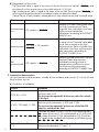
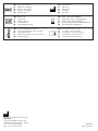
1
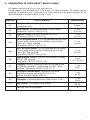
PLATELIA™ RABIES II KIT
Ad Usum Veterinarium
192 TESTS
Ref.: 355-0180
KIT FOR IN VITRO DETECTION AND TITRATION OF IgG ANTI
RABIES VIRUS GLYCOPROTEIN IN DOGS, CATS AND FOXES
SERUM.
All manufactured and commercialized reagents are under complete quality
system starting from reception of raw materials to the final commercialization
of the product.
Each lot is submitted to a quality control and released on the market when
conforming to the acceptance criteria.
Fitness for purpose validated and certified by OIE
Registration number: 20070101
User’s manual
TABLE OF CONTENTS
1 - INTENDED USE
2 - INTEREST OF PLATELIA™ RABIES II KIT
3 - PRINCIPLE OF THE PLATELIA™ RABIES II ASSAY
4 - COMPOSITION OF THE PLATELIA™ RABIES II KIT
5 - PREPARATION OF THE REAGENTS
6 - STORAGE - SHELF-LIFE
7 - SAMPLES
8 - ASSAY PROTOCOL
9 - CALCULATION AND INTERPRETATION OF THE RESULTS
10 - LIMITATIONS OF THE TEST
11 - EQUIPMENT AND MATERIAL REQUIRED BUT NOT SUPPLIED
12 - PRECAUTIONS
13 - LITERATURE
2
1 - INTENDED USE
The PLATELIA™ RABIES II kit is an in vitro diagnostic ELISA test allowing detection and
titration of IgG anti rabies virus glycoprotein in animal (dogs, cats and foxes) serum.
2 - INTEREST OF THE PLATELIA™ RABIES II KIT
Rabies is one of the oldest viral diseases afflicting humans and animals. Rabies virus is a
highly neurotropic virus, usually causing fatal encephalitis in mammals. The virus is mostly
transmitted through the saliva via bites from rabid animals. Immunization and control
programs for the protection of animal populations from rabies virus have resulted in the
elimination of the disease in several Western-European countries and in some countries in
South America.
As of July 3rd 2004, a new European regulation applies to international movements of
domestic carnivores from rabies-infected to rabies-free countries. Domestic carnivores are
allowed to travel if their serum contain at least 0.5 IU/ml (International Units/ml) of rabies
neutralising antibodies.
Anti-rabies antibodies titration has several practical applications :
• Individual serology for international trade purposes: anti-rabies antibodies are measured
in specialized laboratories in order to determine the degree of immunity of vaccinated
animals (cats and dogs). A level of antibody equal to or superior to 0.5 IU/ml is
considered by OIE experts as an acceptable seroconversion level above which the
vaccination could be considered as successful.
• Confirmation of vaccinated status during a vaccination campaign: control of the antirabies antibodies enables indirect assessment of the effectiveness of oral vaccination
campaign in wildlife (foxes).
3 - PRINCIPLE OF THE PLATELIA™ RABIES II ASSAY
PLATELIA™ RABIES II is an immuno-enzymatic technique for the detection of rabies virus antiglycoprotein antibodies. This assay can be carried out on the serum of several species of
animals - dog, cat and fox.
The test is based on the use of a solid phase enzyme immunoassay technique referred to as an
indirect ELISA. A microplate is coated with rabies glycoprotein extracted from the inactivated
and purified virus membrane. The enzymatic conjugate consists of a protein A from
Staphylococcus aureus coupled with peroxidase. Positive controls, calibrated against OIE
standard, allow the qualitative or quantitative determination of anti-rabies antibody titre in the
serum.
Implementation of the test comprises the following reaction steps:
1 - The unknown sera as well as the calibrated Positive Controls or the Quantification
Standards are distributed in the glycoprotein coated wells of the microplates. During
incubation of one hour at 37°C, anti-rabies antibodies present in the sample bind to
the glycoprotein coated to the microplate wells. After incubation, unbound antibodies
and other serum proteins are removed by washings.
2 - The conjugate (protein A labelled with peroxidase) is added to the microplate wells.
During a second incubation of one hour at 37°C, the labelled protein A binds to the
anti-rabies-antibody-antigen complexes attached to the microplate wells. The unbound
conjugate is removed by washings.
3
3 - The presence of immune complex is demonstrated by the addition of a solution
containing a peroxidase substrate and a chromogen, initiating a colour development
reaction.
4 - After 30 min. incubation at room temperature, the enzymatic reaction is stopped by
addition of a solution H2SO4 1N. The optical density reading obtained with a
spectrophotometer set at 450 - 620 nm is proportional to the amount of anti-rabies
antibodies present in the samples. A standard curve is constructed using the
Quantification standards (S1 to S6), obtained by serial dilutions of the R4b calibrated
Positive Controls.
The optical density values for the unknown samples are compared with the Positive Controls.
Sera titres in quantification tests are obtained after a direct reading on the standard curve
and are expressed as Equivalent units per ml (EU/ml), unit equivalent to the international
units defined by seroneutralization.
4
4 - COMPOSITION OF THE PLATELIA™ RABIES II ASSAY
All reagents are exclusively for in vitro veterinary use.
Enough reagents are provided for 2 x 96 assays. On each microplate, 90 samples can be
analysed if a qualitative test is performed. Up to 80 samples can be quantified precisely for
rabies antibodies if a quantification system is used.
LABELLING
TYPE OF REAGENT
PRESENTATION
R1
Microplate: 12 strips of 8 wells sensitized with the rabies
virus glycoprotein
2 plates
R2
Wash solution: 10-fold concentrated Tris-NaCl buffer
Preservative: ProClin™ 300 (0.01%)
1 vial
(250 ml)
R3
Negative control: Non reactive control TRIS-EDTA
Preservative: ProClin™ 300 (0.1%)
1 vial
(0.6 ml)
R4a
R4b
R6
0,5 EU/ml Positive control: 0.5 EU/ml calibrated positive control
Glycine buffer containing BSA and dog serum with antirabies IgG. Yellow coloured.
Preservative: ProClin™ 300 (0.1%)
4 EU/ ml Positive control: 4 EU/ml calibrated positive control
Glycine buffer containing BSA and dog serum with antirabies IgG. Blue coloured
Preservative: ProClin™ 300 (0.1%)
Sample diluent: Ready-to use TRIS - EDTA buffer for sample
dilution. Red coloured.
Preservative: ProClin™ 300 (0.1%)
R7
Conjugate: Solution containing Protein A-Peroxidase and
purified bovine protein. Concentrated 10 times. Green
coloured. Preservative: ProClin™ 300 (0.1%)
R8
Peroxydase substrate buffer: Solution of citric acid
and sodium acetate containing 0.015% H2O2
and 4% dimethylsulfoxide (DMSO)
R9
Chromogen: Tetramethylbenzidine (TMB) solution 0.25%
R10
Stop solution: 1 N sulphuric acid solution
Adhesive films for microplates
1 vial
(0.6 ml)
1 vial
(0.6 ml)
2 vials
(2 x 125 ml)
1 vial
(3 ml)
1 vial
(60 ml)
1 vial
(5 ml)
1 vial
(28 ml)
6
5
5 - PREPARATION OF REAGENTS
Note:
Before use, allow reagents to reach room temperature (+18 to +30°C) for 30 minutes.
Homogenize the reagents by gently mixing, before opening.
1 - Ready-to-use reagents
• Microplates (R1)
Prior to use, let the microplate adjust to room temperature (+18°C to +30°C) in its
protective packaging to avoid any water condensation in the wells. Any unused strips
should be immediately returned to the bag, tightly close the bag after expelling any
air, then store at +2°C to +8°C.
• Sample diluent (R6)
• Stop solution (R10)
2 - Reagents to be reconstituted
• Wash solution (R2)
Dilute the solution 1/10 in distilled water (example 100 ml of reagent R2 in 900 ml of
distilled water). 500 ml are required for a plate.
• Negative control (R3)
Dilute the solution 1/100 in R6.
• 0.5 EU/ml Positive control (R4a)
Dilute the R4a positive control 1/100 in R6
• 4 EU/ml Positive control (R4b)
Dilute the R4b positive control 1/100 in R6
• Conjugate (R7)
The solution is concentrated 10 times. To prepare diluted conjugate solution, add 1 volume
of concentrated conjugate to 9 volumes of prepared 1X wash solution (R2). 11 ml is
required for a full microplate.
• Enzymatic development solution (R8 + R9)
Dilute reagent R9 to 1/11 in the reagent R8 (example: 0.1 ml of reagent R9 in 1 ml of
reagent R8) bearing in mind that 11 ml of enzymatic revelation solution is sufficient for
1 microplate. Homogenize gently. Avoid using a Vortex® agitator.
3 - Preparation of the Quantification Standards for a quantification assay
Each quantification assay using PLATELIA™ RABIES II kit should include 6 Quantification
standards – from S1 to S6.
The calibrated R4b Positive control (4 EU/ml) corresponds to the S6 Quantification standard.
Serial dilutions out of the R4b reagent allow the preparation of S5 to S1 Quantification
standards. The dilutions are performed using the sample diluent (reagent R6).
6
Quantification
standards
S6
S5
S4
S3
S2
S1
R4b diluted to 1/100
S6 diluted to 1/2
S5 diluted to 1/2
S4 diluted to 1/2
S3 diluted to 1/2
S2 diluted to 1/2
Concentrations obtained by serial
dilutions of the R4b Positive control
4 EU/ml
2 EU/ml
1 EU/ml
0.5 EU/ml
0.25 EU/ml
0.125 EU/ml
6 - STORAGE - SHELF LIFE
As supplied, all the components of the PLATELIA™ RABIES II kit are stored at +2°to +8°C and
can be used until the expiry date indicated on the kit. Reagent R2 (10 X) can be stored at
+2°C to +25°C before reconstitution.
The shelf-lives of the reagents after preparation are as follow:
REAGENT
R1
Microplate
R2
Diluted
wash solution
R7
Diluted
conjugate
solution
R8 + R9
Reconstituted
development
solution
REMARKS
SHELF-LIFE
After opening the sealed foil
lined bag, the unused strips
should immediately be put
back in the bag and the bag
again sealed. The desiccant
should remain in the foil bag.
1 month at
+2°C to +8°C
2 weeks at
+2°C to +8°C
The diluted conjugate solution
should preferably be used
immediately
8 hours at
+2°C to +8°C
6 hours at room temperature
(+18°C to +30°C) always
protected from light
7 - SAMPLES
• The PLATELIA™ RABIES II test has been developed for application to animal (dogs, cats
and foxes) serum.
• The assay is carried out on sera after a 1/100 dilution in R6 reagent.
• The samples are stored at +2°to +8°C if the detection is carried out within 24 hours or can
be stored at -20°C for 6 months. The samples can be submitted to 3 cycles of freezing and
thawing. Previously frozen samples should be thoroughly mixed after thawing prior to
testing.
NB: Eliminate, if necessary by centrifuging, the fibrin particles or aggregates in suspension
that can provide false positive results.
7
8 - ASSAY PROTOCOL
Strictly follow the recommended protocol.
Use the negative and the positive controls for each test run and on each microplate in order
to validate the quality of the detection in the qualitative assay. If quantification is performed,
the negative control and the Quantification standards should be deposited on each plate. In
both cases, follow the recommended microplate set-up.
Use good laboratory practices.
1. Remove the microplate rack and the required number of rows (R1) from the protective
packaging. Replace the unused rows with the desiccated bag in the microplate sachet
and hermetically close it.
2. Carefully establish the sample distribution and identification plan as follow:
Microplate set-up for Qualitative assay:
A
1
2
R3
E3
3
4
B
R3
E4
C
R4a
E5
D
R4a
E6
E
R4b
E7
F
R4b
E8
G
E1
E9
H
E2
E10…
1
2
3
4
R3
S4
E1
E9
5
6
7
8
9
10
11
12
9
10
11
12
Microplate set-up for Quantitative assay:
A
8
B
R3
S4
E2 E10…
C
R4a
S3
E3
D
R4a
S3
E4
E
S6
S2
E5
F
S6
S2
E6
G
S5
S1
E7
H
S5
S1
E8
5
6
7
8
3. For the qualitative assay, dilute the R3, R4a and R4b controls and the unknown sera
to 1/100 in R6 reagent (e.g.: 10 μl of sample in 990 μl of dilution solution).
4. For the quantitative assay, prepare the quantification standards (refer to chapter 5.3)
and dilute the R3, R4a controls and the unknown sera to 1/100 in R6 reagent (e.g.
10 μl of sample in 990 μl of dilution solution).
5. Distribute 100 μl of diluted samples, controls and Quantification standards in the
correspondent microplate wells according to the pre-established distribution plan.
6. Cover the microplate with adhesive film (cut the sheet if necessary). Press firmly all
over the plate to ensure a tight seal.
7. Incubate the strips at 37 ± 2°C for 60 minutes ± 5 minutes.
8. Prepare the wash solution (R2) [refer to chapter 5]
9. Prepare the conjugate solution (R7), as described in chapter 5 before the end of the
first incubation.
10. Remove the adhesive film. Perform 3 wash cycles. Optimal washing conditions are
obtained with PW40, PW41 or 1575 Bio-Rad plate washers with program TSE 3.
Do not let the microplate stand for more than 5 minutes after the last wash cycle. Dry
by inversion on absorbent paper before the following step.
11. Distribute 100 μl of the conjugate solution (R7) into each well. Cover with a new
adhesive film and incubate for 60 minutes ± 5 minutes at 37 ± 2°C.
12. Prepare the enzymatic development solution (R8+R9) as described in chapter 5, just
before use.
13. Remove the adhesive film, perform 5 wash cycles. Optimal washing conditions are
obtained with PW40, PW41 or 1575 Bio-Rad plate washers with programs TSE 5
Do not let the microplate stand for more than 5 minutes after the last wash cycle. Dry
by inversion on absorbent paper before the following step.
14. Away from direct light, quickly distribute 100 μl of the enzymatic development
solution (R8 + R9) into each well and incubate the plate in darkness at room
temperature (+18 to +30°C) for 30 minutes ± 5 minutes.
NB: Do not use adhesive film during this incubation.
15. Add 100 μl of the stop solution (R10) to each well according to the same sequence
and same distribution rate as for the enzymatic development solution.
16. Thoroughly wipe the bottom of the plate. Read the optical density at 450 - 620 nm
(Dual wavelength mode) within 30 minutes after stopping the reaction (the strips must
always be protected from light before reading).
17. Before recording the results, check that the reading complies with the distribution and
identification plan for the plates and samples.
9
10
PLATE
NAME: TSE 3
Manifold
PLATE
Manifold
-
-
-
Method 2
-
-
0.3
ASP. HOR.
POS.
1.4
BOT : SHAPE
Flat
0.3
CENTERING
-
800
-
-
Plate
Plate
WASH
-
BOTTOM
ASP.
-
Yes
Yes
0.3
0.3
-
BOT VERT.
POS.
9.5
ASP. VER.
POS.
13.5
9.5
B.W. VERT.
POS.
6
HORIZONTAL
SPEED
-
800
-
-
Yes
0.3
Met.
CROS
ASP.
VOLUME
MODE
(Method)
SW ASP. TIME
Plate
BOTTOM
ASP.
Yes
STRIPS
Plate
WASH
-
1,2,3,4,
5,6,7,8,9,
10,11,12
-
-
-
-
-
1,2,3,4,
5,6,7,8,9,
10,11,12
-
CROS
ASP.
Met.
VOLUME
MODE
SW ASP. TIME
(Method)
STRIPS
PLATE NAME: FLAT 01 (PW40/PW41) - FLAT 03 (1575)
-
Method 1
Main
Flat 01 (PW40/PW41) 1*8 (PW40/1575)
parameter
2*8 (PW41)
Flat 03 (1575)
EDIT Mode
finction
-
Method 2
NAME: TSE 5
-
Method 1
Main
Flat 01 (PW40/PW41) 1*8 (PW40/1575)
parameter
2*8 (PW41)
Flat 03 (1575)
EDIT Mode
finction
MICROPLATE WASHER PARAMETERS
-
2.5
-
-
0
-
-
W1
-
8
-
-
-
1
-
-
-
-
-
-
-
-
6
9
DISP. UPW.
SPEED
-
-
-
-
-
-
1
5
-
Nr OF
CYCLES
1
3
-
Nr OF
CYCLES
6
9
1
SHAKING
AMPLITUDE
-
0
0
-
-
MET.
INTER
30 (PW41)
45 (PW40/1575)
SOAKING
-
0
30 (PW41)
45 (PW40/1575)
0
-
MET.
INTER
-
SOAKING
BOT. DOWNW. BOT. UPWARD
SPEED
SPEED
1
-
-
BOT. WASH BOTTOM BOT. ASP. SHAKE
NUMBER
TIME
NUMBER TIME
-
-
-
BOT. WASH BOTTOM BOT. ASP. SHAKE
NUMBER
TIME
NUMBER TIME
ASP. DOWNW.
SPEED
-
0
-
LIQUID FLOW
-
W1
-
LIQUID FLOW
VERTICAL
SPEED
OVER
FLOW
-
2.5
-
OVER
FLOW
-
-
-
KIT
INTER
-
-
-
KIT
INTER
9
SHAKING
SPEED
-
-
1
Nr OF
KITS
-
-
1
Nr OF
KITS
9 - CALCULATION AND INTERPRETATION OF THE RESULTS
The results are given in Optical Densities (ODs) after reading of the microplate at 450 - 620 nm.
1. Qualitative determination
For the qualitative determination, include all the controls (R3, R4a and R4b) for each test
run.
a) Conditions of validation
Criteria
Validation
OD R3(i) < 0.05
The absorbance of each individual negative control must
be lower than 0.05.
The test must be repeated if at least one value lies outside
of this limit.
0.300 ≤ OD R4a(i) ≤ 1.200
Each individual value of the R4a positive control OD must
be between 0.300 and 1.200.
The test must be repeated if at least one of the R4a OD
value lies outside of this limit.
1.500 ≤ OD R4b(i) ≤ 3.500
Each individual value of the R4b positive control optical
densities must be between 1.500 and 3.500. However,
a maximum of one individual aberrant value can be
eliminated when its optical density is lower than 1.500 or
higher than 3.500.
The test must be repeated if the two R4b positive control
OD lie outside of this limit.
11
b) Interpretation of the results
The Threshold Value is equal to the mean of the two R4a Positive controls (OD R4a) and
corresponds to the seroconversion threshold value at 0.5 EU/ml.
High seroconversion Value is equal to the mean of the two R4b Positive control (OD R4b), or
to the single R4b positive control if one aberrant value has been eliminated.
Optical Density of each sample is compared to this High seroconversion and Threshold Value.
Criteria
Seroconverted
+++
Seroconverted
Not
seroconverted
Result
Validation
OD sample > (OD R4b)
Samples with OD value greater than
the High Seroconversion Value
originate from individuals who have
highly seroconverted after
vaccination according to PLATELIA™
RABIES II test.
(OD R4a) ≤ OD sample ≤
(OD R4b)
Samples with OD value greater than
or equal to the Threshold Value and
lower than or equal to the High
Seroconversion Value originate from
individuals who have seroconverted
after vaccination according to
PLATELIA™ RABIES II test.
OD sample < (OD R4a)
Samples with OD value lower than the
Threshold Value originate from
individuals presenting a level of
seroconversion insufficient to assess
the efficiency of the vaccination
according to PLATELIA™ RABIES II test.
2. Quantitative determination
For the quantitative determination, include all the standards and controls (S1 to S6, R3 and
R4a) for each test run.
a) Conditions of validation
Criteria
OD R3(i) < 0.05
0.300 ≤ OD R4a(i) ≤ 1.200
12
Validation
The absorbance of each individual negative control must
be lower than 0.05.
The test must be repeated if at least one value lies outside
of this limit.
Each individual value of the R4a positive control optical
densities must be between 0.300 and 1.200 .
The test must be repeated if at least one of the R4a OD
value lies outside of this limit.
S1 < S2 < S3 < S4 < S5 < S6
Calculate the mean OD for S1 to S6 in the following way:
S1 = mean of the two S1 ODs (corresponds to 0.125 EU/ml).
S2 = mean of the two S2 ODs (corresponds to 0.25 EU/ml) etc.
The signals of the standards must increase in the following way:
S1 < S2 < S3 < S4 < S5 < S6 .
0.7 ≤ S3/R4a ODs ≤
1.3
The ratio between S3 (mean S3 standard) and R4a (mean
R4a) must be between 0.7 and 1.3.
b) Interpretation of the results
The Threshold Value is equal to the mean of the two OD of the S3 Quantification
standard (S3) . The S3 Quantification standard corresponds to the seroconversion
threshold value 0.5 EU/ml.
The quantity of anti-rabies antibodies in a sample is determined by comparing the
optical density of the sample to a standard curve.
Sera titres are expressed as Equivalent units per ml (EU/ml), unit equivalent to the
international units defined by seroneutralization.
Drawing the standard curve:
For manual data reduction, use graph paper and plot the mean values of the OD readings of the
Quantification standards (S1 to S6) on the vertical (y) axes. Plot the corresponding concentrations in
EU/ml on the horizontal (x) axes. Draw a series of line segments which goes through the 6 points.
For automated data reduction, a “point-to-point” function is used to construct a curve from the OD
readings obtained for the standards.
A quantitative result for the anti-rabies antibodies titre for an unknown sample can be delivered if
the OD value for the sample ranges between the S1 (0.125 EU/ml) and the S6 (4 EU/ml)
Optical Density mean values. In this case, the unknown sample titre is found using the Standard
curve. On the Standard curve, find the value corresponding to the OD reading of the sample on
the y-axis and draw a horizontal line to the standard curve. At the point of the intersection with
the standard curve, draw a vertical line to the x-axis. Read the corresponding concentration in
EU/ml.
If the OD value of the unknown sample is higher than the mean Optical Density value for the S6
standard, a precise quantification cannot be performed. If a precise quantification is required,
proceed to 1/10 dilution or more of the sample and perform the assay again in order to have an
Optical Density in the interval of the Standard curve.
Note: An interpretation and conversion tool of OD values into titres is available on the CD
(Rabies QT-ELISA BIORAD-Vers.200712.A.XLS) for users who are not equipped with Bio-Rad
readers.
General comprehension of the results:
Results in Optical Density
for the unknown sample
Titre for the unknown
sample (X)
Interpretation of the result
OD sample > S6
X > 4 EU/ml
High seroconversion level.
If a precise titre is required, the
sample must be diluted before
repeating the assay.
S3 ≤ OD sample ≤ S6
X in EU/ml (0.5 - 4 EU/ml)
Sufficient seroconversion level.
S1 ≤ OD sample < S3
X in EU/ml (0.125 - 0.5 EU/ml)
Insufficient seroconversion level
according to PLATELIA™ RABIES II
test.
OD sample < S1
-
Undetectable seroconversion.
13
10 - LIMITATIONS OF THE TEST
Insufficient or undetectable seroconversion can possibly be reported in recently vaccinated
animals, due to delayed immune response. Similarly to the official neutralising antibody
titration method and in accordance with regulations (EC) No 998/2003 effective July 3,
2004, the PLATELIA™ RABIES II test should be carried out on samples taken at least thirty
days after vaccination.
11 - EQUIPMENT AND MATERIAL REQUIRED BUT NOT SUPPLIED
Equipment
• Microplate reader equipped with 450 and 620 nm filters (*)
• Microplate incubator set at 37°C ± 2°C.
• Manual, semi-automatic or automatic microplate washer (*)
• Vortex® mixer.
(*) Contact us for detailed information about Bio-Rad instruments validated by our technical
departments.
Material
• Automatic or semi-automatic adjustable or preset pipettes or micropipettes to measure and
deliver 10 to 1000 μl and 1, 2 and 10 ml.
• Graduated cylinders of 25 ml, 50 ml, 100 ml, and 1 000 ml capacity.
• Disposable tubes.
• Distilled or deionized water.
• Sodium hypochloride (bleach).
• Absorbent paper.
• Goggles or safety glasses.
• Disposable latex gloves.
• Container for biohazard waste.
12 - PRECAUTIONS
1. Precautions
The quality of the results depends on correct implementation of the following good
laboratory practices:
• Reagents must be stored at +2°C to +8°C.
• Do not use expired reagents.
• Do not mix reagents from different lots within a given test run with the exception of
generic reagents R2, R8, R9, R10.
• Before use, wait 30 minutes for the reagents to adjust to the laboratory temperature.
• Thoroughly reconstitute reagents, avoiding any contaminations.
• Do not perform the test in the presence of reactive vapors (acids, alkalis, aldehydes)
or dust that could alter enzyme activity of the conjugate.
• Use glassware thoroughly washed and rinsed with deionized water or preferably,
disposable material.
• Do not leave the microplate more than 5 minutes between the end of washing
operation and the reagent distribution.
• Check the accuracy of the pipettes and the good operation of the apparatus used.
• Never use the same container to distribute conjugate and the development solution.
14
• The enzyme reaction is very sensitive to metal or metallic ions. Consequently, do not allow
any metal element to come in contact with the various conjugate or substrate solution.
• The development solution (substrate buffer + chromogen) must be colourless. The
appearance of a blue colour within a few minutes after reconstitution of the
development solution indicates that the reagent can not be used and must be
replaced. Preparation of this solution can be made in a clean disposable plastic tray
or glass container that has first been pre-washed with 1N HCl, and rinsed
thoroughly with distilled water and dried. Store the solution in the dark.
• Use a new pipette tip for each sample.
• Do not change the assay procedure.
• Washing of the wells is a crucial step in the procedure: respect the recommended
number of washing cycles and make sure that all wells are completely filled, then
emptied. Inadequate washing can give incorrect results.
2. Hygiene and safety instructions
Generally, hygiene conditions, biosafety measures and good laboratory practices must be
in agreement with recommendation of regular authorities of the country.
All the reagents of the kit are intended for “in vitro” veterinary diagnostic use - dog, cat
and fox sera.
• Wear disposable gloves when handling reagents and samples. Thoroughly wash
your hands after sample and reagent handling.
• Do not pipette with the mouth.
• Consider any material directly in contact with the samples and the reagents, as well
as the wash solutions, as infectious material.
• Avoid spilling samples or solutions containing samples.
• Spills may be rinsed with bleach diluted to 10% Chl. If the contaminating fluid is
acid, spills must be initially neutralized with sodium bicarbonate, then cleaned with
bleach and dried with absorbent paper. The material used for cleaning must be
discarded after decontamination.
• Samples and contaminated material and products should be discarded after
decontamination
- either by immersion in bleach at the final concentration at 5% of sodium
hypochloride for 30 min.
- or by autoclaving at 121°C for 2 hours minimum.
Caution: Do not introduce solutions containing sodium hypochloride into the
autoclave.
• Chemicals should be handled and disposed of in accordance with Good Laboratory
Practice.
• Reagents containing 0.1% ProClin™ 300 are classified as irritating preparations
according to European legislation.
Xi
(0.1% ProClin™ 300)
R: 43
May cause sensitisation by skin contact.
S: 28-37 After contact with skin, wash immediately with plenty of water. Wear
suitable gloves.
15
13 - LITERATURE
1. ATANASIU P., SAVY V. and PERRIN P. 1977. Epreuve immuno-enzymatique pour la recherche
rapide des anticorps antirabiques. Ann. Inst. Pasteur Microbiol. 128 A, 489-498.
2. ATANASIU P., SAVY V. and GIBERT C. 1978. Rapid immunoenzymatic technique for titration
of rabies antibodies IgG and IgM results. Med. Microbiol. Immunol. 166, 201-208.
3. ATANASIU P. and PERRIN P. 1979. Microméthode immuno-enzymatique de titrage des
anticorps antirabiques : utilisation de la glycoprotéine rabique et de la protéine A conjuguées à
la péroxydase. Ann. Inst. Pasteur Microbiol. A, 257-268.
4. ATANASIU P., PERRIN P. and DELAGNEAU J.F. 1980. Use of an enzyme immunoassay with
protein A for rabies antigen and antibody determination. Develop. Biol. Standard. 46, 207215.
5. AVRAMEAS S. and TERNYNCK T. 1971. Peroxidase labelled antibody and Fab conjugates
with enhanced intracellular penetration. Immunochemistry 8, 1175-1179.
6. BIDERFIELD P., GHETIE V. and SJOQUIST J. 1975. Demonstration and assaying of IgG
antibodies in tissues and on cells by labelled staphylococcal protein A. J. Immunol. Meth. 6,
249-259.
7. BOURHY H., SUREAU P. 1989. Comparative field evaluation of the fluorescent antibody test,
virus isolation from tissue culture, and enzymes immunodiagnosis for rapid laboratory diagnosis
of rabies. J. Clin. Microbiol. 27, 519-523.
8. BRIGGS D.J., SMITH J.S., MUELLER F.L., SCHWENKE J., DAVIS R.D., GORDON C.R,
SCHWEITZER K., ORCIARI L.A., YAGER P.A., RUPPRECHT C.E.. 1998. A comparison of two
serological methods for detecting the immune response after rabies vaccination in dogs and
cats being exported to rabies-free areas. Biologicals 26, 347-355.
9. CLIQUET F., AUBERT M., SAGNE L. 1998. Development of a fluorescent antibody virus
neutralization test (FAVN test) for the quantitation of rabies-neutralising antibody. J. Immunol.
Methods 212, 79-87.
10. CLIQUET F, L. SAGNE, J.L. SCHEREFFER, M.F.A. AUBERT. 2000. ELISA test for rabies antibody
titration in orally vaccinated foxes sampled in the fields. Vaccine 18, 3272-3279.
11. CLIQUET F., VERDIER Y., SAGNE L., AUBERT M., SCHEREFFER J.L., SELVE M., WASNIEWSKI
M, SERVAT A. 2003. Neutralising antibody titration in 25,000 sera of dogs and cats
vaccinated against rabies in France, in the framework of the new regulations that offer an
alternative to quarantine. Rev. Sci. Tech. Off. Int. Epiz. 22 (3), 857-866.
12. CLIQUET F., MÜLLER T, MUTINELLI F., BROCHIER B., AUBERT M. and all. 2003.
Standardisation and establishment of a rabies ELISA test in European laboratories for assessing
the efficacy of oral fox vaccination campaigns. Vaccine 21, 2986-2993.
13. CLIQUET F, Mc ELHINNEY L.M., SERVAT A., BOUCHER J.M., LOWINGS J.P., GODDARD T.,
MANSFIELD K.L., FOOKS A.R. 2004. Development of a qualitative indirect ELISA for the
measurement of rabies virus-specific antibodies from vaccinated dogs and cats. J. Vir. Methods
117, 1-8.
16
14. ENGVALL E. and PERLMANN P. 1971. Enzyme-linked immunosorbent assay (ELISA):
Quantitative assay of immunoglobulin G. Immunochemistry 8, 871-874.
15. FEYSSAGUET M., DACHEUX L, AUDRY L., COMPOINT A., MORIZE JL. , BLANCHARD I.,
BOURHY H. 2007. Multicenter comparative study of a new ELISA, PLATELIA™ RABIES II, for
the detection and titration of anti-rabies glycoprotein antibodies and comparison with the rapid
fluorescent focus inhibition test (RFFIT) on human samples from vaccinated and non-vaccinated
people. Vaccine 25(12):2244-51
16. FORSGREN A. and SJOQUIST J. 1966. "Protein A" from S. aureus. I. Pseudo-immune reaction
with human g-globulin. J. Immunol. 97, 822-827.
17. GRASSI M., WANDELER A.I., PETERHANS E. 1989. Enzyme-Linked Immunosorbent Assay for
Determination of the Envelope Glycoprotein of Rabies Virus. J. Clin. Microbiol. 27 (5), 899902.
18. PERRIN P., VERSMISSE P., DELAGNEAU J.F., LUCAS G., ROLLIN P.E. and SUREAU P. 1986.
The influence of the type of immunosorbent on rabies antibody EIA: advantages of purified
glycoprotein over whole virus. J. Biol. Standard. 14, 95-102.
19. SERVAT A., CLIQUET F. 2006. Collaborative study to evaluate a new ELISA test to monitor the
effectiveness of rabies vaccination in domestic carnivores. Virus Res. 120, 17-27.
20. SERVAT A., FEYSSAGUET M., BLANCHARD I., BOUE F., CLIQUET F. 2007. A quantitative
indirect ELISA to monitor the effectiveness of rabies vaccination in domestic and wild carnivores.
J Immunol Methods 318(1-2):1-10
21. STANTIC-PAVLINIC M., HOSTNIK P., LEVICNIK-STEZINAR S., ZALETEL-KRAGELJ L. 2006.
Vaccination against rabies and protective antibodies : comparison of ELISA and fluorescent
antibody virus neutralization (FAVN) assays. Veterinarski Archiv 76 (4), 281-289.
22. WRIGHT C., WILLIAM K.J., SJODAHL J., BURTON D.R. and DWEK R.A. 1977. The interaction
of protein A and the Fc fragment of rabbit immunoglobulin G as probed by complement-fixation
and nuclear magnetic resonance studies. Bioch. J. 167, 661-668.
23. Manual of standards for diagnostic tests and vaccines. OIE 2000. Chapters 1.1.3 and 2.2.
17
(US)
(F)
(E)
(I)
(D)
-
Catalogue number
Référence catalogue
Número de catálogo
Numero di catalogo
Bestellnummer
(US)
(F)
(E)
(I)
(D)
-
Manufacturer
Fabricant
Fabricante
Produttore
Hersteller
(US)
(F)
(E)
(I)
(D)
-
Batch code
Code du lot
Código de lote
Codice del lotto
Chargen-Bezeichnung
(US)
(F)
(E)
(I)
(D)
-
Expiry date YYYY/MM/DD
Date de peremption AAAA/MM/JJ
Estable hasta AAAA/MM/DD
Da utilizzare prima del AAAA/MM/GG
Verwendbar bis JJJJ/MM/TT
(US)
(F)
(E)
(I)
(D)
-
Storage temperature limitation
Limites de températures de stockage
Temperatura límite
Limiti di temperatura di conservazione
Lagertemperatur
(US)
(F)
(E)
(I)
(D)
-
Consult Instruction for use
Consulter le mode d'emploi
Consulte las instrucciones de uso
Consultare le istruzioni per uso
Siehe Gebrauchsanweisung
Made and distributed in France by
Bio-Rad
3, Boulevard Raymond Poincaré
92430 Marnes-La-Coquette - France
Tel.: +33 1 47 95 60 00
Fax.: +33 1 47 41 91 33
04/2009
Code : 862196