1
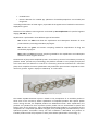
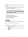
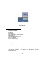
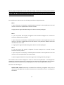
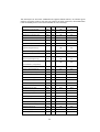
CLART SeptiBac DETECTION AND MOLECULAR IDENTIFICATION OF BACTERIA AND FUNGI CAUSING SEPSIS FOR IN VITRO DIAGNOSTICS 1 CLART SeptiBac CLART® SeptiBac is under protection of patent family corresponding to International PCT Patent Application WO2015055768. CLART®, CLART-Strip®, CAR®, SAICLART® and SEPTIBAC® are registered Trademarks of GENOMICA.. For further information and questions do not hesitate to visit www.genomica.com GENOMICA, S.A.U. Parque Empresarial Alvento, Edificio B Calle Vía de los Poblados, 1 – 1ª planta 28033 Madrid, Spain www.genomica.com Version 5 July 2015 2 CONTENTS: 1. GLOSSARY OF TERMS 2. DESCRIPTION 3. KIT COMPONENTS AND STORAGE 3.1. Amplification reagents 3.2. Visualization reagents 3.3. Other components 4. REQUIRED AND NOT SUPPLIED MATERIAL 4.1. Reagents and material 4.2. Equipment 5. HANDLING PROCEEDINGS AND RECOMMENDATIONS 5.1. General recommendations 5.2. Extraction precautions 5.3. Visualization precautions 6. SAMPLES: BLOOD CULTURES 7. WORKING PROTOCOLS 7.1. Automatic extraction of genetic material. 7.2. Amplification reaction. 7.3. Visualization of amplified product. 8. RESULTS READING 9. RESULTS INTERPRETING 10. TECHNICAL AND WORKING SPECIFICATIONS 11. BIBLIOGRAPHY 3 1. GLOSSARY OF TERMS Please, check handling instructions Expiry date Sanitary product for In vitro diagnostics Batch 25ºC Store at room temperature 20ºC 8ºC Store between 4 ºC and 8 ºC 4ºC -18ºC Store between –30 ºC and –18 ºC - 30ºC 4 2.- DESCRIPTION CLART SeptiBac detects the presence of the following gram-positive, gram-negative bacteria and fungi causing bacteriemia, fungaemia, sepsis or septic shock in positive blood cultures. • • • • • • • • • • • • • • • • • • • • • • • • • • • • • • • • • • • • • • • Staphylococcus aureus* Staphylococcus epidermidis* Staphylococcus hominis* Staphylococcus haemolyticus* Streptococcus pyogenes/dysgalactiae Streptococcus pneumoniae Streptococcus mitis Streptococcus agalactiae Streptococcus sanguinis/parasanguinis Streptococcus de grupo milleri (including S. constellatus y S. anginosus). Detección genérica para Streptococcus spp. Enterococcus faecium Enterococcus faecalis Gram positive cocci (CGPs) Listeria monocytogenes Clostridium perfringens Escherichia coli Klebsiella pneumoniae Klebsiella oxytoca Klebsiella spp.(K. pneumoniae, K, oxytoca) Salmonella enterica Enterobacter cloacae Enterobacter aerogenes Enterobacter spp. (E. cloacae, E. aerogenes) Citrobacter freundii Citrobacter spp. (C. freundii, C. koserii) Serratia marcescens/plimutica Proteus vulgaris/penneri Proteus mirabilis Proteus spp. (P. vulgaris, P. mirabilis, P. penneri) Haemophilus influenzae Haemophilus spp. (H. influenzae, H. parainfluenzae) Acinetobacter baumanii Bacteroides fragilis Bacteroides spp. (B. fragilis, B. faecis) Pseudomonas spp (P. aeruginosa, P. putida, P. stuartii ) Stenotrophomonas maltophilia Candida albicans Candida glabrata 5 • • Candida krusei Generic detection for Candida spp. (detection of Candida parapsilosis not included) and fungal taxa. *Including the detection of mecA region, responsible of the appearance of methicillin resistance in Staphylococci genus. Detection of the different microorganisms is achieved by PCR amplification of a specific fragment ranging 149-500 bp. size. Amplification is performed in three different types of PCR tubes: Mix 1 tubes are white and allow the amplification and subsequent detection of Gram positive bacteria, excluding Clostridium perfringens. Mix 2 tubes are green and contain everything needed for amplification of fungi and Clostridium perfringens. Mix 3 tubes are blue and contain everything needed for the amplification and subsequent detection of Gram negative bacteria. The detection of the product amplified by PCR is carried out by means of a low-density microarray platform: CLART® (Clinical Arrays Technology). The platform is based on a very simple principle, but at the same time cost effective. It consists of a microarray printed at the bottom of a microtiter plate, which simplifies the entire hybridization and visualization process when compared to classic microarray systems. Figure 1 displays a CLART-Strip® or CS of 8 wells. Figure 1: CLART-Strip® in 8-well strip format (CS). The CLART® SeptiBac detection system is based on the precipitation of an insoluble product in those areas of the microarray where hybridization of amplified products with specific probes occurs. During the PCR, the amplified products are labeled with biotin. After amplification, the products are hybridized to their respective specific probes that are immobilized at specific and known areas of the microarray. These immobilised biotinilated products are recognized by the streptavidin of a streptavidin-peroxidase conjugate, thus providing with peroxidase activity to the hybridised products. Peroxidase activity will then metabolise o-Dianisidine and produce an insoluble product which will precipitate in those places where hybridisation occurred (Fig. 2). 6 Figure 2: Diagram of the visualization method. Probes, immobilized on the surface, capture their complementary biotin-labeled amplified products. With the help of the biotin, the conjugate binds, in this case streptavidin-HRP (HorseRadish Peroxidase). Due to the HRP action, the o-dianisidine substrate produces a precipitation on the hybridization site. 7 3. KIT COMPONENTS AND STORAGE CLART SeptiBac Kit contains enough reagents for the amplification and the analysis of 48 clinical samples. The reagents are provided in two different boxes, depending on the temperature they should be stored at. All the reagents provided are stable under the appropriate conditions until the indicated expiration date. 3.1. Amplification reagents They are delivered and should be storage at -20ºC. Amplification tubes, are provided ready to use. They contain 45 µl of reaction mix. Only the required number should be thawed on ice at any given time while the remaininds should be kept at –20ºC. Three different amplification tubes are included in the kit: -Mix 1: White tube. Amplification of Staphylococcus aureus, Staphylococcus epidermidis, Staphylococcus hominis, Staphylococcus haemolyticus, Streptococcus pyogenes/dysgalactiae, Streptococcus pneumoniae, Streptococcus mitis, Streptococcus agalactiae, Streptococcus sanguinis/parasanguinis, Streptococcus milleri group (including S. constellatus and S. anginosus), Streptococcus spp., Enterococcus faecium, Enterococcus faecalis,Listeria monocytogenes and mecA. Amplification control is included. -Mix 2: Green tube. Amplification of Clostridium perfringens, Candida albicans, Candida glabrata, Candida krusei, Candida spp., and generic marker for detecting fungi. Extraction and amplification controls are included in this tube. -Mix 3: Blue tube. Amplification of Escherichia coli, Klebsiella (K. pneumoniae, K, oxytoca), Salmonella enterica, Enterobacter (E. cloacae, E. aerogenes), Citrobacter (C. freundii, C. koserii), Serratia (S. marcescens, S. plimutica), Proteus (P. vulgaris, P. mirabilis, P. penneri), Haemophilus (H. influenzae, H. parainfluenzae), Acinetobacter baumanii, Bacteroides (B. fragilis, B. faecis), Pseudomonas (P. aeruginosa, P. putida, P. stuartii ), Stenotrophomonas maltophilia. Extraction and amplification controls are included in this tube. Note: The kit package includes an adhesive and irreversible indicator of temperature, the appearance of a reddish colour in the display window indicates that, at some time, products have exceeded the storage temperature of -20ºC and should not be used. 3.2. Visualization reagents. Visualization kit is shipped and should be storage at 4ºC 8 WARNING!: Once received the kit, the strips, CLART-Strip® (CS), should be removed from the box and stored at room temperature, provided that the temperature in the lab does not exceed 25 °C. • CLART-Strip® (CS) (including the specific probes). These are delivered in a sealed envelope. After opening, the envelope should be closed and stored at room temperature, protected from light exposure. The kit contains two different strips identified as Type 1 CS and Type 2 CS: - Type 1·CS: only intended for visualizing the amplification products coming from the tubes Mix1 (white) and Mix2 tubes (green), providing results for the identification of Gram positive bacteria and fungi. For the proper interpretation of results, both tubes must be amplified and visualized. - Type 2·CS: intended only for the visualization of the amplification product derived from Mix3 tubes (blue), providing results for the identification of Gram negative bacteria. • SH (Hybridization solution). Store at 4ºC. • DC (Conjugate diluent). Store at 4ºC. • CJ (Conjugate). Store at 4ºC. Centrifuge briefly before using. • RE (Developer). Store at 4ºC and protected from light. • TL (Washing buffer). Store at 4ºC. • Support and lid for the 8-well strip. 3.3 Other components • CAR® (CLINICAL ARRAY READER): which allows the reading and automatic interpretation up to 12 CS, which means, a total amount of 96 samples. This platform is manufactured exclusively for GENOMICA kits use only. • SAICLART®: software developed by GENOMICA for image processing. • • CLART® SeptiBac Software: It is specific for CLART® SeptiBac, designed and validated by GENOMICA for each specific array that composes the kit. - Software 1: To be used exclusively for the analysis of Type 1·CS: Gram positive bacteria and fungi. - Software 2: To be used exclusively for the analysis of Type 2·CS: Gram negative bacteria. 9 Figure 3: CAR® reader 4. MATERIAL REQUIRED AND NOT PROVIDED 4.1. Reagents and materials. • Distilled water • Disposable gloves • Positive displacement or filtered pipette tips. • Bowl of chipped ice. • Sterile Eppendorf type tubes (1.5 ml). • Racks for 1.5 ml tubes. • Racks for 0.5 ml/0.2 ml tubes. • Saline buffer (NaCl 0.9%) 4.2. Equipments • Microcentrifuge. • Thermocycler. • Biosafety cabinet type II. • Three adjustable micropipettes (1-20 µl, 20-200 µl and 200-1000 µl) for use in the extraction area. • One adjustable micropipette (1-20 µl) for adding extracted products in to amplification tubes. • Three adjustable micropipettes (1-20 µl, 20-200 µl and 200-1000 µl) for use in the visualization laboratory. 10 • Thermoblocks (at 25°C, 30°C and 59ºC) with an adjustable agitation source and compatible with microtitter plates. • Vortex. • Vacuum system. • Automatic extractor unit. 5. HANDLING PROCEEDINGS AND RECOMMENDATIONS Very important in order to avoid potential contaminations!!: read this section carefully before beginning any work. 5.1. General recommendations 1. The procedure should be performed in two physically separated areas. This will avoid contamination of samples with previously amplified products. Each area should have its own, identified working materials (pipettes, tips, tubes, racks, gloves etc.) which should never leave the assigned area. 1. Pre-PCR area: This area is dedicated to DNA extraction and sample preparation. This preparation must be done within an adequate biosafety cabinet and observing the broader sterilization measures possible to avoid potential contamination. 2. Post-PCR area: This area is dedicated to carry out amplification and visualization of the amplified product. The material in this area has never come into contact with the extraction area. Avoid going to the pre-PCR area after having worked in the area of postPCR. 2. Use gloves at all times. It is recommended to change gloves quite frequently and, it is mandatory to change them prior to start working in each of the above-mentioned areas. New gloves should be used for the preparation of the amplification tubes and every time DNA is added to them. The user working in the pre-PCR area should use skin disinfectant solutions to prevent contamination from epidermal flora. In addition, gloves should completely cover the skin once disinfected, without leaving the forearm exposed. 3. Clean working areas (laboratory benches, hoods, grids, pipettes, thermocycler) thoroughly with 10% diluted disinfectant following every sample batch processing; it is mandatory to disinfect all working areas preventing contaminations. 4. Always use pipette tips containing a filter or use positive displacement pipettes to avoid contamination. 11 5. Use disposable and autoclaved laboratory materials. 6. Do not mix reagents from two different tubes, even though they belong to the same lot. 7. Close reagent tubes immediately after use, as this will avoid contamination. 8. Dispose of micropipette tips after use. 9. GENOMICA does not assume any responsibility for those results obtained when performing other samples different from those mentioned. 5.2. Extraction precautions. Many of the microorganisms in CLART® SeptiBac kit are part of the normal physiological human flora, therefore maximal precautions should be observed when extracting the genetic material from clinical samples. • Wear gloves at any time. Gloves should completely cover the skin once disinfected, without leaving the forearm exposed. • Put on gloves with fingers touching the edge of them. • Clean work surfaces with 10% diluted bleach. • Turn on laminar flow and UV light at least 20 minutes before extraction. • The preparation of the samples before extraction should be performed inside the biosafety cabinet. 5.3 Visualization precautions. 1. Prevent the pipette tip or vacuum system from touching the bottom of the well since it might damage the microarray printed on it. 2. It is recommended to add all solutions to the side wall of the CS; never directly to the bottom. 3. Add the hybridization solution (SH) immediately before adding the denaturalize PCR products. 4. Using CS there will be a slight residual volume left avoiding the array to dry out. 5. After incubating with the conjugate (CJ), it is very important to wash thoroughly the array in order to prevent any remaining residues from reacting unspecific with the developer (RE), thus generating an unspecific precipitate that might lead to misinterpretation of results. 12 6. Avoid forming bubbles on the surface of the microarray when adding the different solutions. 7. Keep the bottom of the CS clean in order to avoid possible interference when reading the results. 8. When visualizing the image in the reader, confirm that position markers appear and that there are no bubbles or spots interfering with the reading. If required, clean the bottom of the tube with cellulose paper. 6. SAMPLES: BLOOD CULTURES The CLART SeptiBac kit has been designed and validated for its use in POSITIVE BLOOD CULTURES; GENOMICA is not responsible for the results when using other samples. The kit has been validated using both, positive blood cultures with activated carbon and positive blood cultures with resin. Blood cultures can be stored at 4 ° C for 24 h and at -20 ° C (preferably at -80 ° C) for an undefined period. 7. WORKING PROTOCOL CLART® SeptiBac Kit has been validated for automatic DNA extraction. Performing a manual extraction procedures may affect the final result. 7.1. Automatic DNA extraction: 1. Sample preparation before extraction. Recommended for all automatic equipments. • Include a negative control in each samples serie to be performed. The negative control should be a negative blood culture, and should be processed like the rest of the samples. • Mix by turning the blood culture. Transfer 1 ml of blood culture to one 1.5 ml Eppendorf type tube and centrifuge for 2 min at 1500rpm. Note: Manipulations of blood cultures must be performed in biosafety cabinets, using sterile material. • Take the supernatant into a sterile 1.5 ml tube and discard the pellet. Note: This step eliminates active carbon excess and / or resins present in the blood culture. It is sometimes difficult to visualize the pellet and therefore the supernatant should be taken carefully. 13 • Centrifuge the supernatant at 13000rpm for 5 min to concentrate the bacterial and fungal cells that might be in suspension.. • Discard supernatant and resuspend in 1 ml of saline solution (sodium chloride 0.9%). Resuspend vigorously. • Transfer 1 ml to the extractor. Sometimes the activated carbon and resins create agglomerates. Do not transfer these agglomerates to the extractor since they might obstruct the extractor pipettes. 2. Internal lysis and DNA extraction in NucliSENSTM easyMAG equipment from Biomérièux: follow the computer's user guide. The elution volume selected is 80 l. This volume may vary depending on the automatic extractor unit used. 3. Once the extraction is made, transfer 80 0 of eluted DNA with the micropipette into an Eppendorf tubes of 1.5 ml. Use 5 µl for every tube of amplification and store the rest at -20° C. It is important to include an extraction negative control to verify that the samples have not been contaminated during the extraction, amplification or visualization processes, thus giving rise to a false positive. 7.2. Amplification reaction Specific recommendations for amplification: • Work in pre-PCR area, always using cabinets and following the recommentations described in paragraph 5.1. • Add the DNA to the amplification tubes always in the cabinet. During the process keep the tubes well separated and refrigerated. 1. Thaw three reaction tubes per sample (white, green and blue) on ice. Do not use temperatures above 37ºC for thawing reagents. 2. Centrifuge the reaction tubes in a microcentrifuge so that all the liquid goes to the bottom (if no adapters are available to hold the reaction tubes, they can be placed in larger tubes with their caps removed). 3. Add 5 µl of extracted DNA from each sample to each amplification tube and resuspend several times with a micropipette. Leave the tubes refrigerated at any time. 5. Program the thermocycler as follows: 14 1 cycle 40 cycles 95ºC 15 min 95ºC 15 sec 55ºC 15 sec 68ºC 45 sec 1 cycle 68ºC 10 min 4ºC (maintained) until tube collection (optional) 6. Start the program and place the amplification tubes in the thermocycler just when the block is above 90ºC. This minimises any non-specific amplifications due to hybridization occuring below the reaction temperature. The amplification process lasts about 2,5 hours, although this can vary depending on the thermocycler used. Keep the amplified product at 4 °C if used immediately or at -20 °C if they will be used after several days. 7.3. Visualization Protocol for CLART-Strip® (CS) Specific recommendations before starting the visualisation process: THE PROTOCOL DESCRIBED BELOW SHOULD BE FOLLOWED IN THE POST-PCR AREA. NEVER TAKE THE AMPLIFIED PRODUCT INTO THE PRE-PCR AREA. 1. Turn on the CAR® (CLINICAL ARRAY READER) at the beginning of the process. The calibration of the equipment takes a few minutes and samples references must also be entered in the program before reading. The unit must be ready at the time of reading to avoid unnecessary delays that may lead to overexposed arrays. 2. Switch on the thermomixer at 59ºC at least 60 min before starting the hybridization process. 3. Allows the SH (Hybridization Solution) at room temperature. 4. PREPARE THE WASHING SOLUTION BEFORE EACH RUN. DO NOT USE PREVIOUS SOLUTIONS OR ANY REMAINING FROM PREVIOUS ASSAYS. 5. Before starting the denaturing program, wash the thermocycler with 10%diluted bleach. During the denaturing process, place the amplification tubes separated from each other into the thermocycler. Do not denature for more than10 minutes. 6. It is not necessary to use filter tips during the visualization process, only when adding amplified products to every well. However, a different tip for each sample and for every reagent must be used. This precaution must also be undertaken for the TL buffer. 15 7. The 8-tip combs used with the aspiration pumps must be discarded after use or decontaminated with 10% bleach solution after each assay. Make sure that the vacuum pump works properly and do not let remaining liquid in the wells. 8. All the buffers must be thoroughly aspirated from the wells without touching the bottom. 9 Conserve revealed solution from light and not use after expiration date. In If a precipitate appears, product must be discarded VISUALIZATION: SELECT THE ARRAY TYPE WHERE THE DIFFERENT AMPLIFICATION TUBES WILL BE VISUALIZED: Type 1·CS: Only intended for the visualization of the amplification products coming from tubes Mix1 (white) and Mix2 (green), providing results for the identification of Gram positive bacteria and fungi. For the proper interpretation of the results it is necessary to visualize both tubes (mixes 1 and 2) in the same well. Type 2·CS: Only intended for visualization of the amplification products coming from Mix3 tubes (blue), providing results for the identification of Gram negative bacteria. For the proper interpretation of the results it is necessary to visualize the mix 3 tube. 1. Denaturation: use the thermocycler to denature the PCR products. For this step, place the already amplified tubes in the thermocycler and incubate at 95 ° C for 10 minutes. Program the thermocycler for 15 minutes and remove the tubes after 10 minutes in order to keep them at 95ºC. Remove the tubes from incubation at 95 ° C and place immediately in a container with ice. It is recommended not to exceed 10 min denaturation time. 2. Prepare TL diluted Solution: For 8 wells (one strip) add 1 ml of TL solution to 9 ml of distilled water. This will make up 10 ml of diluted TL solution necessary for one strip. 3. Pre-washing of CS: Add 200 µl of diluted TL Solution to every well and mix it up 10 to 15 times with a multichannel pipette avoiding the contact with the array. It is recommended to prewash the strips while denaturation process is performed, keeping the arrays on washing buffer until adding the denaturate samples. Remove the diluted TL Solution using a pipette or preferably a vacuum system. 16 The CS should not contain washing solution residues. Under no circumstances, should CSs be allowed to dry out for a long period of time. 4. Hybridization: Hybridization solution (SH) must be heated at 59ºC in order to dissolve crystallized salts. Add 100 µl of SH buffer (avoiding the appearance of foam) on each well. For each array add: a. Type 1·CS: add 5 µl of the denatured PCR product from both, mix 1 (white tube) and mix 2 (green tube). b. Type 2·CS: add 5 µl of the denatured PCR product from mix 3 (blue tube) Mix properly with the pipette avoiding touching the array and incubate the strip, covered with the transparent plastic lid in the thermomixer for 30 minutes at 59 oC, shaking at 550 rpm. We recommend adding the amplified products on each strip independently and separately from the rest to avoid contaminations. Remove the CSs and remove the SH Solution using a pipette or a vacuum system. Program the heating block at 30ºC and leave it running so that it can be used later on step 6. You can remove the lid from the heating block to let it cool down quicker. 5. Double washing:. Wash the wells twice by adding 200 µl of diluted TL Solution and mix it 10 to 15 times with the pipette. Remove the diluted TL Solution using a pipette or a vacuum system, without leaving any remanent volume. Repeat the process. This step must be carried out with different tips for each well in both washed. In case the heating block has not reached a temperature of 30ºC when you get to this step, leave the CSs filled with diluted TL Solution until the heating block reaches the necessary temperature. 6. Blocking and conjugate: It is recommended to spin the CJ buffer for 10 seconds before using. Prepare the diluted CJ solution 15 minutes before hybridization time is over and keep it on ice until its use. For one strip (8 wells) adds 7.5 µl of high affinity CJ buffer to 1 ml of DC buffer . Remove the diluted TL buffer avoiding the array to dry out and add 100 µl of diluted CJ buffer to each well. Incubate in the thermomixer at 30oC, 550 rpm, for 10 minutes exactly. After this incubation, take the strip and remove the diluted CJ buffer immediately with the pump. (Set the thermomixer at 25oC and no shaking for step 8. Remove the lid to speed up the cooling). 7. Triple Washing: Remove the diluted CJ buffer and add straight away 200 µl of TL diluted solution per well. Mix it 10 to 15 times with the pipette and remove the diluted TL buffer with the pump without drying the array out. Repeat this washing twice and leave the CS with 200 µl of TL buffer at RT for 5-10 minutes or until the thermomixer has reached 25oC. 17 It is very important that the diluted CJ buffer is completely washed off. Any remaining buffer could react with the RE buffer producing an unspecific signal. 8. Developing with RE buffer: remove the diluted TL buffer without drying the array out and add 100 µl of RE buffer per well. Incubate in the thermomixer at 25oC for 5 minutes without agitation. Attention! It is very important to use the thermomixer without agitation in this step. 9. Remove the RE buffer with the pump. The array must be dry at this time. 10. CAR® (CLINICAL ARRAY READER): Place the plate normally on the tray and the CAR® will take and analyse the arrays automatically. Firstly: Type 1·CS: Select software 1, CLART® SeptiBac 1 for the interpretation of Gram positive bacteria and fungi results. Secondly: Type2·CS: Select software 2, CLART® SeptiBac 2 for the interpretation of Gram negative bacteria results. 8. RESULTS READING The processing of the data obtained in each analysis is completely automatic. The reading/analysis equipment will provide a report with the results. 9. RESULTS INTERPRETATION One of the main drawbacks of genomic amplification is the utilization of poor quality DNA samples (too short DNA fragments, degradation of DNA, or loss of DNA during extraction) or the presence of DNA polymerase inhibitors (e.g., haemoglobin, remains of paraffin wax, salts etc.) in the samples to be analyzed, thus interfering with the genomic amplification and resulting in false negatives. However, the CLART SeptiBac eliminates false negatives by integrating different internal controls in the amplification tubes (Mix1,Mix2 and Mix3) which are indicators of the amplification efficiency. On top of that, a genomic control (extraction control) has been included in Mix2 and Mix3 tubes avoiding false negatives due to a innefifcient extraction procedure. 18 For the proper interpretation of the results, the sample for detection of Gram-positive bacteria and fungi must be processed in the amplification tubes Mix1 and Mix2 and visualized in Type 1·CS, while the sample for detection of Gram negative bacteria must be processed in the amplification tubes Mix 3 and visualized in Type 2·CS. Each amplification tube contains the following amplification oligonucleotides: Mix 1: • A pair of primers that amplify a modified plasmid included in the amplification tube and used as a control for amplification of the PCR reaction. •Target-specific oligonucleotides designed to detect microbial pathogens. Mix 2: • A pair of primers that amplify a fragment of the human CFTR gene as a control for extraction of DNA from the patient. • A pair of primers that amplify a modified plasmid included in the amplification tube and used as a control for amplification of the PCR reaction. This plasmid is different from that included in the mix 1 tube. • Target-specific oligonucleotides designed to detect microbial pathogens. Mix 3: • A pair of primers that amplify a fragment of human CFTR gene as a control for DNA extraction from the patient. • A pair of primers that amplify a modified plasmid included in the amplification tube and used as control in amplification of the PCR reaction. • Specific oligonucleotides for the specific targets for detecting bacterial pathogens. The reaction tube has been designed in order to favour the amplification of microorganisms comparing to the other two controls. Among these two controls, the amplification of the genomic DNA will be performed preferentially comparing to the control of the amplification reaction. The reason for this design is: Genomic DNA control would only be essential for confirming a negative result, since it informs you that DNA from the patient was present in the sample even if no microorganism was found. 19 Internal PCR control would only be essential when no amplification in the tube has been performed, because it will help to distinguish between an inhibited PCR and a sample where no DNA is present. There are two possibilities that may lead to a NOT VALID result: • Not valid extraction: The amplification of the microbial target and/or the amplification of the extraction and amplification controls may be disturbed due to the presence of amplification inhibitors or due to a mechanical mistake during extraction. The process has completely to be repeated. • Not valid amplification: The microbial presence in one tube and the absence of amplification in the other indicates a valid extraction procedure but indicates the presence of a problem during amplification. The amplification with both tubes has to be repeated. There are three possibilities that may lead to an UNCERTAIN result: • In those cases when replicates of an array probe are very different from each other. • In co-infections of more than 3 bacteria for Mix 1 and Mix 3, and more than 2 microorganisms for Mix 2. • When the signal intensity of non-normalized absorbance is found at the detection limit of the technique, which range is set by the software for each type of microorganism. In cases of erroneous visualization of those amplification products in not specified CS according to the type of sample, following possible interpretations should be considered: Case 1: Visualization of an amplification product coming from tube mix 1 (white) in Type 2·CS: the result will be PCR inhibited. an Case 2: Visualization of an amplification product coming from tube mix 2 (green) in a Type 2·CS: the result will be Negative (in case of genomic control presence) or PCR inhibited (in case of no genomic control missed). Case 3: Visualization of an amplification product coming from a tube mix 3 (blue) an Type 1·CS: the result will be Negative (in case of (in case of genomic control presence) PCR inhibited (in case of no genomic control missed). 10. TECHNICAL AND WORKING SPECIFICATIONS 10.1. Known sources of interference: 20 in or Certain substances can interfere with the CLART SeptiBac kit. They are mainly substances inhibiting enzyme mixing and, thus, inhibiting the amplification reaction. The most common factors are as follows: • Use of unsuitable samples. The analysis of any kind of clinical samples other than those specified in the manual of the CLART SeptiBac kit, as well as incorrect sampling, can produce an inconclusive or invalid analysis result due to the lack of amplification as a consequence of sample shortage or inhibited reaction. • Inadequate storage of samples can influence the result of the analysis. If samples are subject to conditions that can result in the degradation of their DNA, the result of the analysis might lead to a false negative. • The presence of either hemoglobin, active carbon or resins after extracting DNA might lead to PCR inhibitions 10.2 Technical specifications: Analytical parameters: • Analytical sensitivity. Analytical sensitivity of microorganism presented in Table 1 was determined via amplification of a series of DNA dilutions of recombinant plasmids. Each one of them contains the inserted amplified product (including the complementary part of the detection-specific probe). The visualization step was performed in CLART-Strip®, obtaining same results which are summarized in the following table (Table 1). Microorganism Nr. of recombinant plasmid copies per PCR reaction Klebsiella pneumoniae Serratia marcescens 10 Proteus mirabilis Proteus vulgaris Haemophilus influenzae Enterobacter aerogenes 100 Citrobacter freundii 21 Salmonella enterica Acinetobacter baumanii Bacteriodes fragilis Stenotrophomonas maltophilia Streptococcus pyogenes Streptococcus sanguinis Staphylococcus aureus Staphylococcus haemolyticus Staphylococcus hominis Candida albicans Candida krusei Clostridium perfringens Klebsiella oxytoca Pseudomonas aeruginosa Enterococcus faecium Streptococcus agalactiae Listeria monocytogenes Streptococcus pneumoniae 1000 Streptococcus anginosus Streptococcus mitis Staphylococcus epidermidis mecA Candida glabrata 22 Candida spp. Universal Hongos Enterobacter cloacae Escherichia coli 1000.000 Enterococcus faecalis Table 1. Number of copies of recombinant plasmid necessary to obtain 100% sensitivity in detecting each of the microorganisms. • Analytical specificity. Several specificity experiments were conducted with 35 recombinant plasmids, no nonspecific detection of other microorganisms other than that is to be determined was observed. Therefore, it can be considered that the technique achieves an almost 100% analytical specificity. Diagnostic parameters. In order to determine the diagnostic parameters of the kit CLART® SeptiBac, comparative studies against blood culture characterization were performed. These comparisons were performed in collaboration with two Spanish hospitals and one Portuguese hospital • Microbiology Service of the University Hospital Germans Trías i Pujol, Badalona, Barcelona. • Microbiology Service of the University Hospital Ramon y Cajal, Madrid. • Pathology and Laboratory Medicine of the Egas Moniz Hospital, Lisbon, Portugal. Starting from blood cultures, genetic material was extracted and analyzed for the presence of each of the microorganisms described in Table 2. We analyzed a total of 132 samples positive for Gram positive bacteria and fungi and 137 samples positive for Gram negative bacteria. It should be noted that samples with coinfections were found, so that the total amount of microorganisms analyzed is higher than the number of samples analyzed. CLART® SeptiBac results were compared to the results of the reference technique (Blood culture + characterization). In case of discrepancies, the results of sequencing were considered as the valid results. In those specific cases, where this information was not available, the discrepancies were evaluated by Nested-PCR and further DNA sequencing. • Diagnostic specificity. 23 The technique has also been validated with negative blood cultures, co-infection gram negative and gram positive, and with the presence of other organisms, whose detection is not covered by the kit. The results showed no cross reactivity. NPV PPV Sensibility Specificity Staphylococcus aureus (16) 100 100 100 100 Staphylococcus epidermidis (45) 98.5 100 95.6 100 Staphylococcus hominis (25) 100 100 100 100 Staphylococcus haemolyticus (3) 99.4 100 66.7 100 Resistencia mecA (65) 92.3 91.8 86.2 95.6 Streptococcus pneumoniae (17) 99.4 94.1 100 Streptococcus 100 100 100 100 100 pyogenes/dysgalactiae (5) Streptococcus agalactiae (6) 100 100 100 100 Streptococcus mitis (2) 100 100 100 100 sanguinis/parasanguinis (5) 100 100 100 100 Streptococcus grupo milleri (1) 100 100 100 100 Streptococcus (S. constellatus y S. anginosus) Enterococcus faecium (9) 100 100 100 100 Listeria monocytogenes (5) 99.4 80 80 100 Enterococcus faecalis (9) 99.4 100 88.9 100 Clostridium perfringens (2) 100 100 100 100 Candida albicans (5) 100 100 100 100 Candida glabrata (7) 100 87.5 100 99.4 Bacteroides spp. (5) 100 100 100 100 Bacteroides fragilis (4) 100 80 100 99.4 Pseudomonas spp. (32) 98.7 93.7 93.8 98.7 Proteus mirabilis (11) 100 91.7 100 99.4 Proteus vulgaris (1) 100 100 100 100 Proteus spp. (1) 100 50 100 99.4 E.coli (45) 100 100 100 100 Stenotrophomonas (8) 98.9 100 75 100 Klebsiella pneumoniae (26) 100 100 100 100 Klebsiella oxytoca (2) 100 100 100 100 Klebsiella spp (1) 100 100 100 100 Serratia (7) 100 87.5 100 99.4 24 Enterobacter cloacae (19) 100 100 100 100 Enterobacter aerogenes (3) 100 100 100 100 Salmonella (13) 100 100 100 100 Citrobacter freundii (2) 100 100 100 100 Citrobacter spp. (1) 100 100 100 100 Haemophylus influenzae (2) 100 100 100 100 Haemophylus spp. (1) 100 50 100 99.5 ® Table 2. Diagnostic sensitivity and specificity of CLART SeptiBac for each microorganism. PPV: positive predictive value. NPV: negative predictive value. • Diagnostic reproducibilty and repeatability. The obtained data are as follows: For the detection of Gram positive bacteria and fungi : • Reproducibility: 95.4% (n= 44 samples). • Repeatability: 96.9% (n= 44 samples). For the detection of Gram negative bacteria: • Reproducibility: 94.1% (n= 51 samples). • Repeatability: 98.6% (n= 49 samples). Samples have been processed starting from the extraction step. 25 11. BIBLIOGRAPHY 1. “Diseño y optimización de un sistema de detección molecular por microarrays para la rápida identificación de bacterias grampositivas y hongos en hemocultivos positivos”. Moraga A. I., Salazar, O., Jordana LLuch E., Martró E., Molinos, S., Giménez M., Cospedal R., Ausina, V., and M. L. Villahermosa. Enfermedades Infecciosas y Microbiología Clínica. Vol 29, 33-34 (2011). 15th Congreso de la Sociedad Española de Microbiología Clínica (SEIMC). Malaga, 1-4 Junio 2011. 2. “Identification and characterization of bacterial pathogens and fungi causing blood infections (sepsis) by DNA microarrays”. Moraga A. I., Salazar, O., Manjón N., Jordana E., Martró E., Giménez M., Cospedal R., Ausina, V., and M. L. Villahermosa. Proceedings of the 22nd European Congress of Clinical Microbiology and Infectious Diseases, ECCMID 2012. London, UK, March 30th-April 2nd 2012. 3. “Diseño y optimización de un sistema de detección molecular por microarrays para la rápida identificación de bacterias gram-negativas en hemocultivos positivos”. Salazar Torres O., Manjon Vega N., Moraga Quintanilla A.I., Jordana Lluch E., Martro Catala E., Molinos Abos S., Gimenez Perez M, Cospedal Garcia R., Ausina Ruiz V., Villahermosa Jaen M.L.. Congreso de la Sociedad Española de Microbiología Clínica (SEIMC). Bilbap, 9-11 Mayo 2012. 26