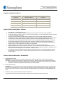
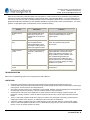
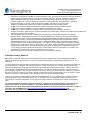
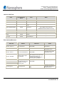
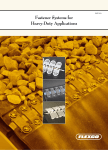
1
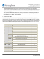
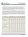
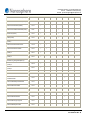
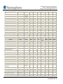
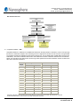
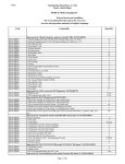
Customer Service or Technical Service:
Phone: 1-888-837-4436 (toll free)
E-Mail: [email protected]
Verigene® Respiratory Virus Plus Nucleic Acid Test on the Verigene® System
20-005-020 (Test Kit) ● 20-012-020 (Amplification Kit)
INTENDED USE
The Verigene® Respiratory Virus Plus Nucleic Acid Test (RV+) on the Verigene® System is a qualitative nucleic acid multiplex test
intended to simultaneously detect and identify multiple respiratory virus nucleic acids in nasopharyngeal (NP) swab specimens
from individuals with signs and symptoms of respiratory tract infection. The following virus types and subtypes are identified using
the RV+: Influenza A, Influenza A subtype H1, Influenza A subtype H3, 2009 H1N1, Influenza B, Respiratory Syncytial Virus
(RSV) subtype A, and RSV subtype B. The test is not intended to detect Influenza C virus. Detecting and identifying specific viral
nucleic acids from individuals exhibiting signs and symptoms of respiratory infection aids in the diagnosis of respiratory viral
infection, if used in conjunction with other clinical and laboratory findings.
Negative results for Influenza A, Influenza B, or RSV do not preclude influenza virus or RSV infection and should not be used as
the sole basis for diagnosis, treatment, or patient management decisions. Conversely, positive results do not rule-out bacterial
infection or co-infection with other viruses. The agent detected may not be the definite cause of disease. The use of additional
laboratory testing and clinical presentation must be considered in order to obtain the final diagnosis of respiratory viral infection.
Performance characteristics for Influenza A Virus were established when Influenza A/H3, A/H1, and 2009 H1N1 were the
predominant Influenza A viruses circulating. These characteristics may vary when other Influenza A viruses are emerging.
If infection with a novel Influenza A virus is suspected based on current clinical and epidemiological screening criteria
recommended by public health authorities, specimens should be collected with appropriate infection control precautions used
specifically for novel virulent influenza viruses and sent to state or local health department for testing. Viral culture should not be
attempted in these cases unless a BSL 3+ facility is available to receive and culture specimens.
BACKGROUND INFORMATION AND CLINICAL UTILITY
The respiratory tract is one of the most common sites for infections as it comes into contact with pathogens frequently.
Influenza A and B viruses and RSV are collectively responsible for a majority of respiratory illnesses and cause significant
1, 2, 3, 4
morbidity and mortality.
Infections with Influenza A and B viruses often result in the respiratory illness commonly referred to as the ‘flu.’ Flu is highly
contagious, and, according to the CDC, 5-20% of the population contract the flu each year. Over 200,000 people are
5
hospitalized, and between 3,000 and 49,000 people die of complications each year, depending on the severity of the season.
Symptoms include fever, cough, headache, body aches, congestion, and fatigue. Flu can lead to serious complications such as
pneumonia, bronchitis, sinus infections, and a general worsening of chronic conditions.6
In the spring of 2009 a novel quadruple-reassortant virus, now known as 2009 H1N1 Influenza, emerged in North America and
quickly spread, becoming a global pandemic by the summer of 2009.7 According to CDC estimates, the virus infected between
8
43 million and 89 million people between April 2009 and April 2010. Importantly, this Influenza A subtype was found to be
9
susceptible to the antiviral drug oseltamivir (brand name Tamiflu), while antiviral resistance varied among other Influenza A
10
subtypes. Thus, treatment decisions may be impacted by the timely availability of Influenza A subtyping information.
Respiratory Syncytial Virus (RSV) infection is the most common cause of bronchiolitis and pneumonia in children under 1 year of
age in the United States. Each year 75,000 to 125,000 children in this age group are hospitalized due to RSV infection.
Symptoms of RSV infection include coughing, sneezing, runny nose, fever, and decrease in appetite. RSV is also recognized as
a serious contributor to respiratory ailments in the aged and immunocompromised demographic. Flu and RSV occur as seasonal
5, 11
outbreaks in the United States, generally starting as early as October or November and ending as late as April or May.
Page 1 of 27
027-00024-02 Rev. B
Customer Service or Technical Service:
Phone: 1-888-837-4436 (toll free)
E-Mail: [email protected]
PRINCIPLES AND PROCEDURES OF THE RV+ AND THE VERIGENE SYSTEM
The RV+ identifies virus-specific nucleic acids for Influenza A virus, Influenza B virus, and Respiratory Syncytial Virus (RSV).
The RV+ targets the following genes within the viruses: matrix gene (Influenza A); hemagglutinin gene (Influenza A subtypes H1
and H3); nucleoprotein gene (Influenza A subtype 2009 H1N1); non-structural gene (Influenza B); polymerase gene (RSV A and
RSV B). For each target, a set of primers amplifies a region of the gene, and a set of probes located within the amplified region
detects the generated amplicons by using gold nanoparticle-based detection technology.
The entire RV+ is performed on the Verigene® System, which is a bench-top ‘sample-to-result’ molecular diagnostics workstation
consisting of two instruments: the Verigene® Processor SP and the Verigene® Reader. The Verigene Processor SP automates
the following steps of the RV+: (i) Sample Preparation - Magnetic bead-based viral RNA isolation from nasopharyngeal swab
specimens obtained from symptomatic patients; (ii) Target Amplification – Multiplex RT-PCR-based amplification of the eluted
viral RNA to generate virus-specific amplicons; (iii) Verigene Hybridization Test – Gold nanoparticle probe-based hybridization of
the virus-specific amplicons on a microarray. Gold nanoparticle probes bound specifically to target-containing spots on the
microarray are silver-enhanced and light scatter from the spots is measured on the Verigene Reader and further analyzed to
make decisions regarding the presence (Detected) or absence (Not Detected) of a virus/analyte. The Verigene Reader also
serves as the user interface and stores and tracks sample information throughout the assay process.
The Verigene Processor SP utilizes single-use disposables to perform the RV+, including an Extraction Tray, Amplification Tray,
and Verigene Test Cartridge. A separate Tip Holder Assembly contains two pipette tips that are used to transfer and mix
reagents during the assay. The user tests a sample by loading the single-use disposables into the Verigene Processor SP and
pipetting the sample into the Extraction Tray. The user initiates the test protocol on the Verigene Reader by scanning or entering
the barcode ID located on the Test Cartridge along with sample information. Following assay completion, the user collects data
on the Verigene Reader by scanning the barcode ID on the Test Cartridge and inserting it into the Verigene Reader for analysis.
MATERIALS PROVIDED
A.
Verigene® RV+ Test Kit; Catalog number 20-005-020
•
20 Verigene® RV+ Test Cartridges
•
20 Verigene® RV+ Extraction Trays (with Tip Holder Assemblies)
B.
Verigene® RV+ Amplification Kit; Catalog number 20-012-020
•
20 Verigene® RV+ Amplification Trays
MATERIALS NEEDED BUT NOT PROVIDED
A.
Instruments and Equipment
•
Verigene® Reader; Catalog number 10-0000-02
•
Verigene® Processor SP with Amplification; Catalog number 10-0000-07
•
-70°C and -20°C freezer
•
2 – 8°C refrigerator
•
Micro-pipettors & tips
•
Mini-centrifuge
•
Cooling block
B.
Consumables and Reagents
•
Nylon or Rayon tipped nasopharyngeal swabs (Copan Innovation)
•
Universal Transport Medium (Catalog number 330C; Copan Innovation); Micro Test M5 Viral Transport Medium
(Catalog number R12515; Remel, Inc.)
Page 2 of 27
027-00024-02 Rev. B
Customer Service or Technical Service:
Phone: 1-888-837-4436 (toll free)
E-Mail: [email protected]
STORAGE, HANDLING, STABILITY
Component
Storage Conditions
Comments
Extraction Tray
2 – 8°C
Do not freeze.
Amplification Tray
≤-20°C
Keep frozen.
Test Cartridge
2 – 8°C
Do not freeze.
Tip Holder Assembly
2 – 30°C
Room Temperature.
PRECAUTIONS AND WARNINGS – GENERAL
•
•
•
•
•
•
•
•
•
The RV+ is for in vitro diagnostic use only.
The performance of the test with viruses infecting swine and other animal hosts has not been established.
Federal law restricts this device to sale by or on the order of a physician, or to a clinical laboratory; its use is restricted
to, by, or on the order of a physician.
Performance characteristics of the RV+ have been determined only with nasopharyngeal swab specimens.
If infection with a novel Influenza A virus is suspected based on current clinical and epidemiological screening criteria
recommended by public health authorities, specimens should be collected with appropriate infection control
precautions for novel virulent influenza viruses and sent to state or local health department for testing. Viral culture
should not be attempted in these cases unless a BSL 3+ facility is available to receive and culture specimens.
Handle supplies, reagents, and kits with powder-free gloves at all times to avoid contamination and change gloves
between removal of used disposables and loading of new disposables.
Handle samples carefully. Open one tube or sample at a time to prevent sample contamination.
Biological samples such as tissues, body fluids, and blood of humans and other animals are potentially infectious.
When handling and/or transporting human respiratory specimens, follow all applicable regulations mandated by local,
state/provincial, and federal agencies for the transport of etiologic agents.
The detection of viral nucleic acid is dependent upon proper specimen collection, handling, transportation, storage
and preparation, including extraction. Failure to observe proper procedures in any one of these steps can lead to
incorrect results.
PRECAUTIONS AND WARNINGS – INSTRUMENTS
A.
General Instrument Safety
WARNING: Use this product only as specified in this document. Using this instrument in a manner not specified by
Nanosphere may result in personal injury or damage to the instrument. Ensure that anyone who operates the instrument:
•
Received instructions in both general safety practices for laboratories and specific safety practices for the instrument.
•
Reads and understands all applicable Material Safety Data Sheets (MSDS).
B.
Electrical Shock Hazard
WARNING: Severe electrical shock can result from operating the instrument without its instrument covers or back panels in
place. Do not remove instrument covers or panels. High-voltage contacts are exposed when instrument covers or panels
are removed from the instrument. If service is required, contact Nanosphere Technical Support at 1-888-VERIGENE (8374436).
C. Maintenance of the Verigene Reader and Verigene Processor SP
For general maintenance and cleaning instructions, please refer to the Verigene System User’s Manual.
Page 3 of 27
027-00024-02 Rev. B
Customer Service or Technical Service:
Phone: 1-888-837-4436 (toll free)
E-Mail: [email protected]
PRECAUTIONS AND WARNINGS – REAGENTS AND TEST CARTRIDGES
A.
Toxicity of Reagents
•
Exposure to chemicals sealed inside the Test Cartridge is hazardous in case of skin contact and of ingestion.
Protective disposable gloves, laboratory coats, and eye protection should be worn when handling specimens,
Extraction Trays, Amplification Trays, and Verigene Test Cartridges.
•
See Material Safety Data Sheets (MSDS) for toxicity information. Material Safety Data Sheets (MSDS) are available
upon request from Nanosphere, Inc.
B.
Waste Disposal
•
Dispose unused reagents and waste in accordance with federal, state, and local regulations.
METHODS
A.
Specimen Collection & Storage
•
Use Nylon or Rayon tipped nasopharyngeal swabs for specimen collection.
•
Place swab into Viral Transport Medium. Break swab shaft and cap the tube.
•
Inadequate or inappropriate specimen collection, storage, or transport may yield false negative results.
•
Training in specimen collection and handling is highly recommended because of the importance of specimen quality.
•
Store specimens refrigerated at 2 – 8ºC for up to 72 hours before processing. Store leftover specimens at < -70ºC.
•
Transport human respiratory specimens refrigerated at 2 – 8ºC. When transporting human respiratory specimens,
ensure that all applicable regulations for the transport of etiologic agents are met.
B. RV+ Test Procedure
This section has step-wise instructions for testing samples using the RV+ assay on the Verigene System. Please refer to the
Verigene System User’s Manual for additional details on performing tests on the Verigene System including daily maintenance.
1.
Create a Verigene Session
a. Login to the system as a ‘user’.
b. From the Menu Bar, Session tab, select Start New Session. The Session Setup window will appear.
c. Touch the Session ID button and enter information by using the data entry keyboard. The Session ID can be
any unique identifier in a format defined by the lab. The operator ID is automatically entered as the currently
logged in operator.
d. Touch the Processing option on the Navigation Bar at the bottom of the screen.
2.
Performing a RV+ test
a. For each sample to be tested, retrieve an RV+ Test Kit and an RV+ Amplification Kit from storage and set aside
at room temperature. Each RV+ Test requires an RV+ Test Cartridge, RV+ Extraction Tray (and Tip Holder
Assembly), and an RV+ Amplification Tray.
b. Open the Drawer Assembly of the Verigene Processor SP by pressing the open/close button on the front of the
instrument.
c. Lift the Drawer Clamp so that the trays and Tip Holder Assembly can be inserted into the Verigene Processor
SP.
d. Shake the Extraction Tray to mix the reagents and tap to settle the reagents. Place the Extraction Tray onto the
Extraction Module.
e. Remove the Tip Holder Assembly from the plastic pouch and place the assembly onto the Tip Module.
f. Thaw the Amplification Tray, gently vortex, and tap to settle the reagents. Place the Amplification Tray onto the
Amplification Module.
g. Lower the Drawer Clamp over the trays and latch it to secure the trays in their positions. NOTE: If the Drawer
Clamp is not latched appropriately, the drawer will not close.
h. Set the RV+ Test Cartridge on a level surface and remove the cover that seals the Test Cartridge by holding
the Substrate Holder handle with the left hand, pressing the palm of the right hand along one edge, and using
fingers to lift up the opposite edge.
Page 4 of 27
027-00024-02 Rev. B
Customer Service or Technical Service:
Phone: 1-888-837-4436 (toll free)
E-Mail: [email protected]
i.
Before loading the Test Cartridge into the Verigene Processor SP, scan the RV+ Test Cartridge identification
number into the Verigene Reader using the attached barcode scanner. Upon being prompted, enter sample
information into the Verigene Reader.
j. The Verigene Reader will display the viral targets for which results will be reported upon successful completion
of the assay. De-select any undesired targets from the list. NOTE: Once the test process is initiated the results
for any de-selected targets cannot be retrieved
k. Select viruses and/or virus subtypes to be tested.
l. Tap the Test Cartridge and insert into Verigene Processor SP. NOTE: If the Test Cartridge is not seated
properly, the drawer will not close.
m. Pipette 200 μL of sample into the Sample Well of the Extraction Tray.
n. Close the drawer of the Verigene Processor SP by pressing the open/close button on the front of the
instrument. The Verigene System will perform a series of checks and automatically initiate the test process.
o. Once the test process is complete, remove the RV+ Test Cartridge from the Verigene Processor SP.
p. Remove the Reagent Pack from the Substrate Holder.
NOTE: Handle only the Substrate Holder. Do not touch the substrate’s surface.
q. Re-scan the RV+ Test Cartridge identification number into the Verigene Reader using the attached barcode
scanner.
r. Immediately before analysis, remove the protective tape from the back of the Substrate Holder and insert the
Substrate Holder into the Verigene Reader for analysis. Refer to the Verigene System User’s Manual for
specific instructions on starting the analysis and obtaining results.
s. Open the Drawer Clamp and remove the used trays and Tip Holder Assembly from the Verigene Processor SP.
t. Dispose of each consumable in the appropriate waste receptacle.
QUALITY CONTROL
Quality control, as a component of an overall quality assurance program, consists of tests and procedures for monitoring and
evaluating the analytical performance of a measurement system to ensure the reliability of patient test results.
A.
Quality Control - Verigene System
The Verigene System uses a series of automated on-line quality measurements to monitor instrument functionality, software
performance, fluidics, test conditions, reagent integrity, and procedural steps each time a test is performed. A series of
automated on-line procedural checks guide the user through the testing process each time a test is performed. RV+ test
barcode and sample information are linked upon entry into the Verigene Reader to help prevent misreporting of results.
B.
Quality Control – RV+
The RV+ is a ‘sample-to-result’ detection system wherein viral RNA is isolated from nasopharyngeal swabs and amplified
prior to specific detection on a microarray housed within the Test Cartridge. All reagents are prepackaged in single use
disposable reagent trays and cartridges to prevent reagent dispensing errors. Several layers of controls built into the RV+
ensure that failures at any step within the RV+ are identified during the procedure or in the end-point image analysis of the
Test Cartridge.
An MS2 bacteriophage sample processing control (IC2) is added to each sample automatically prior to extraction to monitor
failure in the Sample Extraction and Target Amplification steps (see Table below). An Inhibition Control (IC1) is included in
the Primer Mix reagent and added automatically to monitor PCR inhibition. A valid ‘Call’ is made only after both inhibition
control (IC1) and process control (IC2) are verified during analysis of each test, signifying that the extraction and target
amplification processes performed correctly. If IC1 or IC2 are ‘Not Detected’ a ‘No Call – INT CTL 1’ or a ‘No Call – INT CTL
2’ is provided respectively. If both IC1 and IC2 are ‘Not Detected’, a ‘NO CALL – INT CTL’ result is provided. The following
exception exists: IC2 detection alone is sufficient for a valid call if any of the viral targets are also detected; there is no
requirement for IC1 to also be ‘Detected’. The recourse for the ‘No Call – INT CTL decision is to repeat the RV+ test. In
addition, separate internal positive and negative controls are present within each Test Cartridge that inform regarding the
proper functioning of the Test Cartridge.
C. External Controls
Regardless of the choice of quality control materials, all external quality control requirements and testing should be
performed in conformance with local, state, and federal regulations or accreditation organizations as applicable and should
Page 5 of 27
027-00024-02 Rev. B
Customer Service or Technical Service:
Phone: 1-888-837-4436 (toll free)
E-Mail: [email protected]
follow the user’s laboratory’s standard quality control procedures. External Controls can be prepared by using Influenza A,
Influenza B, and RSV viral particles diluted in negative clinical matrix or Viral Transport Media. It is recommended that the
user refer to CLSI document C24-A2, Statistical Quality Control for Quantitative Measurements: Principles and Definitions:
[Approved Guideline-Second Edition] or other published guidelines for general quality control recommendations. For further
guidance on appropriate quality control practices, refer to 42CFR 493.1202(c).
Control
Description
Function
Inhibition Control
(IC1)
Double-stranded DNA target
present in the primer mix.
Amplified with every RT-PCR
reaction.
Controls for PCR inhibition due to sampleor process-related inhibitors or due to
reagent failures.
Process Control
(IC2)
MS2 bacteriophage with an
intact viral RNA genome.
Added automatically to each
test sample including external
positive and negative controls.
Controls for sample isolation step (or
nucleic acid extraction step) and the
RT-PCR step.
External Positive
Control
Any of the three viral particles
(Influenza A, Influenza B, or
RSV).
Serve as external controls for the extraction,
target amplification, and detection steps.
Used to verify reagent performance: (a)
during installation, system validation, and
when troubleshooting dictates (see
Verigene System User’s Manual); (b) to
verify the performance of a new lot/batch of
reagents; (c) when the integrity of storage
conditions is in question.
External Negative
Control
Viral Transport Media
Controls for reagent and/or environmental
contamination.
TROUBLESHOOTING
Refer to the Troubleshooting section of the Verigene System User’s Manual.
LIMITATIONS
•
•
•
•
•
•
•
Performance characteristics of this product were determined with nasopharyngeal swab specimens only.
A trained health care professional should interpret assay results together with the patient’s medical history, clinical signs
and symptoms, and the results of other diagnostic tests.
Viral nucleic acid may persist in vivo, independent of virus viability. Detection of analyte target(s) does not imply that the
corresponding virus(es) are infectious, or are the causative agents for clinical symptoms.
The detection of viral nucleic acid is dependent on proper specimen collection, handling, transport, storage, and
preparation, including extraction. Failure to observe proper procedures in any of these steps could lead to incorrect
results.
There is a risk of false negative results due to sequence variants in the viral targets of the assay, procedural errors,
amplification inhibitors in the specimen, or inadequate viral concentration for amplification.
A specimen yielding a negative result may contain respiratory viruses other than Influenza A, Influenza B, or RSV.
There is a risk of false positive results due to cross-contamination by target viruses, their nucleic acids or amplified
product, or from non-specific signals in the assay.
Page 6 of 27
027-00024-02 Rev. B
Customer Service or Technical Service:
Phone: 1-888-837-4436 (toll free)
E-Mail: [email protected]
•
•
•
•
•
•
•
•
Performance characteristics of the RV+ in a paired comparison study of fresh and 3x ‘freeze thaw’ cycles demonstrated
positive percent agreement of 100% for Influenza A, Influenza A subtype H1, Influenza A subtype H3, Influenza A
subtype 2009 H1N1, Influenza B, RSV A and RSV B. The negative percent agreement was 100% Influenza A,
Influenza A subtype H1, Influenza A subtype H3, Influenza A subtype 2009 H1N1, Influenza B, RSV A and RSV B.
However, freeze-thaw cycles may compromise specimen stability, especially at very low virus titers.
The RV+ is a qualitative test and does not provide the quantitative value of the detected organism.
The RV+ has not been evaluated for patients without signs and symptoms of upper respiratory infection.
The RV+ has not been evaluated for monitoring treatment of Influenza or RSV infection.
The RV+ has not been evaluated for screening of blood or blood product for the presence of influenza.
The affect of interfering substances has only been evaluated for those listed within. Interference by substances other than those
described can lead to erroneous results.
The assay performance has not been established in individuals who received nasally administered or injectable
influenza vaccine. FluMist Influenza Vaccine Live, Intranasal (MedImmune) contains live attenuated reassortants of
each of the three strains: A/California/7/2009 (H1N1), A/Perth/16/2009 (H3N2), and B/Brisbane/60/2008. FluMist is
administered intranasally. FluLaval (GlaxoSmithKline/ID Biomedical) is administered as an intramuscular shot and
contains inactivated strains of each of the three strains: A/California/7/2009 (H1N1), A/Victoria/210/2009 (H3N2) (an
A/Perth/16/2009-like virus), and B/Brisbane/60/2008. In the dilution series for FluMist, Influenza A and the H1 and H3
5
subtypes were not detected at or below an approximate 1:5x10 dilution while Influenza B was not detected at or below
6
an approximate 1:5x10 dilution. In the dilution series for FluLaval, the H1 subtype was not detected at or below the
5
6
1:1x10 dilution, Influenza A and the H3 subtype were not detected at or below an approximate 1:1x10 dilution, and
7
Influenza B was not detected at or below an approximate 1:1x10 dilution.
The assay performance has not been established in immunocompromised individuals.
INTERPRETATION OF RESULTS
RV+ provides a qualitative result for the presence (Detected) or absence (Not Detected) of the following viruses/analytes:
Influenza A, Influenza B, RSV A, RSV B. For Influenza A positive samples, RV+ provides subtyping information as H1, H3, or
2009 H1N1.
In order to obtain results, the intensities from target-specific oligonucleotide spots on the microarray are imaged and analyzed on
the Verigene Reader. Additionally, two Internal Controls, IC1 – inhibition control and IC2 – process controls guide decisions
regarding the validity of the test process and their presence is verified before a valid result is provided. Together, they ensure
that the isolation and amplification steps performed satisfactorily. If the internal controls fail verification, a No Call – INT CTL 1
(for IC1 failure) or a No Call – INT CTL 2 (for IC2 failure) or a No Call – INT CTL (for failure of both IC1 and IC2) are provided. If
the internal controls are verified, the presence or absence of individual viruses is reported based on the cut-off criteria. For each
valid test, a ‘Detected’ or ‘Not Detected’ result is provided for each of the viruses/analytes in the RV+.
If the specimen contains a novel Influenza A virus that is unsubtypeable for H1, H3, or 2009 H1N1, RV+ provides a Detected
result for Influenza A and a Not Detected result for H1, H3, and 2009 H1N1; the unsubtypeable result for Influenza A is an
inferred result or ‘inferred unsubtypeable’. A fresh specimen should be tested for confirmation of the ‘inferred unsubtypeable’
result.
For assays that differentiate between Influenza A subtypes, a positive test result for Influenza A in the presence of a
negative test result for an Influenza subtype (i.e., H1 or H3 or 2009 H1N1) necessitates immediate notification of
appropriate local, state or federal public health authorities to determine necessary measures for verification of results in
accordance with the MMWR notice (http://www.cdc.gov/mmwr/preview/mmwrhtml/mm5613a4.htm and
http://www.cste.org/ps/2007pdfs/novelfluanndssjan10final23.pdf), to determine whether the questionable Flu A
specimen represents a novel strain of Influenza A.
Page 7 of 27
027-00024-02 Rev. B
Customer Service or Technical Service:
Phone: 1-888-837-4436 (toll free)
E-Mail: [email protected]
Calls for Valid Tests
Virus
Internal Controls
Status
Calls
Notes
INFA/H1
Verified
Influenza A; H1
--
INFA/H3
Verified
Influenza A; H3
--
INFA/2009H1N1
Verified
Influenza A; 2009
H1N1
--
INFA UNSUBTYPEABLE
Verified
Influenza A
‘Inferred unsubtypeable’ Influenza A strain - rare
event. Repeat recommended (see
INTERPRETATION OF RESULTS section)
INFB
Verified
Influenza B
--
RSVA
Verified
RSV A
--
RSVB
Verified
RSV B
--
No Target
Verified
Not Detected for all
viruses/analytes
--
Error Calls and Recourse
Error Call
Reason
Recourse
Notes
No Call – INT CTL 1
IC1 Not Detected.
Repeat RV+
Inhibition during Target
Amplification
No Call – INT CTL 2
IC 2 Not Detected
Repeat RV+
Processing and/or Target
Amplification Issues
No Call – INT CTL
IC1 and IC2 Not Detected
Repeat RV+
Processing and/or Target
Amplification Issues
Reader unable to image
Test Substrate
Ensure protective silver tape has
been removed from the back of the
Test Substrate. Adjust Test
Substrate, repeat image analysis. If
the call persists, repeat RV+
--
An inability to obtain the
test result because of high
variability in the targetspecific signals.
Repeat RV+
--
Pre-analytical error
Power cycle ProcessorSP. Repeat
RV+.
No Call – NO GRID
No Call – VARIATION
No Call – BKGD
No Call – NEG CTL
Processing Error
Internal checks within the
Processor SP detected an
unexpected event.
Page 8 of 27
027-00024-02 Rev. B
Customer Service or Technical Service:
Phone: 1-888-837-4436 (toll free)
E-Mail: [email protected]
EXPECTED VALUES
While the timing and duration of influenza and RSV seasons can vary, the fall and winter months are the peak times in the US.
RSV infections in the US typically occur during annual community outbreaks in the late fall, winter and early spring, and there
may be variation in the timing of outbreaks between regions and between communities in the same region.
During the 2008-2009 and 2009-2010 flu seasons, 21% of 519,543 samples and 20% of 456,302 samples tested for influenza
were positive for either Influenza A or Influenza B [based on data from laboratories in the US which collaborated with World
12
Health Organization (WHO) and National Respiratory and Enteric Virus Surveillance System (NREVSS)]. During the 200613
14
2007 and 2007-2008 flu seasons, the prevalence was 13% of 179,268 samples tested and 18% of 225,329 samples tested,
respectively.
According to data reported to the NREVSS, the prevalence of RSV was 15%15 in the 404,798 samples tested for RSV during the
2008-2009 season and 16%16 in the 369,944 samples tested during the 2007-2008 season.
In the RV+ multi-site methods comparison study, which analyzed 1022 samples collected during the 2008-2009 and 2009-2010
flu season, the prevalence of Influenza A was 30.0%, for Influenza B was 4.3%, and of RSV was 10.2%. No dual infections were
detected by culture and DFA, but 0.2% (or 2 specimens) of the total infections were found to contain dual infections by the RV+
(and confirmed subsequently by bi-directional sequencing); one specimen was positive for RSV A and RSV B and a second
specimen was positive for Influenza A and RSV A. It is recommended that the samples undergo repeat testing if nucleic acids
from all three analytes, Influenza A, Influenza B, and RSV are detected in a single sample.
The age distribution of the patient population in the RV+ multi-site methods comparison study is presented in the Table below:
Age Categorization (yrs)
Number of Subjects
Proportion (%)
0-2
269
26.3%
3-5
118
11.5%
6-11
147
14.4%
12-18
124
12.1%
19-64
297
29.1%
≥ 65
67
6.6%
All Ages
1022
100%
PERFORMANCE CHARACTERISTICS OF RV+
A.
Method Comparison Studies for RV+
A total of 1022 nasopharyngeal swab specimens were collected prospectively during the 2008-2009 and 2009-2010
respiratory seasons for routine influenza or RSV testing by DFA/culture methods. The residual specimens were frozen and
later tested at three clinical sites (between Site 1 – 314; Site 2 – 385; Site 3 – 323), using the RV+. The results for each
target, including the Sensitivity and Specificity, for the comparison study, are presented in the following Tables. The RV+
performance was compared to a culture-based reference method followed by direct fluorescent antibody (DFA) identification
of all culture positive specimens. A small number of specimens (see footnotes in the 2x2 tables) were tested by using an
Page 9 of 27
027-00024-02 Rev. B
Customer Service or Technical Service:
Phone: 1-888-837-4436 (toll free)
E-Mail: [email protected]
FDA-cleared Nucleic Acid Amplification Test (NAAT). Subtyping results for Influenza A and RSV specimens and discordant
results between the RV+ and the reference method were confirmed and/or analyzed respectively by using bi-directional
sequencing at an independent reference laboratory (described in Table footnotes).
Samples with an initial ‘No Call’ result were re-tested successfully by following the recommendations in the ‘Interpretation of
Results’ section. A total of 34 samples (3.3%) generated a “No Call” result; all but two of the samples (0.2%) resolved upon
retest. A total of 25 samples (2.4%) resulted in a pre-analysis error (‘pre-ae’). A ‘pre-ae’ may occur due to a number of
causes including a user- initiated procedure termination (for example, procedure termination upon realizing that an incorrect
sample was loaded) or an instrument-initiated procedure termination (for example, because internal checks detect an
unexpected event), or non-availability of the Test Substrate for analysis (due to test consumable breakage, etc). All the
specimens with an initial ‘pre-ae’ result resolved upon retest.
Influenza A
A: Influenza A Results: RV+ vs. Culture/DFA
Culture/DFA
INFA
Positive
RV+
Positive
311
Negative
4
Total
a, c
d, e, f
315
Negative
Total
Total
48 b
359
Sensitivity = 98.7% (96.8%-99.5%) 95% CI
659
663
Specificity = 93.2% (91.1%-94.8%) 95% CI
707
1022
g
B: Influenza A Subtype H3 Results: RV+ vs. Culture/DFA/Sequencing
Culture/DFA/Sequencing
INFA/H3
RV+
Positive
Negative
Total
Total
Positive
108
0
108
Sensitivity = 100% (96.6%-100%) 95% CI
Negative
0
909
909
Specificity = 100% (99.6%-100%) 95% CI
Total
108
909
1017
Page 10 of 27
027-00024-02 Rev. B
Customer Service or Technical Service:
Phone: 1-888-837-4436 (toll free)
E-Mail: [email protected]
C: Influenza A Subtype H1 Results: RV+ vs. Culture/DFA/Sequencing
Culture/DFA/Sequencing
INFA/H1
RV+
Positive
Negative
Total
Positive
39
1
40
Sensitivity = 100% (91.0%-100%) 95% CI
Negative
0
977
977
Specificity = 99.9% (99.4%-100%) 95% CI
Total
39
978
1017
c
Total
D: Influenza A Subtype 2009 H1N1 Results: RV+ vs. Culture/DFA/Sequencing
Culture/DFA/Sequencing
INFA/2009 H1N1
RV+
Positive
Negative
Total
Total
Positive
206
0
206
Sensitivity = 99.5% (97.3%-99.9%) 95% CI
Negative
1
810
811
Specificity = 100% (99.5%-100%) 95% CI
Total
207
810
1017
c
E: Influenza A and Subtype Discordant Summary
a
1 specimen was positive for Influenza A by both culture and RV+. No sub-typing was observed on the RV+ result.
Also, the specimen failed subtype sequencing for H1, H3 and 2009 H1N1. b 4 specimens were Influenza A positive
by RV+. No sub-typing was observed on the RV+. All 4 specimens were culture negative and failed sequencing. c 1
specimen was Influenza A/H1 by RV+ and positive for Influenza A by culture. Sequencing resulted in a positive
result for Influenza A/2009H1N1. d 1 specimen was negative by RV+ for Flu A and positive for Flu B. Initial
culture/DFA of the specimen was Flu A positive. By sequencing, the specimen was positive for Flu B and negative
for Flu A. By NAAT and upon repeat culture/DFA, the specimen was positive for Flu B and negative for Flu A and
e
1 specimen was negative for Flu A and positive for RSV B by RV+. By initial culture/DFA, the specimen
RSV.
was Flu A positive. By sequencing the specimen was positive for RSV B and negative for Flu A. By NAAT and upon
repeat culture/DFA the specimen was positive for RSV and negative for Flu A and Flu B. f 2 specimens were
negative by RV+ but Flu A positive by culture. By sequencing both specimens were negative for Flu A. Both
g
specimens were negative for Flu A (and negative for Flu B and RSV B) by NAAT. 5 specimens failed sequencing
or were not subtyped by the RV+ assay and are not included in the Subtyping Tables.
Page 11 of 27
027-00024-02 Rev. B
Customer Service or Technical Service:
Phone: 1-888-837-4436 (toll free)
E-Mail: [email protected]
Influenza B
A: Influenza B Results: RV+ vs. Culture/DFA results
Culture/DFA
INFB
Positive
RV+
Negative
Positive
43
3
Negative
0
Total
43
a, b
Total
Total
46
Sensitivity = 100% (91.8%-100%) 95% CI
976
976
Specificity = 99.7% (99.1%-99.9%) 95% CI
979
1022
B: Influenza B Discordant Summary
a
1 specimen was positive for Influenza B by RV+ and negative for Influenza B by culture/DFA. This specimen was
tested by NAAT assay and found to be positive for Influenza B. Specimen was positive for Influenza B by
sequencing and upon repeat culture/DFA. b 2 specimens were positive for Influenza B by RV+ and negative for
Influenza B by culture. Both specimens were positive for Influenza B by sequencing.
RSV
A: RSV Results: RV+ vs. Culture/DFA results
Culture/DFA
RSV
Positive
RV+
Total
Total
a, d
109
Sensitivity = 97.2% (92.1%-99.0%) 95% CI
b, c
910
913
Specificity = 99.5% (98.7%-99.8%) 95% CI
107
915
1022
Positive
104
Negative
3
Total
Negative
5
e
Page 12 of 27
027-00024-02 Rev. B
Customer Service or Technical Service:
Phone: 1-888-837-4436 (toll free)
E-Mail: [email protected]
B: RSV A Subtype Results: RV+ vs. Culture/DFA/Sequencing
Culture/DFA/Sequencing
RSV A
Positive
RV+
f
Negative
Total
0
57
Sensitivity = 100% (93.7%-100%) 95% CI
Specificity = 100% (99.6%-100%) 95% CI
Positive
57
Negative
0
962
962
Total
57
962
1019
Total
C: RSV B Subtype Results: RV+ vs. Culture/DFA/Sequencing
Culture/DFA/Sequencing
RSV B
Positive
RV+
f
Negative
Total
0
53
Sensitivity = 100% (93.2%-100%) 95% CI
Specificity = 99.9% (99.6%-100%) 95% CI
Positive
53
Negative
0
966
966
Total
53
966
1019
Total
D: RSV Discordant Summary
a
1 specimen was positive for RSV B by RV+ and by sequencing but negative for RSV by culture/DFA. Specimen
was positive for RSV by NAAT and upon repeat culture/DFA. b 1 specimen was negative for RSV by RV+ and by
c
sequencing but positive for RSV by culture and NAAT assay. 2 specimens were negative for RSV by RV+ and
positive for RSV by culture. The specimens were negative for RSV by NAAT assay and failed sequencing. d 4
specimens were positive for RSV A or RSV B by RV+ but negative by culture: 1 was positive for RSV A and 3 were
positive for RSV B by RV+ and by sequencing. e 3 specimens failed subtype sequencing and are not included in the
f
Subtyping Tables. 1 specimen was positive for both RSV A and RSV B (dual infection) by RV+ and was culture
positive for RSV. By sequencing specimen was positive for both RSV A and RSV B.
Page 13 of 27
027-00024-02 Rev. B
Customer Service or Technical Service:
Phone: 1-888-837-4436 (toll free)
E-Mail: [email protected]
B.
Reproducibility / Precision Studies
The Reproducibility/Precision study was conducted at three sites (two external and one internal) to investigate the
inter-laboratory reproducibility of the RV+. The reproducibility panels (see Table below) were developed by using 6 unique
virus strains that together represented all of the analytes in the RV+. The 6 virus strains were combined to generate
combinations such that each virus strain was represented at 3 levels: HN- High Negative, LP - Low Positive, and
MP - Moderate Positive. For two of the unique samples, virus strains were combined as in the case of INFA/H3 and RSVA
and Influenza B and RSV B. For the Reproducibility/Precision study, the Test Panel comprised the 12 unique samples in
duplicate for a total of 24 samples. The Test Panel samples were then divided equally into Panel A (12 samples) and
Panel B (12 samples).
Viral Panel
Virus Strain
Level
High Negative
INFB + RSVB
Low Positive
Moderate Positive
A
High Negative
INFA/2009H1N1-MT
Low Positive
Moderate Positive
High Negative
INFA/H1-MT
Low Positive
Moderate Positive
B
High Negative
INFA/H3 + RSVA
Low Positive
Moderate Positive
Samples
1
2
3
4
5
6
7
8
9
10
11
12
1
2
3
4
5
6
7
8
9
10
11
12
At the two external study sites, Test Panel A and Test Panel B were tested on separate days. Testing each day involved 2
operators testing the same Test Panel in two replicate runs. Both Test Panels A and B were tested for a total testing period
of six non-consecutive days.
The internal site ran a 12-day precision study and utilized the same four unique samples at the three levels. The complete
Test Panel (Test Panel A and B) was tested separately by two operators. The precision study was run for a total testing
period of 12 non-consecutive days. The cumulative results from the Reproducibility/Precision Studies are summarized.
Data is presented for each individual site and then combined to provide collective results. The Table contains the agreement
between the expected results and the obtained results for each virus strain in the Test Panel. These are grouped further
based on their concentration levels (MP, LP, and HN). NOTE: Though some samples in the Test Panels contained
combinations of viruses, results are stratified by individual virus strains.
Page 14 of 27
027-00024-02 Rev. B
Customer Service or Technical Service:
Phone: 1-888-837-4436 (toll free)
E-Mail: [email protected]
Site
Specific Panel
Member
INFA/H1-MT
INFA/H3
INFA/2009H1N1
-MT
INFB
RSVA
RSVB
a
Site 1
Site 2
Site 3
All 3 Sites
Level
Agreement
Agreement
Agreement
Total
Agreement
%
Agreement
95% CI
MP
12/12
12/12
70/72
97.2%
90.4% - 99.2%
LP
12/12
12/12
48/48
72/72
100%
94.9% - 100%
HN
11/12
3
12/12
48/48
71/72
98.6%
92.5% - 99.8%
MP
12/12
12/12
48/48
72/72
100%
94.9% - 100%
LP
12/12
12/12
47/48
4
71/72
98.6%
92.5% - 99.8%
HN
12/12
12/12
48/48
72/72
100%
94.9% - 100%
MP
12/12
12/12
48/48
72/72
100%
94.9% - 100%
LP
12/12
12/12
48/48
72/72
100%
94.9% - 100%
HN
12/12
12/12
47/48
5
71/72
98.6%
92.5% - 99.8%
MP
12/12
12/12
48/48
72/72
100%
94.9% - 100%
LP
12/12
12/12
48/48
72/72
100%
94.9% - 100%
HN
12/12
11/12
a
48/48
71/72
98.6%
92.5% - 99.8%
MP
12/12
12/12
48/48
72/72
100%
94.9% - 100%
LP
12/12
12/12
70/72
97.2%
90.4% - 99.2%
HN
12/12
12/12
48/48
72/72
100%
94.9% - 100%
MP
12/12
12/12
48/48
72/72
100%
94.9% - 100%
LP
11/12
c
12/12
47/48
d
70/72
97.2%
90.4% - 99.4%
HN
11/12
e
12/12
48/48
71/72
98.6%
92.5% - 99.8%
46/48
46/48
1,2
b,4
b
Expected Calls: One (1) INFB + RSVB HN detected Influenza B. One (1) INFA/H3 + RSVA LP sample detected Influenza A and H3 but
c
d
did not detect RSV A. One (1) INFB + RSVB LP sample detected Influenza B but did not detect RSV B. One INFB + RSVB LP sample
e
detected Influenza B but did not detect RSV B. One INFB + RSVB HN sample detected RSV B but did not detect Influenza B. Additional
1
Positive Calls: One (1) INFA/H1-MT MP sample detected both Influenza A and H1 as expected, but also detected H3 which was
2
unexpected. One (1) INFA/H1-MT MP sample detected both Influenza A and H1 as expected, but also detected H3, Flu B and RSV A
3
which was unexpected. One (1) INFA/H1-MT HN sample detected Influenza A, but did not detect H1 which was expected as this is a high
4
negative sample. However, RSV A was also detected which was unexpected. One INFA/H3 + RSVA LP sample detected both
Page 15 of 27
027-00024-02 Rev. B
Customer Service or Technical Service:
Phone: 1-888-837-4436 (toll free)
E-Mail: [email protected]
5
Influenza A, H3 and RSV A as expected for a low positive sample, but also detected 2009 H1N1 which was not expected. One
INFA/2009H1N1-MT HN sample did not detect Influenza A and 2009 H1N1 which was expected as this is a high negative sample.
However, Influenza B and RSV B were also detected which was not expected. NOTE: All the samples with additional positive calls gave the
expected results for the intended virus; there were no mis-calls. The source of the additional positives is likely cross-contamination artifacts
during the Test Panel preparation.
There were 14 “No Calls” and 2 “pre-analytical errors” in the study. These 16 samples were repeat tested successfully. In
this study the “No Call” rate was 1.6% (14/864) and the “pre-analysis error” failure rate was 0.2% (2/864). Out of the 864
samples tested, the percent agreement for all panel members for the combined sites ranged from 97.2%-100% (95% CI
range from 90.3% - 99.7% to 95.0% - 100.0%, respectively).
C. Analytical Sensitivity – RV+
The analytical sensitivity of RV+ was assessed and confirmed by using virus strains with established titers to represent all
analytes in the test. In order to determine the Limits of Detections (LODs), the strains were serially diluted into negative pools of
nasopharyngeal (NP) swab samples around the estimated LOD and replicates were tested in the RV+. The lowest concentration
level where all the replicates were detected was chosen as the LOD. For confirmation, 20 replicate samples were prepared at the
LOD level chosen. Confirmation of LOD required that at least 19 of the 20 samples tested gave a ‘Detected’ call for the analyte.
The confirmed LOD levels for the different analytes and the associated virus strains are presented.
Virus
INFA
Analyte
INFA
H1
2009H1N1
H3
INFB
INFB
Strain
LOD (TCID50/ML)
Influenza A/Virginia/01/2006 (H1N1)
1
Influenza A/2009H1N1 Clinical Isolate (H1N1)
10
Influenza A/Brisbane/59/2007 Clinical Isolate (H1N1)
5
Influenza A/California/04/2009 (H1N1)
1
Influenza A/Wisconsin/67/05 (H3N2)
0.1
Influenza A/Virginia/01/2006 (H1N1)
5
Influenza A/Brisbane/59/2007 Clinical Isolate (H1N1)
5
Influenza A/2009H1N1 Clinical Isolate (H1N1)
10
Influenza A/California/04/2009 (H1N1)
1
Influenza A/Wisconsin/67/05 (H3N2)
0.1
Influenza B/Wisconsin/2/2006
0.1
Influenza B/Florida/02/06
1
RSVA
RSVA
RSV A Strain A2
10
RSVB
RSVB
RSV B Strain Wash/18537/62
1
Page 16 of 27
027-00024-02 Rev. B
Customer Service or Technical Service:
Phone: 1-888-837-4436 (toll free)
E-Mail: [email protected]
D. Analytical Reactivity
The analytical reactivity of the RV+ was evaluated using multiple strains of Influenza A (7 seasonal H1 strains, 7 seasonal H3
strains, 4 2009 H1N1 strains, 2 swine-origin H1N1 strains, 3 H5N1 strain, and 2 H7N7 strain), 8 Influenza B strains, 2 RSV A
strains and 3 RSV B strains. Viral strains for Influenza A were selected to include strains representing temporal and
geographical diversity. In order to assess analytical reactivity, cultured and titered strains of Influenza A/H3, Influenza A/H1,
Influenza A/2009H1N1, Influenza B, RSV A, and RSV B were diluted in sample matrix (Universal Transport Medium, Copan)
at the concentrations listed in the adjoining Table and tested in triplicate in the RV+. All the Influenza A/H3, Influenza A/H1,
and Influenza A/2009 H1N1 strains yielded expected subtyping results.
Two swine-origin H1N1 strains (of non-human origin), Influenza A/Swine/Iowa/15/30 and Influenza A/Swine/1976/31 gave a
‘Detected’ result for Influenza A and for H1 but gave a ‘Not Detected’ result for 2009 H1N1. Higher concentrations (1- and 2logs) did not cross-react with 2009 H1N1 demonstrating good assay specificity. The H5 and H7 Influenza strains in this study
were grown in culture, titered, and inactivated before they were tested in the RV+. The H5 and H7 strains gave a ‘Detected’
result for Influenza A and a ‘Not Detected’ result for H1, H3, and 2009 H1N1. As expected, these strains were unsubtypeable
in the RV+. Due to the inactivation process used for the H5 and H7 strains, a loss in the original titer was noted. While lower
dilutions than those reported in the Table below were not tested, higher dilutions (1- and 2-logs higher concentrations) were
examined. No cross-reactivity with H1, H3, or 2009 H1N1 was observed at any of these higher dilutions, again demonstrating
good assay specificity.
Virus Strains
Titer
(TCID50/mL)
INFA
H1
H3
2009
H1N1
INFB
RSVA
RSVB
Influenza A
Influenza A/New Caledonia/20/99
(H1N1)
1X10
2
+
+
-
-
-
-
-
Influenza A/PR/8/34 (H1N1)
1X10
2
+
+
-
-
-
-
-
Influenza A1/FM/1/47 (H1N1)
5X10
2
+
+
-
-
-
-
-
Influenza A/NWS/33 (H1N1)
1X10
2
+
+
-
-
-
-
-
Influenza A1/Denver/1/57 (H1N1)
1X10
3
+
+
-
-
-
-
-
Influenza A/Hawaii/15/01 (H1N1)
1X10
2
+
+
-
-
-
-
-
Influenza A/Brisbane/59/2007
Clinical Isolate (H1N1)
1X10
2
+
+
-
-
-
-
-
Influenza A/ Virginia/01/2006 (H1N1)
1X10
2
+
+
-
-
-
-
-
Influenza A/Port Chalmers/1/73
(H3N2)
1X10
2
+
-
+
-
-
-
-
Influenza A/Hong Kong/8/68 (H3N2)
1X10
2
+
-
+
-
-
-
-
Influenza A/Aichi2/68 (H3N2)
1X10
2
+
-
+
-
-
-
-
Influenza A/Victoria/3/75 (H3N2)
1X10
2
+
-
+
-
-
-
-
Page 17 of 27
027-00024-02 Rev. B
Customer Service or Technical Service:
Phone: 1-888-837-4436 (toll free)
E-Mail: [email protected]
Influenza A/Wisconsin/67/05 (H3N2)
1X10
2
+
-
+
-
-
-
-
Influenza A/Hiroshima/52/05 (H3N2)
1X10
2
+
-
+
-
-
-
-
Influenza A/NY/55/04 (H3N2)
1X10
2
+
-
+
-
-
-
-
Influenza A/California/04/2009 (H1N1)
1X10
2
+
-
-
+
-
-
-
Influenza A/2009H1N1/PSF1 Clinical
Isolate 09 (H1N1)
1X10
2
+
-
-
+
-
-
-
Influenza A/Wisconsin/629-D01606/
2009 (H1N1)
1X10
2
+
-
-
+
-
-
-
Influenza A/2009H1N1 Clinical Isolate
(H1N1)
1X10
2
+
-
-
+
-
-
-
Influenza A/Swine/Iowa/15/30*
1X10
2
+
+
-
-
-
-
-
Influenza A/Swine/1976/31*
1X10
2
+
+
-
-
-
-
-
Influenza A/Duck/Hunan/795/02
(H5N1)**
9X10
2
+
-
-
-
-
-
-
Influenza A/Chicken/Korea/IS/2006
(H5N1)**
3X10
3
+
-
-
-
-
-
-
Influenza A/Scaly Breasted
Munia/Hong Kong/2006 (H5N1)**
4X10
1
+
-
-
-
-
-
-
Influenza A/Netherlands/219/2003
(H7N7)**
3X10
4
+
-
-
-
-
-
-
Influenza A/New York/107/2003
(H7N2)**
5X10
0
+
-
-
-
-
-
-
Influenza B/ Wisconsin/2/2006
1X10
2
-
-
-
-
+
-
-
Influenza B/
Florida/02/2006
1X10
2
-
-
-
-
+
-
-
Influenza B/Malaysia/2506/2004
1X10
2
-
-
-
-
+
-
-
Influenza B/Ohio/1/2005
1X10
2
-
-
-
-
+
-
-
Influenza B/ Lee/40
1X10
2
-
-
-
-
+
-
-
Influenza B/GL/1739/54
1X10
2
-
-
-
-
+
-
-
Influenza B/Taiwan/2/62
1X10
2
-
-
-
-
+
-
-
Influenza B/Hong Kong/5/72
1X10
2
-
-
-
-
+
-
-
Influenza B/Maryland/1/59
1X10
2
-
-
-
-
+
-
-
Influenza B
Page 18 of 27
027-00024-02 Rev. B
Customer Service or Technical Service:
Phone: 1-888-837-4436 (toll free)
E-Mail: [email protected]
RSV A
2
-
-
-
-
-
+
-
1X10
2
-
-
-
-
-
+
-
RSV B Strain Wash/18537/62
1X10
2
-
-
-
-
-
-
+
RSV B Strain B-1 Wild Type
1X10
2
-
-
-
-
-
-
+
RSV B Strain 9320
1X10
2
-
-
-
-
-
-
+
RSV A Strain A2
1X10
RSV A Strain Long
RSV B
3
4
* In addition to the above testing, both swine-origin H1N1 strains were tested at 1- and 2-logs higher concentrations (1x10 and 1x10
TCID50/mL). No cross-reactivity to 2009 H1N1 was observed. ** In addition to the above testing, the H5 and H7 strains were tested at
1- and 2-logs higher concentration. No cross-reactivity to any sub-types was observed.
E.
Analytical Specificity/Cross-Reactivity
Analytical specificity studies were performed to assess potential cross-reactivity of the RV+ with respiratory pathogens and
other microorganisms commonly present in the respiratory tract. A total of 53 organisms of interest were identified as
respiratory pathogens with which the majority of the population may be infected. These organisms included 24 bacterial
strains that were tested between 105 to107 cfu/mL and 29 virus strains that were tested between 104 to106 TCID50/mL. The
microorganisms were diluted into a sample matrix (Universal Transport Medium, Copan) at the concentration levels described
in the Table below and tested subsequently in the RV+.
In all the samples tested, a valid test result was provided indicating that the microorganisms did not negatively effect the RV+
processing. Significantly, all the microorganisms tested gave a ‘Not Detected’ call for each analyte on the RV+; the
microorganisms tested did not demonstrate cross reactivity with any of the analytes in the RV+.
Viruses
Human Adenovirus Type 1
Human Adenovirus Type 2
Human Adenovirus Type 3
INFA
H1
H3
2009
H1N1
INFB
RSVA
RSVB
5
-
-
-
-
-
-
-
1.0x10
5
-
-
-
-
-
-
-
1.0x10
6
-
-
-
-
-
-
-
-
-
-
-
-
-
-
Strain
pfu/mL
Adenoid 71
6.2x10
Adenoid 6
G.B.
Human Adenovirus Type 4
RI-67
1.0x10
6
Human Adenovirus Type 5
Adenoid 75
5.5x10
6
-
-
-
-
-
-
-
6.2x10
5
-
-
-
-
-
-
-
1.9x10
5
-
-
-
-
-
-
-
5.5x10
6
-
-
-
-
-
-
-
3.5x10
5
-
-
-
-
-
-
-
1.1x10
5
-
-
-
-
-
-
-
1.1x10
6
-
-
-
-
-
-
-
1.1x10
5
-
-
-
-
-
-
-
9.9x10
4
-
-
-
-
-
-
-
2.0x10
5
-
-
-
-
-
-
-
Human Adenovirus Type 7
Human Adenovirus Type 11
Human Adenovirus Type 14
Human Adenovirus Type 31
Human Adenovirus Type 35
Human Coronavirus (OC43)
Human coronavirus (229E)
Human coronavirus (NL63)
Cytomegalovirus
Gomen
Slobitski
de Wit
1315
holden
OC43
229E
NL63
68-1
Page 19 of 27
027-00024-02 Rev. B
Customer Service or Technical Service:
Phone: 1-888-837-4436 (toll free)
E-Mail: [email protected]
Enterovirus, H. coxsackievirus
B4
J.V.B.
Enterovirus, echovirus 11
Epstein Barr Virus
Herpes Simplex virus Type 1
Measles
Metapneumovirus
Metapneumovirus
Mumps virus
Human Parainfluenza Type 1
Human Parainfluenza Type 2
Human Parainfluenza Type 3
Human Parainfluenza Type 4
Human Rhinovirus Type 1A
-
-
-
-
-
-
-
1.4x10
5
-
-
-
-
-
-
-
-
-
-
-
-
-
-
9
2.2x10
cp/ml
MacIntyre
2.4x10
5
-
-
-
-
-
-
-
1.1x10
6
-
-
-
-
-
-
-
1.1x10
4
-
-
-
-
-
-
-
1.1x10
4
-
-
-
-
-
-
-
3.5x10
5
-
-
-
-
-
-
-
1.1x10
4
-
-
-
-
-
-
-
1.2x10
5
-
-
-
-
-
-
-
2.2x10
5
-
-
-
-
-
-
-
1.1x10
5
-
-
-
-
-
-
-
7.0x10
5
-
-
-
-
-
-
-
1.1x10
5
-
-
-
-
-
-
-
8.0x10
5
-
-
-
-
-
-
-
INFA
H1
H3
2009
H1N1
INFB
RSVA
RSVB
A1
A2
B2
Enders
C35
Greer
C243
M-25
2060
Varicella-Zoster virus
Bacteria
6
B95-8
Edmonston
Metapneumovirus 21
1.9x10
Strain
cfu/mL
3.0x10
6
-
-
-
-
-
-
-
3.0x10
5
-
-
-
-
-
-
-
3.7x10
7
-
-
-
-
-
-
-
1.1x10
6
-
-
-
-
-
-
-
Corynebacterium
pseudodiphtheriticum
8.2x10
7
-
-
-
-
-
-
-
Escherichia coli
1.5x10
7
-
-
-
-
-
-
-
1.0x10
7
-
-
-
-
-
-
-
5.0x10
5
-
-
-
-
-
-
-
1.1x10
6
-
-
-
-
-
-
-
2.1x10
6
-
-
-
-
-
-
-
1.2x10
6
-
-
-
-
-
-
-
6.5x10
7
-
-
-
-
-
-
-
Mycoplasma pneumoniae
3.0x10
7
-
-
-
-
-
-
-
Mycobacterium tuberculosis,
attenuated
2.6x10
6
-
-
-
-
-
-
-
Neisseria gonorrhoeae
5.0x10
6
-
-
-
-
-
-
-
1.0x10
5
-
-
-
-
-
-
-
2.0x10
6
-
-
-
-
-
-
-
3.5x10
6
-
-
-
-
-
-
-
6.1x10
7
-
-
-
-
-
-
-
Acinetobacter Baumannii
Bordetella bronchiseptica
Bordetella pertussis
Chlamydia pneumoniae
CM-1
Haemophilus influenzae
Klebsiella pneumoniae
Lactobacillus acidophilus
Legionella pneumophila
Listeria innocua
Moraxella catarrhalis
Neisseria meningitidis
Proteus vulgaris
Pseudomonas aeruginosa
Staphylococcus aureus
Boston 41501
Page 20 of 27
027-00024-02 Rev. B
Customer Service or Technical Service:
Phone: 1-888-837-4436 (toll free)
E-Mail: [email protected]
Staphylococcus epidermidis
Streptococcus pneumoniae
Streptococcus agalactiae
Streptococcus pyogenes
Streptococcus salivarius
F.
O90R
3.7x10
6
-
-
-
-
-
-
-
9.0x10
6
-
-
-
-
-
-
-
2.0x10
6
-
-
-
-
-
-
-
1.3x10
6
-
-
-
-
-
-
-
5.5x10
7
-
-
-
-
-
-
-
Determination of Clinical Cut-Off and Call Algorithm
The RV+ uses a microarray-based platform in which the viruses, the inhibition control (IC1) and process control (IC2) are
represented by recognition elements on the Test Substrate. The recognition elements or spots are virus-specific
oligonucleotide sequences that bind to the amplified viral targets, which in turn bind to gold nanoparticle probes via additional
recognition elements. A gold nanoparticle probe-specific signal enhancement reaction deposits silver at the virus-specific
spots. The scatter from the spots is detected by the Verigene Reader and registered as signal intensity. In addition to the
above recognition elements, the Test Substrate has spots specific to positive control (PC) and negative control (NC). Three
conditions were identified that together served as a single set of clinical cutoff criteria.
Condition 1: Noise Threshold
Condition 2: Normalized ‘Ratio to Negative Control’ (Ratio-to-NC) – intensity at the virus-specific recognition element
normalized against the intensity values at the negative control elements.
Condition 3: Normalized ‘Ratio to Positive Control’ (Ratio-to-PC) – intensity at the virus-specific recognition element
normalized against the intensity values at the positive control elements.
The Noise Threshold was determined empirically. The cut-offs for the normalized ratios, ‘Ratio-to-NC’ and the ‘Ratio-to-PC’,
were determined by using ROC curves. For a positive ‘Detected’ decision the following criteria apply:
Condition 1: Signal intensity is above the noise threshold
Condition 2: Ratio-to-NC > 0.85
Condition 3: Ratio-to-PC ≥ -0.4
If any one of these criteria is not met, a negative ‘Not Detected’ decision is provided. Criteria set for each of the three
conditions are required to be met for a ‘Detected’ call.
For a result, the decision tree verifies the presence of IC1 and IC2 in conjunction with each of the viruses (see Schematic).
Both IC1 and IC2 signal intensities have to meet the detection criteria for a valid call. A valid ‘Call’ is made only after both
inhibition control (IC1) and process control (IC2) are verified during analysis of each test, signifying that the extraction and
target amplification processes performed correctly. If IC1 or IC2 are ‘Not Detected’ a ‘No Call – INT CTL 1’ or a ‘No Call –
INT CTL 2’ is provided respectively. If both IC1 and IC2 are ‘Not Detected’, a ‘NO CALL – INT CTL’ result is provided. The
following exception exists: IC2 detection alone is sufficient for a valid call if any of the viral targets are also detected; there is
no requirement for IC1 to also be ‘Detected’. The recourse for the ‘No Call – INT CTL’ decision is to repeat the RV+ test. In
addition, separate internal positive and negative controls are present within each Test Cartridge that inform regarding the
proper functioning of the Test Cartridge.
Page 21 of 27
027-00024-02 Rev. B
Customer Service or Technical Service:
Phone: 1-888-837-4436 (toll free)
E-Mail: [email protected]
RV+ Decision Process
RV+
Decision Process
Internal Controls fail
verification
No Call – INT CTL
Repeat RV+
Verify Internal Controls
Internal Controls verified
proceed to viral identification
IF
Target Signal > Noise Threshold AND
Ratio-to-NC > 0.85 AND
Ratio-to-PC ≥ -0.4
IF
Target Signal < Noise Threshold OR
Ratio-to-NC ≤ 0.85 OR
Ratio-to-PC < -0.4
Viral Agent
Detected
Viral Agent
Not Detected
G. Competitive Inhibition – RV+
Competitive inhibition or interference in the RV+ was assessed in clinically relevant co-infections. A set of 12 unique ‘high
titer-low titer’ combination samples, each containing a virus at a high titer and another virus at a low titer, were generated.
The samples represented combinations of Influenza A subtypes (H1, H3, and 2009 H1N1) and RSV A/B viruses (see the
Table below). The levels for individual strains were chosen based on the limits of detection determined for the individual
strains. The high titers for the strains were at least 3 log orders higher than their limits of detection and the low titers were
held close to the limits of detection.
Sample
1
2
High Titer
Virus
RSVA
RSVA
Low Titer
TCID50/mL
Virus
TCID50/mL
2X10
4
INFA/H3
5
2X10
4
INFA/H1
25
INFA/2009H1N1
10
3
RSVA
2X10
4
4
RSVB
5X10
3
INFA/H3
5
5X10
3
INFA/H1
25
5X10
3
5
6
7
8
9
10
11
12
RSVB
RSVB
INFA/2009H1N1
10
2.5X10
3
RSVA
100
INFA/H3
2.5X10
3
RSVB
5
INFA/H1
2X10
4
RSVA
100
2X10
4
RSVB
5
1X10
4
RSVA
100
1X10
4
RSVB
5
INFA/H3
INFA/H1
INFA/2009H1N1
INFA/2009H1N1
Viral Strains: INFA/H3: Influenza A/Wisconsin/67/05; INFA/H1: Influenza A/Virgina/01/2006; INFA/2009H1N1: Influenza
A/California/04/2009; RSV A: RSV A Strain A2; RSVB: RSV B/Wash/18537/62
Page 22 of 27
027-00024-02 Rev. B
Customer Service or Technical Service:
Phone: 1-888-837-4436 (toll free)
E-Mail: [email protected]
H. Fresh vs. Frozen
Freezing virus-containing samples can potentially compromise viral integrity, which may translate to a lower effective
concentration in frozen samples. In order to determine the impact of freezing samples on RV+ performance, unfrozen
negative nasopharyngeal swab samples were obtained and spiked with Influenza A, Influenza B, and RSV viruses to
represent all the analytes in the RV+. Each virus was tested at multiple concentrations. Paired comparisons were conducted
for each sample by testing it before and after freezing. The impact of 2 additional freeze-thaw cycles on the RV+ (for a total of
3 freeze-thaw cycles) was also assessed.
The ‘Fresh-vs-Frozen’ comparison study involved testing a total of 60 negative nasopharyngeal (NP) swab samples across 6
unique virus types representing all the targets and analytes in the RV+. The negative NP swab samples were not pooled;
each virus was represented by 10 unique NP swab samples (10 x 6 = 60 unique samples). Each virus type was tested at 2 or
3 different concentrations to represent levels ~1 log above the limits of detection, ~2 logs above the limit of detection, and
greater than 2 logs above the limit of detection for all but 2 viruses.
Each unique sample was tested 4 times: once while fresh or unfrozen and once after each of the three (3) freeze-thaw cycles.
Logistically, after a sample was spiked with the appropriate virus, an aliquot of this sample was tested in the RV+ while the
sample was ‘fresh.’ The residual sample was frozen by placing the sample at or below -70 °C. For the first freeze thaw, the
residual sample was thawed and an aliquot was tested in the RV+. This constituted the first ‘freeze-thaw’ cycle. This process
was repeated for the second and third ‘freeze-thaw’ cycles. Thus, the study involved testing 60 samples x 4 separate tests (1
fresh + 3 frozen) = 240 samples. The performance of each virus/analyte after each freeze-thaw cycle was compared to the
corresponding result obtained when the sample was ‘fresh’ or unfrozen. The results from this paired comparison showed
100% agreement between the results obtained while the sample was ‘fresh’ and those obtained after each freeze-thaw cycle
at all the concentration levels tested.
Overall, the results showed that the RV+ detected the viruses/analytes correctly even after the sample was subjected to 3
sets of ‘freeze-thaw’ cycles. The viral analytes yielded a positive percentage agreement of 100% after each freeze/thaw cycle
when compared to the results from the corresponding ‘fresh’ samples.
I.
Carry-Over / Cross-Contamination Study
The carry-over study was performed to assess the carry over/cross-contamination of the RV+ by alternately running ‘High
Positive’ samples followed by ‘High Negative’ samples. Multiple Verigene Processors SP s used in the study and each
Processor SP was subjected to a comprehensive set of tests.
Based on the collective data, there was no evidence of cross-contamination. Moreover, there was no evidence of any
cross over within the Verigene Processor SP modules when high titer samples were alternated with low titer samples.
J.
Interferences
The potential inhibitory effect of interfering substances or interferents that may be encountered in nasopharyngeal specimens
on the RV+ was assessed. The viral strains and the titers used in the studies are listed along with the interferents and the
amount employed in the tests in the following two Tables.
Unique Sample
Viral Strain
Expected ‘Detected’ Calls
Concentration
(TCID50/mL)
Influenza A/Wisconsin/67/05
Influenza A; H3
RSV A/ Strain A2
RSV A
Influenza B/Wisconsin/2/06
Influenza B
5
RSV B/Wash/18537/62
RSV B
10
INFA/H1
Influenza A/Virginia/01/2006
Influenza A; H1
20
INFA/2009 H1N1
A/California/04/2009
Influenza A; 2009 H1N1
20
INFA/H3 + RSV A
INFB + RSV B
2
100
Page 23 of 27
027-00024-02 Rev. B
Customer Service or Technical Service:
Phone: 1-888-837-4436 (toll free)
E-Mail: [email protected]
Active Interferent
Source
Amount
No Interferent
Universal Transport Media
Not Applicable
Human Blood
Human Blood
5% v/v
Phenylephrine
WalFour Nasal Spray
10% v/v
Oxymetazoline
Anefrin Nasal Spray
10% v/v
NaCl
Saline Nasal Spray
10% v/v
Luffa opperculata
Similasan Sinus Relief
1.0% v/v
Benzocoaine
Anbesol
0.5% v/v of sample
Beclomethasone dipropionate
Beclomethasone dipropionate
16 μg/mL
Dexamethasone
Dexamethasone
50 μg/mL
Flunisolide
Flunisolide
58 μg/mL
Triamcinolone acetonide
Triamcinolone acetonide
5.5 μg/mL
Budesonide
Budesonide
25 μg/mL
Mometasone Furoate
Mometasone Furoate
2.5 μg/mL
Fluticasone propionate
Fluticasone propionate
5 μg/mL
Sulphur
Boiron
4.5 mg/mL
Menthol
Menthol
0.5 mg/mL
Mupirocin
Mupirocin
5 μg/mL
Tobramycin
Tobramycin
0.150 mg/mL
Mucin
Mucin
0.1 mg/mL
Oseltamamivir Phosphate
Tamiflu
33 μg/mL
Zanamivir*
Relenza
10 mg/mL
Fluticasone furoate
Veramyst
10% v/v
Galphimia Glauca Homeopathic Remedy
Boiron
115 μg/mL
Histaminum Hydrochloricum
Boiron
115 μg/mL
Medimmune
▪Influenza A and subtypes H1 and
H3: No detection at or below
5
~5x10 dilution.*
▪Influenza B: No detection at or
6
below ~5x10 dilution.*
GlaxoSmithKline/ID Biomedical
▪Influenza A and subtypes H1 and
H3: No detection at or below
6
~1x10 dilution.*
▪Influenza B: No detection at or
7
below ~1x10 dilution.*
FluMist Influenza Vaccine Live,
Intranasal
FluLaval
* See below for additional information.
In addition to the interferents, the impact of two sets of Influenza vaccines in the RV+ was also assessed. FluMist Influenza
Vaccine Live, Intranasal (MedImmune) contains live attenuated reassortants of each of the three strains:
A/California/07/2009 (H1N1), A/Perth/16/2009 (H3N2), and B/Brisbane/60/2008. FluMist is administered intranasally.
FluLaval (GlaxoSmithKline/ID Biomedical) is administered as an intramuscular shot and contains inactivated strains of each
Page 24 of 27
027-00024-02 Rev. B
Customer Service or Technical Service:
Phone: 1-888-837-4436 (toll free)
E-Mail: [email protected]
of the three strains: A/California/7/2009 (H1N1), A/Victoria/210/2009 (H3N2) (an A/Perth/16/2009-like virus), and
B/Brisbane/60/2008. Both vaccine samples were diluted into a sample matrix, Universal Transport Medium (Copan), and log
dilution series were generated for each vaccine.
Both Influenza vaccines gave ‘Detected’ signals in the RV+ for Influenza A, H3, H1, and Influenza B. As the viruses in these
vaccines were reassortants, neither vaccines gave a ‘Detected’ call for 2009 H1N1. In the dilution series for FluMist,
5
Influenza A and the H1 and H3 subtypes were not detected at or below an approximate 1:5x10 dilution while Influenza B
6
was not detected at or below an approximate 1:5x10 dilution (see Table). In the dilution series for FluLaval, the H1 subtype
5
was not detected at or below the 1:1x10 dilution, Influenza A and the H3 subtype were not detected at or below an
6
approximate 1:1x10 dilution, and Influenza B was not detected at or below an approximate 1:1x10⁷ dilution.
In all the other samples tested, a valid test result was provided indicating that the interferents did not present a problem
during the different steps of the RV+. Moreover, in each of these samples the expected calls were obtained for each of the
analytes tested; none of the interferents at the tested concentration prevented the RV+ from making the correct ‘Detected’
calls for the virus strains and analytes present in samples.
CONTACT INFORMATION
Nanosphere, Inc.
4088 Commercial Avenue
Northbrook, IL 60062
Customer Service and Technical Service:
Phone: 1-888-837-4436 (toll free),
E-Mail: [email protected]
TEST KIT LABELING
The contents of a Test Kit may use symbols defined below on labels.
Catalog number
Use by YYYY-MM-DD
Batch code
Serial number
In vitro diagnostic medical device
Page 25 of 27
027-00024-02 Rev. B
Customer Service or Technical Service:
Phone: 1-888-837-4436 (toll free)
E-Mail: [email protected]
PATENTS AND TRADEMARKS
The Verigene® Reader may be protected by US patent 7,110,585, and other pending US and foreign patent applications. The
Verigene Processor and SP Processor may be protected by US patent 7,396,677 and 7,625,746 and other pending US and
foreign patent applications. The Verigene Test Cartridge and/or its method use may be protected by one or more of the following
US patents: 6,506,564; 6,602,669; 6,645,721; 6,673,548; 6,677,122; 6,720,147; 6,730,269; 6,750,016; 6,767,702; 6,759,199;
6,812,334; 6,818,753, 6,903,207; 6,962,786; 6,986,989; 7,695,952 and other pending US and foreign patent applications.
Methods for analysis of results by the Verigene Reader are made possible under license of US patents 5,599,668 and 5,843,651
owned by Abbott Laboratories.
The use of this product in relation to the manufacture or use of nucleic acid arrays may be covered by one or more of the
following patents owned by Oxford Gene Technology Limited or Oxford Genet Technology IP Limited (together “OGT”): US
patents 6,054,270, 5,700,637; European patent 0,373,203; Japan patents 3,393,528 and 3,386,391 and pending patents. The
purchase of this product does not confer the purchaser any rights or licenses under any of OGT’s patents.
This product is sold under licensing arrangements between Nanosphere, Inc. and Life Technologies Corporation IP Holdings,
Inc. The purchase of this product conveys to the buyer limited, non-transferable rights to use the Platinum® Tfi One-Step q-RT
PCR SuperMix (which includes SuperScript® III Reverse Transcriptase) and UDG decontamination technology owned by Life
Technologies, wherein both the Platinum® Tfi One-Step q-RT PCR SuperMix and the UDG decontamination technology are
solely for activities by the purchaser in detection of respiratory infectious agents within the field of human diagnostics. No other
rights are conveyed. Further information on purchasing licenses may be obtained by contacting the Licensing Department, Life
Technologies Corporation, 5791 Van Allen Way, Carlsbad, CA 92008. Email: [email protected]
Verigene® and the Nanosphere logo are registered trademarks of Nanosphere, Inc.
Platinum® and SuperScript® are registered trademarks of Life Technologies Corporation.
Copyright ©2010 Nanosphere, Inc. All rights reserved.
NOTICE TO RECIPIENTS ABOUT LIMITED LICENSE OR RELATED
The receipt of this product from Nanosphere, Inc. or its authorized distributor includes limited, non-exclusive license under patent
rights held by Nanosphere, Inc. Such license is solely for the purposes of using this product to perform the proprietary nucleic
acid analysis method for which it was intended from Nanosphere, Inc. or its authorized distributor. For avoidance of doubt, the
foregoing license does not include rights to use this product for agriculture or veterinary medicine applications. Except as
expressly provided in this paragraph, no other license is granted expressly, impliedly, or by estoppels.
LIMITED PRODUCT WARRANTY
Nanosphere, Inc. warrants that this product will meet the specifications stated on the product information sheet. If any component
of this product does not conform to these specifications, Nanosphere, Inc. will at its sole discretion, as its sole and exclusive
liability and as the users sole and exclusive remedy, replace the product at no charge or refund the cost of the product; provided
that notice of nonconformance is given to Nanosphere, Inc. within sixty (60) days of receipt of the product.
This warranty limits Nanosphere, Inc. liability to the replacement of this product or refund of the cost of the product. NO OTHER
WARRANTIES OF ANY KIND, EXPRESS OR IMPLIED, INCLUDING WITHOUT LIMITATION IMPLIED WARRANTY OF
MERCHANTABILITY OR FITNESS FOR A PARTICULAR PURPOSE OR NON-INFRINGMENT, ARE PROVIDED BY
NANOSPHERE, INC. Nanosphere, Inc. shall have no liability for any direct, indirect, consequential or incidental damages arising
out of the use, the results of use or the inability to use this product and its components.
Page 26 of 27
027-00024-02 Rev. B
Customer Service or Technical Service:
Phone: 1-888-837-4436 (toll free)
E-Mail: [email protected]
BIBLIOGRAPHY
1
Thompson, W.W., Shay, D.K.; Weintrau, E., et. al. Mortality associated with influenza and respiratory syncytial virus in the
United States. JAMA 2003; 289; 179-186.
2
Jansen, A.G.S.C., Sanders, E.A.M., Hoes, A.W., et. al. Influenza- and respiratory syncytial virus-associated mortality and
hospitalizations. Eur. Respir. J. 2007; 30; 1158-1166.
3
Falsey, A.R., Hennessey, P.A., Formica, M.A., et. al. Respiratory syncytial virus infection in elderly and high-risk adults. New
Engl. J. Med. 2005; 352; 1749-1759.
4
Mahoney, J. B. Detection of Respiratory Viruses by Molecular Methods. Clin. Microbiol. Rev. 2008; 21; 716-747.
5
“Q&A: Seasonal Influenza (Flu): The Disease,” Centers for Disease Control and Prevention. Updated September 10, 2010.
Retrieved October 22, 2010 from http://www.cdc.gov/flu/about/qa/disease.htm
6
“Seasonal Influenza (Flu) – Flu Symptoms & Severity,” Centers for Disease Control and Prevention. Updated September 8,
2010. Retrieved October 22, 2010 from http://www.cdc.gov/flu/about/disease/symptoms.htm
7
“The 2009 H1N1 Pandemic Summary Highlights, April 2009-April 2010,” Centers for Disease Control and Prevention. Updated
August 3, 2010. Retrieved October 29, 2010 from http://www.cdc.gov/h1n1flu/cdcresponse.htm
8
“CDC Estimates of 2009 H1N1 Influenza Cases, Hospitalizations and Deaths,” Centers for Disease Control and Prevention.
Updated May 14, 2010. Retrieved October 29, 2010 from http://www.cdc.gov/h1n1flu/estimates_2009_h1n1.htm
9
“Key Facts About Antiviral Drugs and Influenza (Flu),” Centers for Disease Control and Prevention. Updated September 9,
2009. Retrieved October 29, 2010 from http://www.cdc.gov/flu/protect/antiviral/keyfacts.htm
10
Centers for Disease Control and Prevention. 2008. Update: Influenza Activity—United States, September 28-November 29,
2008. Respiratory Syncytial Virus Activity – United States, July 2008-December 2009. MMWR 57(49): 1329-1332.
11
“RSV: Frequently Asked Questions,” Centers for Disease Control and Prevention. Updated October 17, 2008. Retrieved
October 22, 2010 from http://www.cdc.gov/rsv/about/faq.html
12
“Seasonal Influenza (Flu) – Past Weekly Surveillance Reports,” Centers for Disease Control and Prevention website. Updated October
15, 2010. Retrieved October 26, 2010 from http://www.cdc.gov/flu/weekly/pastreports.htm
13
“2006-07 U.S. Influenza Season Summary,” Centers for Disease Control and Prevention website. Report prepared August 10, 2007.
Retrieved December 5, 2008 from http://www.cdc.gov/flu/weekly/weeklyarchives2006-2007/06-07summary.htm
14
“2007-08 U.S. Influenza Season Summary,” Centers for Disease Control and Prevention website. Report updated October 6, 2008.
Retrieved December 5, 2008 from http://www.cdc.gov/flu/weekly/weeklyarchives2007-2008/07-08summary.htm
15
Centers for Disease Control and Prevention. 2010. Respiratory Syncytial Virus Activity – United States, July 2008-December
2009. MMWR 59(8): 230-233.
16
Centers for Disease Control and Prevention. 2008. Brief Report – Respiratory Syncytial Virus Activity – United States, July
2007-December 2008. MMWR 57(50): 1355-1358.
Page 27 of 27
027-00024-02 Rev. B