1
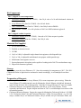
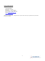
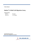
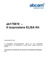
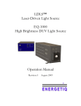
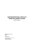
Product Manual Total Cholesterol Assay Kit (Colorimetric) Catalog Number STA-384 192 assays FOR RESEARCH USE ONLY Not for use in diagnostic procedures Introduction Cholesterol is a lipid sterol that is produced in and transported throughout the bloodstream in eukaryotes. Cholesterol is a critical compound used in the structure of cell membranes, hormones, and cell signaling. It is an essential component of animal cell structure in order to maintain permeability and fluidity. Cholesterol is a precursor for steroid hormones including the adrenal gland hormones cortisol and aldosterone, sex hormones progesterone, estrogens, and testosterone, and bile acids and vitamin D. Cholesterol is transported throughout the body within lipoproteins, which have cell-specific signals that direct the lipids they transport to certain tissues. For this reason, lipoproteins exist in different forms within the blood based on their density. These include chylomicrons, very-low density lipoproteins (VLDLs), low-density lipoproteins (LDLs), intermediate-density lipoproteins (IDLs), and high-density lipoproteins (HDLs). The higher the lipid content within a lipoprotein, the lower its density. Cholesterol exists within a lipoprotein as a free alcohol and as a fatty cholesteryl ester, which is the predominant form of cholesterol transport and storage. Determining circulatory levels of lipoproteins is critical to the diagnosis of lipid transport disorders. High levels of cholesterol and cholesteryl esters (hypercholesterolemia) have been associated with cardiovascular disease such as atherosclerosis and heart disease, although lower levels (hypocholesterolemia) may be associated with cancer, depression, or respiratory diseases. Cell Biolabs’ Total Cholesterol Assay Kit is a simple colorimetric assay that measures the amount of total cholesterol present in plasma, serum, tissue homogenates, or cell lysates in a 96-well microtiter plate format. The assay will detect total cholesterol (cholesteryl esters plus free cholesterol) in the presence of cholesterol esterase or only free cholesterol in the absence of the esterase enzyme. Each kit provides sufficient reagents to perform up to 192 assays, including blanks, cholesterol standards and unknown samples. Sample cholesterol concentrations are determined by comparison with a known cholesterol standard. Cholesteryl esters can be quantified by subtracting the free cholesterol values from the total cholesterol value. Assay Principle Cell Biolabs’ Total Cholesterol Assay Kit measures the total cholesterol within serum, plasma, lysate, or tissue samples. The assay is based on the enzyme driven reaction that quantifies both cholesterol esters and free cholesterol. Cholesterol esters are hydrolyzed via cholesterol esterase into cholesterol, which is then oxidized by cholesterol oxidase into the ketone cholest-4-en-3-one plus hydrogen peroxide. The hydrogen peroxide is then detected with a highly specific colorimetric probe. Horseradish peroxidase catalyzes the reaction between the probe and hydrogen peroxide, which bind in a 1:1 ratio. Samples are compared to a known concentration of cholesterol standard in a 96-well microtiter plate format. Samples and standards are incubated for 45 minutes and then read with a standard 96-well colorimetric plate reader (Figure 1). 2 Figure 1. Colorimetric Cholesterol Assay Principle Related Products 1. STA-241: Human Low Density Lipoprotein 2. STA-242: Human Very Low Density Lipoprotein 3. STA-243: Human High Density Lipoprotein 4. STA-361: Human ApoAI and ApoB Duplex ELISA Kit 5. STA-362: Human ApoAI ELISA Kit 6. STA-363: Human ApoAII ELISA Kit 7. STA-364: Human ApoCI ELISA Kit 8. STA-365: Human ApoCII ELISA Kit 9. STA-366: Human ApoCIII ELISA Kit 10. STA-367: Human ApoE ELISA Kit 11. STA-368: Human ApoB-100 ELISA Kit 12. STA-369: OxiSelect™ Human Oxidized LDL ELISA Kit (MDA-LDL Quantitation) 13. STA-390: Total Cholesterol Assay Kit (Fluorometric) 14. STA-391: HDL and LDL/VLDL Cholesterol Assay Kit 3 Kit Components Box 1 (shipped at room temperature) 1. Cholesterol Standard (Part No. 239001): One 50 µL tube of a 10 mM cholesterol solution in ethanol. 2. Assay Diluent (5X) (Part No. 239002): One 100 mL bottle. 3. 50X Colorimetric Probe (Part No. 238401): One 200 µL tube in DMSO. 4. HRP (Part No. 234402): Two 100 μL tubes of 100 U/mL HRP solution in glycerol. Box 2 (shipped on blue ice packs) 1. Cholesterol Esterase (Part No. 239003): One tube of 10 Units enzyme in powder. 2. Cholesterol Oxidase (Part No. 239004): One 200 µL tube. Materials Not Supplied 1. 96-well microtiter plates 2. Distilled or deionized water 3. 1X PBS 4. 10 μL to 1000 μL adjustable single channel micropipettes with disposable tips 5. 50 μL to 300 μL adjustable multichannel micropipette with disposable tips 6. Multichannel micropipette reservoir 7. Spectrophotometric microplate reader capable of reading in the 540-570 nm absorbance range. 8. Superoxide dismutase (optional) Storage Upon receipt, store the Assay Diluent at 4ºC. Store the remaining kit components at -20ºC. The 50X Colorimetric Probe is light sensitive and must be stored accordingly. Avoid multiple freeze/thaw cycles. Preparation of Reagents 1X Assay Diluent: Warm the Assay Diluent (5X) to room temperature prior to using. Dilute the Assay Diluent (5X) with deionized water by diluting the 100 mL Diluent with 400 mL deionized water for 500 mL total. Mix to homogeneity. Store the 1X Assay Diluent at 4ºC up to six months. Cholesterol Esterase: Reconstitute the powder with 200 μL of 1X Assay Diluent. Vortex vigorously until dissolved. Prepare aliquots and store at -20ºC to avoid multiple freeze thaws of the reconstituted powder. Cholesterol Reaction Reagent: Prepare the reagent by diluting the Cholesterol Oxidase 1:50, HRP 1:50, Colorimetric Probe 1:50, and Cholesterol Esterase 1:250 in 1X Assay Diluent. (e.g. For 100 assays, combine 100 μL of Cholesterol Oxidase, 100 μL of HRP, 100 μL Colorimetric Probe, and 4 20 μL Cholesterol Esterase with 1X Assay Diluent to 5 mL total solution). Mix thoroughly and protect the solution from light. For best results, place the Cholesterol Reaction Reagent on ice and use within 30 minutes of preparation. Do not store the Cholesterol Reaction Reagent solution. Notes: 1. If testing for the concentration of free cholesterol is needed only, omit the addition of Cholesterol Esterase from the Cholesterol Reaction Reagent solution. 2. The Colorimetric Probe is light sensitive and must be stored accordingly. Preparation of Samples Samples should be assayed immediately or stored at -80ºC prior to performing the assay. Optimal experimental conditions for samples must be determined by the investigator. The following recommendations are only guidelines and may be altered to optimize or complement the user’s experimental design. A set of serial dilutions is recommended for samples to achieve optimal assay results and minimize possible interfering compounds. Run proper controls as necessary. Always run a standard curve with samples. Tissue Lysates: For 10 mg of tissue, extract with 200 μL of a mixture of chloroform : isopropanol : NP-40 (7:11:0.1) in a micro-homogenizer. Centrifuge the extract 10 minutes at 15,000 x g. Transfer the liquid (organic phase) to a new tube, taking care to avoid the pellet. Air dry at 50ºC to remove the chloroform. Put samples under vacuum for 30 minutes to remove the trace amounts of organic solvent. Dissolve the dried lipids in 200 μL of 1X Assay Diluent with sonicating and vortexing until the solution is homogenous (the solution may appear cloudy). This extraction procedure may be scaled up if larger sample amounts are desired. Use 1 - 50 μL of extracted sample per assay. Next, adjust the volume to 50 μL per well with 1X Assay Diluent. For unknown samples, we suggest testing different amounts of samples to ensure that the readings are within the linear portion of the standard curve. Cell Lysates: Wash cells 3 times with cold PBS prior to lysis. For 106 cells, extract with 200 μL of a mixture of chloroform : isopropanol : NP-40 (7:11:0.1) in a micro-homogenizer. Centrifuge the extract 10 minutes at 15,000 x g. Transfer the liquid (organic phase) to a new tube, taking care to avoid the pellet. Air dry at 50ºC to remove the chloroform. Put samples under vacuum for 30 minutes to remove the trace amounts of organic solvent. Dissolve the dried lipids in 200 μL of 1X Assay Diluent with sonicating and vortexing until the solution is homogenous (the solution may appear cloudy). This extraction procedure may be scaled up if larger sample amounts are desired. Use 1 - 50 μL of extracted sample per assay. Next, adjust the volume to 50 μL per well with 1X Assay Diluent. For unknown samples, we suggest testing different amounts of samples to ensure that the readings are within the linear portion of the standard curve. Serum: Collect blood in a tube with no anticoagulant. Allow the blood to clot at room temperature for 30 minutes. Centrifuge at 2500 x g for 20 minutes. Remove the serum layer and store on ice. Avoid disturbing the white buffy layer. Aliquot samples for testing and store at -80ºC. Perform dilutions in 1X Assay Diluent. Serum samples must be diluted at least 1:100 to 1:200 with Assay Diluent. This will provide values within the range of the standard curve. Cholesterol levels in serum average about 3% higher in value than in the corresponding plasma pair (Ref. 2). Plasma: Avoid hemolyzed and lipemic blood samples. Collect blood with heparin or citrate and centrifuge at 2000 x g and 4ºC for 10 minutes. Remove the plasma layer and store on ice. Avoid disturbing the white buffy layer. Aliquot samples for testing and store at -80ºC. Perform dilutions 5 in 1X Assay Diluent. Plasma samples must be diluted at least 1:100 to 1:200 with Assay Diluent. This will provide values within the range of the standard curve. Notes: 1. Samples with NADH concentrations above 10 μM and glutathione concentrations above 50 μM will oxidize the probe and could result in erroneous readings. To minimize this interference, it is recommended that superoxide dismutase (SOD) be added to the reaction at a final concentration of 40 U/mL. 2. Avoid samples containing DTT or β-mercaptoethanol since the colorimetric probe is not stable in the presence of thiols (above 10 μM). Preparation of Cholesterol Standard Curve 1. Prepare fresh cholesterol standards before use by diluting in 1X Assay Diluent. First, dilute the stock Cholesterol Standard 10 mM solution 1:40 in 1X Assay Diluent for a 250 µM solution. (eg. add 25 µL of the stock 10 mM standard to 975 µL of 1X Assay Diluent). Vortex thoroughly. Use the diluted Cholesterol Standards promptly. 2. Use this 250 µM solution to prepare a series of the remaining cholesterol standards according to Table 1 below. Tubes 1 2 3 4 5 6 7 8 9 10 250 µM Cholesterol Standard (µL) 1000 500 of Tube #1 500 of Tube #2 500 of Tube #3 500 of Tube #4 500 of Tube #5 500 of Tube #6 500 of Tube #7 500 of Tube #8 0 1X Assay Diluent (µL) 0 500 500 500 500 500 500 500 500 1000 Resulting Cholesterol Concentration (µM) 250 125 62.5 31.3 15.6 7.8 3.9 1.9 1.0 0 Table 1. Preparation of Cholesterol Standards. Note: Do not store diluted cholesterol standard solutions. Assay Protocol Each cholesterol standard and sample should be assayed in duplicate or triplicate. A freshly prepared standard curve should be used each time the assay is performed. 1. Add 50 µL of the diluted cholesterol standards or samples to a 96-well microtiter plate. 2. Add 50 µL of the prepared Cholesterol Reaction Reagent to each well and mix the well contents thoroughly. 3. Cover the plate wells to protect the reaction from light. Incubate the plate for 45 minutes at 37ºC. 6 4. IMMEDIATELY read the plate with a spectrophotometric microplate reader in the 540-570 nm range. 5. Calculate the concentration of cholesterol within samples by comparing the sample absorbance values to the cholesterol standard curve. Example of Results The following figures demonstrate typical Total Cholesterol Assay results. One should use the data below for reference only. This data should not be used to interpret or calculate actual sample results. Figure 2: Cholesterol Standard Curve. Calculation of Results 1. Calculate the average absorbance values for every standard, control, and sample. Subtract the average zero standard value from itself and all standard and sample values. This is the corrected absorbance. 2. Plot the corrected absorbance for the standards against the final concentration of the cholesterol standards from Table 1 to determine the best curve. See Figure 2 for an example standard curve. 3. Determine the cholesterol concentration of the samples with the equation obtained from the linear regression analysis of the standard curve. Substitute the corrected absorbance values for each sample. Remember to account for dilution factors. Sample corrected absorbance Total Cholesterol (µM) = x Sample dilution Slope Cholesteryl Ester (µM) = Total Cholesterol - Free Cholesterol 7 Note: For the conversion of results from µM to mg/dl, divide the cholesterol concentration (µM) by 25.9. References 1. Admundson, D.M., et al. (1999) J. Biochem. Biophys. Meth. 38: 43-52. 2. Cholesterol and Triglyceride concentrations in Serum/Plasma Pairs. (1977) Clin. Chem. 23: 60-63. 3. Fossati, P., et al. (1982) Clin. Chem. 28: 2077-2080. 4. Ledwozyw, A., et al. (1986) Clin. Chim. Acta. 155: 275-284. 5. Lee, S.M. et al. (2008) Lipids 43: 419-429. Recent Product Citations 1. Parisi, O. I. et al. (2015). Sericin/poly (ethylcyanoacrylate) nanospheres by interfacial polymerization for enhanced bioefficacy of fenofibrate: in vitro and in vivo studies. Biomacromolecules. doi: 10.1021/acs.biomac.5b00746. 2. Mignarri, A. et al. (2015). Evaluation of cholesterol metabolism in cerebrotendinous xanthomatosis. J Inherit Metab Dis. doi: 10.1007/s10545-015-9873-1. 3. Moon, Y. S. et al. (2014). Lipid-modulating effects of aqueous extract of Rubus occidentalis in hepatocarcinoma HepG2 cells. American Journal of Bioscience 2:64-69. 4. Prévéraud, D. P. et al. (2014). Effect of the type of dietary triacylglycerol fatty acids on αtocopherol concentration in plasma and tissues of growing pigs. J Anim Sci. 92:4972-4980. 5. Raveendran,W. et al. (2014). H1-antihistamines exacerbate high-fat diet-induced hepatic steatosis in wild-type but not in apolipoprotein E knockout mice. Am J Physiol Gastrointest Liver Physiol. 307:G219-G228. 6. Marino, A. et al. (2014). ITCH deficiency protects from diet-induced obesity. Diabetes. 63:550-561. 7. Mathews, E, et al. (2014). Mutation of 3-hydroxy-3-methylglutaryl CoA synthase I reveals requirements for isoprenoid and cholesterol synthesis in oligodendrocyte migration arrest, axon wrapping, and myelin gene expression. J. Neurosci. 34:3402-3412. Warranty These products are warranted to perform as described in their labeling and in Cell Biolabs literature when used in accordance with their instructions. THERE ARE NO WARRANTIES THAT EXTEND BEYOND THIS EXPRESSED WARRANTY AND CELL BIOLABS DISCLAIMS ANY IMPLIED WARRANTY OF MERCHANTABILITY OR WARRANTY OF FITNESS FOR PARTICULAR PURPOSE. CELL BIOLABS’s sole obligation and purchaser’s exclusive remedy for breach of this warranty shall be, at the option of CELL BIOLABS, to repair or replace the products. In no event shall CELL BIOLABS be liable for any proximate, incidental or consequential damages in connection with the products. 8 Contact Information Cell Biolabs, Inc. 7758 Arjons Drive San Diego, CA 92126 Worldwide: +1 858 271-6500 USA Toll-Free: 1-888-CBL-0505 E-mail: [email protected] www.cellbiolabs.com 2012-2015: Cell Biolabs, Inc. - All rights reserved. No part of these works may be reproduced in any form without permissions in writing. 9