1
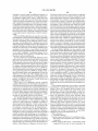
US008491488B1
(12) United States Patent
Criley et a].
(54)
METHOD AND SYSTEM FOR IDENTIFYING
7,248,923 B2
7,424,321 B2
7,438,689 B2
CARDIOPULMONARY FINDINGS BY USING
A HEART AND LUNG SOUNDS BUILDER
(75)
Inventors: Stuart Ross Criley, Palos Verdes
Estates, CA (U S); John Michael Criley,
Sr., Palos Verdes Estates, CA (US)
Laboratories, LLC, Rolling Hills
Estates, CA (US)
Notice:
U.S.C. 154(b) by 0 days.
(21) App1.No.: 13/252,023
(22) Filed:
7/2007 Maile et a1.
9/2008 Wariar et a1.
10/2008 Munk
4/2009
7,559,901 B2
7/2009 Maile et a1.
Kushnir et a1.
7,611,471 B2 *
11/2009
7,615,012 B2
7,645,141 B2
11/2009 Von Arx et a1.
1/2010 Lecat
B2
2/2010 Siejko et a1.
3/2010 Carlson et al.
6/2010 Patangay et al.
8/2010 Carlson et al.
10/2010
12/2010
2/2011
4/2011
5/2011
5/2011
6/2011
7/2011
............. .. 600/528
Thiagarajan ................ .. 600/528
B2
B2
B2
B2
B2
B2
B2
B2
B2
7,962,210 B2
7,972,275 B2
Subject to any disclaimer, the term of this
patent is extended or adjusted under 35
Jul. 23, 2013
7,517,319 B2 *
7,662,104
7,670,298
7,736,319
7,780,606
7,806,833
7,853,327
7,883,470
7,922,669
7,938,781
7,951,087
(73) Assignee: Blaufuss Medical Multimedia
(*)
US 8,491,488 B1
(10) Patent N0.:
(45) Date of Patent:
Thiagarajan et al.
Patangay et al.
Scheiner et al.
Zhang et a1.
Carlson et al.
Siejko et a1.
Daum et a1.
Siejko et a1.
(Continued)
Oct. 3, 2011
OTHER PUBLICATIONS
Related U.S. Application Data
(60)
Littman Cardiac ExamVirtual Trainer CD-ROM application manual,
Provisional application No. 61/389,164, ?led on Oct.
2007.
1, 2010, provisional application No. 61/542,293, ?led
(Continued)
on Oct. 3, 2011.
Primary Examiner * Joseph Stoklosa
(51)
Int. Cl.
(74) Attorney, Agent, or Firm * Peters Verny, LLP
(2006.01)
A61B 5/02
(52) U.S. Cl.
USPC
(58)
(57)
........................................................ ..
600/528
Field of Classi?cation Search
USPC
........................................................ ..
600/528
See application ?le for complete search history.
(56)
References Cited
ABSTRACT
A system is provided for creating a sound pro?le that matches
sounds produced by a patient during a physical examination,
such as a cardiac or pulmonary examination. A user selects
multiple sounds from a library and combines them to form the
pro?le Which may then be modi?ed by the addition of further
sounds, adjustments to their relative timing, duration, loud
ness, and so forth. The re?nement continues iteratively, and
U.S. PATENT DOCUMENTS
5,687,738 A * 11/1997 Shapiro et al.
5,957,866 A *
9/1999
6,220,866 B1
6,527,559 B2
4/2001 Amend et a1.
3/2003 Yoshii et al.
7,107,095 B2*
9/2006
after each change the pro?le is provided by the system to the
.... ..
600/528
Shapiro et al. .............. .. 600/586
Manolas ..................... .. 600/513
user, for example, as a phonocardiogram for comparison
against the sounds observed during the examination.
12 Claims, 6 Drawing Sheets
w E66 for timing
4"“- 8611126659933)! 0?
niwnaeatziisgmm
Emmi button 13?)!
Link 212 awnings
.
'
~ 5:13am met
' £21211 m éiftamn?a
Link to imp page 1 v
diagnosis
has? button
seiezted £00;
011212111103
HSM
Murmur 122mm: 80%
ESM
US 8,491,488 B1
Page 2
US. PATENT DOCUMENTS
8,000,780 B2
8,007,442 B2
8,012,098 B2
8/2011 Wariar et al.
8/2011 Carlson et al.
9/2011 Maile et al.
8,034,000 B2
10/2011 Zhang et al.
8,084,001 B2
8,096,954 B2
12/2011 Burns et al.
1/2012 Stahmann et al.
8,108,034
8,133,187
8,162,844
8,167,811
8,211,034
8,277,389
2004/0092846
2005/0048455
2005/0119585
B2
B2
B2
B2
B2
B2
A1
A1
A1
1/2012
3/2012
4/2012
5/2012
7/2012
10/2012
5/ 2004
3/ 2005
6/2005
Patangay et al.
Holmstrom et al.
Carlson et al.
Scheiner et al.
Patangay et al.
Carlson et al.
Watrous
HayamiZu et al.
Watrous
2008/0232605 A1
9/2008 Bagha
2009/0117527 A1
5/2009 Lecat
OTHER PUBLICATIONS
Adolph, R. J ., et al., “Teaching selective attention to the cardiac cycle:
The Cardio-gator,” Am. Hit. 1., Aug. 1971.
Cardionics CardioSim VII Sales Literature, Aug. 2008.
Cardionics CardioSim VII Sales Literature, 1995.
Cardionics Learning Cardiac Auscultation CD-ROM application
Sales Literature, 2003.
iMurmur iPhone app Product information, 2010.
iMurmur iPhone app Product information, 2009.
Levinson, D. “A New Audiovisual Aid to Heart-Sound Instruction,”
JAMA, Sep. 1968.
Easy Auscultation Website, http://WWW.easyauscultation.com/,
MedEdu LLC, 2010.
Pinnacle Heart Sounds Tutor Website, http://WWW.pinnacletec.com/
medical-training-products/Heart-Sounds-TUTORhtml, 1990.
Cardioscan User Manual, Zargis Medical, 2009.
Vukanovic -Criley, J. M., et al., “Competency in Cardiac Examination
Skills in Medical Students, Trainees, Physicians, and Faculty,”Arch.
Intern. Med., vol. 166, 2006.
Vukanovic-Criley, J. M., et al., “Con?dential Testing of Cardiac
Examination Competency in Cardiology and Noncardiology Faculty
and Trainees: A Multicenter Study,” Clin. Cardiol., vol. 33, No. 12,
pp. 738-45, 2010.
Criley, J .M., “Beyond Heart Sounds, vol. 1” Companion Study
Guide, 2000.
* cited by examiner
US. Patent
Jul. 23, 2013
Sheet 1 of6
Audio Input
Network Interface ‘—
l
_,
v
US 8,491,488 B1
User Input Device
v
Processor
1
>
Memory
Sound Library
I
I
Audio output
—>
_>
_
Audio Mixer
_>
<-
DX Engine
Display Output
Follow on Dx
GUI
Questions Engine
Graphics Library
Explanatory Text
Explanatory Graphics
<
Auowed Sound
Combinations Matrix
Sound Display
_>
Control Panel
Sounds | DX mode
Help
I
Settings
Differential DX
_>
Training &
“sing Engine
*
Tutoria| Modu|e
‘-
_> Patient Findings
a ‘9m F'nd'ngs
Saved combinations
Train/Test Scores
Uplink Module
—> Save Combinations 4
Module
Patient Findings
Power
Source
Test scores
FIG. 1
US. Patent
Jul. 23, 2013
Sheet 2 of6
US 8,491,488 B1
\\\“\
\....
US. Patent
Jul. 23, 2013
g‘Eisa;?mQEu
FIG. 2
Sheet 3 of6
US 8,491,488 B1
US. Patent
Jul. 23, 2013
Sheet 4 of6
FIG. 3
US 8,491,488 B1
US. Patent
Jul. 23, 2013
Sheet 5 of6
FIG. 4
US 8,491,488 B1
US. Patent
Jul. 23, 2013
8Y%‘v
B
U Sp w
Sheet 6 of6
US 8,491,488 B1
AO rt.m S+1. 9 n O S .m
V
Pulmonic Stenosis
FIG. 5
w
\\
US 8,491,488 B1
1
2
METHOD AND SYSTEM FOR IDENTIFYING
CARDIOPULMONARY FINDINGS BY USING
A HEART AND LUNG SOUNDS BUILDER
that match the sounds heard from a patient during physical
examination With a stethoscope, alloWing the selection of
different pulseWave contours that match the arterial, venous,
or precordial impulses seen on a patient during physical
examination, and alloWing selection of different respiratory
CROSS-REFERENCE TO RELATED
APPLICATIONS
patterns that match the respiration seen and heard from a
patient during physical examination.
This application claims the bene?t of US. Provisional
Patent Application No. 61/389,164 ?led on Oct. 1, 2010 and
Various embodiments of the invention are used in human
and veterinary medicine as a tool to aid in identi?cation of a
also entitled “Method and System for Identifying Cardiopul
monary Findings by Using a Heart and Lung Sounds
Builder,” and the bene?t of US. Provisional Patent Applica
patient’s cardiopulmonary ?ndings. These embodiments
include a computing device con?gured for selecting a com
bination of sounds, murmurs, and/or lung sounds, visual
examples of arterial, venous, and precordial impulses, and
respiratory patterns, and combining the selected sounds and
tion No. 61/542,293 ?led on Oct. 3, 2011 and entitled “Arte
rial and Venous Pulsations, Precordial Impulses and Respira
tory Pattern Finder,” both of Which are incorporated herein by
visual examples into an audio and audiovisual sequence. The
audio sequence is checked for accuracy by direct comparison
reference.
to sounds heard in a patient. The video sequence [animation
BACKGROUND OF THE INVENTION
Echocardiography Was initially developed in the mid-20th
Century and its adoption in the subsequent decades has
or example patient video] is checked for accuracy by direct
comparison With the patient. The selection is quickly re?ned
20
selected ?ndings and patient ?ndings has been achieved.
strongly in?uenced hoW clinicians are trained and hoW they
These ?ndings can be documented locally on the computing
device, as Well as securely uploaded to a database, and linked
subsequently perform cardiac examinations of patients. A
readily available, non-invasive method of imaging the beating
heart that could provide critical reinforcement of What Was
to the patient’s electronic health record. In some embodi
25
heard through the stethoscope should have enhanced physical
examination skills, but in fact the opposite has happened.
diagnoses based on a user’s selection of heart or lung sounds.
Work in reverse, listing diagnoses and then playing the appro
priate audiovisual ?ndings, including clinical variations that
30
are encountered across the spectrum of acute versus chronic,
mild versus severe, congenital versus acquired. Finally, in
some embodiments the computing device is con?gured to
function in a training or testing mode, playing a patient
opportunities for bedside teaching rounds. Today, hospital
admissions are short and intensely focused, With feWer oppor
tunities for trainees to learn and practice bedside examination
ments the computing device is con?gured to list differential
In some embodiments the computing device is con?gured to
Cardiac examination skills have declined since the advent of
echocardiography, a function of overreliance on technology
and the present healthcare environment that emphasiZes rapid
delivery of care. Several decades ago, patients’ hospital stays
Were long, providing trainees and their instructors frequent
by an iterative process until a satisfactory match betWeen
ronment, further amplify the problem if their oWn cardiac
recording, and prompting the user to identify correctly the
heart sounds and murmurs, breath and lung sounds, pulsa
tions in the neck and precordium, and respiratory patterns,
grading the user entries, and storing the results locally or
examination skills are not Well developed. In the absence of
remotely.
35
skills Attending physicians, having been trained in this envi
bedside training With patients, audio recordings of heart
sounds has served as a poor substitute, and as a result clini
40
BRIEF DESCRIPTION OF DRAWINGS
cians noW commonly close their eyes While conducting car
diopulmonary examination, shutting out important visual and
FIG. 1 is a schematic representation of a sound builder
palpable cues exhibited by the patient. Multicenter studies of
system according to various embodiments of the present
cardiac examination skills document a rise in test scores until
the third year of medical school, but no further improvement
thereafter despite years in residency training, or even further
45
years in practice. (See, e.g., Arch Intern Med (166):610-617,
(2006) and Clin Cardiol (33;12):738-745 (2010)) Indeed,
full-time internal medicine professors perform no better in
tests of cardiac examination skills than the third-year medical
students they teach. Compounding the problem is a lack of
created through the Sound Builder application and displayed
50
critical reinforcement When auscultating patients. Critical
reinforcement implies a commitment to con?rming or refut
ing one’s bedside diagnostic impressions by critical revieW
and correlation With available imaging and/ or hemodynamic
55
studies performed on that patient. Unfortunately, patient
With a corresponding ECG through a user interface according
to various embodiments of the present invention.
FIG. 3 is a schematic representation of common listening
locations for stethoscopic cardiac examination.
FIG. 4 is a graphical representation of an alert provided by
the Sound Builder application and displayed through a user
interface according to various embodiments of the present
invention.
FIG. 5 is a graphical representation of a differential diag
exposure Without critical reinforcement seems to be the norm
for the average medical resident, explaining their lack of
advancement in examination skills despite clinical encoun
ters With hundreds of patients.
invention.
FIG. 1.5 is a graphical representation of a control panel
provided through a user interface according to various
embodiments of the present invention.
FIG. 2 is a graphical representation of a phonocardiogram
nosis screen provided by the Sound Builder application and
60
displayed through a user interface according to various
embodiments of the present invention.
SUMMARY
DETAILED DESCRIPTION
This invention relates to audiovisual display on a digital
computer system, inpar‘ticular, visual as Well as audio display
of cardiac and lung auscultation, alloWing the selection of
individual sound components to create custom combinations
65
FIG. 1 illustrates the general architecture of the sound
builder system as embodied in an electronic computing
device con?gured to execute a Sound Builder application. In
US 8,491,488 B1
3
4
some embodiments the electronic computing device includes
is added to the playback, the visual display of the phonocar
diogram shows the new component in its proper temporal
a cellular telephone, digital pad, personal digital assistant,
personal computer, and/or the like. Audio input is received
location, and the selected button lights up to show the user
which components are already active.
from an external source: from a wired audio connection to a
microphone and/or stethoscope; from a wireless connection
The Sound Builder application is con?gured to generate
via the Network Interface (e. g., WiFi, Bluetooth, USB); and/
the control panel which provides the user with menus such as
or from a live transmission or recorded transmission of a
a component-based menu and a lesion-based menu. The com
patient recording.
ponent-based menu allows the user to select sounds, mur
User input is received via a user input device. The user
input device uses the user input to select sound components,
to create a combination. The lesion-based menu allows the
murs, and murmur combinations (e.g., to-fro murmurs) used
switch to text, help, settings, or diagnosis sections (Dx) of the
user to select a lesion and optionally to further modify the
lesion. Selected lesions can be acute or chronic and vary from
mild to severe. The control panel can also be con?gured to
allow the user to vary or specify the heart rate and the listening
location.
In various embodiments some or all of the features below
can be selected and/or controlled. A ?rst heart sound (S1) is
Sound Builder application, to submit answers to training/test
questions, to record patient ?ndings, or to control the volume,
temporal location, tempo of the sound combinations, and/or
the like.
In some embodiments, selected sound combinations are
checked against an Allowed Combinations Matrix before
playing. Combinations may be saved for later recall. Patient
?ndings may be saved and/or uploaded via the Network Inter
face.
In some embodiments, sound components are recalled
from Memory and sent to the Audio Mixer before being sent
selected, for instance, by recording the location on the patient
at which the sound is heard, as the location will in?uence the
20
of different types, such as single and split (e. g., physiological,
to Audio Output. The Display Output is updated to re?ect the
paradoxical, or persistent), loudness can vary from absent to
soft to loud, and their timings can be adjusted from early
new sound combination chosen by the user.
The following is a step-by-step description of how various
embodiments of the invention may be used. The computing
sound. Sound components, their loudness, and their relative
timings can also be selected for S 1. Sound components can be
25
through the expecting timing to late.
Second (S2), third (S3), and fourth (S4) sounds are simi
device of the invention can be con?gured to perform some or
larly selected. In some embodiments, the quality of S3 can be
all of these steps. The various alternatives provided are not
meant to be limiting. Other alternatives are considered to be
within the scope of the invention.
A user listens to a patient’s heart sounds, for example,
ness can be varied from soft to medium to loud. The loudness
of S4 can also be varied from soft to medium to loud and the
selected, such as dull, rumbling, and knocking, and the loud
30
through an acoustic stethoscope, through an electronic
stethoscope, from a received live transmission via computer
network, from a recorded transmission via computer net
work, from a locally stored recording, or from a remotely
stored recording. Transmission methods to reach the user’s
35
ear include, for example, (1) via headset (wired or wireless)
attached or paired to an electronic stethoscope, (2) via head
phones (wired or wireless) attached or paired to an electronic
stethoscope, (3) via a wired or wireless speaker placed under
valsihigher left atrial/ left ventricular pressure gradient,
40
transmitting stethoscope or system.
shifted relative to the timing of other sounds such that, for
The user launches the Sound Builder application on the
instance, the S l -MSC interval can vary, with shorter intervals
45
a computer readable medium and executed by a processor of
the computing device. The Sound Builder application option
ally comprises a smartphone application, a website-based
program, an application installed on a personal computer, an
implying more severe mitral stenosis; and longer intervalsi
lower left atrial/left ventricular gradient, implying less severe
mitral stenosis. The MSC can be selected for the mitral valve
or the tricuspid valve. The MSC timing can likewise be
or attached to an acoustic or electronic stethoscope, and (4)
via external audio input and/ or electronic interface with the
computing device. The Sound Builder application can be
implemented in hardware, ?rmware and/or software store on
location (left or right ventricular) can be recorded.
Other selectable sounds can include an opening snap (OS),
an ejection sound (ES) which can be aortic or pulmonic, and
a mild systolic click (MSC). The OS loudness can vary from
soft to loud, and the OS timing can be shifted relative to the
timing of other sounds. For example, the S2-OS interval can
vary either continuously or in discrete steps; shorter inter
50
application installed on a personal digital assistant, an embed
ded application of an electronic stethoscope.
implying an earlier systolic prolapse, and longer intervals
implying a later systolic prolapse. The MSC can be given an
accompanying murmur. For example, mitral or tricuspid
regurgitation may occur with prolapse, causing a murmur that
immediately follows the click. The timing of this murmur
depends upon the timing of the click, such that the murmur
can be holosystolic, midsystolic, or late systolic.
Other selectable sounds comprise murmurs such as dias
tolic murmurs, systolic murmurs, and continuous and to-fro
The user selects a sound-murmur combination, for
example, by pressing inputs (e.g., on-screen buttons). The
murmurs. Selectable diastolic murmurs include early dias
nents and mixes the audio tracks dynamically to create a
tolic, mid diastolic, and presystolic murmurs, and as above,
for each the location on the patient is speci?ed. For early
repeating heart and/or breath sounds. This audio playback
diastolic murmurs, quality (harsh or high-pitched), shape (de
optionally continues uninterrupted until a further sound, mur
crescendo or crescendo-decrescendo), and duration (long and
tapering or short and abruptly terminated) can be selected.
sound builder takes the one or more selected audio compo
55
mur, or breath sound is added or removed, or until the user
halts the playback.
FIG. 1.5 shows an exemplary control panel provided by the
Sound Builder application executing on an electronic com
puting device. In some embodiments, the user starts with a
blank slate, or more speci?cally, a simple S1 S2 combination.
By adding sounds or murmurs, a combination of sounds is
built up to match what is heard in the patient. Each time a
button is selected on the control panel, the sound component
60
For mid diastolic murmurs quality and loudness can be
selected, and for presystolic murmurs shape (crescendo),
loudness, and accompanying sounds and murmur can be
selected. Accompanying sounds and murmur can comprise a
mid diastolic murmur, an OS, and a loud Sl.
65
Selectable systolic murmurs include holosystolic, early
systolic, mid systolic, and late systolic murmurs, and as
above, for each the location on the patient is speci?ed. For
US 8,491,488 B1
5
6
each, too, the quality is selectable (harsh, blowing, or musi
cal). The holosystolic shape can be equal intensity, cre
scendo-decrescendo, or decrescendo. The early systolic and
late systolic shapes can be decrescendo, While the mid sys
ness and quality of heart sounds and murmurs, and some
sounds and murmurs are only detectable in speci?c locations
on the patient’s chest, and only With either the stethoscope
bell or diaphragm. Also, the quality of the same murmur can
be harsh or musical depending upon the location of the stetho
scope. The location of the stethoscope can be indicated on the
screen by text and/or by an indicator on a chest diagram.
SWitching to a different location Will cause the selected sound
tolic shape can be equal intensity or crescendo-decrescendo.
The duration of the late systolic murmur is also variable.
For continuous murmurs location on the patient is speci
?ed. Timing is also controllable as the peak of murmur inten
sity varies depending upon the etiology. To-fro murmurs
comprise semilunar and atrio -ventricular murmurs, both have
systolic and diastolic components, and for both the location
combination to update. For example, moving from the cardiac
apex (5th left intercostal at the midclavicular line) to the 2nd
right intercostal space Will cause S3 or S4 to disappear, and
on the patient is speci?ed. Semilunar to-fro murmurs can be
aortic or pulmonic, and either can be characterized as to-FRO
moving from the cardiac apex (5th left intercostal at the
midclavicular line) to the 2nd left intercostal space Will cause
MDM and PSM to disappear.
The Sound Builder application can provide the user With an
option of focusing on similar heart sounds and murmurs
(louder diastolic component) or TO-fro (louder systolic com
ponent). Aortic to-fro murmurs can also be Austin Flint mur
murs, While pulmonic to-fro murmurs can be right-sidedAus
tin Flint murmurs. Atrio-ventricular to-fro murmurs can be
mitral or tricuspid.
FIG. 2 illustrates a sound sequence assembled from sound
20
(cardiac base), and S1 S2 S3 (cardiac apex). Extra sounds
components, according to various embodiments of the inven
tion. The sound sequence displayed shoWs the ECG, phono
cardiogram, and the phases of the cardiac cycle, systole and
diastole. Control buttons sWitch the display to settings, help,
explanatory text, and the relevant differential diagnoses for
near Sl include Sl ES S2 (cardiac apex or sometimes base),
Split S1 S2 (cardiac apex), and S4 S1 S2 (cardiac apex).
The Sound Builder application is optionally con?gured to
25
the rate may increase, but the sounds and murmurs them
account for one or more of the folloWing maneuvers and
perturbations. The sound sequence can be varied depending
this sound combination. Slider controls alloW the user to vary
the heart rate and the playback volume.
In some embodiments the Sound Builder application is
con?gured to alloW user control of heart rate, rhythm, and
regularity. Heart rate can be varied to re?ect different patient
conditions, such as sloWer heart rates for relatively longer
diastoles and faster heart rates for relatively shorter diastoles.
Heart sounds and murmurs can be affected by changes in
rhythm, for example, cannon Wave, post-extrasystolic beat
augments/does not augment murmurs, and atrial ?brillation
(long or short diastoles). The frequency of the sound or mur
mur is not generally affected by the heart rate; for this reason,
combinations While changing the location. For instance, extra
sounds near S2 include S1 S2 OS (cardiac base), Sl Split S2
on user-selectable factors such as respiration, posture, extra
30
systolic beat, hand grip, and Valsalva maneuver. Options for
respiration include apnea, inspiration, and expiration, the lat
ter tWo of Which can augment murmurs, augment sounds, and
split sounds. Posture options include supine, left lateral decu
bitus, sitting upright, sitting While leaning forWard, standing,
squatting, and passive leg elevation. The extrasystolic beat
35
can augment or not augment the murmur. Hand grip options
include control, grip, and release, While options for the Val
salva maneuver include control, strain, release, and recovery.
40
FIG. 4 illustrates an alert provided to the user through the
Sound Builder interface When an implausible combination is
selected, as not all combinations of heart sounds and murmurs
selves must be changed in temporal location (and often dura
are physiologically possible. An exemplary implausible com
tion) Without altering the frequency spectrum. Using discrete
bination is an opening snap (OS) and a third heart sound (S3).
sound components alloWs the heart rate to be varied up or
doWn Without adversely affecting the frequency of the play
These sounds are not likely to occur simultaneously in a
45
back.
FIG. 3 illustrates a chest diagram With circles indicating
four common listening locations for a stethoscope during
mitral valve, While a third heart sound usually requires a Wide
open mitral valve. In various embodiments a caution triangle
appears in the sound display, and the user is alerted With a
notice.
cardiac examination (other listening positions are possible).
The numbers indicate intercostal spaces. Locations are gen
erally noted as left or right, and by intercostal space. In the
Sound Builder application, the location of the current heart
sounds recording can be noted by this chest diagram, as Well
as alternate listening areas. For breath and lung sounds, a
50
55
user selects combinations that are speci?cally heard at one or
tWo locations, the sound builder updates the displayed loca
60
As noted, the location of the stethoscope chest piece, and
shoWn beloW in Table 1. Adding more than one example of
each murmur or sound Will increase the combinations, but
more importantly, increase the verisimilitude of the combi
nations. For example, the ?rst heart sound of mitral stenosis is
louder than normal; thus, a loud ?rst heart sound of mitral
sound/murmur combination to change What Would be
expected to be heard in a patientiin many cases removing
the choice of stethoscope bell or diaphragm, affects the loud
combinations, a sounds and murmurs matrix includes 75,344
valid combinations. The Sound Builder application can auto
matically screen implausible selections by consulting a
Sound-Murmur Plausibility Matrix, an example of Which is
similar diagram is used. The user can change to a neW listen
the extra sound or murmur altogether.
It is noted that With 20 heart sound and murmur compo
nents, there are an excessive number of possible combina
tions, but by limiting the combinations to only alloWable
ing position by selecting a different circle. In addition, if the
tion to shoW Where these sounds and murmurs Would be
heard. Manually sWitching to a neW location Will cause the
patient since the opening snap requires a narroWed, stenotic
65
stenosis Would be substituted for a normal ?rst heart sound.
As another example, the midsystolic murmur of aortic steno
sis can sound harsh When listening at the cardiac base, but
musical at the cardiac apex.
US 8,491,488 B1
TABLE 1
Allowed
TFM
“l” is a valid combination;
“0” is an invalid combination
right), femoral (left or right), popliteal (left or right), and post
The user can look at and listen to (as well as palpate) the
created combination. The Sound Builder application seam
40
tibial (left or right). Contour can include normal, quick, ?ick
lessly loops the audio of the created combination, and the user
ing upstroke, bounding, collapsing, slow rising, or distorted,
may choose to listen via headphones, a wireless headset, an
electronic stethoscope, or a wired or wireless speaker
attached to an acoustic stethoscope, for example. While a
when palpating pulse at peripheral vessels. These pulse con
loudspeaker can be used, it is noted that loudspeakers can
distort audio.
tours can be displayed as waveforms or as audiovisual ani
45
(systolic rise occurs close in time with ventricular systole) or
distal to the heart (systolic rise is delayed, and the pulse
contour is distorted).
The user can also observe the ECG waveform and the
phonocardiogram, and also the selected buttons on the pro
vided intcrfacc. Thc ECG wavcform can indicate, for
example, the onset of systole. The ECG waveform may
morph, consistent with the sound-murmur combination
selected. The phonocardiogram provides a visual depiction of
Thc uscr can compare the combination to thc paticnt’s
50
heart sounds. Two methods for making the comparison
involve either switching ear pieces or not switching ear
55
(no confusing artifacts), and to enhance recognition at small
resolutions. The combination selected can be indicated, in
some embodiments, by labeled buttons in an ON state (e.g., a
white-colored button). The last button selected is optionally
highlighted a different color (e.g., a yellow-colored button).
pieces. Where switching ear pieces is employed, the Sound
Builder application plays the selected audio combination and
a stethoscope or playback device plays the patient’s heart
sounds on a separate system. Alternatively, the Sound Builder
application can be switched between playing the selected
60
audio combination through a set of ear pieces and either a live
transmission or a recording of the patient’s heart sounds. In
another alternative, the Sound Builder system includes an
external speaker, and the user moves the stethoscope between
the patient’s chest and the external speaker to make the com
65
parison.
As noted, the user can palpate the created combination. In
some embodiments, a signal to drive a palpable pulse will
accompany the audio. Some embodiments provide a stereo
output where one channel contains the audio and the other
channel contains the pulse signal, including modulation and
DC voltage. Locations include carotid (left or right), apical
(apex beat), parastemal, brachial (left or right), radial (left or
heart sounds. For instance, by switching back and forth to the
patient’s heart sounds, the user can check whether the
selected combination is a reasonable match to the patient’s
heart sounds and murmurs and can be selected to show the
selected combination, the actual heart sounds and murmurs of
the patient, as well as cartoon depictions of idealized forms
mations of the neck and precordium. Intensity and timing are
additional options, where timing can be proximal to the heart
A good match with the patient will not require further
modi?cation of the combination. A poor match will prompt
US 8,491,488 B1
9
10
the user to modify his selected combination. Errors in under
standing the de?nitions can lead to erroneous selections, for
Which are often considered different in origin from harsh
murmurs, When in fact they have the same etiology.
example, the user may confuse a continuous murmur With a
Diastolic murmurs that are often confused include EDM
to-fro murmur. Failure to identify systole can also lead to
erroneous selections, for instance, the user may confuse a
vs. MDM, EDM long vs. CM, and EDM With MDM (Austin
systolic murmur With a diastolic murmur.
confused include Sl MSC LSM S2, S1 ES MSM S2, and S4
S1 HSM S2.
The best match result of this re?nement process may be
de?nitive or may contain ambiguous features. In the latter
instance, for example, the user may not be able to determine
Flint) vs. EDM. Sound/murmur combinations that are often
In various embodiments the Sound Builder application
provides an info section that the user can access by pressing
the info button, for instance, in order to learn more about each
sound/murmur choice. The ?rst page displayed in the info
Whether an extra sound near S1 is S4-Sl, Sl-ES, or a split Sl.
section can be linked to the last button the user selected on the
Over-speci?city may not be appropriate When the ?ndings
cannot be ?rmly established, and alternative ?ndings can be
stored With equal Weight, or With preferential Weights
assigned to the alternatives.
control panel. Additional pages that describe other control
panel choices can be made available from a table of contents,
or by pressing previous or next buttons.
Similarly, various embodiments the Sound Builder appli
cation provide a help section that can be accessed, for
example, by pressing the help button to display the help
section on the screen. The help section can provide a descrip
tion each button’s appearance and function, program How, a
20
description of each section, the role of the differential diag
nosis section (including limitations and When alternative
diagnoses should be considered), recommended customiZed
settings based on the user’s preferences, and cardiac exami
nation tips and tricks to improve detection and identi?cation
example, Sl ES MSM S2 has a short list of potential diag
noses, either bicuspid aortic stenosis or aortic stenosis. The
Sound Builder application can help narroW the choices fur
ther With queries to the user. For example, the user can be
25
of ?ndings.
Various embodiments the Sound Builder application can
also provide the user the ability to customiZe certain settings.
Examples of customiZable settings include the display, Wam
ings, and preferences for sending and receiving data.
asked to report the contour of the carotid pulse (for aortic
stenosis the pulse is parvus et tardus), to compare 2nd right vs.
2nd left intercostal listening areas, or the user can be
prompted to learn more about the clinical setting by suggest
ing intervieW questions for the patient.
30
The Sound Builder application can also provide the user
the ability to modify a combination to create a closer match to
the patient’ s sounds, for example, through an interactive pro
cess, possibly over several iterations, of selecting a combina
tion, comparing the combination to the patient, and modify
FIG. 5 illustrates a differential diagnosis list that can be
provided to the user as a list of potential differential diagnoses
based on the ?nal combination entered. The Dx button turns
green, in some embodiments, to indicate that the list of poten
tial diagnoses has become very narroW. In the illustrated
The Sound Builder application may create the Dx list by
consulting a Sounds-Murmurs Dx Matrix that contains likely
diagnoses mapped to the selected sounds and murmurs. This
list of differential diagnoses can be dynamically updated
based upon further input from the user, including listening
35
location, patient history, and vital signs (e.g., pulse contour,
heart rate, body temperature, blood pres sure). Patient history
can comprise the patient’s chief complaint, social history
(e.g., smoking, IV drug abuse), any childhood illness, and
country of origin (e.g., Where rheumatic fever is still preva
ing the combination, to hone in on a best match to the patient
?ndings.
The Sound Builder application can also offer plausible
alternatives to aid in the step of modifying the combination.
For instance, sound combinations near S1 and S2 are often 40 lent). The Dx list can be sorted by plausibility, or severity, and
confused, heart sounds are often not heard When a murmur is
each Dx entry can be linked to explanatory text, illustrations,
and audio or audiovisual examples of patient recordings.
present, systolic murmurs are often confused, diastolic mur
The Sound Builder application, in some embodiments,
suggests additional actions to narroW the differential diagno
murs are often confused, continuous murmurs are often con
fused With to-fro murmurs, certain sound/murmur combina
tions are commonly confused, and confusion of systole and
45
Regarding sound combinations near Sl, the combinations
ofS4 and S1, split and S1, S1 and ES, S1 and early MSC are
commonly confused. Regarding sound combinations near S2,
the combinations of late MSC and S2, split and S2, S2 and
OS, and S2 and S3 are commonly confused. Heart sounds that
sis, including altering the listening location, sWitching from
stethoscope bell to diaphragm, postural maneuvers, and the
postextrasystolic beat. Altering the listening location can
diastole leads to a large number of errors.
help, for example, to differentiate betWeen Whether an extra
50
heart sound near S1 is an S4, a split S1, or an ejection sound.
If the extra sound is heard at locations in addition to the
cardiac apex, it is likely to be an ejection sound, With a softer
Sl preceding it. LikeWise, listening at the apex With the
stethoscope bell Will most likely detect the loW-frequency S4,
are often not heard When a murmur is present include S1 and
S2 With HSM, S3 With HSM or another murmur, and MDIVI/
but sWitching to the stethoscope diaphragm (Which ?lters loW
S3 rumble With EDM (Austin Flint).
HSM. Here, right-sided murmurs may in fact be midsystolic,
frequency sounds) should make the extra sound go aWay.
Postural maneuvers can help detect the midsystolic click of
but run into or past the aortic valve closure sound (usually the
mitral valve prolapse Which can occur earlier or later as a
loudest component of S2) tricking the user into thinking the
murmur is holosystolic. An ejection sound (ES) may precede
the MSM, tricking the user into thinking the murmur begins
function of standing and squatting and as a function of the
Valsalva maneuver. The postextrasystolic beat (the beat that
Systolic murmurs that are often confused include MSM vs.
55
60
with S1, and concluding the murmur is ESM or HSM. Cre
scendo-decrescendo holosystolic murmurs are possible, and
may confuse the user Who expects this shape of murmur to be
exclusively midsystolic. Other systolic murmurs that are
often confused include MSM vs. ESM (both end before S2),
LSM vs. MSM vs. HSM (regurgitation folloWing the click of
mitral valve prolapse), and musical murmurs (Gallavardin)
65
folloWs a preventricular contraction, or PVC) can narroW the
origin of a systolic murmur. Systolic murmurs that augment
folloWing a PVC are generally con?ned to the ventricular
out?oW tract such as aortic stenosis and hypertrophic cardi
omyopathy, While systolic murmurs that do not augment
include the holosystolic murmur of mitral regurgitation.
The Sound Builder application, in some embodiments,
suggests items in the patient’ s history or the clinical setting to
US 8,491,488 B1
11
12
investigate to narrow further the differential diagnosis. As
both ears give the brain cues as to the location of a particular
sound. These audio cues can be added to existing audio With
examples, a patient presenting With Worsening dyspnea dur
ing pregnancy suggests mitral stenosis, While being from
head-related transfer functions (or similar methods) to create
the perception that a particular sound is emitting from a
certain areas of the World (e.g., the Paci?c Rim and Latin
America) can implicate rheumatic fever. As another example,
certain position in space. For example, positioning the audio
aortic regurgitation secondary to endocarditis can be due to
location of S1 and S2 behind the user’ s head, and positioning
the murmurs in front, can help the user identify the sounds,
and differentiate them from the murmurs. Likewise, the pres
ence of an extra heart sound can be highlighted by placing the
IV drug abuse With infected needles. As yet another example,
a third heart sound can be physiologic or pathologic in origin,
Where a physiologic third heart sound is common in athletes,
the healthy young, and pregnancy, While a pathologic third
heart sound can be due to ventricular dysfunction secondary
extra sound in yet another position (e.g., left or right). Merg
ing the sounds and murmurs back into monoaural space Will
re?ect What is heard through the stethoscope, While expand
ing the sounds to highlight components aids in their identi?
to myocardial infarction, hypertension, or dilated cardiomy
opathy.
cation. Another aid in identi?cation is matching visual infor
mation (either video of patient or computer animation of an
The Sound Builder application can assist the user to docu
ment the patient’ s cardiac examination ?ndings by appending
the patient name, ID number, location (of the patient), time
and date to the ?ndings, and by adding the patient’s history,
vital signs, and other patient data. Listening location can be
added by selecting a listening area off of a chest diagram. The
examination ?ndings can indicate that the ?nal selection sat
is?ed the user that these are the ?ndings established upon
examination of the patient, or may indicate that the ?nal
selection contains appropriate ambiguity Where a ?nding can
not be ?rmly established. Documented ?ndings can be
uploaded securely With encryption to a database that stores
the patient’s electronic health record.
In some instances, the Sound Builder application can be
avatar) to the sounds, to aid in recognition. Human hearing
becomes more sensitive and speci?c if a sound is accompa
nied by a visual cue. Animation or video of pulsations in the
neck or precordium, or movement of the chest during respi
20
Will aid the user in identifying individual sound components.
The Sound Builder application can also comprise a lung
sounds builder module that functions analogously and With
the same technology as the heart sounds builder module illus
25
nents in order to create a matching combination. The user can
panel. Here, the user starts With a diagnosis, and the Sound
While selecting calci?c aortic stenosis Will cause the applica
tion to produce the pattern of S l, MSM, S2. Similarly, select
ing Well-tolerated chronic aortic regurgitation Will cause the
30
the selected combination With the patient’s lung sounds and
modi?es the selected combination as necessary to improve
35
lung sounds builder suggests additional listening locations,
40
lung sounds that are associated With the selected patient con
dition.
examples. Mitral stenosis may be tolerated by the patient at a
45
indicated by an opening snap and a barely detectable mid
diastolic murmur. The more severe mitral stenosis is indicated
by loud Sl, MDM, perhaps no PSM, and longer S2-OS inter
50
ured to facilitate methods to improve identi?cation of indi
vidual sound components. As one example, by adding and
ments and applications beyond those described herein
Without departing from the broader spirit and scope of the
speci?cation. The speci?cation and draWings are, accord
ingly, to be regarded as illustrative rather than restrictive. As
used herein, logic refers to hardWare, ?rmWare, or a combi
55
murs. With S1 S2 S3, often the user does not perceive the
loW-frequency, loW-intensity S3, thus, by adding and remov
nation of hardWare and softWare such as a processor and
computer readable instructions stored on a computer readable
medium, but logic as used herein speci?cally excludes soft
Ware alone. It Will be recogniZed that the terms “comprising,”
“including,” and “having,” as used herein, are speci?cally
ing S3 While S1 and S2 remain can improve detection by
giving the user a visual cue When S3 is present on the phono
cardiogram, as Well as by playing the extra sound. As another
In the foregoing speci?cation, the invention is described
With reference to speci?c embodiments thereof, but those
skilled in the art Will recogniZe that the invention is not
limited thereto. Various features and aspects of the above
described invention may be used individually or jointly. Fur
ther, the invention can be utiliZed in any number of environ
val. The very severe mitral stenosis is indicated by a louder
removing a single component to highlight its presence or
absence against the background of other sounds and mur
maneuvers, and items in the patient’s history or clinical set
ting to narroW further the differential diagnosis. In various
embodiments, the lung sounds builder can be used in reverse:
starting With a condition, and playing back the spectrum of
Mitral stenosis and regurgitation provide additional
and later Sl, PSM, MDM, and short S2-OS interval.
The Sound Builder application optionally may be con?g
the match to the patient’s lung sounds. The lung sounds
builder is con?gured to suggest differential diagnoses based
on the combination selected. In various embodiments, the
regurgitation, MDM at the apex.
sloW heart rate, become more severe With moderate heart rate,
or very severe With fast heart rate. Tolerated mitral stenosis is
also select different patient postures (e.g., supine, upright),
respiratory patterns (normal, tachypnea, Cheyne-Stokes), or
patient maneuvers (e. g., egophony). The user then compares
application to produce long EDM, selecting acute severe
aortic regurgitation Will produce short EDM, MSM, Sl (soft
or absent), and selecting Austin Flint aortic regurgitation Will
produce, in addition to the sounds of acute severe aortic
trated above, but employing breath sounds components.
Here, the user listens to the patient’ s chest for lung sounds and
consults the lung sounds builder to select lung sound compo
used in reverse by using a lesion-based menu on the control
Builder application then reproduces the spectrum of ?ndings
associated With that lesion for comparison against the patient.
For instance, selecting congenital aortic stenosis Will cause
the application to produce the pattern of S1, ES, MSM, S2,
ration, When displayed in time With the heart and lung sounds,
60
intended to be read as open-ended terms of art.
example, positional audio processing can be employed to
What is claimed is:
place components in three-dimensional space, so that the user
can perceive some sounds as occurring in front, to the side, or
behind the ears. Human hearing is very sensitive to positional
1. A method comprising:
selecting, using an input device of a computing system, a
cues that occur as sounds are modi?ed as they re?ect off 65
external surfaces, as Well as the portions of the external ear. In
components;
selecting, using the input device, a second bodily sound
addition, subtle differences in frequency and arrival time to
component from the menu of sound components, the
?rst bodily sound component from a menu of sound
US 8,491,488 B1
14
13
?rst and second bodily sound components being repre
sentative of ?rst and second sounds of a stethoscopic
7. The method of claim 1 further comprising an act of
reading a list, the list including the names of differential
examination;
diagnoses, the list being read from the display of the comput
observing on a display of the computing system, and lis
ing system.
tening to, a repeating graphical representation of the ?rst
8. The method of claim 4 further comprising vieWing on the
display of the computing system an ECG proximate to the
graphical representation of the ?rst and second sound com
and second sound components; and
simulating an abnormality by iteratively modifying and
observing the graphical representation.
ponents.
2. The method of claim 1 Wherein the ?rst bodily sound
component is a ?rst lung sound component and Wherein the
second bodily sound component is a second lung sound com
9. The method of claim 1 Wherein listening to the repeating
graphical representation of the ?rst and second sound com
ponents includes hearing the ?rst and second sound compo
nents spatially separated from one another.
10. The method of claim 4 Wherein the ?rst and second
ponent.
3. The method of claim 2 Wherein selecting the ?rst and
second bodily sound components from the menu of sound
sounds are heart sounds S1 and S2.
components comprises selecting a respiratory pattern from a
menu of respiratory patterns.
4. The method of claim 1 Wherein the graphical represen
tation of the ?rst and second sound components comprises a
11. The method of claim 1 further comprising physically
examining a patient, Wherein the ?rst and second sound com
ponents are selected to be representative of ?rst and second
sounds heard during a stethoscopic examination of the
phonocardiogram.
5. The method of claim 1 Wherein modifying the graphical
representation comprises selecting a third sound.
6. The method of claim 1 Wherein modifying the graphical
representation comprises changing a relative timing of the
?rst and second sounds.
20
patient.
12. The method of claim 11 further comprising uploading,
from the computing system, ?ndings from the physical
examination to an electronic health record for the patient.
*
*
*
*
*