1
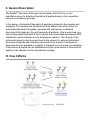
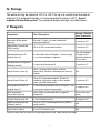
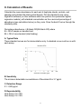
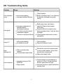
RayBio® Human/Mouse/Rat Neurokinin
B Enzyme Immunoassay Kit
Catalog #: EIA-NEB, EIAM-NEB, EIAR-NEB
User Manual
Last revised December 1, 2015
Caution:
Extraordinarily useful information enclosed
ISO 13485 Certified
3607 Parkway Lane, Suite 100
Norcross, GA 30092
Tel: 1-888-494-8555 (Toll Free) or 770-729-2992, Fax:770-206-2393
Web: www.RayBiotech.com, Email: [email protected]
1
Table of Contents
Section
Page #
I.
Introduction
3
II.
General Description
4
III.
How It Works
4
IV.
Storage
5
V.
Reagents
5
VI.
Additional Materials Required
6
VII.
Reagent Preparation
A. Preparation of Plate and Anti-Neurokinin B Antibody
B. Preparation of Biotinylated Peptide (Item F)
C. Preparation of Standards
D. Preparation of Positive Control
E. Preparation of Samples
F. Preparation of Wash Buffer and HRP-Strep
6
6
7
8
9
9
10
VIII.
Assay Procedure
10
IX.
Assay Procedure Summary
11
X.
Calculation of Results
A. Typical Data
B. Sensitivity
C. Detection Range
D. Reproducibility
E. Assay Diagram
12
12
12
12
12
13
XI.
Specificity
14
XII.
Select Publications
14
XIII.
Troubleshooting Guide
15
Please read the entire manual carefully before starting your experiment
2
I. Introduction
Neurokinin B is a peptide hormone belonging to the tachykinin family, which also
includes Substance P and Neurokinin A. All the peptides from tachykinin families
are derived from two preprotachykinin genes - the PPT-A gene and PPT-B gene.
The former encodes Substance P. Neurokinin A & Neuropeptide K, and the latter
encodes Neurokinin B.
Neurokinin B has shown important roles in regulating immune and neurological
functions in humans. It is found in higher concentration in women suffering from preeclampsia during pregnancy. It is reported to stimulate the production of
immunoglobulins in peripheral B lymphocytes via its NK-3 receptor. Recently
Neurokinin B, along with its NK-3 receptor, has shown to play a critical role as a key
neuroregulatory switcher on human puberty, governed by the brain through the
release of the hormone GnRH, which starts a series of processes that ultimately
leads to the production of sex hormones.
Neurokinin B has shown potential clinical application as a biomarker in preeclampsia during pregnancy and onset of puberty.
3
II. General Description
The RayBio® Neurokinin B Enzyme Immunoassay (EIA) Kit is an in vitro
quantitative assay for detecting Neurokinin B peptide based on the competitive
enzyme immunoassay principle.
In this assay, a biotinylated Neurokinin B peptide is spiked into the samples and
standards. The samples and standards are then added to the plate, where the
biotinylated Neurokinin B peptide competes with endogenous (unlabeled)
Neurokinin B for binding to the anti-Neurokinin B antibody. After a wash step, any
bound biotinylated Neurokinin B then interacts with horseradish peroxidase (HRP)streptavidin, which catalyzes a color development reaction. The intensity of the
colorimetric signal is directly proportional to the amount of captured biotinylated
Neurokinin B peptide and inversely proportional to the amount of endogenous
Neurokinin B in the standard or samples. A standard curve of known concentration
of Neurokinin B peptide can be established and the concentration of Neurokinin B
peptide in the samples can be calculated accordingly.
III. How It Works
4
IV. Storage
The entire kit may be stored at -20°C to -80°C for up to 6 months from the date of
shipment. For extended storage, it is recommended to store at -80°C. Avoid
repeated freeze-thaw cycles. For prepared reagent storage, see table below.
V. Reagents
Component
Size / Description
Storage / Stability
After Preparation
Neurokinin B Microplate
(Item A)
96 wells (12 strips x 8 wells) coated with
secondary antibody.
1 month at 4°C*
Wash Buffer Concentrate
(20X) (Item B)
25 ml of 20X concentrated solution.
1 month at 4°C
Standard Neurokinin B
Peptide (Item C)
2 vials of Neurokinin B Peptide. 1 vial is enough
to run each standard in duplicate.
The first standard:
2-3 days at 4°C
Additional dilutions:
Do not store
Anti-Neurokinin B Polyclonal
Antibody (Item N)
2 vials of anti-Neurokinin B.
1 month at 4°C
Assay Diluent A (Item D)
30 ml, contains 0.09% sodium azide as
preservative. Diluent for standards and serum or
plasma.
N/A
Assay Diluent B (Item E)
15 ml of 5X concentrated buffer. Diluent for
standards, cell culture media or other sample
types, and HRP-Streptavidin.
1 month at 4°C
Biotinylated Neurokinin B
Peptide (Item F)
2 vials of Biotinylated Neurokinin B Peptide, 1 vial
is enough to assay the whole plate.
2-3 days at 4°C
HRP-Streptavidin
Concentrate (Item G)
600 µl 400X concentrated HRP-conjugated
streptavidin.
Do not store and
reuse
Positive Control (Item M)
1 vial of Positive Control.
2-3 days at 4°C
TMB One-Step Substrate
Reagent (Item H)
12 ml of 3,3,5,5'-tetramethylbenzidine (TMB) in
buffer solution.
N/A
Stop Solution (Item I)
8 ml of 0.2 M sulfuric acid.
N/A
*Return unused wells to the pouch containing desiccant pack, reseal along entire edge.
5
VI. Additional Materials Required
1.
2.
3.
4.
5.
6.
7.
8.
9.
10.
11.
Microplate reader capable of measuring absorbance at 450 nm
Precision pipettes to deliver 2 µl to 1 ml volumes
Adjustable 1-25 ml pipettes for reagent preparation
100 ml and 1 liter graduated cylinders
Absorbent paper
Distilled or deionized water
SigmaPlot software (or other software which can perform four-parameter
logistic regression models)
Tubes to prepare standard or sample dilutions
Orbital shaker
Aluminum foil
Plastic wrap
VII. Reagent Preparation
Keep kit reagents on ice during reagent preparation steps.
Note: Assay Diluent A should be used for dilution of samples, Item F and Item C
when testing plasma or serum samples. 1X Assay Diluent B should be used for
dilution of samples, Item F and Item C when testing cell culture media or other
sample types.
A. Preparation of Plate and Anti-Neurokinin B Antibody
1. Equilibrate plate to room temperature before opening the sealed pouch.
2. Label removable 8-well strips as appropriate for your experiment.
3. 5X Assay Diluent B (Item E) should be diluted 5-fold with deionized or distilled
water.
4. Briefly centrifuge the anti-Neurokinin B antibody vial (Item N) Then add 50 µl of
1X Assay Diluent B to the vial to prepare the antibody concentrate. Pipette up
and down to mix gently.
5. The antibody concentrate should then be diluted 100-fold with 1X Assay
Diluent B. This is your anti-Neurokinin B antibody working solution, which will
be used in step 2 of Assay Procedure (Section VIII).
6
Note: The following steps may be done during the antibody incubation procedure
(step 2 of Assay Procedure)
B. Preparation of Biotinylated Neurokinin B (Item F)
5. Briefly centrifuge the vial of Biotinylated Neurokinin B (Item F) before use.
6. See the image below for proper preparation of Item F. Transfer the entire
contents of the Item F vial into a tube containing 10 ml of the appropriate
Assay Diluent. This is your Working Stock of Item F. Pipette up and down to
mix gently. The final concentration of biotinylated Neurokinin B will be 20
pg/ml.
a. Second Dilution of Item F for Standards: Add 2 ml of Working Stock Item
F to 2 ml of the appropriate Assay Diluent. The final concentration of
biotinylated Neurokinin B will be 10 pg/ml.
b. Second Dilution of Item F for Positive Control: Add 100 µl of Working
Stock Item F to 100 µl of the prepared Positive Control (Item M). (See
section D for Positive Control preparation) The final concentration of
biotinylated Neurokinin B will be 10 pg/ml.
c. Second Dilution of Item F for samples: Add 125 µl of Working Stock Item
F to 125 µl of prepared sample (see section E for sample preparation).
This is a 2-fold dilution of your sample. The final concentration of
biotinylated Neurokinin B will be 10 pg/ml.
7
C. Preparation of Standards
7. Label 6 microtubes with the following concentrations: 1000 pg/ml, 100 pg/ml,
10 pg/ml, 1 pg/ml, 0.1 pg/ml and 0 pg/ml. Pipette 450 µl of biotinylated
Neurokinin B Item F working solution (prepapred in step 6a) into each tube,
except the 1,000 pg/ml (leave this one empty).
It is very important to make sure the concentration of biotinylated Neurokinin B is
10 pg/ml in all standards.
8. Briefly centrifuge the vial of Neurokinin B Standard (Item C). Pipette 8 µl of
Item C and 792 µl of 10 pg/ml biotinylated Neurokinin B working solution
(prepared in step 6a) into the tube labeled 1000 pg/ml. Mix thoroughly. This
solution serves as the first standard (1000 pg/ml Neurokinin B standard, 10
pg/ml biotinylated Neurokinin B).
9. To make the 100 pg/ml standard, pipette 50 µl of the 1000 pg/ml Neurokinin B
standard into the tube labeled 100 pg/ml. Mix thoroughly.
10. Repeat this step with each successive concentration, preparing a dilution
series as shown in the illustration below. Each time, use 450 µl of biotinylated
Neurokinin B and 50 µl of the prior concentration until the 0.1 pg/ml is reached.
Mix each tube thoroughly before the next transfer.
8
D. Positive Control Preparation
11. Briefly centrifuge the Positive Control vial (Item M).
12. Refer to step 6b. This is a 2-fold dilution of the Positive Control. The final
concentration of biotinylated Neurokinin B should still be 10 pg/ml.
The Positive Control is a cell culture media sample that serves as a system control
to verify that the kit components are working. The resulting OD will not be used in
any calculations; if no positive competition is observed please contact RayBiotech
Technical Support. The Positive Control may be diluted further if desired, but be
sure the final concentration of biotinylated Neurokinin B is 10 pg/ml.
E. Sample Preparation
13. If you wish to perform a 2-fold dilution of your sample, proceed to step 6c. If
you wish to perform a higher dilution of your sample, dilute your sample with
the appropriate Assay Diluent before performing step 6c.
EXAMPLE (to make a 4-fold dilution of sample):
a. Dilute sample 2-fold (62.5 µl of sample + 62.5 µl of the appropriate Assay
Diluent.).
b. Perform step 6c (125 µl of working solution Item F + 125 µl of sample
prepared above).
The total volume is 250 µl, enough for duplicate wells on the microplate.
It is very important to make sure the final concentration of the biotinylated Neurokinin
B is 10 pg/ml.
Note: Optimal sample dilution factors should be determined empirically, however you
may reference below for recommended dilution factors for serum: Human=2X
Mouse=2X Rat=2X.
If you have any questions regarding the recommendended dilutions you may contact
technical support at 888-494-8555 or [email protected].
9
F. Preparation of Wash Buffer and HRP
14. If Item B (20X Wash Concentrate) contains visible crystals, warm to room
temperature and mix gently until dissolved.
15. Dilute 20 ml of Wash Buffer Concentrate into deionized or distilled water to yield
400 ml of 1X Wash Buffer.
16. Briefly centrifuge the HRP-Streptavidin vial (Item G) before use.
17. Dilute the HRP-Streptavidin concentrate 400-fold with 1X Assay Diluent B.
Note: do not use Assay Diluent A for HRP-Streptavidin preparation in step 17
VIII. Assay Procedure
1. Keep kit reagents on ice during reagent preparation steps. It is recommended
that all standards and samples be run at least in duplicate.
2. Add 100 µl of Anti-Neurokinin B Antibody (Item N) (See Reagent Preparation
step 3) to each well. Incubate for 1.5 hours at room temperature with gentle
shaking (1-2 cycle/sec). You may also incubate overnight at 4ºC.
3. Discard the solution and wash wells 4 times with 1X Wash Solution Buffer (200300 µl each). Washing may be done with a multichannel pipette or an automated
plate washer. Complete removal of liquid at each step is essential to good assay
performance. After the last wash, remove any remaining Wash Buffer by
aspirating or decanting. Invert the plate and blot it against clean paper towels.
4. Add 100 µl of each standard (see Reagent Preparation Section C), Positive
Control (see Reagent Preparation Section D) and sample (see Reagent
Preparation Section E) in appropriate wells. Be sure to include a blank well
(Assay Diluent only). Cover wells and incubate for 2.5 hours at room
temperature with gentle shaking (1-2 cycles/sec) overnight or at 4ºC.
5. Discard the solution and wash 4 times as directed in Step 3.
6. Add 100 µl of prepared HRP-Streptavidin solution (see Reagent Preparation
step 7) to each well. Incubate for 45 minutes at room temperature with gentle
10
shaking. It is recommended that incubation time should not be shorter or longer
than 45 minutes.
7. Discard the solution and wash 4 times as directed in Step 3.
8. Add 100 µl of TMB One-Step Substrate Reagent (Item H) to each well. Incubate
for 30 minutes at room temperature in the dark with gentle shaking (1-2
cycles/sec).
9. Add 50 µl of Stop Solution (Item I) to each well. Read at 450 nm immediately.
IX. Assay Procedure Summary
1. Prepare all reagents, samples and standards as instructed.
2. Add 100 µl anti-Neurokinin B to each well. Incubate 1.5 hours at room
temperature or overnight at 4ºC.
3. Add 100 µl standard or sample to each well. Incubate 2.5 hours at room
temperature or overnight at 4ºC.
4. Add 100 µl prepared Streptavidin solution. Incubate 45 minutes at room
temperature.
5. Add 100 µl TMB One-Step Substrate Reagent to each well. Incubate 30 minutes
at room temperature.
6. Add 50 µl Stop Solution to each well. Read at 450 nm immediately.
11
X. Calculation of Results
Calculate the mean absorbance for each set of duplicate stands, controls, and
samples and subtract the blank optical density. Plot the standard curve using
SigmaPlot software (or other software which can perform four-parameter logistic
regression models), with standard concentration on the x-axis and percentage of
absorbance (see calculation below) on the y-axis. Draw the best-fit curve through the
standard points.
Percentage absorbance = (B-blank OD)/B 0-blank OD) where
B = OD of sample or standard and
B0 = OD of zero standard (total binding)
A. Typical Data
These standard curves are for demonstration only. A standard curve must be run with
each assay.
B. Sensitivity
The minimum detectable concentrations of Neurokinin B is 3.7 pg/ml.
C. Detection Range
0.1-1,000 pg/ml
D. Reproducibility
Intra-Assay: CV<10%
Inter-Assay: CV<15%
12
E. Assay Diagram
Recommended Plate Layout:
Key:
Blank = Buffer Only
Total Binding = Biotin-Neurokinin B only
Standard 1 = 1000 pg/ml
Standard 2 = 100 pg/ml
Standard 3 = 10 pg/ml
Standard 4 = 1 pg/ml
Standard 5 = 0.1 pg/ml
Pos Control = Biotin with Item M
13
XI. Specificity
Cross Reactivity: This EIA kit shows no cross-reactivity with any of the cytokines
tested: Ghrelin, Nesfatin, Angiotensin II, NPY and APC.
XIV. Select EIA Publications
1. Plum L, Lin HV, Dutia R, Tanaka J, Aizawa KS, et al. The Obesity Susceptibility
Gene Carboxypeptidase E Links FoxO1 Signaling in Hypothalamic Proopiomelanocortin Neurons with Regulation of Food Intake. Nature Med.
2009;15(10):1195-1201. (Ghrelin EIA, EIA-GHR-1)
2. Hug C, Lodish HF. Visfatin: a new adipokine. Science. 2005; 307(5708):366-7.
3. Kim MK. Crystal structure of visfatin/pre-B cell colony-enhancing factor
1/nicotinamide phosphoribosyltransferase, free and in complex with the anticancer agent FK-866. J Mol Biol. 2006; 362(1):66-77.
4. Revollo, J.R., et al. The NAD biosynthesis pathway mediated by nicotinamide
phosphoribosyltransferase regulates Sir2 activity in mammalian cells. J. Biol.
Chem. 2004; 279: 50754-50763.
5. Oh-I S, Shimizu H, Satoh T, et al. Identification of nesfatin-1 as a satiety
molecule in the hypothalamus. Nature 2006; 443 (7112): 709-12.
6. Zhang J, Ren P, Avsian-Kretchmer O, Luo C, Rauch R, Klein C, Hsueh A.
Obestatin, a peptide encoded by the ghrelin gene, opposes ghrelin's effects on
food intake. Science 2005; 310 (5750): 996-9.
7. Cummings D, Weigle D, Frayo R, Breen P, Ma M, Dellinger E, Purnell J. Plasma
ghrelin levels after diet-induced weight loss or gastric bypass surgery. N Engl J
Med 2002; 346 (21): 1623-30.
8. Tschop M, Smiley DL, Heiman ML. Ghrelin induces adiposity in rodents. Nature
2002; 407 (6806): 908-913.9. Kojima M, Hosoda H, Date Y, Nakazato M, Matsuo
H, Kangawa K. Ghrelin is a growth-hormone-releasing acylated peptide from
stomach. Nature 1999; 402 (6762): 656-60.
14
XIII. Troubleshooting Guide
Problem
Cause
Solution
Inaccurate pipetting
Improper standard dilution
Check pipettes
Briefly centrifuge Item C and dissolve
the powder thoroughly by gently
mixing
Low signal
Improper preparation of
standard and/or
biotinylated antibody
Too brief incubation times
Inadequate reagent
volumes or improper dilution
Briefly spin down vials before
opening. Dissolve the powder
thoroughly.
Ensure sufficient incubation time;
assay procedure step 2 may be done
overnight
Check pipettes and ensure correct
preparation
Large CV
Inaccurate pipetting
Air bubbles in wells
Check pipettes
Remove bubbles in wells
High
background
Plate is insufficiently
washed
Contaminated wash buffer
Review the manual for proper wash.
If using a plate washer, ensure that
all ports are unobstructed.
Make fresh wash buffer
Improper storage of the
ELISA kit
Stop solution
Follow storage recomendations in
sections IV and V. Keep substrate
solution protected from light.
Add stop solution to each well before
reading plate
Poor standard
curve
Low sensitivity
15
RayBio® ELISA Kits
Over 2,000 ELISA kits available, visit www.RayBiotech.com/ELISA-Kits.html for
details.
This product is for research use only.
©2015 RayBiotech, Inc
16