1
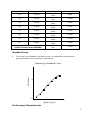
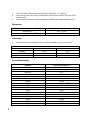
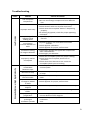
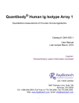
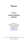
AssayMaxTM Human IgA ELISA Kit Assaypro LLC 3400 Harry S Truman Blvd St. Charles, MO 63301 T (636) 447-9175 F (636) 395-7419 www.assaypro.com For any questions regarding troubleshooting or performing the assay, please contact our support team at [email protected]. Thank you for choosing Assaypro. Assay Summary Step 1. Add 50 µl of Standard or Sample per well. Incubate 2 hours. Step 2. Wash, then add 50 µl of Biotinylated Antibody per well. Incubate 1 hour. Step 3. Wash, then add 50 µl of SP Conjugate per well. Incubate 30 minutes. Step 4. Wash, then add 50 µl of Chromogen Substrate per well. Incubate 20 minutes. Step 5. Add 50 µl of Stop Solution per well. Read at 450 nm immediately. Symbol Key Consult instructions for use. H G F E D C B A 1 2 3 4 5 6 7 8 9 10 11 12 Assay Template Human Immunoglobulin A (IgA) ELISA Kit Catalog No. EI7001-1 Sample insert for reference use only Introduction Human Immunoglobulin A (IgA) is the most abundant antibody isotype in mucosal secretions and exists in two subclasses IgA1 and IgA2 (1). While circulating serum IgA1 occurs mainly in the monomeric 160 kDa form (2), mucosal secretary IgA2 is in dimeric form and serves as the first line of defense against microorganisms through immune exclusion (3). Selective IgA deficiency is the most common primary immunodeficiency observed by a maturation defect in B cells to produce IgA (4). Principle of the Assay The AssayMax Human IgA ELISA (Enzyme-Linked Immunosorbent Assay) kit is designed for detection of human IgA in plasma, serum, urine, saliva, milk, CSF, and cell culture samples. This assay employs a quantitative sandwich enzyme immunoassay technique that measures human IgA in less than 4 hours. A polyclonal antibody specific for human IgA has been pre-coated onto a 96-well microplate with removable strips. IgA in standards and samples is sandwiched by the immobilized antibody and the biotinylated polyclonal antibody specific for IgA, which is recognized by a streptavidin-peroxidase conjugate. All unbound material is washed away and a peroxidase enzyme substrate is added. The color development is stopped and the intensity of the color is measured. Caution and Warning This product is for Research Use Only and is Not For Use In Diagnostic Procedures. Prepare all reagents (working diluent buffer, wash buffer, standard, biotinylated antibody, and SP conjugate) as instructed, prior to running the assay. Prepare all samples prior to running the assay. The dilution factors for the samples are suggested in this insert. However, the user should determine the optimal dilution factor. Spin down the SP conjugate vial and the biotinylated antibody vial before opening and using contents. The Stop Solution is an acidic solution. The kit should not be used beyond the expiration date. 1 Reagents Human IgA Microplate: A 96-well polystyrene microplate (12 strips of 8 wells) coated with a polyclonal antibody against human IgA. Sealing Tapes: Each kit contains 3 precut, pressure sensitive sealing tapes that can be cut to fit the format of the individual assay. Human IgA Standard: Human IgA in a buffered protein base (200 ng, lyophilized). Biotinylated Human IgA Antibody (60x): A 60-fold concentrated biotinylated polyclonal antibody against IgA (100 l). MIX Diluent Concentrate (10x): A 10-fold concentrated buffered protein base (30 ml). Wash Buffer Concentrate (20x): A 20-fold concentrated buffered surfactant (30 ml, 2 bottles). Streptavidin-Peroxidase Conjugate (SP Conjugate): A 100-fold concentrate (80 l). Chromogen Substrate: A ready-to-use stabilized peroxidase chromogen substrate tetramethylbenzidine (8 ml). Stop Solution: A 0.5 N hydrochloric acid to stop the chromogen substrate reaction (12 ml). Storage Condition Upon arrival, immediately store components of the kit at recommended temperatures up to the expiration date. Store SP Conjugate and Biotinylated Antibody at -20°C. Store Microplate, Diluent Concentrate (10x), Wash Buffer, Stop Solution, and Chromogen Substrate at 2-8°C. Unused microplate wells may be returned to the foil pouch with the desiccant packs and resealed. May be stored for up to 30 days in a vacuum desiccator. Diluent (1x) may be stored for up to 30 days at 2-8°C. Store Standard at 2-8°C before reconstituting with Diluent and at -20°C after reconstituting with Diluent. Other Supplies Required 2 Microplate reader capable of measuring absorbance at 450 nm. Pipettes (1-20 l, 20-200 l, 200-1000 l, and multiple channel). Deionized or distilled reagent grade water. Sample Collection, Preparation, and Storage Plasma: Collect plasma using one-tenth volume of 0.1 M sodium citrate as an anticoagulant. Centrifuge samples at 3000 x g for 10 minutes. Dilute samples 1:80000 into MIX Diluent or within the range of 1:20000 to 1:200000, and assay. Depending on application needs, user should determine proper dilutions. The undiluted samples can be stored at -20°C or below for up to 3 months. Avoid repeated freeze-thaw cycles (EDTA or Heparin can also be used as an anticoagulant). Serum: Samples should be collected into a serum separator tube. After clot formation, centrifuge samples at 3000 x g for 10 minutes, and remove serum. Dilute samples 1:80000 into MIX Diluent and assay or within the range of 1:20000 to 1:200000, and assay. Depending on application needs, user should determine proper dilutions. The undiluted samples can be stored at -20°C or below for up to 3 months. Avoid repeated freeze-thaw cycles. Cell Culture Supernatants: Centrifuge cell culture media at 3000 x g for 10 minutes to remove debris. Collect supernatants and assay. Store the remaining samples at -20°C or below. Avoid repeated freeze-thaw cycles. Urine: Collect urine using sample pot. Centrifuge samples at 800 x g for 10 minutes. Dilute urine 1:20 with MIX Diluent or within the range of 1:10 to 1:100, and assay. Depending on application needs, user should determine proper dilutions. The undiluted samples can be stored at -20°C or below for up to 3 months. Avoid repeated freeze-thaw cycles. Saliva: Collect saliva using sample tube. Centrifuge samples at 800 x g for 10 minutes. Dilute saliva 1:2000 with MIX Diluent or within the range of 1:1000 to 1:10000, and assay. Depending on application needs, user should determine proper dilutions. The undiluted samples can be stored at -20°C or below for up to 3 months. Avoid repeated freeze-thaw cycles. Milk: Collect milk using sample tube. Centrifuge samples at 800 x g for 10 minutes. Dilute milk 1:10000 with MIX Diluent or within the range of 1:2000 to 1:40000, and assay. Depending on application needs, user should determine proper dilutions. The undiluted samples can be stored at -20°C or below for up to 3 months. Avoid repeated freeze-thaw cycles. CSF: Collect cerebrospinal fluid (CSF) using sample pot. Centrifuge samples at 3000 x g for 10 minutes. Dilute samples 1:500 into MIX Diluent or within the range of 1:200 to 1:2000, and assay. Depending on application needs, user should determine proper dilutions. The undiluted samples can be stored at -80°C for up to 3 months. Avoid repeated freeze-thaw cycles. Refer to Sample Dilution Guidelines below for further instruction. 3 Guidelines for Dilutions of 1:100 or Greater (for reference only; please follow the insert for specific dilution suggested) 1:100 1:10000 A) 4 ul sample: 396 µl buffer(100x) = 100 fold dilution A) B) Assuming the needed volume is less than or equal to 400 µl. Assuming the needed volume is less than or equal to 400 µl. 1:1000 A) B) 4 µl sample : 396 µl buffer (100x) 24 µl of A : 216 µl buffer (10x) = 1000 fold dilution Assuming the needed volume is less than or equal to 240 µl. 4 µl sample : 396 µl buffer (100x) 4 µl of A : 396 µl buffer (100x) = 10000 fold dilution 1:100000 A) B) C) 4 µl sample : 396 µl buffer (100x) 4 µl of A : 396 µl buffer (100x) 24 µl of B : 216 µl buffer (10x) = 100000 fold dilution Assuming the needed volume is less than or equal to 240 µl. Reagent Preparation 4 Freshly dilute all reagents and bring all reagents to room temperature before use. MIX Diluent Concentrate (10x): If crystals have formed in the concentrate, mix gently until the crystals have completely dissolved. Dilute the MIX Diluent Concentrate 1:10 with reagent grade water. Store for up to 30 days at 2-8°C. Standard Curve: Reconstitute the 200 ng (24 mU/ml) of Human IgA Standard with 2 ml of MIX Diluent to generate a 100 ng/ml (12 mU/ml) standard stock solution. Allow the standard to sit for 10 minutes with gentle agitation prior to making dilutions. Prepare duplicate or triplicate standard points by serially diluting the standard stock solution (100 ng/ml) 1:2 with MIX Diluent to produce 50, 25, 12.5, 6.25, 3.125, 1.563, and 0.781 ng/ml solutions. MIX Diluent serves as the zero standard (0 ng/ml). Any remaining solution should be frozen at -20°C and use within 30 days. Standard Point P1 P2 P3 P4 P5 P6 P7 P8 Dilution 1 part Standard (100 ng/ml) + 1 part MIX Diluent 1 part P1 + 1 part MIX Diluent 1 part P2 + 1 part MIX Diluent 1 part P3 + 1 part MIX Diluent 1 part P4 + 1 part MIX Diluent 1 part P5 + 1 part MIX Diluent 1 part P6 + 1 part MIX Diluent MIX Diluent [IgA] (ng/ml) [IgA] (mU/ml) 50.00 6.000 25.00 12.50 6.250 3.125 1.563 0.781 0.000 3.000 1.500 0.750 0.375 0.188 0.094 0.000 Biotinylated Human IgA Antibody (60x): Spin down the antibody briefly and dilute the desired amount of the antibody 1:60 with MIX Diluent. Any remaining solution should be frozen at -20°C. Wash Buffer Concentrate (20x): If crystals have formed in the concentrate, mix gently until the crystals have completely dissolved. Dilute the Wash Buffer Concentrate 1:20 with reagent grade water. SP Conjugate (100x): Spin down the SP Conjugate briefly and dilute the desired amount of the conjugate 1:100 with MIX Diluent. Any remaining solution should be frozen at -20°C. Assay Procedure Prepare all reagents, standard solutions, and samples as instructed. Bring all reagents to room temperature before use. The assay is performed at room temperature (20-25°C). Remove excess microplate strips from the plate frame and return them immediately to the foil pouch with desiccants inside. Reseal the pouch securely to minimize exposure to water vapor and store in a vacuum desiccator. Add 50 l of Human IgA Standard or sample per well. Cover wells with a sealing tape and incubate for 2 hours. Start the timer after the last addition. Wash five times with 200 l of Wash Buffer manually. Invert the plate each time and decant the contents; hit 4-5 times on absorbent material to completely remove the liquid. If using a machine, wash six times with 300 l of Wash Buffer and then invert the plate, decanting the contents; hit 4-5 times on absorbent material to completely remove the liquid. Add 50 l of Biotinylated Human IgA Antibody to each well and incubate for 1 hour. Wash the microplate as described above. 5 Add 50 l of Streptavidin-Peroxidase Conjugate to each well and incubate for 30 minutes. Turn on the microplate reader and set up the program in advance. Wash the microplate as described above. Add 50 l of Chromogen Substrate per well and incubate for 20 minutes or till the optimal blue color density develops. Gently tap plate to ensure thorough mixing and break the bubbles in the well with pipette tip. Add 50 l of Stop Solution to each well. The color will change from blue to yellow. Read the absorbance on a microplate reader at a wavelength of 450 nm immediately. If wavelength correction is available, subtract readings at 570 nm from those at 450 nm to correct optical imperfections. Otherwise, read the plate at 450 nm only. Please note that some unstable black particles may be generated at high concentration points after stopping the reaction for about 10 minutes which will reduce the readings. Data Analysis Calculate the mean value of the duplicate or triplicate readings for each standard and sample. To generate a standard curve, plot the graph using the standard concentrations on the x-axis and the corresponding mean 450 nm absorbance on the y-axis. The best-fit line can be determined by regression analysis using four-parameter or log-log logistic curve-fit. Determine the unknown sample concentration from the Standard Curve and multiply the value by the dilution factor. Typical Data 6 The typical data is provided for reference only. Individual laboratory means may vary from the values listed. Variations between laboratories may be caused by technique differences. Standard Point ng/ml P1 50.00 P2 25.00 P3 12.50 P4 6.250 P5 3.125 P6 1.563 P7 0.781 P8 0.000 Sample: Pool Normal, Sodium Citrate Plasma (80000x) OD Average OD 1.885 1.777 1.492 1.427 0.965 0.936 0.586 0.556 0.331 0.326 0.202 0.189 0.134 0.126 0.056 0.060 1.831 1.460 0.951 0.571 0.329 0.195 0.130 0.058 1.154 1.151 1.153 Standard Curve The curve is provided for illustration only. A standard curve should be generated each time the assay is performed. OD 450 nm Human IgA Standard Curve 1.0 0.1 1 10 100 [hIgA] (ng/ml) Performance Characteristics 7 The minimum detectable dose of IgA is typically ~ 0.7 ng/ml. Intra-assay and inter-assay coefficients of variation were 5.0% and 7.2% respectively. Kit standard has been calibrated against WHO International Reference. Recovery Standard Added Value Recovery % Average Recovery % 3.13 – 25 ng/ml 85 – 113% 98% Linearity Plasma and serum samples were serially-diluted to test for linearity. Average Percentage of Expected Value (%) Sample Dilution Plasma Serum 1:40000 92% 91% 1:80000 99% 100% 1:160000 105% 103% Cross-Reactivity Species Canine Bovine Monkey Mouse Rat Swine Rabbit Immunoglobulins IgM IgA1 IgA2 IgG1 IgG2 IgG3 IgG4 IgD IgE 8 Cross Reactivity (%) None None <5% None None None None Cross Reactivity (%) <5% 100% 100% <1% None None None <1% <1% Troubleshooting Issue Causes Use of expired components Low Precision Improper wash step Splashing of reagents while loading wells Inconsistent volumes loaded into wells Insufficient mixing of reagent dilutions Unexpectedly Low or High Signal Intensity Improperly sealed microplate Microplate was left unattended between steps Omission of step Steps performed in incorrect order Insufficient amount of reagents added to wells Wash step was skipped Improper wash buffer Improper reagent preparation Insufficient or prolonged incubation periods Course of Action Check the expiration date listed before use. Do not interchange components from different lots. Check that the correct wash buffer is being used. Check that all wells are dry after aspiration. Check that the microplate washer is dispensing properly. If washing by pipette, check for proper pipetting technique. Pipette properly in a controlled and careful manner. Pipette properly in a controlled and careful manner. Check pipette calibration. Check pipette for proper performance. Thoroughly agitate the lyophilized components after reconstitution. Thoroughly mix dilutions. Check the microplate pouch for proper sealing. Check that the microplate pouch has no punctures. Check that three desiccants are inside the microplate pouch prior to sealing. Each step of the procedure should be performed uninterrupted. Consult the provided procedure for complete list of steps. Consult the provided procedure for the correct order. Check pipette calibration. Check pipette for proper performance. Consult the provided procedure for all wash steps. Check that the correct wash buffer is being used. Consult reagent preparation section for the correct dilutions of all reagents. Consult the provided procedure for correct incubation time. 9 Deficient Standard Curve Fit Non-optimal sample dilution Contamination of reagents Contents of wells evaporate Improper pipetting Insufficient mixing of reagent dilutions Sandwich ELISA: If samples generate OD values higher than the highest standard point (P1), dilute samples further and repeat the assay. Competitive ELISA: If samples generate OD values lower than the highest standard point (P1), dilute samples further and repeat the assay. User should determine the optimal dilution factor for samples. A new tip must be used for each addition of different samples or reagents during the assay procedure. Verify that the sealing film is firmly in place before placing the assay in the incubator or at room temperature. Pipette properly in a controlled and careful manner. Check pipette calibration. Check pipette for proper performance. Thoroughly agitate the lyophilized components after reconstitution. Thoroughly mix dilutions. References (1) (2) (3) (4) Delacroix DL et al. (1982) Immunology 47:383-385 Kerr MA (1990) Biochem. J. 271:285-296 Corthésy B (2007) J Immunol. 178(1):27-32 Yel L (2010) J Clin Immunol. 30(1):10–16 Version 1.8R Related Products EI7200-1 AssayMax Human IgG ELISA Kit (Plasma, Serum, Urine, Milk, Saliva, and Cell Culture samples) EI7201-1 AssayMax Human IgG3 ELISA Kit (Plasma, Serum, Urine, Milk, Saliva, and Cell Culture samples) EI7301-1 AssayMax Human IgM ELISA Kit (Plasma, Serum, Urine, Milk, Saliva, and Cell Culture samples) EI7800-1 AssayMax Human IgD ELISA Kit (Plasma, Serum, Urine, Milk, Saliva, and Cell Culture samples) www.assaypro.com • e-mail: [email protected] 10