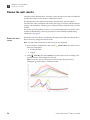
1
USER GUIDE
Protein Thermal Shift™ Studies
Using Protein Thermal Shift™ reagents and
Protein Thermal Shift™ Software v1.0
Publication Part Number 4461808 Rev. A
Revision Date May 2011
For Research Use Only. Not intended for any animal or human therapeutic or diagnostic use.
Information in this document is subject to change without notice.
APPLIED BIOSYSTEMS DISCLAIMS ALL WARRANTIES WITH RESPECT TO THIS DOCUMENT, EXPRESSED OR IMPLIED, INCLUDING BUT NOT LIMITED TO
THOSE OF MERCHANTABILITY OR FITNESS FOR A PARTICULAR PURPOSE. TO THE FULLEST EXTENT ALLOWED BY LAW, IN NO EVENT SHALL APPLIED
BIOSYSTEMS BE LIABLE, WHETHER IN CONTRACT, TORT, WARRANTY, OR UNDER ANY STATUTE OR ON ANY OTHER BASIS FOR SPECIAL, INCIDENTAL,
INDIRECT, PUNITIVE, MULTIPLE OR CONSEQUENTIAL DAMAGES IN CONNECTION WITH OR ARISING FROM THIS DOCUMENT, INCLUDING BUT NOT
LIMITED TO THE USE THEREOF, WHETHER OR NOT FORESEEABLE AND WHETHER OR NOT APPLIED BIOSYSTEMS IS ADVISED OF THE POSSIBILITY OF
SUCH DAMAGES.
LIMITED USE LABEL LICENSE: RESEARCH USE ONLY
The purchase of this product conveys to the purchaser the limited, non-transferable right to use the purchased amount of the product only to perform internal
research for the sole benefit of the purchaser. No right to resell this product or any of its components is conveyed expressly, by implication, or by estoppel.
This product is for internal research purposes only and is not for use in commercial applications of any kind, including, without limitation, quality control and
commercial services such as reporting the results of purchaser's activities for a fee or other form of consideration. For information on obtaining additional
rights, please contact [email protected] or Out Licensing, Life Technologies, 5791 Van Allen Way, Carlsbad, California 92008.
TRADEMARKS
The trademarks mentioned herein are the property of Life Technologies Corporation or their respective owners.
Microsoft and Windows are registered trademarks of Microsoft Corporation. Intel and Pentium are registered trademarks of Intel Corporation.
© 2011 Life Technologies Corporation. All rights reserved.
Part Number 4461808 Rev. A
May 2011
Contents
About This Guide . . . . . . . . . . . . . . . . . . . . . . . . . . . . . . . . . . . . . . . . . . . . . . . . . 5
Purpose . . . . . . . . . . . . . . . . . . . . . . . . . . . . . . . . . . . . . . . . . . . . . . . . . . . . . . . . . . . . . . . . . . . . . . . . . . . . . 5
User attention words . . . . . . . . . . . . . . . . . . . . . . . . . . . . . . . . . . . . . . . . . . . . . . . . . . . . . . . . . . . . . . . . . . 5
■ CHAPTER 1
Getting Started with a Protein Thermal Shift™ Study . . . . 7
Product information . . . . . . . . . . . . . . . . . . . . . . . . . . . . . . . . . . . . . . . . . . . . . . . . . . . . . . . . . . . . . . . . . . . 8
General guidelines . . . . . . . . . . . . . . . . . . . . . . . . . . . . . . . . . . . . . . . . . . . . . . . . . . . . . . . . . . . . . . . . . . . . 9
Create and set up an experiment file for the instrument run . . . . . . . . . . . . . . . . . . . . . . . . . . . . . . . 12
Prepare the protein melt reactions . . . . . . . . . . . . . . . . . . . . . . . . . . . . . . . . . . . . . . . . . . . . . . . . . . . . . 18
Run the protein melt reactions . . . . . . . . . . . . . . . . . . . . . . . . . . . . . . . . . . . . . . . . . . . . . . . . . . . . . . . . . 19
Set up the analysis . . . . . . . . . . . . . . . . . . . . . . . . . . . . . . . . . . . . . . . . . . . . . . . . . . . . . . . . . . . . . . . . . . . 21
Review the well results . . . . . . . . . . . . . . . . . . . . . . . . . . . . . . . . . . . . . . . . . . . . . . . . . . . . . . . . . . . . . . . 24
Review the replicate results . . . . . . . . . . . . . . . . . . . . . . . . . . . . . . . . . . . . . . . . . . . . . . . . . . . . . . . . . . . 29
■ CHAPTER 2
Buffer Screening Studies . . . . . . . . . . . . . . . . . . . . . . . . . . . 35
General guidelines . . . . . . . . . . . . . . . . . . . . . . . . . . . . . . . . . . . . . . . . . . . . . . . . . . . . . . . . . . . . . . . . . . . 36
Create and set up an experiment file for the instrument run . . . . . . . . . . . . . . . . . . . . . . . . . . . . . . . 36
Prepare the protein melt reactions . . . . . . . . . . . . . . . . . . . . . . . . . . . . . . . . . . . . . . . . . . . . . . . . . . . . . 38
Run the protein melt reactions . . . . . . . . . . . . . . . . . . . . . . . . . . . . . . . . . . . . . . . . . . . . . . . . . . . . . . . . . 39
Set up the analysis . . . . . . . . . . . . . . . . . . . . . . . . . . . . . . . . . . . . . . . . . . . . . . . . . . . . . . . . . . . . . . . . . . . 40
Review the well results . . . . . . . . . . . . . . . . . . . . . . . . . . . . . . . . . . . . . . . . . . . . . . . . . . . . . . . . . . . . . . . 42
Review the replicate results . . . . . . . . . . . . . . . . . . . . . . . . . . . . . . . . . . . . . . . . . . . . . . . . . . . . . . . . . . . 46
■ CHAPTER 3
Mutation Screening Studies . . . . . . . . . . . . . . . . . . . . . . . . . 51
General guidelines . . . . . . . . . . . . . . . . . . . . . . . . . . . . . . . . . . . . . . . . . . . . . . . . . . . . . . . . . . . . . . . . . . . 52
Create and set up an experiment file for the instrument run . . . . . . . . . . . . . . . . . . . . . . . . . . . . . . . 52
Prepare the protein melt reactions . . . . . . . . . . . . . . . . . . . . . . . . . . . . . . . . . . . . . . . . . . . . . . . . . . . . . 54
Run the protein melt reactions . . . . . . . . . . . . . . . . . . . . . . . . . . . . . . . . . . . . . . . . . . . . . . . . . . . . . . . . . 55
Set up the analysis . . . . . . . . . . . . . . . . . . . . . . . . . . . . . . . . . . . . . . . . . . . . . . . . . . . . . . . . . . . . . . . . . . . 56
Review the well results . . . . . . . . . . . . . . . . . . . . . . . . . . . . . . . . . . . . . . . . . . . . . . . . . . . . . . . . . . . . . . . 58
Review the replicate results . . . . . . . . . . . . . . . . . . . . . . . . . . . . . . . . . . . . . . . . . . . . . . . . . . . . . . . . . . . 62
Protein Thermal Shift™ Studies User Guide
3
Contents
■ CHAPTER 4
Ligand Screening Studies . . . . . . . . . . . . . . . . . . . . . . . . . . 67
General guidelines . . . . . . . . . . . . . . . . . . . . . . . . . . . . . . . . . . . . . . . . . . . . . . . . . . . . . . . . . . . . . . . . . . . 68
Create and set up an experiment file for the instrument run . . . . . . . . . . . . . . . . . . . . . . . . . . . . . . . . 68
Prepare the protein melt reactions . . . . . . . . . . . . . . . . . . . . . . . . . . . . . . . . . . . . . . . . . . . . . . . . . . . . . . 70
Run the protein melt reactions . . . . . . . . . . . . . . . . . . . . . . . . . . . . . . . . . . . . . . . . . . . . . . . . . . . . . . . . . 71
Set up the analysis . . . . . . . . . . . . . . . . . . . . . . . . . . . . . . . . . . . . . . . . . . . . . . . . . . . . . . . . . . . . . . . . . . . . 72
Review the well results . . . . . . . . . . . . . . . . . . . . . . . . . . . . . . . . . . . . . . . . . . . . . . . . . . . . . . . . . . . . . . . . 74
Review the replicate results . . . . . . . . . . . . . . . . . . . . . . . . . . . . . . . . . . . . . . . . . . . . . . . . . . . . . . . . . . . . 78
■ CHAPTER 5
Troubleshooting . . . . . . . . . . . . . . . . . . . . . . . . . . . . . . . . . . . 83
Examples of symptoms . . . . . . . . . . . . . . . . . . . . . . . . . . . . . . . . . . . . . . . . . . . . . . . . . . . . . . . . . . . . . . . 85
■ APPENDIX A
Supplemental Information . . . . . . . . . . . . . . . . . . . . . . . . 87
Hardware recommendations . . . . . . . . . . . . . . . . . . . . . . . . . . . . . . . . . . . . . . . . . . . . . . . . . . . . . . . . . . . 87
Boltzmann fitting method . . . . . . . . . . . . . . . . . . . . . . . . . . . . . . . . . . . . . . . . . . . . . . . . . . . . . . . . . . . . . . 88
■ APPENDIX B
Software Warranty Information . . . . . . . . . . . . . . . . . . . . 89
Computer configuration . . . . . . . . . . . . . . . . . . . . . . . . . . . . . . . . . . . . . . . . . . . . . . . . . . . . . . . . . . . . . . . 89
Limited product warranty . . . . . . . . . . . . . . . . . . . . . . . . . . . . . . . . . . . . . . . . . . . . . . . . . . . . . . . . . . . . . . 89
■ APPENDIX C
Ordering Information . . . . . . . . . . . . . . . . . . . . . . . . . . . . . 91
■ APPENDIX D
Safety . . . . . . . . . . . . . . . . . . . . . . . . . . . . . . . . . . . . . . . . . . . 93
Chemical safety . . . . . . . . . . . . . . . . . . . . . . . . . . . . . . . . . . . . . . . . . . . . . . . . . . . . . . . . . . . . . . . . . . . . . . 93
Biological hazard safety . . . . . . . . . . . . . . . . . . . . . . . . . . . . . . . . . . . . . . . . . . . . . . . . . . . . . . . . . . . . . . . 94
Documentation and Support . . . . . . . . . . . . . . . . . . . . . . . . . . . . . . . . . . . . . 95
Related documentation . . . . . . . . . . . . . . . . . . . . . . . . . . . . . . . . . . . . . . . . . . . . . . . . . . . . . . . . . . . . . . . 95
Obtaining information from the Help system . . . . . . . . . . . . . . . . . . . . . . . . . . . . . . . . . . . . . . . . . . . . . 95
Obtaining SDSs . . . . . . . . . . . . . . . . . . . . . . . . . . . . . . . . . . . . . . . . . . . . . . . . . . . . . . . . . . . . . . . . . . . . . . 96
Obtaining support . . . . . . . . . . . . . . . . . . . . . . . . . . . . . . . . . . . . . . . . . . . . . . . . . . . . . . . . . . . . . . . . . . . . 96
Glossary . . . . . . . . . . . . . . . . . . . . . . . . . . . . . . . . . . . . . . . . . . . . . . . . . . . . . . 97
4
Protein Thermal Shift™ Studies User Guide
About This Guide
IMPORTANT! Before using this product, read and understand the information the
“Safety” appendix in this document.
Purpose
This guide is designed to help you quickly learn how to perform Protein Thermal
Shift™ studies using Protein Thermal Shift™ reagents, Applied Biosystems Real-Time
PCR Systems, and Protein Thermal Shift™ Software v1.0. This guide provides step-bystep procedures on:
• How to perform an example ligand titration study using the Protein Thermal
Shift™ Starter Kit and the example experiment files installed with the Protein
Thermal Shift™ Software.
• How to perform and troubleshoot your buffer screening, mutation screening, and
ligand screening studies using Protein Thermal Shift™ reagents and Protein
Thermal Shift™ Software v1.0.
User attention words
Five user attention words may appear in this document. Each word implies a
particular level of observation or action as described below:
Note: Provides information that may be of interest or help but is not critical to the use
of the product.
IMPORTANT! Provides information that is necessary for proper instrument operation
or accurate chemistry kit use.
CAUTION! Indicates a potentially hazardous situation that, if not avoided, may
result in minor or moderate injury. It may also be used to alert against unsafe
practices.
WARNING! Indicates a potentially hazardous situation that, if not avoided,
could result in death or serious injury.
DANGER! Indicates an imminently hazardous situation that, if not avoided,
will result in death or serious injury.
Protein Thermal Shift™ Studies User Guide
5
About This Guide
User attention words
6
Protein Thermal Shift™ Studies User Guide
1
Getting Started with a
Protein Thermal Shift™ Study
Perform a Protein Thermal Shift™ study to screen for buffers, mutations, or ligands
that affect the thermal stability of the protein of interest. With Protein Thermal Shift™
studies, you can determine optimal protein storage conditions, perform functional
characterization, or improve the success rates of protein purification and
crystallization.
This chapter provides instructions for using the Protein Thermal Shift™ Starter Kit to
perform an example ligand titration study. In the example ligand titration study, you
titrate a ligand to determine the concentration that increases the thermal stability of the
protein.
Perform an example ligand titration study
Create and set up an experiment file for the instrument run:
• ViiA™ 7 System (page 12)
• StepOne™ or StepOnePlus™ System (page 14)
• 7500 or 7500 Fast System (page 16)
Prepare the protein melt reactions (page 18)
Run the protein melt reactions (page 19)
Set up the analysis (page 21)
Review the well results (page 24)
Review the replicate results (page 29)
Protein Thermal Shift™ Studies User Guide
7
1
Getting Started with a Protein Thermal Shift™ Study
Product information
Product information
The Protein Thermal Shift™ Starter Kit includes buffer, dye, a control protein, and a
control ligand.
Purpose of the
starter kit
After you follow the instructions in this chapter to learn how to perform a Protein
Thermal Shift™ study, you can continue to use the starter kit components:
Use the Protein Thermal Shift™ Buffer and the Protein Thermal Shift™ Dye in your
protein melt reactions. The buffer and dye included in the starter kit are identical to the
buffer and dye in the Protein Thermal Shift™ Dye Kit.
The Protein Thermal Shift™ Starter Kit contains two boxes.
Starter kit contents
and storage
Part
number
4462263
Box
Quantity
Component
Storage conditions
Protein
Thermal
Shift™ Dye Kit
Sufficient for
Protein Thermal Shift™ Buffer
2000 reactions
Protein Thermal Shift™ Dye
Protein
Thermal
Shift™ Starter
Kit
Sufficient for
100 reactions
Protein Thermal Shift™ Control Ligand
Room temperature (RT,
18°C to 25°C)
–15°C to –25°C
Protein Thermal Shift™ Control Protein
Purpose of the dye
kit
After you learn how to perform a Protein Thermal Shift™ study, you can use the
Protein Thermal Shift™ Dye Kit. The Protein Thermal Shift™ Dye Kit contains buffer
and dye to perform the Protein Thermal Shift™ reactions with your proteins of interest.
Dye kit contents
and storage
The Protein Thermal Shift™ Dye Kit contains one box.
Part number
4461146
8
Quantity
Sufficient for
2000 reactions
Component
Storage conditions
Protein Thermal Shift™ Buffer
Protein Thermal
Shift™
Dye
Room temperature (RT,
18°C to 25°C)
Protein Thermal Shift™ Studies User Guide
Chapter 1 Getting Started with a Protein Thermal Shift™ Study
General guidelines
Materials required
but not included
1
For the Safety Data Sheet (SDS) of any chemical not distributed by Life Technologies,
contact the chemical manufacturer. Before handling any chemicals, refer to the SDS
provided by the manufacturer, and observe all relevant precautions.
Item
Source
Applied Biosystems Real-Time PCR System
Applied Biosystems Protein Thermal
Shift™
Life Technologies
Software v1.0
Life Technologies
Applied Biosystems
MicroAmp®
Optical Reaction Plates
Life Technologies
Applied Biosystems
MicroAmp®
Optical Adhesive Film
Life Technologies
Centrifuge with plate adapters
Major laboratory
suppliers (MLS)
Microcentrifuge
MLS
Microcentrifuge tubes
MLS
Pipettors and pipette tips
MLS
Vortexer
MLS
Water
MLS
General guidelines
Protein
considerations
Before you perform a Protein Thermal Shift™ study, first consider whether the protein
of interest is a suitable candidate for this method:
• Is the protein thermally stable, as determined by other methods? If the Tm for the
protein is greater than 98°C, the protein melt reactions will produce melt curves
with no distinct melt phase. You may observe flat fluorescence signals, a decrease
in signal, or high signals, depending on the protein. You may need to use other
methods to screen for conditions that increase thermal stability of the protein.
For thermally stable proteins, you may consider performing Protein Thermal
Shift™ studies to screen for buffers, ligands, or mutations that decrease thermal
stability of the protein.
• In the native state of a protein, are there external hydrophobic sites? If so, you
may observe a high initial background signal. Conversely, if the protein does not
contain sufficient hydrophobic residues, you may observe low fluorescence
signals.
• Does the protein of interest comprise more than one domain or form oligomers? If
so, you may observe a multi-state model of unfolding and multiple melt phases in
the melt curves.
– For proteins that comprise more than one domain, you may consider
separate expression of just the domain with the active site.
– For proteins that form oligomers, you can perform a buffer/additive
screening study to identify conditions in which the protein unfolding
follows a two-state model.
Protein Thermal Shift™ Studies User Guide
9
1
Getting Started with a Protein Thermal Shift™ Study
General guidelines
Experimental
conditions
We recommend that you begin with the experimental conditions that we provide. If
you do not observe clear, well-resolved melt phases in the melt curves and a
reasonable rise in fluorescence relative to the NPC wells, you can optimize the
experimental conditions according to your protein:
• Protein:dye ratio: If you observe high initial fluorescence or small transitional
increase in your signal, then we recommend that you perform a titration study
with the protein and Protein Thermal Shift™ Dye to identify the optimal
protein:dye ratio.
• Ramp speed and ramp rate: Start with the Fast ramp speed and the
recommended ramp rate, then decrease the ramp speed and/or ramp rate as
needed to obtain well-resolved melt phases.
In Protein Thermal Shift™ studies, the ramp speed affects protein unfolding and
the resulting melt curves. With some proteins, we have observed low resolution
of the melt phases in the melt curves with the Fast ramp speed and high ramp
rates. For these proteins, we observed improved resolution of the melt phases
when we used the Standard ramp speed and decreased ramp rates.
• Thermal profile: If you observe that the Tm is close to the beginning or close to
the end of the melt curve, adjust the temperature range so that the Tm is in the
middle of the temperature range and you are able to observe a complete protein
melt.
Plate layout
Because of possible plate-to-plate variability, we recommend that you set up the plates
so that each plate contains a reference group and an analysis group does not span
multiple plates.
Replicates
For all Protein Thermal Shift™ studies, we recommend that you prepare at least 4
replicates of each protein melt reaction to ensure statistically significant results.
Controls
For Protein Thermal Shift™ studies, we recommend that you prepare controls:
• No Protein Control (NPC): Protein melt reactions that contain only buffer, water,
and dye.
NPC wells with high fluorescence signals and melt phases may indicate
contamination in wells or protein melt reactions.
• Ligand Only Control (LOC): Protein melt reactions that contain only ligand,
buffer, water, and dye.
LOC wells with melt profiles distinct from NPC wells may indicate ligand-dye
interactions. Ligands that bind the dye and affect the fluorescence levels may
mask the presence or absence of protein-dye interactions.
10
Protein Thermal Shift™ Studies User Guide
Chapter 1 Getting Started with a Protein Thermal Shift™ Study
General guidelines
Supported
real-time PCR
systems
1
Melt curve experiment files from the following Applied Biosystems Real-Time PCR
Systems and system software are supported:
Applied Biosystems Real-Time PCR System
System software versions
ViiA™ 7 Real-Time PCR System
ViiA™ 7 Software v1.0 and v1.1
StepOne™ and StepOnePlus™ Real-Time PCR Systems
StepOne™ Software v2.1 and v2.2
7500 and 7500 Fast Real-Time PCR Systems
7500 Software v2.0.4 and v2.0.5
Note: Other system software versions for the real-time PCR systems listed may be
accepted by the Protein Thermal Shift™ Software. By default, a warning message is
displayed when you import an experiment file from a system software version that is
not supported.
Real-time PCR
system
maintenance
schedule
Perform routine maintenance of your Applied Biosystems real-time PCR system to
ensure proper operation. Refer to the appropriate user guide for your real-time PCR
system for the maintenance schedule and detailed instructions.
Maintenance
type
Task
Instrument
Clean the surface of the instrument with a lint-free cloth.
Computer
• Check the computer disk space, and archive or back up your
experiment files.
• Power off the computer controlling the instrument, then after 30
seconds, power on the computer.
• Defragment the computer hard drive.
Calibrations
• Perform a background calibration.
• Perform a spatial calibration.
• Perform a dye calibration with ROX™ dye.
Protein Thermal Shift™ Studies User Guide
11
1
Getting Started with a Protein Thermal Shift™ Study
Create and set up an experiment file for the instrument run
Create and set up an experiment file for the instrument run
Create and set up
an experiment file
for the ViiA™ 7
System
Define and assign targets so you can review the melt curves for the replicate groups in
the ViiA™ 7 Software.
1. Select Start > All Programs > Applied Biosystems > ViiA 7 Software> ViiA 7
Software v1.1.
Note: If this is your first time starting the software, read and accept the license
agreement.
2. In the Home screen of the ViiA™ 7 Software, click Experiment Setup.
3. Complete the Experiment Properties screen:
Field
Entry
Experiment Name
Enter a unique Experiment Name using up to 100 letters and/or
numbers.
Note: If you plan to start the run using the instrument
touchscreen, do not enter more than 32 characters for the
Experiment Name and do not include spaces in the name.
Block type
Select the type of block that you are using: 384-Well Block or
96-Well Block (0.2mL)
Experiment type
Melt Curve
Reagent type
Other
Ramp speed
Fast
4. In the Define screen, define the targets (conditions) and dyes:
a. In the Targets pane, enter the target name for each condition, select ROX for
the reporter, and select None for the Quencher.
For the example ligand titration study:
b. For the Passive Reference, select None.
5. In the Assign screen, assign the target and task to wells:
a. For each target, select the wells that contain the control protein, select the
Assign checkbox for Control Protein, then select U for the Task.
b. (Optional) For no protein controls (NPC), select the NPC wells, select the
Assign checkbox for Control Protein, then select N for the Task.
6. Make sure that the well assignments correspond exactly with the arrangement of
reactions in the reaction plate.
12
Protein Thermal Shift™ Studies User Guide
Chapter 1 Getting Started with a Protein Thermal Shift™ Study
Create and set up an experiment file for the instrument run
1
For the example ligation titration study:
IMPORTANT! Setup errors may result in an incorrect grouping of replicates.
7. Complete the Run Method screen to define the melt curve:
a. For the Reaction Volume Per Well, enter 20 (µL).
b. For the ramp mode, select Continuous.
c. Define the thermal profile:
Time
(mm:ss)
Step
Ramp rate
Temp (°C)
1
1.6°C/s
25.0
02:00
2
0.05°C/s
99.0
02:00
Protein Thermal Shift™ Studies User Guide
13
1
Getting Started with a Protein Thermal Shift™ Study
Create and set up an experiment file for the instrument run
8. Select the optical filters for the melt curve:
a. In the Run Method screen, select the Optical Filters tab.
Note: If you do not see the Optical Filters tab, select Tools > Preferences,
then select the Show optical filters for run method checkbox in the Defaults
tab.
b. In the Melt Curve Filter section, select x4(580±10)-m4(623±14) for the
Excitation Filter-Emission Filter.
9. Select File > Save, then enter a file name and select a location for the experiment
file.
Create and set up
an experiment file
for the StepOne™
or StepOnePlus™
System
Define and assign targets so you can review the melt curves for the replicate groups in
the StepOne™ Software.
1. Select Start > All Programs > Applied Biosystems > StepOne™ Software v2.2,
then log into the software.
Note: If you are setting up the experiment file on a computer that is not connected
to the instrument, click Continue without Connection in the Instrument
Connection Failed dialog box.
2. In the Home screen of the StepOne™ Software, click Advanced Setup.
Note: If you do not see the Advanced Setup button, click
Design Wizard button.
below the
3. Complete the Experiment Properties screen:
Field
Entry
Experiment Name
Enter a unique Experiment Name using up to 50 letters and/or
numbers.
Instrument type
Select the type of instrument that you are using: StepOnePlus™
Instrument (96 Wells) or StepOne™ Instrument (48 Wells)
Experiment type
Melt Curve
Reagent type
Other
Ramp speed
Fast
4. In the Plate Setup > Define Targets and Samples screen, define the targets
(conditions):
In the Targets pane, enter the target name for each condition, select ROX for the
reporter, and select None for the Quencher.
For the example ligand titration study:
5. Define the well contents in the Plate Setup > Assign Targets and Samples screen:
a. In the Plate Setup screen, click the Assign Targets and Samples tab.
14
Protein Thermal Shift™ Studies User Guide
Chapter 1 Getting Started with a Protein Thermal Shift™ Study
Create and set up an experiment file for the instrument run
1
b. For each target (conditions), select the wells with those conditions, select the
Assign checkbox for the target, then select U for the Task.
c. (Optional) For no protein controls (NPC), select the NPC wells, select the
Assign checkbox for the NPC target, then select N for the Task.
d. For the passive reference, select None.
For the example ligand titration study (column 12 is empty):
6. Make sure that the well assignments correspond exactly with the arrangement of
reactions in the reaction plate.
IMPORTANT! Setup errors may result in an incorrect grouping of replicates.
7. Complete the Run Method screen to define the melt curve:
a. For the Reaction Volume Per Well, enter 20 (µL).
b. For the ramp mode, select Continuous.
c. Define the thermal profile:
Time
(mm:ss)
Step
Ramp rate
Temp (°C)
1
100%
25.0
02:00
2
1%
99.0
02:00
Protein Thermal Shift™ Studies User Guide
15
1
Getting Started with a Protein Thermal Shift™ Study
Create and set up an experiment file for the instrument run
8. Select File > Save, then enter a file name and select a location for the experiment
file.
Create and set up
an experiment file
for the 7500 or
7500 Fast System
Define and assign targets so you can review the melt curves for the replicate groups in
the 7500 Software.
1. Select Start > All Programs > Applied Biosystems > 7500 Software v2.0.5 to start
the instrument software, then log into the software.
2. In the Home screen of the 7500 Software, click Advanced Setup.
Note: If you do not see the Advanced Setup button, click
Design Wizard button.
below the
3. Complete the Experiment Properties screen:
Field
Entry
Experiment Name
Enter a unique Experiment Name using up to 100 letters and/or
numbers.
Instrument type
7500 Fast (96 Wells) or 7500 (96 Wells)
Experiment type
Melt Curve
Reagent type
Other
Ramp speed
Standard
4. In the Plate Setup > Define Targets and Samples screen, define the targets
(conditions):
In the Targets pane, enter the target name for each condition, select ROX for the
reporter, and select None for the Quencher.
16
Protein Thermal Shift™ Studies User Guide
Chapter 1 Getting Started with a Protein Thermal Shift™ Study
Create and set up an experiment file for the instrument run
1
For the example ligand titration study:
5. Define the well contents in the Plate Setup > Assign Targets and Samples screen:
a. In the Plate Setup screen, click the Assign Targets and Samples tab.
b. For each target (conditions), select the wells with those conditions, select the
Assign checkbox for the target, then select U for the Task.
c. (Optional) For no protein controls (NPC), select the NPC wells, select the
Assign checkbox for the NPC target, then select N for the Task.
d. For the passive reference, select None.
6. Make sure that the well assignments correspond exactly with the arrangement of
reactions in the reaction plate.
IMPORTANT! Setup errors may result in an incorrect grouping of replicates.
7. Complete the Run Method screen to define the melt curve:
a. For the Reaction Volume Per Well, enter 20 (µL).
b. For the ramp mode, select Continuous.
c. Define the thermal profile:
Time
(mm:ss)
Step
Ramp rate
Temp (°C)
1
100%
25.0
02:00
2
1%
99.0
02:00
Protein Thermal Shift™ Studies User Guide
17
1
Getting Started with a Protein Thermal Shift™ Study
Prepare the protein melt reactions
8. Select File > Save, then enter a file name and select a location for the experiment
file.
Prepare the protein melt reactions
About the example
study protein melt
reactions
For the example ligand titration study, prepare 5 sets of protein melt reactions using
the control protein and control ligand in the Protein Thermal Shift™ Starter Kit:
• Control protein and 0.0 mM control ligand
• Control protein and 0.1 mM control ligand
• Control protein and 1.0 mM control ligand
• No protein and 0.0 mM control ligand (NPC)
• No protein and 1.0 mM control ligand (LOC)
Required materials
Required materials for protein melt reactions:
• Protein Thermal Shift™ Dye (1000✕)
• Protein Thermal Shift™ Buffer
• Water
• Protein Thermal Shift™ Control Protein
• Protein Thermal Shift™ Control Ligand
• MicroAmp® Optical Reaction Plate appropriate for your real-time PCR
instrument
• MicroAmp® Optical Adhesive Film appropriate for your reaction plate
18
Protein Thermal Shift™ Studies User Guide
Chapter 1 Getting Started with a Protein Thermal Shift™ Study
Run the protein melt reactions
Prepare the protein
melt reactions
1
We recommend that you prepare at least four replicates of each reaction.
1. Prepare a fresh dilution of Protein Thermal Shift™ Dye (1000✕) to 8✕.
2. Place the appropriate reaction plate or tubes on ice, then prepare the protein melt
reactions:
• Make sure that the arrangement of reactions in the reaction plate
corresponds exactly with the well assignments in the experiment file.
IMPORTANT! Setup errors may result in an incorrect grouping of the data
and incorrect Tm statistics.
• Add reaction components to the plate in the order listed.
Component
Volume
Protein Thermal Shift™ Buffer
5.0 µL
Shift™
Water + 2.0 µL Protein Thermal
Control Protein + Protein Thermal Shift™
Control Ligand (0 mM, 0.1 mM, or 1 mM
final concentration)
12.5 µL
Diluted Protein Thermal Shift™ Dye (8✕)
2.5 µL
Total volume for each control reaction
20.0 µL
3. Pipet each reaction up and down 10 times to mix well.
4. Seal the plate with MicroAmp® Optical Adhesive Film, spin it at 1000 rpm for
1 minute, then place it on ice.
Run the protein melt reactions
Load and run the protein melt reactions on a supported Applied Biosystems
Real-Time PCR System, then analyze and save the experiment file before importing the
experiment file into the Protein Thermal Shift™ Software.
IMPORTANT! Keep the protein melt reactions on ice until you load the instrument.
Load and run the
reactions
Refer to your instrument user guide for detailed instructions on how to transfer
experiment files to the instrument computer or instrument, how to operate the
instrument, how to start the run from an instrument touchscreen, and how to monitor
the run.
1. If necessary, transfer the experiment file that you created for the run to the
computer that is connected to the instrument.
2. In the Home screen of the Real-Time PCR System Software, click Open, then
select the experiment file you created for the run.
3. Load the reaction plate into the instrument.
4. In the Real-Time PCR System Software, click Run in the navigation pane, then
click START RUN.
Protein Thermal Shift™ Studies User Guide
19
1
Getting Started with a Protein Thermal Shift™ Study
Run the protein melt reactions
Review the melt
curves
Using the Real-Time PCR System Software, open the experiment file from the
completed instrument run, analyze and save the experiment file, then review the melt
curves.
Note: You must analyze and save the experiment file in the Real-Time PCR System
Software before you can import it into the Protein Thermal Shift™ Software.
Some common troubleshooting causes are provided here. For more detailed
troubleshooting information, see page 83.
1. In the Home screen of the Real-Time PCR System Software, click Open, then
select the experiment file from the instrument run.
2. View the melt curves:
Real-Time PCR
System Software
View the melt curve
ViiA™ 7 Software
Click Analysis > Melt Curve Plot in the navigation pane,
select Normalized Reporter from the Plot dropdown list,
then select Target from the Color dropdown list.
StepOne™ Software or
7500 Software
Click Analysis > Melt Curve in the navigation pane, select
Normalized Reporter from the Plot dropdown list, then
select Target from the Color dropdown list.
Melt curves for the ligand screening example file from the ViiA™ 7 System:
3. Review the melt curves:
• Do you see fluorescence signals in all of the sample wells?
No fluorescence signals in the sample wells may indicate missing dye or
protein or an instrument problem.
• Do you see flat fluorescence levels in the NPC wells?
High fluorescence levels in the NPC wells may indicate protein
contamination in the wells or protein melt reactions; or it may indicate that
the dye interacts with a component in the buffer.
• Do you see flat fluorescence levels in the LOC wells?
With the Protein Thermal Shift™ Control Ligand, high fluorescence levels in
the LOC wells but not in the NPC wells may indicate protein contamination
in the control ligand.
20
Protein Thermal Shift™ Studies User Guide
Chapter 1 Getting Started with a Protein Thermal Shift™ Study
Set up the analysis
1
With your own samples, high fluorescence levels in the LOC wells but not in
the NPC wells may indicate protein contamination in the control ligand or
ligand-dye interactions.
• Do the replicates have similar melt curves?
4. Save, then close the experiment file.
Note: The melt curves in the real-time PCR software may not exactly match the melt
curves in the Protein Thermal Shift™ Software. When the experiment files are
imported into the Protein Thermal Shift™ Software, the Protein Thermal Shift™
Software reduces the noise in the fluorescence data.
Set up the analysis
This section provides instructions for setting up the protein thermal shift study using
Protein Thermal Shift™ Software v1.0 and the example ligand screening files that are
installed with the software.
For more information about how to use the software, refer to the Protein Thermal Shift™
Software Help.
Start the analysis
software
Create a study
Select Start > All Programs > Applied Biosystems > Protein Thermal Shift Software
v1.0.
1. In the Home screen, click
Create Study in the toolbar.
2. In the Setup > Properties screen, enter a unique name for the study.
Note: The Study Name cannot be more than 100 characters and cannot contain
these characters: / \ * “ ? < > | . ,
3. For the Instrument, select the instrument that you used to run the protein melt
reactions.
IMPORTANT! The instrument selection must match the instrument type that you
used to run the protein melt reactions and generate the experiment files.
Experiment file
guidelines
Guidelines for the experiment files that you import into the study:
• The experiment file must contain analyzed and saved melt curve data from a
complete melt curve run.
• The instrument type in the experiment file must match the instrument type
selected for the study.
Note: The data needed for the study are extracted from the *.eds file. The Protein
Thermal Shift™ Software does not create, modify, save, or export *.eds files.
Protein Thermal Shift™ Studies User Guide
21
1
Getting Started with a Protein Thermal Shift™ Study
Set up the analysis
Set up the example
ligand titration
study
First import the example experiment file (*.eds), then set up the plate using the
example plate template file (*.csv).
The example files are located in <drive>:\Program Files\Applied Biosystems\Protein
Thermal Shift Software\examples, where <drive> is where you installed the software.
1. Go to the Setup > Experiment Files screen, click
Import, then select the ligand
screening example file (*.eds) for the instrument type that you selected for the
study:
• Ligand_Screening_Example_ViiA7.eds
• Ligand_Screening_Example_StepOnePlus.eds
• Ligand_Screening_Example_7500.eds
Note: The experiment file name and data are imported. Plate setup information is
not imported.
2. Click
Load Plate Template, then select the ligand screening plate template file
(*.csv) for the instrument type that you selected for the study:
• Ligand_Screening_Example_Setup_ViiA7.csv
• Ligand_Screening_Example_Setup_StepOnePlus.csv
• Ligand_Screening_Example_Setup_7500.csv
The conditions, task, and analysis group are assigned to the wells that contained
protein melt reactions.
3. Make sure that the condition assignments correspond exactly with the contents of
the reaction plate.
IMPORTANT! Setup errors may result in an incorrect grouping of the data and
incorrect Tm statistics.
4. Select the replicate group to use as the reference:
a. Select the wells that contain 0.1 mM ligand:
Instrument type
ViiA™ 7 System
Click the B row header to select row B (wells B1–B24).
StepOnePlus™
Click-drag the 4 column header to select columns 4–
6 (wells A4–A6, B4–B6, C4–C6, D4–D6, E4–E6, F4–F6,
G4–G6, H4–H6).
System
or 7500 Fast System
b. Click
5. Click
About the analysis
settings
Select wells
Assign, then select Reference as the Task.
Save in the toolbar to save and analyze the study.
With the analysis settings, you can adjust how positive hits are determined and specify
settings for applying flags. If you generate a study template from the study file, the
analysis settings for the study are saved in the study template.
Although the flags and the positive hits can be useful ways to quickly scan the results,
we recommend that you review the values in the tables, review the plots, and review
the replicates carefully.
22
Protein Thermal Shift™ Studies User Guide
Chapter 1 Getting Started with a Protein Thermal Shift™ Study
Set up the analysis
Review the analysis
settings
1
Review and edit the analysis settings to optimize the analysis for your study.
1. Click
Analysis Settings in the toolbar.
2. On the Positive Hit tab, specify the ΔTm-Boltzmann and ΔTm-Derivative values to
indicate a positive hit:
• Select > to identify conditions that increase protein thermal stability, or
select < to identify conditions that decrease protein thermal stability.
• Enter the number of degrees (°C) of Tm shift relative to the reference to
indicate a positive hit.
3. On the Flags tab, specify settings for applying flags:
a. Select the flags to use in the analysis.
b. For the High Background, High NPC, Low Signal, and Poor Fit flags, specify
the condition and threshold for applying the flag.
4. Apply the analysis settings and analyze:
• Click Apply to apply the analysis settings and reanalyze while keeping the
Analysis Settings dialog box open.
or
• Click OK to apply the analysis settings, reanalyze, and close the Analysis
Settings.
Protein Thermal Shift™ Studies User Guide
23
1
Getting Started with a Protein Thermal Shift™ Study
Review the well results
Review the well results
Using the Protein Thermal Shift™ Software, review the melt curves and well table and
optimize the analysis in the Analysis > Well Results screen.
The Well Results screen displays fluorescence and derivative melt curve plots,
calculated Tm values, individual well results, and flags. As necessary, edit the analysis
settings, edit the baseline, edit the region of analysis, edit the analysis mode, and omit
outliers.
This section provides guidance on how to review and interpret the well results. Some
common troubleshooting causes are provided. For more detailed troubleshooting
information, see page 83.
About the melt
curve plots
In the Well Results screen, the fluorescence data are plotted as fluorescence melt curves
and as derivative melt curves. The regions of analysis (ROAs), Boltzmann Tm values,
and derivative Tm values are displayed in the melt curve plots.
Boltzmann Tm
Derivative Tm
Fluorescence
melt curves
Derivative melt
curves
ROA start
ROA end
The fluorescence melt curves show noise-reduced fluorescence data from the protein
melt curve run. The fluorescence melt curves and the Boltzmann method are used to
determine the Boltzmann Tm.
The derivative melt curves are calculated using a first derivative of the fluorescence
data at each temperature. The derivative melt curve is used to identify melt phases, or
regions of analysis (ROAs), from which the derivative Tm is calculated. In Protein
Thermal Shift™ Software, up to six ROAs and derivative Tm values can be
automatically detected or manually defined for each melt curve.
Because the Boltzmann Tm and derivative Tm values are calculated independently of
each other, the values may be dissimilar.
24
Protein Thermal Shift™ Studies User Guide
Chapter 1 Getting Started with a Protein Thermal Shift™ Study
Review the well results
About the
Boltzmann fit curve
1
The Boltzmann fit curve is calculated for the region of analysis (ROA) according to the
Boltzmann equation and is plotted in the fluorescence melt curve plot as a dark green
curve.
Fluorescence melt curve
Boltzmann fit curve (dark green)
The Boltzmann fit value corresponds to how similar the fluorescence melt curve and
the Boltzmann fit curve are.
Note: When you define the ROA manually, you may observe a gap between the ROA
start or end temperature and the start or end of the Boltzmann fit curve. The gap
occurs if the defined ROA start or end temperature does not correspond exactly with a
fluorescence datapoint.
Review the melt
curves
Review the melt curves to visualize the fluorescence and derivative fluorescence data
in the example ligand titration study.
Note: For NPC wells, the derivative melt curves are not displayed.
1. In the Analysis > Well Results screen, select
Show in Plot, then select to show
these plot components:
• Unselected Wells
• Legend
2. Select
Color By, then select Ligand to color the melt curves according to the
ligand condition value assigned for the well.
Note: To set the color for each ligand to make the plots easier to distinguish, go to
the Setup > Conditions screen.
3. For each replicate group, select all the wells in the replicate group, then review
the fluorescence levels in the melt curves:
• For the NPC wells, do you observe a rise in fluorescence? If so, the wells or
protein melt reactions may be contaminated with protein or the dye may
interact with a buffer component.
• For the LOC wells, do you observe a rise in fluorescence? A rise in
fluorescence in LOC wells but not in NPC wells may indicate protein
contamination in the ligand or ligand-dye interactions.
Protein Thermal Shift™ Studies User Guide
25
1
Getting Started with a Protein Thermal Shift™ Study
Review the well results
• For sample or reference wells, do you observe flat melt curves? If so,
condition assignments may be incorrect or a component is missing from the
protein melt reactions.
• Within each replicate group, are the fluorescence melt curves similar to each
other? Within each replicate group, are the derivative melt curves similar to
each other? If the melt curves for the replicates are dissimilar, pipetting
errors may have occurred during reaction setup or condition assignments
may be incorrect.
About the analysis
mode
In the Protein Thermal Shift™ Software, the analysis mode is the method for defining
the regions of analysis (ROA) and determining the derivative Tm for a melt curve.
The analysis mode is displayed in the Analysis Mode column of the Well Table in the
Analysis screens.
Analysis mode
Auto: Single Tm
Description
The software detects one melt phase, defines one ROA, and
determines one derivative Tm within the ROA.
By default, the melt curves are analyzed using the Auto: Single Tm
analysis mode.
Auto: Multiple Tm
The software detects more than one melt phase, defines an ROA for
each melt phase, and determines one derivative Tm for each ROA.
The software can detect up to six ROAs for each melt curve.
If the derivative melt curve shows multiple melt phases, change the
analysis mode to Auto: Multiple Tm.
Manual
You define the ROA within the software, then the software determines
one derivative Tm for each ROA that you defined. You can define up
to six ROAs for each melt curve.
If you edit or delete an ROA detected by the software, the analysis
mode is changed to Manual.
Set the analysis
mode
In the example ligand titration study, the melt curves should show one melt phase. If
you see more than one peak, the wells or protein melt reactions may be contaminated
with protein.
In your own studies, you may observe multiple melt phases. If the derivative melt
curves for the replicate group show multiple melt phases, set the analysis mode to
Auto: Multiple Tm, then review the derivative melt curves.
1. In the Well Table or in the melt curve plot, select the wells with multiple melt
phases.
2. Click
3. Click
Auto Analysis Options, then select Auto: Multiple Tm.
Analyze to reanalyze using the Auto: Multiple Tm analysis mode.
4. Review the number of melt phases (peaks) in the derivative melt curves:
• Do all replicate groups have the same number of melt phases?
• For each replicate group, are there outliers with a different number of melt
phases than the other samples in the replicate group? You may consider
omitting outliers from analysis.
26
Protein Thermal Shift™ Studies User Guide
Chapter 1 Getting Started with a Protein Thermal Shift™ Study
Review the well results
Review the regions
of analysis (ROA)
1
Review the ROAs detected by the software. If necessary, edit the ROAs.
Note: If no melt phases are detected, no ROAs are defined. Negative controls should
have no melt phases and no ROAs.
1. In the Analysis > Well Results screen, confirm that each ROA meets the following
criteria:
• For melt curves with one melt phase, the curve within the ROA resembles a
sigmoidal profile.
• At the start temperature, the signal is relatively flat.
• At the end temperature, the signal has already reached its maximum.
2. For each replicate group, edit the ROAs so that all of the wells in the replicate
group have the same ROA:
a. Select the replicates, click
(Define ROA) in the toolbar above the melt
curve plots, then click-drag an area in one of the plots to define a melt phase
and replace the ROA. Repeat for each melt phase you identify.
b. If necessary, adjust the start and end temperatures for each ROA:
• To move the ROA: Starting from within the ROA, click-drag the ROA.
• To adjust the start and end temperatures individually: Click-drag the
ROA start or the end line.
IMPORTANT! Make sure that the fluorescence at the start temperature is
lower than the fluorescence at the end temperature.
3. Click
Analyze to reanalyze using the edited ROAs.
Note: After you edit ROAs, the analysis mode changes from Auto to Manual.
Review the flags
and Tm values
Review the flags and Tm values, then consider editing the analysis settings and/or
omitting wells before you review the replicate results.
1. In the Analysis > Well Results screen, select
Show in Plot, then select to show
these plot components:
• Boltzmann Tm
• Derivative Tm
• Unselected Wells
• Legend
2. Review flagged wells in the Well Table:
a. Click the
(Flag Indicator) column header to sort the wells according to the
number of flags applied to the well.
b. Scroll the table to the right to view the flags that are applied to the wells.
c. For flagged wells, select the well in the Well Table, then review the melt
curves for the well, compared to the other wells in the replicate group.
d. Omit wells from analysis as necessary.
Protein Thermal Shift™ Studies User Guide
27
1
Getting Started with a Protein Thermal Shift™ Study
Review the well results
3. For each replicate group, select the wells in the replicate group, then review the
Tm B (Boltzmann Tm) and the Tm D (derivative Tm) in the Well Table and in the
melt curves:
Note: In the melt curves, the Boltzmann Tm is a green dashed vertical line, and
the derivative Tm is black dotted vertical line.
• Are the Tm B or Tm D values significantly different from the Tm values for
other wells in the replicate group? Do any replicates have melt curves that
are inconsistent with the other melt curves for the replicate group? If so, you
may consider omitting wells from analysis.
• Are the melt curves within the replicate group similar?
4. If you omitted any wells from the analysis, click
Review the
Boltzmann fit
1. In the Analysis > Well Results screen, select
Analyze.
Show in Plot, then select to show
these plot components:
• Boltzmann Fit
• Unselected Wells
• Legend
2. Scroll through each well in the Well Table, then compare the fluorescence melt
curve to the Boltzmann fit curve (dark green thick curve) and review the value in
the B Fit (Boltzmann Fit) column of the Well Table:
• How well does the fluorescence melt curve correspond to the Boltzmann fit
curve?
• Is the B Fit value close to 1?
• Is the B Fit value similar among wells in the replicate group?
Note: When you define the ROA manually, you may observe a gap between the
ROA start or end temperature and the start or end of the Boltzmann fit curve. The
gap occurs if the defined ROA start or end temperature does not correspond
exactly with a fluorescence datapoint.
Example well
results
For the ViiA™ 7 System example ligand titration study using the Protein Thermal
Shift™ Control Protein and the Protein Thermal Shift™ Control Ligand, observe the
following:
• Fluorescence levels are flat in the NPC and LOC wells.
• For the sample and reference replicate groups, the fluorescence levels as
displayed in the fluorescence melt curve are not significantly different, so you do
not need to edit the baseline.
• The sample and reference wells contain one peak in the derivative melt curve.
• For each replicate group, the Boltzmann Tm values are similar and there are no
outliers.
• For each replicate group, the derivative Tm values are similar and there are no
outliers.
• For each well, the region of analysis meets the recommended criteria.
28
Protein Thermal Shift™ Studies User Guide
Chapter 1 Getting Started with a Protein Thermal Shift™ Study
Review the replicate results
1
Review the replicate results
Using the Protein Thermal Shift™ Software, review the Tm statistics for the replicate
groups and look for positive hits in the Analysis > Replicate Results screen.
This section provides guidance on how to review and interpret the replicate results.
Some common troubleshooting causes are provided. For more detailed
troubleshooting information, see page 83.
About the Tm
statistics
ΔTm values are calculated for each ROA by subtracting the mean Tm for the replicate
group from the mean Tm for the reference replicate group. If there is more than one
ROA, the ΔTm values are calculated for each ROA in the order that they appear (left to
right).
About the Replicate
Results Plot
The Replicate Results Plot shows Tm statistics for each replicate group and Tm
datapoints for each well in the replicate group. You can select to view the Boltzmann
Tm statistics, derivative Tm statistics, ΔBoltzmann Tm statistics, or ΔDerivative Tm
statistics. The plot shows the calculated Tm or ΔTm value for each sample in the
replicate group. The mean Tm or ΔTm value and the upper and lower 95% confidence
limits are identified by colored diamonds - red diamonds for the reference replicate
groups and blue diamonds for the sample replicate groups.
Single Tm Analysis – Reference replicate group results
Lower 95% confidence limit
Upper 95% confidence limit
Median
Mean
Single Tm Analysis – Sample replicate group results
Lower 95% confidence limit
Upper 95% confidence limit
Median
Mean
Multiple Tm Analysis – Sample replicate group results (one plot for each ROA)
Diamond color is the task color:
Reference
replicate group
Sample
replicate group
Datapoint color is the experiment file color:
Experiment1.eds
Experiment2.eds
Datapoint shape indicates omit status:
Omitted well
Included well
Protein Thermal Shift™ Studies User Guide
29
1
Getting Started with a Protein Thermal Shift™ Study
Review the replicate results
Review the
Boltzmann Tm
statistics
In the Analysis > Replicate Results screen, review the Boltzmann Tm statistics to
evaluate the variability among replicates. The Boltzmann Tm statistics are plotted
along the x-axis for each replicate group.
For the example ligand titration study, we provide instructions for reviewing the
Boltzmann Tm statistics. For your own studies, you may also review derivative Tm
statistics.
1. In the Analysis > Replicate Results screen, select
Plot by, then select Tm-
Boltzmann.
Note: Select Tm-Derivative to review the derivative Tm statistics.
2. Specify the condition hierarchy to group the replicate plots and change the order
of conditions in the plot:
Note: Changing the condition hierarchy does not affect the results; it only
changes how the replicate plots are grouped and the order in which the
conditions are displayed in the Replicate Results Plot.
a. Click
Condition Hierarchy above the top right corner of the plot.
b. In the dialog box, select a condition, then use the Up and Down arrows to
change the hierarchy of conditions.
c. For the example ligand titration study, continue to move the conditions up
and down until you obtain this hierarchy:
The condition at the top-most level of the hierarchy is displayed on the farright side of the Replicate Results Plot:
3. Scan the Replicate Results Plot to review the conditions that affect the Tm values
relative to the reference replicate group.
Reference replicate plot (red)
Sample replicate plot (blue)
Sample replicate plot (blue)
30
Protein Thermal Shift™ Studies User Guide
Chapter 1 Getting Started with a Protein Thermal Shift™ Study
Review the replicate results
1
4. Review the Tm statistics for each replicate group in the Replicate Results Plot:
a. Zoom in: Click
, then click-drag an area on the plot one or more times.
b. Move the plot: Click
interest is in view.
, then click-drag the plot until the replicate plot of
c. Place the cursor within the diamond, then wait to view a tooltip with the Tm
statistics for the replicate group.
d. To examine outliers, place the cursor over a datapoint, then wait to view a
tooltip with the well information, experiment file name, and the Tm selected
for the plot.
Note: Click
to restore the default zoom.
5. In the Replicate Groups table, review the Boltzmann Tm statistics for each
replicate group:
• Tm B - Std. Error (standard error of the mean for the Boltzmann Tm): Is the
value low? If the value is high, review the data for each replicate.
• Tm B - Min and Tm B - Max (minimum and maximum Boltzmann Tm
values for the replicate group): Is the range of Boltzmann Tm values for the
replicate group within 1 degree? If the range of Tm values exceeds 1 degree,
review the data for each replicate.
6. Omit outliers as necessary, then click
Protein Thermal Shift™ Studies User Guide
Analyze.
31
1
Getting Started with a Protein Thermal Shift™ Study
Review the replicate results
Review the positive
hits
In the Analysis > Replicate Results screen, review the positive hits to identify the
conditions that produce the maximum effect on thermal stability. Replicate groups
with positive hits have ΔTm values that exceed the threshold set in the analysis
settings.
Note: The positive hits are determined according to the analysis settings that you
specified (page 23).
For the example ligand titration study, review the ΔTm-Boltzmann positive hits. In
your own studies, you may choose to review the ΔTm- Derivative positive hits.
Note: You must specify the reference replicate group to calculate ΔTm values and to
determine positive hits.
1. In the Analysis > Replicate Results screen, select the
select ΔTm - Boltzmann.
2. Select the
Plot by menu, then
Show in Plot menu, then select to show Positive Hits.
3. Scan the Replicate Results Plot for positive hits.
The positive hits are replicate plot(s) within the green shaded area and labeled
with the hit threshold in the bottom right corner of the plot.
Positive hit threshold from analysis settings
Reference replicate plot: ΔTm = 0°C
Positive hit: Mean ΔTm exceeds the hit
threshold
Positive hit region: Mean ΔTm exceeds the hit threshold
4. Review the ΔTm statistics for each replicate group in the Replicate Results Plot:
a. Zoom in: Click
, then click-drag an area on the plot as many times as you
need to magnify the plot.
b. Move the plot: Click
, then click-drag the plot until the replicate plot you
want to review is in view.
32
Protein Thermal Shift™ Studies User Guide
Chapter 1 Getting Started with a Protein Thermal Shift™ Study
Review the replicate results
1
c. Place the cursor over the replicate plot, then wait to view a tooltip with the
ΔTm statistics for the replicate group.
Note: Click
to restore the default zoom.
5. In the Replicate Groups table, review the positive hits (
and review the ΔTm B statistics in the table.
Example replicate
results
) in the Hits B column
For the ViiA™ 7 System example ligand titration study using the starter kit, observe the
following:
• The control ligand stabilizes the protein in a concentration-dependent manner.
• The protein melt reactions that contained 1.0 mM control ligand increased the
thermal stability of the control protein beyond the threshold set in the analysis
settings (ΔTm Boltzmann > 2.0°C).
Next steps
The use of Protein Thermal Shift™ studies to determine the relative thermal stability of
proteins (ΔTm values) is precise and consistent with analysis using calorimetry or
spectroscopy methods. After you perform Protein Thermal Shift™ studies to screen for
and identify positive hits, you can validate the positive hits using calorimetry or
spectroscopy methods to determine the thermodynamic stability.
Protein Thermal Shift™ Studies User Guide
33
1
34
Getting Started with a Protein Thermal Shift™ Study
Review the replicate results
Protein Thermal Shift™ Studies User Guide
2
Buffer Screening Studies
Perform a buffer screening study to identify the optimal buffer, pH, or salt
concentration for storing a protein. Buffer screening may also be used to identify a
buffer system for a protein that aggregates or precipitates from solution or to improve
protein crystal formation for x-ray crystallography.
Perform a buffer screening study
Create and set up an experiment file for the instrument run (page 36)
Prepare the protein melt reactions (page 38)
Run the protein melt reactions (page 39)
Set up the analysis (page 40)
Review the well results (page 42)
Review the replicate results (page 46)
Example experiment files and plate template files are located in the examples folder:
<drive>:\Program Files\Applied Biosystems\Protein Thermal Shift
Software\examples, where <drive> is where you installed the software.
To view the data for the buffer screening example used in this chapter, use the
Buffer_Screening_Example_ViiA7.eds and
Buffer_Screening_Example_Setup_ViiA7.csv files in the ViiA7 Example Files folder.
Protein Thermal Shift™ Studies User Guide
35
2
Buffer Screening Studies
General guidelines
General guidelines
For general guidelines that apply to all Protein Thermal Shift™ studies, see page 9.
Experimental
design
With Protein Thermal Shift™ studies, you can perform a series of buffer screening
studies to identify the optimal buffer, pH, and salt concentration for a protein. You
may also perform a buffer screening study before performing a mutation screening
study or a ligand screening study.
Replicates and
controls
For buffer screening studies, we recommend that you prepare:
•
At least 4 replicates of each reaction
• At least 4 replicates of no protein controls (NPCs)
Create and set up an experiment file for the instrument run
This section contains general settings for creating and setting up an experiment file.
For detailed instructions, see:
Instrument
Page
ViiA™ 7 Real-Time PCR System
page 12
StepOne™
page 14
and
StepOnePlus™
Real-Time PCR Systems
7500 and 7500 Fast Real-Time PCR Systems
Experiment file
settings
Setup
Experiment
properties
page 16
Setting
• Experiment type: Melt Curve
• Reagents: Other
• Ramp speed: Fast or Standard
Target
properties
• Reporter: ROX
Plate layout
• Assign targets to all wells in use
• Quencher: None
• Passive reference: None
Run method
• Reaction Volume Per Well: 20 µL
• Thermal profile:
Step 1, Temp: 25°C, Time: 2 minutes
Step 2, Temp: 99°C, Time: 2 minutes
• Ramp mode: Continuous
• Ramp rate:
– ViiA™ 7 System: Step 1: 1.6°C/s, Step 2: 0.05°C/s
– StepOne™ and StepOnePlus™ Systems and 7500 and 7500 Fast
Systems: 1%
• Optical Filters (ViiA™ 7 System only):
– Excitation Filter: x4(580±10)
– Emission Filter: m4(623±14)
36
Protein Thermal Shift™ Studies User Guide
Chapter 2 Buffer Screening Studies
Create and set up an experiment file for the instrument run
Defining and
assigning targets
2
We recommend that you define and assign targets so you can review the melt curves
for the replicate groups in the Real-Time PCR System Software before importing the
experiment files into the Protein Thermal Shift™ study. Define the following targets:
• One target for each buffer condition
• Define a target for the no protein control (NPC)
IMPORTANT! Make sure that the well assignments correspond exactly with the
arrangement of reactions in the reaction plate. Setup errors may result in an incorrect
grouping of replicates.
Example targets
In the ViiA™ 7 System buffer screening example file, targets were defined for each
buffer and the no protein control (NPC).
Example plate
layout
In this example, columns 9–24 and rows K–P are not in use.
Protein Thermal Shift™ Studies User Guide
37
2
Buffer Screening Studies
Prepare the protein melt reactions
Prepare the protein melt reactions
Protein melt
reaction stability
For consistency in Tm values, we recommend that you keep the protein melt reactions
on ice until you are ready to load the instrument and start the run.
If the protein is thermally stable at ambient temperatures, you may consider preparing
the reactions in advance and leave the reaction plate at ambient temperature, protected
from light. However, the fluorescence levels will decrease over time and the Tm values
will vary, depending on the protein and its thermal stability. If you want to prepare the
reaction plates in advance, we recommend that you first determine the benchtop
stability of your protein melt reactions.
Required materials
Required materials for protein melt reactions:
• Protein Thermal Shift™ Dye (1000✕)
• Protein Thermal Shift™ Buffer
• Water
• Protein
• Buffers and/or buffer components
• MicroAmp® Optical Reaction Plate appropriate for your real-time PCR
instrument
• MicroAmp® Optical Adhesive Film appropriate for your reaction plate
Prepare the protein
melt reactions
We recommend that you prepare four replicates of each reaction.
1. Prepare a fresh dilution of Protein Thermal Shift™ Dye (1000✕) to 8✕.
2. Place the appropriate reaction plate or tubes on ice, then prepare the protein melt
reactions:
• Make sure that the arrangement of reactions in the reaction plate
corresponds exactly with the well assignments in the experiment file.
IMPORTANT! Setup errors may result in an incorrect grouping of the data
and incorrect Tm statistics.
• Add reaction components to the plate in the order listed.
Component
Volume
Protein Thermal Shift™ Buffer
5.0 µL
Water + protein + buffer and/or buffer
components
12.5 µL
Diluted Protein Thermal Shift™ Dye (8✕)
2.5 µL
Total volume for each control reaction
20.0 µL
3. Pipet each reaction up and down 10 times to mix well.
4. Seal the plate with MicroAmp® Optical Adhesive Film, spin it at 1000 rpm for
1 minute, then place it on ice.
38
Protein Thermal Shift™ Studies User Guide
Chapter 2 Buffer Screening Studies
Run the protein melt reactions
2
Run the protein melt reactions
Load and run the protein melt reactions on a supported Applied Biosystems
Real-Time PCR System, then analyze and save the experiment file before importing the
experiment file into the Protein Thermal Shift™ Software.
IMPORTANT! Keep the protein melt reactions on ice until you load the instrument.
Load and run the
reactions
1. If necessary, transfer the experiment file that you created for the run to the
computer that is connected to the instrument.
2. In the Home screen of the Real-Time PCR System Software, click Open, then
select the experiment file you created for the run.
3. Load the reaction plate into the instrument.
4. In the Real-Time PCR System Software, click Run in the navigation pane, then
click START RUN.
Review the melt
curves
Using the Real-Time PCR System Software, open the experiment file from the
completed instrument run, analyze and save the experiment file, then review the melt
curves.
Note: You must analyze and save the experiment file in the Real-Time PCR System
Software before you can import it into the Protein Thermal Shift™ Software.
Some common troubleshooting causes are provided here. For more detailed
troubleshooting information, see page 83.
1. In the Home screen of the Real-Time PCR System Software, click Open, then
select the experiment file from the instrument run.
2. View the melt curves:
Real-time PCR
System Software
View the melt curve
ViiA™ 7 Software
Click Analysis > Melt Curve Plot in the navigation pane,
select Normalized Reporter from the Plot dropdown list,
then select Target from the Color dropdown list.
StepOne™ Software or
7500 Software
Click Analysis > Melt Curve in the navigation pane, select
Normalized Reporter from the Plot dropdown list, then
select Target from the Color dropdown list.
3. Review the melt curves:
• Do you see fluorescence signals in all of the sample wells?
No fluorescence signals in the sample wells may indicate missing dye or
protein or an instrument problem.
• Do you see flat fluorescence levels in the NPC wells?
Protein Thermal Shift™ Studies User Guide
39
2
Buffer Screening Studies
Set up the analysis
High fluorescence levels in the NPC wells may indicate protein
contamination in the wells or protein melt reactions; or it may indicate that
the dye interacts with a component in the buffer.
• Do the replicates have similar melt curves?
4. Save, then close the experiment file.
Note: The melt curves in the real-time PCR software may not exactly match the melt
curves in the Protein Thermal Shift™ Software. When the experiment files are
imported into the Protein Thermal Shift™ Software, the Protein Thermal Shift™
Software reduces the noise in the fluorescence data.
Example melt
curves
Melt curves for the buffer screening example file from the ViiA™ 7 System:
Set up the analysis
This section provides instructions for setting up the protein thermal shift study using
Protein Thermal Shift™ Software v1.0. For more information about how to use the
software, refer to the Protein Thermal Shift™ Software Help.
Setup guidelines
• The experiment files that you import into the study must contain analyzed melt
curve data from a complete melt curve run.
• Set up the analysis group so that it contains experiment files from only one
instrument.
Create and set up
the study
1. In the Home screen of the Protein Thermal Shift™ Software, click
Create
Study.
2. Complete the Setup > Properties screen:
• The Study Name cannot be more than 100 characters and cannot contain
these characters: / \ * “ ? < > | . ,
• The instrument selection must match the instrument type that you used to
run the protein melt reactions and generate the experiment files.
3. In the Setup > Conditions screen, define the conditions and condition values, then
list the analysis groups for your study.
40
Protein Thermal Shift™ Studies User Guide
Chapter 2 Buffer Screening Studies
Set up the analysis
2
4. In the Setup > Experiment Files screen, click
Import, then select the
experiment file (*.eds) for the instrument type that you selected for the study.
Repeat for each experiment file to import into the study.
Note: The experiment file name and data are imported. Plate setup information is
not imported.
5. For each experiment file, assign the conditions, task, and analysis group to the
wells that contained protein melt reactions.
6. Make sure that the condition assignments correspond exactly with the contents of
the reaction plate.
IMPORTANT! Setup errors may result in an incorrect grouping of the data and
incorrect Tm statistics.
7. For each analysis group, assign the reference replicate group:
a. Select the wells for the replicate group to use as the reference.
b. Click
8. Click
Review the analysis
settings
Assign, then select Reference as the Task.
Save in the toolbar to save and analyze the study.
Review and edit the analysis settings to optimize the analysis for your study.
For the examples shown in this user guide, no changes were made to the analysis
settings. If you are reviewing the data in the example studies provided with the
software, try revising the analysis settings to see how the settings affect the positive
hits and the flags.
1. Click
Analysis Settings in the toolbar.
2. On the Positive Hit tab, specify the ΔTm-Boltzmann and ΔTm-Derivative values to
indicate a positive hit:
• Select > to identify buffer conditions that increase protein thermal stability, or
select < to identify buffer conditions that decrease protein thermal stability.
• Enter the number of degrees (°C) of Tm shift relative to the reference to
indicate a positive hit.
3. On the Flags tab, specify settings for applying flags:
a. Select the flags to use in the analysis.
b. For the High Background, High NPC, Low Signal, and Poor Fit flags, specify
the condition and threshold for applying the flag.
4. Apply the analysis settings and analyze:
• Click Apply to apply the analysis settings and reanalyze while keeping the
Analysis Settings dialog box open.
or
• Click OK to apply the analysis settings, reanalyze, and close the Analysis
Settings.
Protein Thermal Shift™ Studies User Guide
41
2
Buffer Screening Studies
Review the well results
Review the well results
Using the Protein Thermal Shift™ Software, review the melt curves and well table and
optimize the analysis in the Analysis > Well Results screen.
The Well Results screen displays fluorescence and derivative melt curve plots,
calculated Tm values, individual well results, and flags. As necessary, edit the analysis
settings, edit the baseline, edit the region of analysis, edit the analysis mode, and omit
outliers.
This section provides guidance on how to review and interpret the well results. Some
common troubleshooting causes are provided. For more detailed troubleshooting
information, see page 83.
Review the melt
curves
Review the melt curve plots to visualize the fluorescence and derivative fluorescence
data. If necessary, change the analysis mode.
Note: For NPC wells, the derivative melt curves are not displayed.
1. In the Analysis > Well Results screen, select
Show in Plot, then select to show
these plot components:
• Unselected Wells
• Legend
2. Select
Color By, then select a buffer condition (for example, Buffer or Salt) to
color the melt curves according to the buffer condition value assigned for the well.
Note: To set the color for each buffer condition to make the plots easier to
distinguish, go to the Setup > Conditions screen.
42
Protein Thermal Shift™ Studies User Guide
Chapter 2 Buffer Screening Studies
Review the well results
2
3. For each replicate group, select all the wells in the replicate group, then review
the fluorescence levels in the melt curves:
• For the NPC wells, do you observe a rise in fluorescence? If so, the wells or
protein melt reactions may be contaminated with protein or the dye may
interact with a buffer component.
• For sample or reference wells, do you observe flat melt curves? If so,
condition assignments may be incorrect or a component is missing from the
protein melt reactions.
• Within each replicate group, are the fluorescence melt curves similar to each
other? Within each replicate group, are the derivative melt curves similar to
each other? If the melt curves for the replicates are dissimilar, pipetting
errors may have occurred during reaction setup or condition assignments
may be incorrect.
4. If the derivative melt curves for the replicate group show multiple melt phases,
set the analysis mode to Auto: Multiple Tm, then review the derivative melt
curves:
a. In the Well Table or in the melt curve plot, select the wells with multiple melt
phases.
b. Click
c. Click
Auto Analysis Options, then select Auto: Multiple Tm.
Analyze to reanalyze using the Auto: Multiple Tm analysis mode.
d. Review the number of melt phases in the derivative melt curves:
• Do all replicate groups have the same number of melt phases?
• For each replicate group, are there outliers with a different number of
melt phases than the other samples in the replicate group? You may
consider omitting outliers from analysis.
Review the regions
of analysis (ROA)
Review the ROAs detected by the software. If necessary, edit the ROAs.
Note: If no melt phases are detected, no ROAs are defined. Negative controls should
have no melt phases and no ROAs.
1. In the Analysis > Well Results screen, confirm that each ROA meets the following
criteria:
• For melt curves with one melt phase, the curve within the ROA resembles a
sigmoidal profile.
• At the start temperature, the signal is relatively flat.
• At the end temperature, the signal has already reached its maximum.
2. For each replicate group, edit the ROAs so that all of the wells in the replicate
group have the same ROAs:
a. Select the replicates, click
(Define ROA) in the toolbar above the melt
curve plots, then click-drag an area in one of the plots to define a melt phase
and replace the ROA. Repeat for each melt phase you identify.
b. If necessary, adjust the start and end temperatures for each ROA:
• To move the ROA: Starting from within the ROA, click-drag the ROA.
• To adjust the start and end temperatures individually: Click-drag the
ROA start or the end line.
Protein Thermal Shift™ Studies User Guide
43
2
Buffer Screening Studies
Review the well results
IMPORTANT! Make sure that the fluorescence at the start temperature is
lower than the fluorescence at the end temperature.
3. Click
Analyze to reanalyze using the edited ROAs.
Note: After you edit ROAs, the analysis mode changes from Auto to Manual.
Review the flags
and Tm values
Review the flags and Tm values, then consider editing the analysis settings and/or
omitting wells before you review the replicate results.
1. In the Analysis > Well Results screen, select
Show in Plot, then select to show
these plot components:
• Boltzmann Tm
• Derivative Tm
• Unselected Wells
• Legend
2. Review flagged wells in the Well Table:
a. Click the
(Flag Indicator) column header to sort the wells according to the
number of flags applied to the well.
b. Scroll the table to the right to view the flags that are applied to the wells.
c. For flagged wells, select the well in the Well Table, then review the melt
curves for the well, compared to the other wells in the replicate group.
d. Omit wells from analysis as necessary.
3. For each replicate group, select all the wells in the replicate group, then review
the Tm B (Boltzmann Tm) and the Tm D (derivative Tm) in the Well Table and in
the melt curves:
Note: In the melt curves, the Boltzmann Tm is a green dashed vertical line, and
the derivative Tm is black dotted vertical line.
• Are the Tm B or Tm D values significantly different from the Tm values for
other wells in the replicate group? Do any replicates have melt curves that
are inconsistent with the other melt curves for the replicate group? If so, you
may consider omitting wells from analysis.
• Are the melt curves within the replicate group similar?
4. If you omitted any wells from the analysis, click
Review the
Boltzmann fit
1. In the Analysis > Well Results screen, select
Analyze.
Show in Plot, then select to show
these plot components:
• Boltzmann Fit
• Unselected Wells
• Legend
44
Protein Thermal Shift™ Studies User Guide
Chapter 2 Buffer Screening Studies
Review the well results
2
2. Scroll through each well in the Well Table, then compare the fluorescence melt
curve to the Boltzmann fit curve (dark green thick curve) and review the value in
the B Fit (Boltzmann Fit) column of the Well Table:
• How well does the fluorescence melt curve correspond to the Boltzmann fit
curve?
• Is the B Fit value close to 1?
• Is the B Fit value similar among wells in the replicate group?
Note: When you define the ROA manually, you may observe a gap between the
ROA start or end temperature and the start or end of the Boltzmann fit curve. The
gap occurs if the defined ROA start or end temperature does not correspond
exactly with a fluorescence datapoint.
Example well
results
For the ViiA™ 7 System example buffer screening study, observe the following:
• Fluorescence levels are flat in the NPC wells.
• For the sample and reference replicate groups, the fluorescence levels as
displayed in the fluorescence melt curve are not significantly different, so you do
not need to edit the baseline.
• The sample and reference wells contain one peak in the derivative melt curve.
• Within each replicate group:
– The fluorescence melt curves and the Boltzmann Tm values are similar.
– The derivative melt curves and the derivative Tm values are similar and
there are no outliers.
• For each well, the region of analysis meets the recommended criteria.
Protein Thermal Shift™ Studies User Guide
45
2
Buffer Screening Studies
Review the replicate results
Review the replicate results
Using the Protein Thermal Shift™ Software, review the Tm statistics for the replicate
groups and look for positive hits in the Analysis > Replicate Results screen.
This section provides guidance on how to review and interpret the replicate results.
Some common troubleshooting causes are provided. For more detailed information on
troubleshooting, see page 83.
Review the Tm
statistics
In the Analysis > Replicate Results screen, review the Tm statistics to evaluate the
variability among replicates. The Tm statistics for each ROA are plotted along the xaxis for each replicate group.
1. In the Analysis > Replicate Results screen, select
Plot by, then select the type
of Tm statistics to review:
• Tm-Boltzmann
• Tm-Derivative
2. Specify the condition hierarchy to group the replicate plots and change the order
of conditions in the plot:
Note: Changing the condition hierarchy does not affect the results; it only
changes how the replicate plots are grouped and the order in which the
conditions are displayed in the Replicate Results Plot.
a. Click
Condition Hierarchy above the top right corner of the plot.
b. In the dialog box, select a condition, then use the Up and Down arrows to
change the hierarchy of conditions.
The condition at the top-most level of the hierarchy is displayed on the farright side of the Replicate Results Plot, and the replicate plots are grouped
according to the top-most condition.
Note: For the example buffer screening study, set up the hierarchy so that Buffer
is at the bottom in the dialog box and displayed on the far-left side of the
Replicate Results Plot.
3. Scan the Replicate Results Plot to review the conditions that affect the Tm values
relative to the reference replicate group.
Reference replicate plot (red)
Sample replicate plot (blue)
Sample replicate plot (blue)
46
Protein Thermal Shift™ Studies User Guide
Chapter 2 Buffer Screening Studies
Review the replicate results
2
4. Review the Tm statistics for each replicate group in the Replicate Results Plot:
a. Zoom in: Click
, then click-drag an area on the plot one or more times.
b. Move the plot: Click
interest is in view.
, then click-drag the plot until the replicate plot of
c. Place the cursor within the diamond, then wait to view a tooltip with the Tm
statistics for the replicate group.
d. To examine outliers, place the cursor over a datapoint, then wait to view a
tooltip with the well information, experiment file name, and the Tm selected
for the plot.
Note: Click
to restore the default zoom.
5. In the Replicate Groups table, review the Tm statistics for each replicate group:
• Std. Error (standard error of the mean for the Tm value): Is the value low? If
the value is high, review the data for each replicate.
• Min and Max (minimum and maximum Tm values for the replicate group):
Is the range of Tm values for the replicate group within 1 degree? If the range
of Tm values exceeds 1 degree, review the data for each replicate.
6. Omit outliers as necessary, then click
Protein Thermal Shift™ Studies User Guide
Analyze.
47
2
Buffer Screening Studies
Review the replicate results
Review the positive
hits
In the Analysis > Replicate Results screen, review the positive hits to identify the
conditions that produce the maximum effect on thermal stability. Replicate groups
with positive hits have ΔTm values that exceed the threshold set in the analysis
settings.
Note: The positive hits are determined according to the analysis settings that you
specified.
Note: You must specify the reference replicate group to calculate ΔTm values and to
determine positive hits.
1. In the Analysis > Replicate Results screen, select
of ΔTm statistics to review:
Plot by, then select the type
• ΔTm-Boltzmann
• ΔTm-Derivative
2. Select the
Show in Plot menu, then select to show Positive Hits.
3. Scan the green shaded area of the Replicate Results Plot for positive hits.
Positive hit threshold from analysis settings
Reference replicate plot: ΔTm = 0°C
Positive hit: Mean ΔTm exceeds the hit
threshold
Positive hit region: Mean ΔTm exceeds the hit threshold
4. Review the ΔTm statistics for each replicate group in the Replicate Results Plot:
a. Zoom in: Click
, then click-drag an area on the plot as many times as you
need to magnify the plot.
b. Move the plot: Click
, then click-drag the plot until the replicate plot you
want to review is in view.
48
Protein Thermal Shift™ Studies User Guide
Chapter 2 Buffer Screening Studies
Review the replicate results
2
c. Place the cursor over the replicate plot, then wait to view a tooltip with the
ΔTm statistics for the replicate group.
Note: Click
to restore the default zoom.
5. In the Replicate Groups table, review the positive hits (
) in the Hits B or Hits D
column and review the ΔTm B or ΔTm D statistics in the table.
Protein Thermal Shift™ Studies User Guide
49
2
50
Buffer Screening Studies
Review the replicate results
Protein Thermal Shift™ Studies User Guide
3
Mutation Screening Studies
Perform a mutation screening study to screen for mutations that increase or decrease
the thermal stability of a protein.
Perform a mutation screening study
Create and set up an experiment file for the instrument run (page 52)
Prepare the protein melt reactions (page 54)
Run the protein melt reactions (page 55)
Set up the analysis (page 56)
Review the well results (page 58)
Review the replicate results (page 62)
Example experiment files and plate template files are located in the examples folder:
<drive>:\Program Files\Applied Biosystems\Protein Thermal Shift
Software\examples, where <drive> is where you installed the software.
To view the data for the buffer screening example used in this chapter, use the
Mutation_Screening_Example_ViiA7.eds and
Mutation_Screening_Example_Setup_ViiA7.csv files in the ViiA7 Example Files folder.
Protein Thermal Shift™ Studies User Guide
51
3
Mutation Screening Studies
General guidelines
General guidelines
For general guidelines that apply to all Protein Thermal Shift™ studies, see page 9.
Experimental
design
Before you perform a mutation screening study, we recommend that you first perform
a buffer screening study to identify a buffer in which the wild type protein is thermally
stable.
Replicates and
controls
For mutation screening studies, we recommend that you prepare:
•
At least 4 replicates of each reaction
• At least 4 replicates of no protein controls (NPCs)
Create and set up an experiment file for the instrument run
This section contains general settings for creating and setting up an experiment file.
For detailed instructions, see:
Instrument
Page
ViiA™ 7 Real-Time PCR System
page 12
StepOne™
page 14
and
StepOnePlus™
Real-Time PCR Systems
7500 and 7500 Fast Real-Time PCR Systems
Experiment file
settings
Setup
Experiment
properties
page 16
Setting
• Experiment type: Melt Curve
• Reagents: Other
• Ramp speed: Fast or Standard
Target
properties
• Reporter: ROX
Plate layout
• Assign targets to all wells in use
• Quencher: None
• Passive reference: None
Run method
• Reaction Volume Per Well: 20 µL
• Thermal profile:
Step 1, Temp: 25°C, Time: 2 minutes
Step 2, Temp: 99°C, Time: 2 minutes
• Ramp mode: Continuous
• Ramp rate:
– ViiA™ 7 System: Step 1: 1.6°C/s, Step 2: 0.05°C/s
– StepOne™ and StepOnePlus™ Systems and 7500 and 7500 Fast
Systems: 1%
• Optical Filters (ViiA™ 7 System only):
– Excitation Filter: x4(580±10)
– Emission Filter: m4(623±14)
52
Protein Thermal Shift™ Studies User Guide
Chapter 3 Mutation Screening Studies
Create and set up an experiment file for the instrument run
Defining and
assigning targets
3
We recommend that you define and assign targets so you can review the melt curves
for the replicate groups in the Real-Time PCR System Software before importing the
experiment files into the Protein Thermal Shift™ study. Define the following targets:
• One target for each protein mutation
• Define a target for the no protein control (NPC)
IMPORTANT! Make sure that the well assignments correspond exactly with the
arrangement of reactions in the reaction plate. Setup errors may result in an incorrect
grouping of replicates.
Example targets
In the ViiA™ 7 System mutation screening example file, targets were defined for each
protein and the no protein control (NPC).
Example plate
layout
In this example, columns 13–24 and rows I–P are not in use.
Protein Thermal Shift™ Studies User Guide
53
3
Mutation Screening Studies
Prepare the protein melt reactions
Prepare the protein melt reactions
Protein melt
reaction stability
For consistency in Tm values, we recommend that you keep the protein melt reactions
on ice until you are ready to load the instrument and start the run.
If the protein is thermally stable at ambient temperatures, you may consider preparing
the reactions in advance and leave the reaction plate at ambient temperature, protected
from light. However, the fluorescence levels will decrease over time and the Tm values
will vary, depending on the protein and its thermal stability. If you want to prepare the
reaction plates in advance, we recommend that you first determine the benchtop
stability of your protein melt reactions.
Required materials
Required materials for protein melt reactions:
• Protein Thermal Shift™ Dye (1000✕)
• Protein Thermal Shift™ Buffer
• Water
• Protein samples
• MicroAmp® Optical Reaction Plate appropriate for your real-time PCR
instrument
• MicroAmp® Optical Adhesive Film appropriate for your reaction plate
Prepare the protein
melt reactions
We recommend that you prepare four replicates of each reaction.
1. Prepare a fresh dilution of Protein Thermal Shift™ Dye (1000✕) to 8✕.
2. Place the appropriate reaction plate or tubes on ice, then prepare the protein melt
reactions:
• Make sure that the arrangement of reactions in the reaction plate
corresponds exactly with the well assignments in the experiment file.
IMPORTANT! Setup errors may result in an incorrect grouping of the data
and incorrect Tm statistics.
• Add reaction components to the plate in the order listed.
Component
Volume
Protein Thermal Shift™ Buffer
5.0 µL
Water + protein
12.5 µL
Diluted Protein Thermal
Shift™
Dye (8✕)
Total volume for each control reaction
2.5 µL
20.0 µL
3. Pipet each reaction up and down 10 times to mix well.
4. Seal the plate with MicroAmp® Optical Adhesive Film, spin it at 1000 rpm for
1 minute, then place it on ice.
54
Protein Thermal Shift™ Studies User Guide
Chapter 3 Mutation Screening Studies
Run the protein melt reactions
3
Run the protein melt reactions
Load and run the protein melt reactions on a supported Applied Biosystems
Real-Time PCR System, then analyze and save the experiment file before importing the
experiment file into the Protein Thermal Shift™ Software.
IMPORTANT! Keep the protein melt reactions on ice until you load the instrument.
Load and run the
reactions
1. If necessary, transfer the experiment file that you created for the run to the
computer that is connected to the instrument.
2. In the Home screen of the Real-Time PCR System Software, click Open, then
select the experiment file you created for the run.
3. Load the reaction plate into the instrument.
4. In the Real-Time PCR System Software, click Run in the navigation pane, then
click START RUN.
Review the melt
curves
Using the Real-Time PCR System Software, open the experiment file from the
completed instrument run, analyze and save the experiment file, then review the melt
curves.
Note: You must analyze and save the experiment file in the Real-Time PCR System
Software before you can import it into the Protein Thermal Shift™ Software.
Some common troubleshooting causes are provided here. For more detailed
troubleshooting information, see page 83.
1. In the Home screen of the Real-Time PCR System Software, click Open, then
select the experiment file from the instrument run.
2. View the melt curves:
Real-time PCR
System Software
View the melt curve
ViiA™ 7 Software
Click Analysis > Melt Curve Plot in the navigation pane,
select Normalized Reporter from the Plot dropdown list,
then select Target from the Color dropdown list.
StepOne™ Software or
7500 Software
Click Analysis > Melt Curve in the navigation pane, select
Normalized Reporter from the Plot dropdown list, then
select Target from the Color dropdown list.
3. Review the melt curves:
• Do you see fluorescence signals in all of the sample wells?
No fluorescence signals in the sample wells may indicate missing dye or
protein or an instrument problem.
• Do you see flat fluorescence levels in the NPC wells?
Protein Thermal Shift™ Studies User Guide
55
3
Mutation Screening Studies
Set up the analysis
High fluorescence levels in the NPC wells may indicate protein
contamination in the wells or protein melt reactions; or it may indicate that
the dye interacts with a component in the buffer.
• Do the replicates have similar melt curves?
4. Save, then close the experiment file.
Note: The melt curves in the real-time PCR software may not exactly match the melt
curves in the Protein Thermal Shift™ Software. When the experiment files are
imported into the Protein Thermal Shift™ Software, the Protein Thermal Shift™
Software reduces the noise in the fluorescence data.
Example melt
curves
Melt curves for the mutation screening example file from the ViiA™ 7 System:
Set up the analysis
This section provides instructions for setting up the protein thermal shift study using
Protein Thermal Shift™ Software v1.0. For more information about how to use the
software, refer to the Protein Thermal Shift™ Software Help.
Setup guidelines
• The experiment files that you import into the study must contain analyzed melt
curve data from a complete melt curve run.
• Set up the analysis group so that it contains experiment files from only one
instrument.
Create and set up
the study
1. In the Home screen of the Protein Thermal Shift™ Software, click
Create
Study.
2. Complete the Setup > Properties screen:
• The Study Name cannot be more than 100 characters and cannot contain
these characters: / \ * “ ? < > | . ,
• The instrument selection must match the instrument type that you used to
run the protein melt reactions and generate the experiment files.
3. In the Setup > Conditions screen, define the conditions and condition values, then
list the analysis groups for your study.
56
Protein Thermal Shift™ Studies User Guide
Chapter 3 Mutation Screening Studies
Set up the analysis
3
4. In the Setup > Experiment Files screen, click
Import, then select the
experiment file (*.eds) for the instrument type that you selected for the study.
Repeat for each experiment file to import into the study.
Note: The experiment file name and data are imported. Plate setup information is
not imported.
5. For each experiment file, assign the conditions, task, and analysis group to the
wells that contained protein melt reactions.
6. Make sure that the condition assignments correspond exactly with the contents of
the reaction plate.
IMPORTANT! Setup errors may result in an incorrect grouping of the data and
incorrect Tm statistics.
7. For each analysis group, assign the reference replicate group:
a. Select the wells for the replicate group to use as the reference.
b. Click
8. Click
Review the analysis
settings
Assign, then select Reference as the Task.
Save in the toolbar to save and analyze the study.
Review and edit the analysis settings to optimize the analysis for your study.
For the examples shown in this user guide, no changes were made to the analysis
settings. If you are reviewing the data in the example studies provided with the
software, try revising the analysis settings to see how the settings affect the positive
hits and the flags.
1. Click
Analysis Settings in the toolbar.
2. On the Positive Hit tab, specify the ΔTm-Boltzmann and ΔTm-Derivative values to
indicate a positive hit:
• Select > to identify mutations that increase protein thermal stability, or
select < to identify mutations that decrease protein thermal stability.
• Enter the number of degrees (°C) of Tm shift relative to the reference to
indicate a positive hit.
3. On the Flags tab, specify settings for applying flags:
a. Select the flags to use in the analysis.
b. For the High Background, High NPC, Low Signal, and Poor Fit flags, specify
the condition and threshold for applying the flag.
4. Apply the analysis settings and analyze:
• Click Apply to apply the analysis settings and reanalyze while keeping the
Analysis Settings dialog box open.
or
• Click OK to apply the analysis settings, reanalyze, and close the Analysis
Settings.
Protein Thermal Shift™ Studies User Guide
57
3
Mutation Screening Studies
Review the well results
Review the well results
Using the Protein Thermal Shift™ Software, review the melt curves and well table and
optimize the analysis in the Analysis > Well Results screen.
The Well Results screen displays fluorescence and derivative melt curve plots,
calculated Tm values, individual well results, and flags. As necessary, edit the analysis
settings, edit the baseline, edit the region of analysis, edit the analysis mode, and omit
outliers.
This section provides guidance on how to review and interpret the well results. Some
common troubleshooting causes are provided. For more detailed troubleshooting
information, see page 83.
Review the melt
curves
Review the melt curve plots to visualize the fluorescence and derivative fluorescence
data. If necessary, change the analysis mode.
Note: For NPC wells, the derivative melt curves are not displayed.
1. In the Analysis > Well Results screen, select
Show in Plot, then select to show
these plot components:
• Unselected Wells
• Legend
2. Select
Color By, then select Protein to color the melt curves according to the
protein condition value assigned for the well.
Note: To set the color for each protein variant to make the plots easier to
distinguish, go to the Setup > Conditions screen.
58
Protein Thermal Shift™ Studies User Guide
Chapter 3 Mutation Screening Studies
Review the well results
3
3. For each replicate group, select all the wells in the replicate group, then review
the fluorescence levels in the melt curves:
• For the NPC wells, do you observe a rise in fluorescence? If so, the wells or
protein melt reactions may be contaminated with protein or the dye may
interact with a buffer component.
• For sample or reference wells, do you observe flat melt curves? If so,
condition assignments may be incorrect or a component is missing from the
protein melt reactions.
• Within each replicate group, are the fluorescence melt curves similar to each
other? Within each replicate group, are the derivative melt curves similar to
each other? If the melt curves for the replicates are dissimilar, pipetting
errors may have occurred during reaction setup or condition assignments
may be incorrect.
4. If the derivative melt curves for the replicate group show multiple melt phases,
set the analysis mode to Auto: Multiple Tm, then review the derivative melt
curves:
a. In the Well Table or in the melt curve plot, select the wells with multiple melt
phases.
b. Click
c. Click
Auto Analysis Options, then select Auto: Multiple Tm.
Analyze to reanalyze using the Auto: Multiple Tm analysis mode.
d. Review the number of melt phases in the derivative melt curves:
• Do all replicate groups have the same number of melt phases?
• For each replicate group, are there outliers with a different number of
melt phases than the other samples in the replicate group? You may
consider omitting outliers from analysis.
Review the regions
of analysis (ROA)
Review the ROAs detected by the software. If necessary, edit the ROAs.
Note: If no melt phases are detected, no ROAs are defined. Negative controls should
have no melt phases and no ROAs.
1. In the Analysis > Well Results screen, confirm that each ROA meets the following
criteria:
• For melt curves with one melt phase, the curve within the ROA resembles a
sigmoidal profile.
• At the start temperature, the signal is relatively flat.
• At the end temperature, the signal has already reached its maximum.
2. For each replicate group, edit the ROAs so that all of the wells in the replicate
group have the same ROAs:
a. Select the replicates, click
(Define ROA) in the toolbar above the melt
curve plots, then click-drag an area in one of the plots to define a melt phase
and replace the ROA. Repeat for each melt phase you identify.
b. If necessary, adjust the start and end temperatures for each ROA:
• To move the ROA: Starting from within the ROA, click-drag the ROA.
• To adjust the start and end temperatures individually: Click-drag the
ROA start or the end line.
Protein Thermal Shift™ Studies User Guide
59
3
Mutation Screening Studies
Review the well results
IMPORTANT! Make sure that the fluorescence at the start temperature is
lower than the fluorescence at the end temperature.
3. Click
Analyze to reanalyze using the edited ROAs.
Note: After you edit ROAs, the analysis mode changes from Auto to Manual.
Review the flags
and Tm values
Review the flags and Tm values, then consider editing the analysis settings and/or
omitting wells before you review the replicate results.
1. In the Analysis > Well Results screen, select
Show in Plot, then select to show
these plot components:
• Boltzmann Tm
• Derivative Tm
• Unselected Wells
• Legend
2. Review flagged wells in the Well Table:
a. Click the
(Flag Indicator) column header to sort the wells according to the
number of flags applied to the well.
b. Scroll the table to the right to view the flags that are applied to the wells.
c. For flagged wells, select the well in the Well Table, then review the melt
curves for the well, compared to the other wells in the replicate group.
d. Omit wells from analysis as necessary.
3. For each replicate group, select all the wells in the replicate group, then review
the Tm B (Boltzmann Tm) and the Tm D (derivative Tm) in the Well Table and in
the melt curves:
Note: In the melt curves, the Boltzmann Tm is a green dashed vertical line, and
the derivative Tm is black dotted vertical line.
• Are the Tm B or Tm D values significantly different from the Tm values for
other wells in the replicate group? Do any replicates have melt curves that
are inconsistent with the other melt curves for the replicate group? If so, you
may consider omitting wells from analysis.
• Are the melt curves within the replicate group similar?
4. If you omitted any wells from the analysis, click
Review the
Boltzmann fit
1. In the Analysis > Well Results screen, select
Analyze.
Show in Plot, then select to show
these plot components:
• Boltzmann Fit
• Unselected Wells
• Legend
60
Protein Thermal Shift™ Studies User Guide
Chapter 3 Mutation Screening Studies
Review the well results
3
2. Scroll through each well in the Well Table, then compare the fluorescence melt
curve to the Boltzmann fit curve (dark green thick curve) and review the value in
the B Fit (Boltzmann Fit) column of the Well Table:
• How well does the fluorescence melt curve correspond to the Boltzmann fit
curve?
• Is the B Fit value close to 1?
• Is the B Fit value similar among wells in the replicate group?
Note: When you define the ROA manually, you may observe a gap between the
ROA start or end temperature and the start or end of the Boltzmann fit curve. The
gap occurs if the defined ROA start or end temperature does not correspond
exactly with a fluorescence datapoint.
Example well
results
For the ViiA™ 7 System example buffer screening study, observe the following:
For the ViiA™ 7 System example mutation screening study, observe the following:
• Fluorescence levels are flat in the NPC wells.
• For the sample and reference replicate groups, the fluorescence levels as
displayed in the fluorescence melt curve are not significantly different, so you do
not need to edit the baseline.
• The sample and reference wells contain one peak in the derivative melt curve.
• Within each replicate group:
– The fluorescence melt curves and the Boltzmann Tm values are similar.
– The derivative melt curves and the derivative Tm values are similar and
there are no outliers.
• For each well, the region of analysis meets the recommended criteria.
Protein Thermal Shift™ Studies User Guide
61
3
Mutation Screening Studies
Review the replicate results
Review the replicate results
Using the Protein Thermal Shift™ Software, review the Tm statistics for the replicate
groups and look for positive hits in the Analysis > Replicate Results screen.
This section provides guidance on how to review and interpret the replicate results.
Some common troubleshooting causes are provided. For more detailed information on
troubleshooting, see page 83.
Review the Tm
statistics
In the Analysis > Replicate Results screen, review the Tm statistics to evaluate the
variability among replicates. The Tm statistics for each ROA are plotted along the xaxis for each replicate group.
1. In the Analysis > Replicate Results screen, select
Plot by, then select the type
of Tm statistics to review:
• Tm-Boltzmann
• Tm-Derivative
2. Specify the condition hierarchy to group the replicate plots and change the order
of conditions in the plot:
Note: Changing the condition hierarchy does not affect the results; it only
changes how the replicate plots are grouped and the order in which the
conditions are displayed in the Replicate Results Plot.
a. Click
Condition Hierarchy above the top right corner of the plot.
b. In the dialog box, select a condition, then use the Up and Down arrows to
change the hierarchy of conditions.
The condition at the top-most level of the hierarchy is displayed on the farright side of the Replicate Results Plot, and the replicate plots are grouped
according to the top-most condition.
Note: For the example mutation screening study, set up the hierarchy so that
Protein is at the bottom in the dialog box and displayed on the far-left side of the
Replicate Results Plot.
3. Scan the Replicate Results Plot to review the conditions that affect the Tm values
relative to the reference replicate group.
Reference replicate plot (red)
Sample replicate plot (blue)
Sample replicate plot (blue)
62
Protein Thermal Shift™ Studies User Guide
Chapter 3 Mutation Screening Studies
Review the replicate results
3
4. Review the Tm statistics for each replicate group in the Replicate Results Plot:
a. Zoom in: Click
, then click-drag an area on the plot one or more times.
b. Move the plot: Click
interest is in view.
, then click-drag the plot until the replicate plot of
c. Place the cursor within the diamond, then wait to view a tooltip with the Tm
statistics for the replicate group.
d. To examine outliers, place the cursor over a datapoint, then wait to view a
tooltip with the well information, experiment file name, and the Tm selected
for the plot.
Note: Click
to restore the default zoom.
5. In the Replicate Groups table, review the Tm statistics for each replicate group:
• Std. Error (standard error of the mean for the Tm value): Is the value low? If
the value is high, review the data for each replicate.
• Min and Max (minimum and maximum Tm values for the replicate group):
Is the range of Tm values for the replicate group within 1 degree? If the range
of Tm values exceeds 1 degree, review the data for each replicate.
6. Omit outliers as necessary, then click
Protein Thermal Shift™ Studies User Guide
Analyze.
63
3
Mutation Screening Studies
Review the replicate results
Review the positive
hits
In the Analysis > Replicate Results screen, review the positive hits to identify the
conditions that produce the maximum effect on thermal stability. Replicate groups
with positive hits have ΔTm values that exceed the threshold set in the analysis
settings.
Note: The positive hits are determined according to the analysis settings that you
specified.
Note: You must specify the reference replicate group to calculate ΔTm values and to
determine positive hits.
1. In the Analysis > Replicate Results screen, select
of ΔTm statistics to review:
Plot by, then select the type
• ΔTm-Boltzmann
• ΔTm-Derivative
2. Select the
Show in Plot menu, then select to show Positive Hits.
3. Scan the green shaded area of the Replicate Results Plot for positive hits.
Positive hit threshold from analysis settings
Reference replicate plot: ΔTm = 0°C
Positive hit: Mean ΔTm exceeds the hit
threshold
Positive hit region: Mean ΔTm exceeds the hit threshold
4. Review the ΔTm statistics for each replicate group in the Replicate Results Plot:
a. Zoom in: Click
, then click-drag an area on the plot as many times as you
need to magnify the plot.
b. Move the plot: Click
, then click-drag the plot until the replicate plot you
want to review is in view.
64
Protein Thermal Shift™ Studies User Guide
Chapter 3 Mutation Screening Studies
Review the replicate results
3
c. Place the cursor over the replicate plot, then wait to view a tooltip with the
ΔTm statistics for the replicate group.
Note: Click
to restore the default zoom.
5. In the Replicate Groups table, review the positive hits (
) in the Hits B or Hits D
column and review the ΔTm B or ΔTm D statistics in the table.
Protein Thermal Shift™ Studies User Guide
65
3
66
Mutation Screening Studies
Review the replicate results
Protein Thermal Shift™ Studies User Guide
4
Ligand Screening Studies
Perform a ligand screening study to identify ligand candidates that stabilize a protein
and lead to successful protein crystallization.
Perform a ligand screening study
Create and set up an experiment file for the instrument run (page 68)
Prepare the protein melt reactions (page 70)
Run the protein melt reactions (page 71)
Set up the analysis (page 72)
Review the well results (page 74)
Review the replicate results (page 78)
Example experiment files and plate template files are located in the examples folder:
<drive>:\Program Files\Applied Biosystems\Protein Thermal Shift
Software\examples, where <drive> is where you installed the software.
To view the data for the ligand screening example used in this chapter, use the
Ligand_Screening_Example_ViiA7.eds and
Ligand_Screening_Example_Setup_ViiA7.csv files in the ViiA7 Example Files folder.
Protein Thermal Shift™ Studies User Guide
67
4
Ligand Screening Studies
General guidelines
General guidelines
For general guidelines that apply to all Protein Thermal Shift™ studies, see page 9.
Experimental
design
Before you perform a ligand screening study, we recommend that you first perform a
buffer screening study to identify a buffer in which the protein is thermally stable.
After you identify ligand candidates, you can perform a ligand titration study to
determine the optimal ligand:protein ratio.
Replicates and
controls
For ligand screening studies, we recommend that you prepare:
•
At least 4 replicates of each reaction
• At least 4 replicates of no protein controls (NPCs)
• At least 4 replicates of ligand only controls (LOCs)
Create and set up an experiment file for the instrument run
This section contains general settings for creating and setting up an experiment file.
For detailed instructions, see:
Instrument
Experiment file
settings
Page
ViiA™ 7 Real-Time PCR System
page 12
StepOne™ and StepOnePlus™ Real-Time PCR Systems
page 14
7500 and 7500 Fast Real-Time PCR Systems
page 16
Setup
Experiment
properties
Setting
• Experiment type: Melt Curve
• Reagents: Other
• Ramp speed: Fast or Standard
Target
properties
• Reporter: ROX
Plate layout
• Assign targets to all wells in use
• Quencher: None
• Passive reference: None
68
Protein Thermal Shift™ Studies User Guide
Chapter 4 Ligand Screening Studies
Create and set up an experiment file for the instrument run
Setup
Run method
4
Setting
• Reaction Volume Per Well: 20 µL
• Thermal profile:
Step 1, Temp: 25°C, Time: 2 minutes
Step 2, Temp: 99°C, Time: 2 minutes
• Ramp mode: Continuous
• Ramp rate:
– ViiA™ 7 System: Step 1: 1.6°C/s, Step 2: 0.05°C/s
– StepOne™ and StepOnePlus™ Systems and 7500 and 7500 Fast
Systems: 1%
• Optical Filters (ViiA™ 7 System only):
– Excitation Filter: x4(580±10)
– Emission Filter: m4(623±14)
Defining and
assigning targets
We recommend that you define and assign targets so you can review the melt curves
for the replicate groups in the Real-Time PCR System Software before importing the
experiment files into the Protein Thermal Shift™ study. Define the following targets:
• One target for each ligand or for each ligand titration
• Define a target for the no protein control (NPC)
• Define a target for the ligand only control (LOC)
IMPORTANT! Make sure that the well assignments correspond exactly with the
arrangement of reactions in the reaction plate. Setup errors may result in an incorrect
grouping of replicates.
Example targets
In the ViiA™ 7 System ligand screening example file, targets were defined for each
concentration of ligand, the ligand only control (LOC), and no protein control (NPC).
Example plate
layout
In this example, rows F–P are not in use.
Protein Thermal Shift™ Studies User Guide
69
4
Ligand Screening Studies
Prepare the protein melt reactions
Prepare the protein melt reactions
Protein melt
reaction stability
For consistency in Tm values, we recommend that you keep the protein melt reactions
on ice until you are ready to load the instrument and start the run.
If the protein is thermally stable at ambient temperatures, you may consider preparing
the reactions in advance and leave the reaction plate at ambient temperature, protected
from light. However, the fluorescence levels will decrease over time and the Tm values
will vary, depending on the protein and its thermal stability. If you want to prepare the
reaction plates in advance, we recommend that you first determine the benchtop
stability of your protein melt reactions.
Required materials
Required materials for protein melt reactions:
• Protein Thermal Shift™ Dye (1000✕)
• Protein Thermal Shift™ Buffer
• Water
• Protein
• Ligands or ligand titrations
• MicroAmp® Optical Reaction Plate appropriate for your real-time PCR
instrument
• MicroAmp® Optical Adhesive Film appropriate for your reaction plate
Prepare the protein
melt reactions
We recommend that you prepare four replicates of each reaction.
1. Prepare a fresh dilution of Protein Thermal Shift™ Dye (1000✕) to 8✕.
2. Place the appropriate reaction plate or tubes on ice, then prepare the protein melt
reactions:
• Make sure that the arrangement of reactions in the reaction plate
corresponds exactly with the well assignments in the experiment file.
IMPORTANT! Setup errors may result in an incorrect grouping of the data
and incorrect Tm statistics.
• Add reaction components to the plate in the order listed.
Component
Volume
Protein Thermal Shift™ Buffer
5.0 µL
Water + protein + ligand
12.5 µL
Diluted Protein Thermal
Shift™
Dye (8✕)
Total volume for each control reaction
2.5 µL
20.0 µL
3. Pipet each reaction up and down 10 times to mix well.
4. Seal the plate with MicroAmp® Optical Adhesive Film, spin it at 1000 rpm for
1 minute, then place it on ice.
70
Protein Thermal Shift™ Studies User Guide
Chapter 4 Ligand Screening Studies
Run the protein melt reactions
4
Run the protein melt reactions
Load and run the protein melt reactions on a supported Applied Biosystems
Real-Time PCR System, then analyze and save the experiment file before importing the
experiment file into the Protein Thermal Shift™ Software.
IMPORTANT! Keep the protein melt reactions on ice until you load the instrument.
Load and run the
reactions
1. If necessary, transfer the experiment file that you created for the run to the
computer that is connected to the instrument.
2. In the Home screen of the Real-Time PCR System Software, click Open, then
select the experiment file you created for the run.
3. Load the reaction plate into the instrument.
4. In the Real-Time PCR System Software, click Run in the navigation pane, then
click START RUN.
Review the melt
curves
Using the Real-Time PCR System Software, open the experiment file from the
completed instrument run, analyze and save the experiment file, then review the melt
curves.
Note: You must analyze and save the experiment file in the Real-Time PCR System
Software before you can import it into the Protein Thermal Shift™ Software.
Some common troubleshooting causes are provided here. For more detailed
troubleshooting information, see page 83.
1. In the Home screen of the Real-Time PCR System Software, click Open, then
select the experiment file from the instrument run.
2. View the melt curves:
Real-time PCR
System Software
View the melt curve
ViiA™ 7 Software
Click Analysis > Melt Curve Plot in the navigation pane,
select Normalized Reporter from the Plot dropdown list,
then select Target from the Color dropdown list.
StepOne™ Software or
7500 Software
Click Analysis > Melt Curve in the navigation pane, select
Normalized Reporter from the Plot dropdown list, then
select Target from the Color dropdown list.
3. Review the melt curves:
• Do you see fluorescence signals in all of the sample wells?
No fluorescence signals in the sample wells may indicate missing dye or
protein or an instrument problem.
• Do you see flat fluorescence levels in the NPC wells?
High fluorescence levels in the NPC wells may indicate protein
contamination in the wells or protein melt reactions; or it may indicate that
the dye interacts with a component in the buffer.
Protein Thermal Shift™ Studies User Guide
71
4
Ligand Screening Studies
Set up the analysis
• Do you see flat fluorescence levels in the LOC wells?
High fluorescence levels in the LOC wells but not in the NPC wells may
indicate protein contamination in the ligand or ligand-dye interactions.
Fluorescence from ligand-dye interactions may mask the protein-dye
interactions.
• Do the replicates have similar melt curves?
4. Save, then close the experiment file.
Note: The melt curves in the real-time PCR software may not exactly match the melt
curves in the Protein Thermal Shift™ Software. When the experiment files are
imported into the Protein Thermal Shift™ Software, the Protein Thermal Shift™
Software reduces the noise in the fluorescence data.
Example melt
curves
Melt curves for the ligand screening example file from the ViiA™ 7 System:
Set up the analysis
This section provides instructions for setting up the protein thermal shift study using
Protein Thermal Shift™ Software v1.0. For more information about how to use the
software, refer to the Protein Thermal Shift™ Software Help.
Setup guidelines
• The experiment files that you import into the study must contain analyzed melt
curve data from a complete melt curve run.
• Set up the analysis group so that it contains experiment files from only one
instrument.
Create and set up
the study
1. In the Home screen of the Protein Thermal Shift™ Software, click
Create
Study.
2. Complete the Setup > Properties screen:
• The Study Name cannot be more than 100 characters and cannot contain
these characters: / \ * “ ? < > | . ,
• The instrument selection must match the instrument type that you used to
run the protein melt reactions and generate the experiment files.
72
Protein Thermal Shift™ Studies User Guide
Chapter 4 Ligand Screening Studies
Set up the analysis
4
3. In the Setup > Conditions screen, define the conditions and condition values, then
list the analysis groups for your study.
4. In the Setup > Experiment Files screen, click
Import, then select the
experiment file (*.eds) for the instrument type that you selected for the study.
Repeat for each experiment file to import into the study.
Note: The experiment file name and data are imported. Plate setup information is
not imported.
5. For each experiment file, assign the conditions, task, and analysis group to the
wells that contained protein melt reactions.
6. Make sure that the condition assignments correspond exactly with the contents of
the reaction plate.
IMPORTANT! Setup errors may result in an incorrect grouping of the data and
incorrect Tm statistics.
7. For each analysis group, assign the reference replicate group:
a. Select the wells for the replicate group to use as the reference.
b. Click
8. Click
Review the analysis
settings
Assign, then select Reference as the Task.
Save in the toolbar to save and analyze the study.
Review and edit the analysis settings to optimize the analysis for your study.
For the examples shown in this user guide, no changes were made to the analysis
settings. If you are reviewing the data in the example studies provided with the
software, try revising the analysis settings to see how the settings affect the positive
hits and the flags.
1. Click
Analysis Settings in the toolbar.
2. On the Positive Hit tab, specify the ΔTm-Boltzmann and ΔTm-Derivative values to
indicate a positive hit:
• Select > to identify ligands that increase protein thermal stability, or select <
to identify ligands that decrease protein thermal stability.
• Enter the number of degrees (°C) of Tm shift relative to the reference to
indicate a positive hit.
3. On the Flags tab, specify settings for applying flags:
a. Select the flags to use in the analysis.
b. For the High Background, High NPC, Low Signal, and Poor Fit flags, specify
the condition and threshold for applying the flag.
4. Apply the analysis settings and analyze:
• Click Apply to apply the analysis settings and reanalyze while keeping the
Analysis Settings dialog box open.
or
• Click OK to apply the analysis settings, reanalyze, and close the Analysis
Settings.
Protein Thermal Shift™ Studies User Guide
73
4
Ligand Screening Studies
Review the well results
Review the well results
Using the Protein Thermal Shift™ Software, review the melt curves and well table and
optimize the analysis in the Analysis > Well Results screen.
The Well Results screen displays fluorescence and derivative melt curve plots,
calculated Tm values, individual well results, and flags. As necessary, edit the analysis
settings, edit the baseline, edit the region of analysis, edit the analysis mode, and omit
outliers.
This section provides guidance on how to review and interpret the well results. Some
common troubleshooting causes are provided. For more detailed troubleshooting
information, see page 83.
Review the melt
curves
Review the melt curve plots to visualize the fluorescence and derivative fluorescence
data. If necessary, change the analysis mode.
Note: For NPC wells, the derivative melt curves are not displayed.
1. In the Analysis > Well Results screen, select
Show in Plot, then select to show
these plot components:
• Unselected Wells
• Legend
2. Select
Color By, then select Ligand to color the melt curves according to the
ligand condition value assigned for the well.
Note: To set the color for each ligand to make the plots easier to distinguish, go to
the Setup > Conditions screen.
3. For each replicate group, select all the wells in the replicate group, then review
the fluorescence levels in the melt curves:
• For the NPC wells, do you observe a rise in fluorescence? If so, the wells or
protein melt reactions may be contaminated with protein or the dye may
interact with a buffer component.
• For the LOC wells, do you observe a rise in fluorescence? A rise in
fluorescence in LOC wells but not in NPC wells may indicate protein
contamination in the ligand or ligand-dye interactions.
74
Protein Thermal Shift™ Studies User Guide
Chapter 4 Ligand Screening Studies
Review the well results
4
• For sample or reference wells, do you observe flat melt curves? If so,
condition assignments may be incorrect or a component is missing from the
protein melt reactions.
• Within each replicate group, are the fluorescence melt curves similar to each
other? Within each replicate group, are the derivative melt curves similar to
each other? If the melt curves for the replicates are dissimilar, pipetting
errors may have occurred during reaction setup or condition assignments
may be incorrect.
4. If the derivative melt curves for the replicate group show multiple melt phases,
set the analysis mode to Auto: Multiple Tm, then review the derivative melt
curves:
a. In the Well Table or in the melt curve plot, select the wells with multiple melt
phases.
b. Click
c. Click
Auto Analysis Options, then select Auto: Multiple Tm.
Analyze to reanalyze using the Auto: Multiple Tm analysis mode.
d. Review the number of melt phases in the derivative melt curves:
• Do all replicate groups have the same number of melt phases?
• For each replicate group, are there outliers with a different number of
melt phases than the other samples in the replicate group? You may
consider omitting outliers from analysis.
Review the regions
of analysis (ROA)
Review the ROAs detected by the software. If necessary, edit the ROAs.
Note: If no melt phases are detected, no ROAs are defined. Negative controls should
have no melt phases and no ROAs.
1. In the Analysis > Well Results screen, confirm that each ROA meets the following
criteria:
• For melt curves with one melt phase, the curve within the ROA resembles a
sigmoidal profile.
• At the start temperature, the signal is relatively flat.
• At the end temperature, the signal has already reached its maximum.
2. For each replicate group, edit the ROAs so that all of the wells in the replicate
group have the same ROAs:
a. Select the replicates, click
(Define ROA) in the toolbar above the melt
curve plots, then click-drag an area in one of the plots to define a melt phase
and replace the ROA. Repeat for each melt phase you identify.
b. If necessary, adjust the start and end temperatures for each ROA:
• To move the ROA: Starting from within the ROA, click-drag the ROA.
• To adjust the start and end temperatures individually: Click-drag the
ROA start or the end line.
IMPORTANT! Make sure that the fluorescence at the start temperature is
lower than the fluorescence at the end temperature.
3. Click
Protein Thermal Shift™ Studies User Guide
Analyze to reanalyze using the edited ROAs.
75
4
Ligand Screening Studies
Review the well results
Note: After you edit ROAs, the analysis mode changes from Auto to Manual.
Review the flags
and Tm values
Review the flags and Tm values, then consider editing the analysis settings and/or
omitting wells before you review the replicate results.
1. In the Analysis > Well Results screen, select
Show in Plot, then select to show
these plot components:
• Boltzmann Tm
• Derivative Tm
• Unselected Wells
• Legend
2. Review flagged wells in the Well Table:
a. Click the
(Flag Indicator) column header to sort the wells according to the
number of flags applied to the well.
b. Scroll the table to the right to view the flags that are applied to the wells.
c. For flagged wells, select the well in the Well Table, then review the melt
curves for the well, compared to the other wells in the replicate group.
d. Omit wells from analysis as necessary.
3. For each replicate group, select all the wells in the replicate group, then review
the Tm B (Boltzmann Tm) and the Tm D (derivative Tm) in the Well Table and in
the melt curves:
Note: In the melt curves, the Boltzmann Tm is a green dashed vertical line, and
the derivative Tm is black dotted vertical line.
• Are the Tm B or Tm D values significantly different from the Tm values for
other wells in the replicate group? Do any replicates have melt curves that
are inconsistent with the other melt curves for the replicate group? If so, you
may consider omitting wells from analysis.
• Are the melt curves within the replicate group similar?
4. If you omitted any wells from the analysis, click
Review the
Boltzmann fit
1. In the Analysis > Well Results screen, select
Analyze.
Show in Plot, then select to show
these plot components:
• Boltzmann Fit
• Unselected Wells
• Legend
2. Scroll through each well in the Well Table, then compare the fluorescence melt
curve to the Boltzmann fit curve (dark green thick curve) and review the value in
the B Fit (Boltzmann Fit) column of the Well Table:
• How well does the fluorescence melt curve correspond to the Boltzmann fit
curve?
• Is the B Fit value close to 1?
• Is the B Fit value similar among wells in the replicate group?
76
Protein Thermal Shift™ Studies User Guide
Chapter 4 Ligand Screening Studies
Review the well results
4
Note: When you define the ROA manually, you may observe a gap between the
ROA start or end temperature and the start or end of the Boltzmann fit curve. The
gap occurs if the defined ROA start or end temperature does not correspond
exactly with a fluorescence datapoint.
Example well
results
For the ViiA™ 7 System example buffer screening study, observe the following:
For the ViiA™ 7 System example ligand screening study, observe the following:
• Flourescence levels are flat in the NPC and LOC wells.
• For the sample and reference replicate groups, the fluorescence levels as
displayed in the fluorescence melt curve are not significantly different, so you do
not need to edit the baseline.
• The sample and reference wells contain one peak in the derivative melt curve.
• Within each replicate group:
– The fluorescence melt curves and the Boltzmann Tm values are similar.
– The derivative melt curves and the derivative Tm values are similar and
there are no outliers.
• For each well, the region of analysis meets the recommended criteria.
Protein Thermal Shift™ Studies User Guide
77
4
Ligand Screening Studies
Review the replicate results
Review the replicate results
Using the Protein Thermal Shift™ Software, review the Tm statistics for the replicate
groups and look for positive hits in the Analysis > Replicate Results screen.
This section provides guidance on how to review and interpret the replicate results.
Some common troubleshooting causes are provided. For more detailed information on
troubleshooting, see page 83.
Review the Tm
statistics
In the Analysis > Replicate Results screen, review the Tm statistics to evaluate the
variability among replicates. The Tm statistics for each ROA are plotted along the xaxis for each replicate group.
1. In the Analysis > Replicate Results screen, select
Plot by, then select the type
of Tm statistics to review:
• Tm-Boltzmann
• Tm-Derivative
2. Specify the condition hierarchy to group the replicate plots and change the order
of conditions in the plot:
Note: Changing the condition hierarchy does not affect the results; it only
changes how the replicate plots are grouped and the order in which the
conditions are displayed in the Replicate Results Plot.
a. Click
Condition Hierarchy above the top right corner of the plot.
b. In the dialog box, select a condition, then use the Up and Down arrows to
change the hierarchy of conditions.
The condition at the top-most level of the hierarchy is displayed on the farright side of the Replicate Results Plot, and the replicate plots are grouped
according to the top-most condition.
Note: For the example ligand screening study, set up the hierarchy so that Ligand
is at the bottom in the dialog box and displayed on the far-left side of the
Replicate Results Plot.
3. Scan the Replicate Results Plot to review the conditions that affect the Tm values
relative to the reference replicate group.
Reference replicate plot (red)
Sample replicate plot (blue)
Sample replicate plot (blue)
78
Protein Thermal Shift™ Studies User Guide
Chapter 4 Ligand Screening Studies
Review the replicate results
4
4. Review the Tm statistics for each replicate group in the Replicate Results Plot:
a. Zoom in: Click
, then click-drag an area on the plot one or more times.
b. Move the plot: Click
interest is in view.
, then click-drag the plot until the replicate plot of
c. Place the cursor within the diamond, then wait to view a tooltip with the Tm
statistics for the replicate group.
d. To examine outliers, place the cursor over a datapoint, then wait to view a
tooltip with the well information, experiment file name, and the Tm selected
for the plot.
Note: Click
to restore the default zoom.
5. In the Replicate Groups table, review the Tm statistics for each replicate group:
• Std. Error (standard error of the mean for the Tm value): Is the value low? If
the value is high, review the data for each replicate.
• Min and Max (minimum and maximum Tm values for the replicate group):
Is the range of Tm values for the replicate group within 1 degree? If the range
of Tm values exceeds 1 degree, review the data for each replicate.
6. Omit outliers as necessary, then click
Protein Thermal Shift™ Studies User Guide
Analyze.
79
4
Ligand Screening Studies
Review the replicate results
Review the positive
hits
In the Analysis > Replicate Results screen, review the positive hits to identify the
conditions that produce the maximum effect on thermal stability. Replicate groups
with positive hits have ΔTm values that exceed the threshold set in the analysis
settings.
Note: The positive hits are determined according to the analysis settings that you
specified.
Note: You must specify the reference replicate group to calculate ΔTm values and to
determine positive hits.
1. In the Analysis > Replicate Results screen, select
of ΔTm statistics to review:
Plot by, then select the type
• ΔTm-Boltzmann
• ΔTm-Derivative
2. Select the
Show in Plot menu, then select to show Positive Hits.
3. Scan the green shaded area of the Replicate Results Plot for positive hits.
Positive hit threshold from analysis settings
Reference replicate plot: ΔTm = 0°C
Positive hit: Mean ΔTm exceeds the hit
threshold
Positive hit region: Mean ΔTm exceeds the hit threshold
4. Review the ΔTm statistics for each replicate group in the Replicate Results Plot:
a. Zoom in: Click
, then click-drag an area on the plot as many times as you
need to magnify the plot.
b. Move the plot: Click
, then click-drag the plot until the replicate plot you
want to review is in view.
80
Protein Thermal Shift™ Studies User Guide
Chapter 4 Ligand Screening Studies
Review the replicate results
4
c. Place the cursor over the replicate plot, then wait to view a tooltip with the
ΔTm statistics for the replicate group.
Note: Click
to restore the default zoom.
5. In the Replicate Groups table, review the positive hits (
) in the Hits B or Hits D
column and review the ΔTm B or ΔTm D statistics in the table.
Protein Thermal Shift™ Studies User Guide
81
4
82
Ligand Screening Studies
Review the replicate results
Protein Thermal Shift™ Studies User Guide
5
Troubleshooting
Possible causes
Native protein has external
hydrophobic sites
Symptom
Recommended action
High initial background signal (page 85)
and/or a small transitional increase in
signal
• The protein may not be a suitable
candidate for Protein Thermal
Shift™ studies.
• Perform protein:dye titration
studies to optimize the protein
concentration and protein:dye ratio.
Protein is heat-stable and the Tm
exceeds the range for Protein
Thermal Shift™ studies (98°C or
higher)
Flat signal or decrease in signal
(page 86)
Protein solution contains high levels
of detergent (>0.02%)
High initial background signal (page 85)
• Use an alternate method to screen
for conditions that increase
thermal stability of the protein.
• You may consider performing
Protein Thermal Shift™ studies to
screen for buffers, ligands, or
mutations that decrease thermal
stability of the protein.
High fluorescence in NPC wells
• Perform protein:dye titration
studies to optimize the protein
concentration and protein:dye ratio.
• Repurify the protein using an
ammonium sulfate precipitation
method. Resolubilize the purified
protein using HEPES buffer or a
buffer with neutral pH, then add
glycerol and DTT.
Buffer component interacts with the
dye
Ligand interacts with the dye
High initial background signal (page 85)
High fluorescence in NPC wells
High fluorescence in LOC wells
• Perform a buffer screening Protein
Thermal Shift™ study to identify
alternative buffer conditions.
High initial background signal (page 85)
Use an alternate method to screen for
conditions that affect thermal stability
of the protein.
High fluorescence in LOC wells
Protein aggregation or the protein is
partially unfolded
• Perform protein:dye titration
studies to optimize the protein
concentration and protein:dye ratio.
High initial background signal (page 85)
Flat signal or decrease in signal
(page 86)
Protein Thermal Shift™ Studies User Guide
• Repeat the study with a fresh
protein sample.
• Perform a buffer screening Protein
Thermal Shift™ study to identify
buffer conditions that increase
thermal stability of the protein,
then repeat the original study using
the new buffer conditions.
83
5
Troubleshooting
Possible causes
Symptom
Recommended action
Protein contamination in the buffer,
buffer component, ligand, or dye
Melt curves in NPC wells
Repeat the study with fresh reagents.
Protein concentration is too low
Melt curves with low relative
fluorescence levels
Perform protein:dye titration studies
to optimize the protein concentration
and protein:dye ratio.
Passive reference is not set to None
in the experiment file
Flat signal or decrease in signal
(page 86)
1. Using your real-time PCR system
software, open the experiment file.
Melt curves in LOC wells
2. Set the passive reference to None.
• ViiA™ 7 Software: Define screen
• StepOne™ Software or 7500
Software: Plate Setup > Assign
Targets and Samples screen
3. Save and reanalyze the experiment.
4. Reimport the experiment file into
the Protein Thermal Shift™ study.
Incorrect filters selected with the
ViiA™ 7 System
84
No fluorescence or very low
fluorescence
Repeat the protein melt reactions and
instrument run, making sure that you
select the correct filters when you set
up the experiment file (see page 12).
Protein Thermal Shift™ Studies User Guide
Chapter 5 Troubleshooting
Examples of symptoms
5
Examples of symptoms
High initial
background signal
In this example, protein samples with high detergent levels produce a high initial
background signal in the melt curves.
Figure 1 Melt curves in ViiA™ 7 Software
High initial background signal in protein
samples with high detergent levels
Normal melt curves in protein samples
with acceptable detergent levels
No rise in fluorescence levels in NPC wells
Figure 2 Melt curves in Protein Thermal Shift™ Software
High initial background
signal in protein samples
with high detergent levels
Normal melt curves in
protein samples with
acceptable detergent levels
No rise in fluorescence
levels in NPC wells
Protein Thermal Shift™ Studies User Guide
85
Troubleshooting
5
Examples of symptoms
Flat signal or
decrease in signal
Passive
reference
In this example, flat signals in the melt curves are observed because the passive
reference is not set to None.
Melt curves in StepOne™ Software
Melt curves in Protein Thermal Shift™ Software
TAMRA
None
86
Protein Thermal Shift™ Studies User Guide
A
Supplemental Information
Hardware recommendations
The Protein Thermal Shift™ Software v1.0 is fully operational when installed on a
computer with the recommended hardware configuration.
Component
Computer
Recommended configuration
• 2.4 GHz CPU
• 2 GB of RAM
• Disk space:
– One hard drive (no partitions): 20 GB free space
– Two hard drives or two partitions: 300 MB free space
on the Programs drive, 20 GB free space on the data
drive, and 1 GB free space on the user files drive
Monitor
Minimum requirements†
• Intel® Pentium® 4 processor
or compatible processor,
1.2 GHz
• 1 GB of RAM
• One hard drive with 10 GB
free space
• 16-inch color monitor
• 1280 × 1024 pixel resolution
for full screen display
• 32-bit color
• 16-inch color monitor
• 1280 × 1024 pixel resolution for full screen display
• 32-bit color
Operating
system
Microsoft® Windows® 7 Operating System 32-bit, Service
Pack 2 or later,
or
Microsoft® Windows® XP
Operating System 32-bit, Service
Pack 2 or later
Microsoft® Windows® 7 Operating System, 32-bit
† The Minimum requirements column lists the lowest specifications that permit the software installation. The minimum requirements
may not provide optimal performance. Life Technologies does not guarantee support of an installation in this environment.
Protein Thermal Shift™ Studies User Guide
87
A
Supplemental Information
Boltzmann fitting method
Boltzmann fitting method
Data from the region of analysis are fit to the Boltzmann equation to generate the Tm.
The Boltzmann equation:
> F post – F pre @
F T = F pre + -----------------------------------------------1+e
Tm – T -------------------C
where:
• F(T) is the fluorescence at a particular temperature
• F(pre) is the fluorescence before the transition or melting at the start of the region
of analysis (ROA)
• F(post) is the fluorescence after the transition or melting at the end of the ROA
• Tm is the melting temperature
• C is the enthalpy of the reaction
88
Protein Thermal Shift™ Studies User Guide
B
Software Warranty Information
Computer configuration
Life Technologies supplies or recommends certain configurations of computer
hardware, software, and peripherals for use with its instrumentation. Life
Technologies reserves the right to decline support for or impose extra charges for
supporting nonstandard computer configurations or components that have not been
supplied or recommended by Life Technologies. Life Technologies also reserves the
right to require that computer hardware and software be restored to the standard
configuration prior to providing service or technical support. For systems that have
built-in computers or processing units, installing unauthorized hardware or software
may void the Warranty or Service Plan.
Limited product warranty
Limited warranty
Life Technologies warrants that for a period of ninety (90) days from the date the
warranty period begins, its Protein Thermal Shift™ Software v1.0 will perform
substantially in accordance with the functions and features described in its
accompanying documentation when properly installed on the instrument system for
which it is designated, and that for a period of ninety (90) days from the date the
warranty period begins, the tapes, diskettes, or other media bearing the software
product will be free of defects in materials and workmanship under normal use. If
buyer believes that it has discovered a failure of the software to satisfy the foregoing
warranty, and if buyer notifies Life Technologies of such failure in writing during the
ninety (90) day warranty period, and if Life Technologies is able to reliably reproduce
such failure, then Life Technologies, at its sole option, will either (i) provide any
software corrections or “bug-fixes” of the identified failure, if and when they become
commercially available, to buyer free of charge, or (ii) notify buyer that Life
Technologies will accept a return of the software from the buyer and, upon such return
and removal of the software from buyer's systems, terminate the license to use the
software and refund the buyer's purchase price for the software. If there is a defect in
the media covered by the above warranty and the media is returned to Life
Technologies within the ninety (90) day warranty period, Life Technologies will
replace the defective media. Life Technologies does not warrant that the software will
meet buyer's requirements or conform exactly to its documentation, or that operation
of the software will be uninterrupted or error free.
Warranty period
effective date
Any applicable warranty period under these sections begins on the earlier of the date
of installation or ninety (90) days from the date of shipment for software installed by
Life Technologies personnel. For all software installed by the buyer or anyone other
than Life Technologies, the applicable warranty period begins the date the software is
delivered to the buyer.
Protein Thermal Shift™ Studies User Guide
89
B
Software Warranty Information
Limited product warranty
Warranty claims
Warranty claims must be made within the applicable warranty period.
Warranty
exceptions
The above warranties do not apply to defects resulting from misuse, neglect, or
accident, including without limitation: operation outside of the environmental or use
specifications, or not in conformance with the instructions for the instrument system,
software, or accessories; improper or inadequate maintenance by the user; installation
of software or interfacing, or use in combination with software or products, not
supplied or authorized by Life Technologies; and modification or repair of the product
not authorized by Life Technologies.
The foregoing provisions set forth Life Technologies' sole and exclusive
representations, warranties, and obligations with respect to its products, and Life
Technologies makes no other warranty of any kind whatsoever, expressed or implied,
including without limitation, warranties of merchantability and fitness for a particular
purpose, whether arising from a statute or otherwise in law or from a course of dealing
or usage of trade, all of which are expressly disclaimed.
Warranty
limitations
The remedies provided herein are the buyer's sole and exclusive remedies. Without
limiting the generality of the foregoing, in no event shall Life Technologies be liable,
whether in contract, tort, warranty, or under any statute (including without limitation
any trade practice, unfair competition, or other statute of similar import) or on any
other basis, for direct, indirect, punitive, incidental, multiple, consequential, or special
damages sustained by the buyer or any other person or entity, whether or not
foreseeable and whether or not Life Technologies is advised of the possibility of such
damages, including without limitation, damages arising from or related to loss of use,
loss of data, failure or interruption in the operation of any equipment or software,
delay in repair or replacement, or for loss of revenue or profits, loss of good will, loss
of business, or other financial loss or personal injury or property damage.
No agent, employee, or representative of Life Technologies has any authority to
modify the terms of this Limited Warranty Statement or to bind Life Technologies to
any affirmation, representation, or warranty concerning the product that is not
contained in this Limited Warranty Statement, and any such modification, affirmation,
representation, or warranty made by any agent, employee, or representative of Life
Technologies will not be binding on Life Technologies unless in a writing signed by an
executive officer of Life Technologies.
This warranty is limited to the buyer of the product from Life Technologies and is not
transferable.
Some countries or jurisdictions limit the scope of or preclude limitations or exclusion
of warranties, of liability, such as liability for gross negligence or willful misconduct, or
of remedies or damages, as or to the extent set forth above. In such countries and
jurisdictions, the limitation or exclusion of warranties, liability, remedies or damages
set forth above shall apply to the fullest extent permitted by law, and shall not apply to
the extent prohibited by law.
90
Protein Thermal Shift™ Studies User Guide
C
Ordering Information
For more information on the Protein Thermal Shift™ reagents and Protein Thermal
Shift™ Software, go to www.appliedbiosystems.com.
Table 1 Products for Protein Thermal Shift™ studies
Item
Protein Thermal
Shift™
Catalog no.
Starter Kit
4462263
Protein Thermal Shift™ Dye Kit
4461146
Protein Thermal Shift™ Software v1.0 – Additional License
4466038
Protein Thermal Shift™ Software v1.0, 10 licenses
4466037
Protein Thermal
Shift™
Studies User Documentation Set
4463373
Table 2 Applied Biosystems Real-Time PCR Systems
Item
Catalog no.
Applied Biosystems ViiA™ 7 Real-Time PCR System:
• ViiA™ 7 Real-Time PCR System with 384-Well Block
•
ViiA™
7 Real-Time PCR System with 96-Well Fast Block
• ViiA™ 7 Real-Time PCR System with 96-Well Block
• 4453536
• 4453535
• 4453534
Applied Biosystems StepOnePlus™ Real-Time PCR System:
• StepOnePlus™ Real-Time PCR System
•
StepOnePlus™
• 4376600
Real-Time PCR System with Laptop
• StepOnePlus™ Real-Time PCR System with Tower
• 4376598
• 4376599
Applied Biosystems StepOne™ Real-Time PCR System:
• StepOne™ Real-Time PCR System
•
StepOne™
• 4376357
Real-Time PCR System with Laptop
• StepOne™ Real-Time PCR System with Tower
• 4376373
• 4376374
Applied Biosystems 7500 Fast Real-Time PCR System:
• 7500 Fast Real-Time PCR System with Dell™ Notebook
• 7500 Fast Real-Time PCR System with
Dell™
Tower
• 4351106
• 4351107
Applied Biosystems 7500 Real-Time PCR System:
• 7500 Real-Time PCR System with Dell™ Notebook
• 7500 Real-Time PCR System with
Protein Thermal Shift™ Studies User Guide
Dell™
Tower
• 4351104
• 4351105
91
C
Ordering Information
Table 3 Plates and accessories for 384-well systems
Item
Catalog no.
Applied Biosystems MicroAmp® Optical Adhesive Film:
• 100 films
• 4311971
• 25 films
• 4360954
Applied Biosystems MicroAmp® Optical 384-Well Reaction Plate,
1000 plates
4343370
Applied Biosystems MicroAmp® Optical 384-Well Reaction Plate with
Barcode:
• 1000 plates
• 4343814
• 500 plates
• 4326270
• 50 plates
• 4309849
Table 4 Plates and accessories for 96-well systems
Item
Catalog no.
Applied Biosystems MicroAmp® Optical Adhesive Film:
• 100 films
• 4311971
• 25 films
• 4360954
MicroAmp®
Applied Biosystems
with Barcode, 0.1 mL
Fast Optical 96-Well Reaction Plate
• 200 plates
• 4366932
• 20 plates
• 4346906
Table 5 Plates and accessories for 48-well systems
Item
Catalog no.
Applied Biosystems MicroAmp® 48-Well Optical Adhesive Film:
• 100 films
• 4375323
• 25 films
Applied Biosystems
20 plates
92
• 4375928
MicroAmp®
Fast Optical 48-Well Reaction Plate,
4375816
Protein Thermal Shift™ Studies User Guide
D
Safety
WARNING! GENERAL SAFETY. Using this product in a manner not specified
in the user documentation may result in personal injury or damage to the
instrument or device. Ensure that anyone using this product has received
instructions in general safety practices for laboratories and the safety
information provided in this document.
• Before using an instrument or device, read and understand the safety
information provided in the user documentation provided by the
manufacturer of the instrument or device.
• Before handling chemicals, read and understand all applicable Safety Data
Sheets (SDSs) and use appropriate personal protective equipment (gloves,
gowns, eye protection, etc). To obtain SDSs, see the “Documentation and
Support” section in this document.
Chemical safety
WARNING! GENERAL CHEMICAL HANDLING. To minimize hazards,
ensure laboratory personnel read and practice the general safety guidelines for
chemical usage, storage, and waste provided below, and consult the relevant
SDS for specific precautions and instructions:
• Read and understand the Safety Data Sheets (SDSs) provided by the
chemical manufacturer before you store, handle, or work with any chemicals
or hazardous materials. To obtain SDSs, see the “Documentation and
Support” section in this document.
• Minimize contact with chemicals. Wear appropriate personal protective
equipment when handling chemicals (for example, safety glasses, gloves, or
protective clothing).
• Minimize the inhalation of chemicals. Do not leave chemical containers
open. Use only with adequate ventilation (for example, fume hood).
• Check regularly for chemical leaks or spills. If a leak or spill occurs, follow
the manufacturer's cleanup procedures as recommended in the SDS.
• Handle chemical wastes in a fume hood.
• Characterize (by analysis if necessary) the waste generated by the particular
applications, reagents, and substrates used in your laboratory.
• Ensure that the waste is stored, transferred, transported, and disposed of
according to all local, state/provincial, and/or national regulations.
• IMPORTANT! Radioactive or biohazardous materials may require special
handling, and disposal limitations may apply.
Protein Thermal Shift™ Studies User Guide
93
D
Safety
Biological hazard safety
Biological hazard safety
WARNING! Potential Biohazard. Depending on the samples used on this
instrument, the surface may be considered a biohazard. Use appropriate
decontamination methods when working with biohazards.
WARNING! BIOHAZARD. Biological samples such as tissues, body fluids,
infectious agents, and blood of humans and other animals have the potential to
transmit infectious diseases. Follow all applicable local, state/provincial, and/or
national regulations. Wear appropriate protective equipment, which includes
but is not limited to: protective eyewear, face shield, clothing/lab coat, and
gloves. All work should be conducted in properly equipped facilities using the
appropriate safety equipment (for example, physical containment devices).
Individuals should be trained according to applicable regulatory and company/
institution requirements before working with potentially infectious materials.
Read and follow the applicable guidelines and/or regulatory requirements in
the following:
In the U.S.:
• U.S. Department of Health and Human Services guidelines published in
Biosafety in Microbiological and Biomedical Laboratories found at:
www.cdc.gov/biosafety
• Occupational Safety and Health Standards, Bloodborne Pathogens
(29 CFR§1910.1030), found at: www.access.gpo.gov/nara/cfr/waisidx_01/
29cfr1910a_01.html
• Your company’s/institution’s Biosafety Program protocols for working with/
handling potentially infectious materials.
• Additional information about biohazard guidelines is available at:
www.cdc.gov
In the EU:
Check local guidelines and legislation on biohazard and biosafety precaution
and refer to the best practices published in the World Health Organization
(WHO) Laboratory Biosafety Manual, third edition, found at: www.who.int/
csr/resources/publications/biosafety/WHO_CDS_CSR_LYO_2004_11/en/
94
Protein Thermal Shift™ Studies User Guide
Documentation and Support
Related documentation
Document
Catalog no.
Description
Protein Thermal Shift™ Studies: Ligand
Screening Quick Reference
4461812
Provides brief, step-by-step procedures for performing
Protein Thermal Shift™ studies to screen for ligands that
affect the thermal stability of the protein of interest.
Protein Thermal Shift™ Studies: Buffer
Screening Quick Reference
4461810
Provides brief, step-by-step procedures for performing
Protein Thermal Shift™ studies to screen for buffers that affect
the thermal stability of the protein of interest.
Protein Thermal Shift™ Studies: Mutation
Screening Quick Reference
4461811
Provides brief, step-by-step procedures for performing
Protein Thermal Shift™ studies to screen for mutations that
affect the thermal stability of the protein of interest.
Protein Thermal Shift™ Software Help
4461805
Provides detailed instructions for using the software to
manage studies, set up the analysis, analyze and review the
data, and to publish the data. Also contains video demos on
how to set up the plate.
Applied Biosystems ViiA™ 7 Real-Time
PCR System User Guide: Calibration,
Maintenance, Networking, and Security
4442661
Explains how to calibrate, maintain, network, and secure the
ViiA™ 7 System.
Applied Biosystems ViiA™ 7 Real-Time
PCR System Getting Started Guide
4441434
Explains how to perform experiments on the ViiA™ 7 System.
Applied Biosystems StepOne™ and
StepOnePlus™ Real-Time PCR Systems
Installation, Networking, and Maintenance
Guide
4376782
Explains how to install, network, and maintain the StepOne™
and StepOnePlus™ Systems.
Applied Biosystems 7500/7500 Fast
Real-Time PCR Systems System
Maintenance Guide
4387777
Explains how to install and maintain the 7500 and 7500 Fast
Systems.
Obtaining information from the Help system
The Protein Thermal Shift™ Software has a Help system that describes how to use each
feature of the user interface. Access the Help system by doing one of the following:
• Click
in the toolbar of a study screen.
• Select HelpHelp Topics.
Protein Thermal Shift™ Studies User Guide
95
Documentation and Support
Obtaining SDSs
You can use the Help system to find topics of interest by:
• Reviewing the table of contents
• Searching for a specific topic
• Searching an alphabetized index
Obtaining SDSs
Safety Data Sheets (SDSs) are available from www.appliedbiosystems.com/sds
Note: For the SDSs of chemicals not distributed by Life Technologies, contact the
chemical manufacturer.
Obtaining support
For the latest services and support information for all locations, go to:
www.appliedbiosystems.com
At the website, you can:
• Access worldwide telephone and fax numbers to contact Technical Support and
Sales facilities
• Search through frequently asked questions (FAQs)
• Submit a question directly to Technical Support
• Search for user documents, SDSs, vector maps and sequences, application notes,
formulations, handbooks, certificates of analysis, citations, and other product
support documents
• Obtain information about customer training
• Download software updates and patches
96
Protein Thermal Shift™ Studies User Guide
Glossary
analysis group
In Protein Thermal Shift™ studies, one reference replicate group and multiple sample
replicate groups analyzed together to calculate ΔTm values and to determine positive
hits.
analysis mode
In the Protein Thermal Shift™ Software, the method of defining the regions of analysis
(ROAs):
• Auto: The software defines the ROA(s):
– Auto: Single Tm: The software detects one ROA and determines one Tm
within the ROA.
– Auto: Multiple Tm: The software detects more than one ROA and
determines one Tm for each ROA.
• Manual: Each ROA is manually defined within the Protein Thermal Shift™
Software
The analysis mode is displayed in the Analysis Mode column of the well table in the
Analysis screens.
Boltzmann fit (B fit)
In Protein Thermal Shift™ studies, a value that indicates how tightly the fluorescence
data within the ROA corresponds to the Boltzmann equation.
Boltzmann Tm
(Tm B)
In Protein Thermal Shift™ studies, the melting temperature (°C) calculated by fitting
data in the region of analysis to the Boltzmann equation.
condition
In Protein Thermal Shift™ studies, a set of values to define a component of the protein
melt reaction. For example, the default conditions in the Protein Thermal Shift™
Software are Protein, Ligand, Buffer, and Salt.
You can define the conditions and the condition values in the Setup > Conditions
screen of the software.
derivative Tm (Tm D)
In Protein Thermal Shift™ studies, the melting temperature (°C) calculated for the
region of analysis using the derivative of the melt curve.
experiment file
An electronic record that contains all information about a particular plate, including
metadata (name, barcode, comments), plate setup (well contents, assay definitions),
run method (thermal profile), run results, analysis settings, analysis results, and other
plate-specific data. The analysis settings and analysis results in the experiment files are
limited to the protein melt data from the real-time PCR instrument. The experiment
files do not contain any settings or results from the protein thermal shift analysis.
Experiment files have the suffixes .eds (experiment document single), .edt (template),
and .edm (multiple).
Protein Thermal Shift™ Studies User Guide
97
Glossary
export
A software feature that allows you to save raw data and analysis results to *.txt or *.csv
files. You can select which data to export, the file format, and the file name and
location.
LOC
Ligand Only Control. Protein melt reactions that contain only ligand, buffer, water,
and dye.
melt curve plot
A plot of fluorescence data collected during the melt curve stage. Melt curve plots are
displayed in the real-time PCR software and the Protein Thermal Shift™ Software.
The melt curves in the real-time PCR software may not exactly match the melt curves
in the Protein Thermal Shift™ Software. When the experiment files are imported into
the Protein Thermal Shift™ Software, the Protein Thermal Shift™ Software reduces the
noise in the fluorescence data.
In the Protein Thermal Shift™ Software, the shape of the melt curve can indicate the
protein thermal stability or multiple states of protein-ligand binding. You can view the
melt curve as determined by fitting the fluorescence data to the Boltzmann equation
(in single Tm mode) or as a differential melt curve (in single Tm mode or in multiple
Tm mode).
melt phase
In Protein Thermal Shift™ studies, a region within the melt profile that shows a nearly
continuous increase in fluorescence followed by a noticeable decrease in stabilization
of fluorescence levels. A melt profile may show one or more melt phases.
melting temperature
(Tm)
The temperature (°C) at which 50% of the protein is folded and 50% of the protein is
melted. The Tm values are displayed in the melt curve plots.
NPC
No Protein Control. Protein melt reactions that contain only buffer, water, and dye.
omit well
An action that you perform in the Protein Thermal Shift™ Software before reanalysis to
omit one or more wells from calculations. Omitted wells are analyzed and can be
displayed in the Replicate Results plot. The results are hidden from the results tables
and are not used to calculate Tm statistics for the associated replicate group. You can
add wells back to the analysis; no information is permanently discarded.
outlier
A measurement that deviates significantly from the measurement of the other
replicates in the replicate group.
plate layout
An illustration of the grid of wells and assigned content in the reaction plate. The
number of rows and columns in the grid corresponds to the instrument reaction block
that you use.
In the Protein Thermal Shift™ Software, you can use the plate layout as a selection tool
to make or view condition assignments or to omit wells from analysis. The plate layout
can be saved as a *.jpg or a *.png image file.
positive hit
98
In Protein Thermal Shift™ studies, a sample replicate group with an average ΔTm
value that exceeds the threshold set in the analysis settings for the study. A positive hit
can identify conditions (for example, buffer, ligand, or mutation) that increase or
decrease protein thermal stability relative to the reference replicate group.
Protein Thermal Shift™ Studies User Guide
Glossary
reference
In Protein Thermal Shift™ studies, the task assigned to one replicate group within an
analysis group. The results from the reference replicate group are used to calculate
ΔTm values for sample replicate groups.
region of analysis
A range of temperatures in the melt curves from which the fluorescence data are used
to calculate the Tm. The region of analysis can be detected by the software (auto
analysis mode) or manually defined by the user (manual analysis mode). Up to 6
regions of analysis can be defined for a well.
replicate group
In Protein Thermal Shift™ studies, a set of identical reactions with the same set of
conditions, the same task, and the same analysis group. We recommend that you
include at least 4 replicates in a replicate group.
ROA
See region of analysis.
study
In the Protein Thermal Shift™ Software, a study contains one or more experiment files
with one or more analysis groups. For each analysis group, the goal of the study is to
identify condition sets that have the greatest ΔTm affect on protein thermal stability
relative to the reference.
task
In the Protein Thermal Shift™ Software, the type of reaction performed in the well.
Available tasks in the software:
• Sample: Wells with protein melt conditions that you are testing
• Reference: Wells with protein melt conditions to use as the basis for calculating
ΔTm values and determining positive hits
• NPC (No Protein Control): Wells that contain no protein
Tm
See melting temperature (Tm).
Tm B
See Boltzmann Tm (Tm B).
Tm D
See derivative Tm (Tm D).
Protein Thermal Shift™ Studies User Guide
99
Headquarters
5791 Van Allen Way | Carlsbad, CA 92008 USA | Phone +1 760 603 7200 | Toll Free in USA 800 955 6288
For support visit www.appliedbiosystems.com/support
www.lifetechnologies.com