1
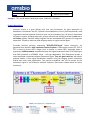
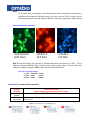
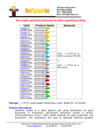
Pre-made Lentiviral Particles for Fluorescent-Target fusion proteins Catalog# LVP673 LVP674 LVP675 LVP676 LVP677 LVP442 LVP443 LVP399-R LVP399-G LVP399-C LVP444-G LVP444-R LVP444-C LVP445-G LVP445-R LVP445-C LVP446-G LVP446-R Product Name Amounts GFP-Luciferase (Puro) fusion lentiviral particles RFP-Luciferase (Puro) fusion lentiviral particles CFP-Luciferase (Puro) fusion lentiviral particles GFP-Luciferase (Neo) fusion lentiviral particles RFP -Luciferase (Neo) fusion lentiviral particles 1x10⁷ IFU/ml x 200ul GFP-RFP fusion control lentiviral particles CFP-RFP fusion control lentiviral particles RFP-LC3 fusion lentiviral particles GFP -LC3 fusion lentiviral particles CFP -LC3 fusion lentiviral particles GFP-Histone 2B fusion lentiviral particles RFP-Histone 2B fusion lentiviral particles CFP-Histone 2B fusion lentiviral particles GFP-Annexin5 fusion Lentiviral particles RFP-Annexin5 fusion Lentiviral particles CFP-Annexin5 fusion Lentiviral particles GFP-Actin fusion Lentiviral particles RFP-Actin fusion Lentiviral particles 1x10⁷ IFU/ml x 200ul 1x10⁷ IFU/ml x 200ul 1x10⁷ IFU/ml x 200ul 1x10⁷ IFU/ml x 200ul 1x10⁷ IFU/ml x 200ul 1x10⁷ IFU/ml x 200ul 1x10⁷ IFU/ml x 200ul 1x10⁷ IFU/ml x 200ul 1x10⁷ IFU/ml x 200ul 1x10⁷ IFU/ml x 200ul 1x10⁷ IFU/ml x 200ul 1x10⁷ IFU/ml x 200ul 1x10⁷ IFU/ml x 200ul 1x10⁷ IFU/ml x 200ul 1x10⁷ IFU/ml x 200ul 1x10⁷ IFU/ml x 200ul 1x10⁷ IFU/ml x 200ul LVP446-C LVP447-G LVP447-R LVP447-C LVP448-G LVP448-R LVP448-C LVP449-G LVP449-R LVP449-C LVP550-R LVP551-R LVP552-R LVP554-R LVP556-R LVP556-G LVP550-C LVP551-C LVP552-C LVP554-C LVP556-C Null-G CFP-Actin fusion Lentiviral particles GFP-TAT fusion Lentiviral particles RFP-TAT fusion Lentiviral particles CFP-TAT fusion Lentiviral particles GFP-hP53 fusion Lentiviral particles RFP-hP53 fusion Lentiviral particles CFP-hP53 fusion Lentiviral particles GFP-Zyxin fusion Lentiviral particles RFP-Zyxin fusion Lentiviral particles CFP-Zyxin fusion Lentiviral particles RFP-CLCN2 fusion Lentiviral particles RFP-KCNN4 fusion Lentiviral particles RFP-TRPV1 fusion Lentiviral particles RFP-TRPC3 fusion Lentiviral particles RFP-CSF1 fusion Lentiviral particles GFP -CSF1 fusion Lentiviral particles CFP-CLCN2 fusion Lentiviral particles CFP-KCNN4 fusion Lentiviral particles CFP-TRPV1 fusion Lentiviral particles CFP-TRPC3 fusion Lentiviral particles CFP-CSF1 fusion Lentiviral particles GFP-Null fusion control 1x10⁷ IFU/ml x 200ul 1x10⁷ IFU/ml x 200ul 1x10⁷ IFU/ml x 200ul 1x10⁷ IFU/ml x 200ul 1x10⁷ IFU/ml x 200ul 1x10⁷ IFU/ml x 200ul 1x10⁷ IFU/ml x 200ul 1x10⁷ IFU/ml x 200ul 1x10⁷ IFU/ml x 200ul 1x10⁷ IFU/ml x 200ul 1x10⁷ IFU/ml x 200ul 1x10⁷ IFU/ml x 200ul 1x10⁷ IFU/ml x 200ul 1x10⁷ IFU/ml x 200ul 1x10⁷ IFU/ml x 200ul 1x10⁷ IFU/ml x 200ul 1x10⁷ IFU/ml x 200ul 1x10⁷ IFU/ml x 200ul 1x10⁷ IFU/ml x 200ul 1x10⁷ IFU/ml x 200ul 1x10⁷ IFU/ml x 200ul 1x10⁷ IFU/ml x 200ul Null-R Null-C RFP-Null fusion control CFP-Null fusion control 1x10⁷ IFU/ml x 200ul 1x10⁷ IFU/ml x 200ul Storage: < -70°C, avoid repeat freeze/thaw cycles. Stable for > 6 months. Product Description: Lentiviral system is a gene delivery tool that uses lentivectors for gene expression or knockdown. Lentivectors are HIV-1 (Human Immunodeficiency Virus 1) derived plasmids, used to generate lentiviral particles (lentivirus) that can be transduced into all kind of mammalian cell types or organs, including stem cells, primary cells and non-dividing cells both in vivo and in cell culture system. Particles stably integrate into the transduced cell’s genome for long term expression. Therefore, lentivirus holds unique promise as a gene transfer agent. Pre-made lentiviral particles, expressing "GFP/RFP/CFP-Target" fusion constructs, are generated from Amsbio’s high expression lentiviral system. A fluorescent protein GFP, RFP or CFP is cloned in frame with a target (such as human or mouse ORF), expressed under a proprietary suCMV promoter that demonstrates the highest expression levels (3-10 fold higher than CMV promoter in pCDNA6.3 vector - cell type dependent). Each fluorescent protein is codon optimized to generate brighter fluorescent signal. They are great tools for: sub-cellular pathway studies, in vivo signal transduction research, living cell imaging, protein interaction studies and many other applications. The positive transduced cells can be sorted via the fluorescent signal or via Puromycin antibiotic selection. (See vector scheme below for vector structures). These Lentiviral Particles are great tool for: Sub-cellular pathway studies; in vivo signal transduction research; Live cell imaging, protein interaction studies and many other applications; The positively transduced cells can be sorted by the fluorescent signal or selected for puromycin / neomycin / blasticidin resistance depending on the product marker selection type. Key features: 1. High titer and robust: Amsbio’s premade lentiviral particles are best in the class, demonstrating the brightest fluorescent signal and strong transduction efficiency. All particles are validated on lot by lot basis and their quality is guaranteed. 2. Delivery of fluorescent labeled targets into hard to transfect cell lines (like primary cells or neuron cells) for long term expression; 3. Very easy to use: Particles are provided in DMEM medium with 10% FBS and 60ug/ml polybrene for ready to use status. Simply add into your cell culture and visualize the fluorescence after 48-92 hours (No need for any additives, components or medium changes); 4. Different fluorescent labeled particles for multi-color application when using multiple different particles in same cells; 5. Easy selection via Fluorescent signal or antibiotic marker. Transduction Protocols: 1) Transduction Protocol for Adhesive cells : Note: Pre-made lentivirus is provided ready to use, so it can be simply added to your cell culture; the amount of virus to add depends on the cell type. For quick transduction, add 50 µl of virus into each well of 24-well-plate where cell density is ~ 50% to 75%. After 72 hours (no need to change medium), visualize positive transduction rate by fluorescence microscopy. For stable cell line generation, passage cells into medium containing antibiotic or perform fluorescence cell sorting followed by antibiotic selection. Day 0: Seed cells in complete medium at the appropriate density and incubate overnight. Note: at the time of transduction, cells should be 50%-75% confluent. For example, seed HeLa cells at 0.5 x 10⁵/ml x 0.5ml in a well of a 24-well plate. Day 1: Remove the culture medium and add 0.5ml fresh, warm, complete medium. Thaw the pre-made lentiviral stock at room temperature and add the appropriate amount of virus stock to obtain the desired MOI. Return cells to 37°C, CO2 incubator. Note: Try to avoid freeze/thaw cycles. If you do not use all of the virus at once, you may re-freeze the virus at -80°C for future use; virus titer will decrease by ~10% for each freeze/thaw cycle. Day 3: At ~72hr after transduction, check the transduction rate by fluorescence microscopy or calculate the exact transduction rate by flow cytometry (FACS or Guava). Day 3 + (optional): Sort transduced cells by FACS, and select for antibiotic resistance. A pilot experiment should be done to determine the antibiotic’s kill curve for your specific cell line (refer to the literature on generation of stable cell lines). 2) Transduction Protocol for Suspension Cells: Grow cells in complete suspension culture medium; use a shaking flask in a CO2 incubator if necessary. Measure cell density. When density has reached ~3 x 10 6 cells/ml, measure viability (should be > 90%). Dilute cells into 1 x 10⁶ cell/ml in complete medium. Day 1: Thaw lentiviral particles at room temperature. Add premade lentiviral particles into diluted cells at a ratio of: 50 to 100 µl virus per 0.5 ml of cells (Note: depending on cell type, you may need to use more or less virus). Grow cells in a shaking flask in a CO2 incubator. Day 2: At 24 hours after transduction, add an equal amount of fresh medium containing relevant antibiotics. Note: amount of antibiotic depends on cell type. Continue growing cells in CO2 incubator. Day 3: At 72 hours after transduction, check fluorescence with a fluorescence microscope or calculate the transduction efficiency using a cell sorter such as FACS or Guava. Sort for fluorescence positive cells and maintain antibiotic selection to generate a stable cell line. Quick transduction examples: LVP442 (50ul) (GFP filter) LVP447-R RFP filter LVP448-C CFP filter Add 50ul each lentivirus into one well in 24-well-plate where cell density is at 50% ~ 75% in different cell types (HEK293, A549, PC3 from left to right). Images taken ~72 hours after virus added (no medium changed). Result: >90% of positive transduced cells. Note: Filter wavelength settings: GFP filter: ~Ex450-490 ~Em525; RFP filter: ~Ex545 ~Em620; CFP filter: ~Ex436 ~Em480; Attachment: Pre-made lentivirus products: Product Category Fluorescent protein Product Description (please Category name to see product's pages) Premade Lentivirus for GFP/ CFP/ YFP/ RFP Human and mouse ORF Premade lentivirus expressing human and mouse ORFs with RFP-Blasticidin fusion dual markers. Luciferase expression CRE recombinase LoxP ColorSwitch TetR inducible expression repressor U T Premade lentivirus for luciferase protein expression: firefly and Renilla with different antibiotic selection markers. Premade lentivirus for expressing nuclear permeant CRE recombinase with different flurescent and antibiotic markers. Premade lentivirus expressing "LoxP-GFP-Stop-LoxP-RFP" cassette, used to monitor the CRE recombination event in vivo. Premade lentivirus expressing TetR (tetracycline regulator) protein, the repressor protein for the inducible expression system. T iPS factors T-antigen Expression Cell Organelle imaging LacZ expression Pre-made shRNA lentivirus microRNA and anti-microRNA lentivirus Negative control lentiviruses Other Enzyme expression Premade lentivirus for human and mouse iPS (Myc, NANOG, OCT4, SOX2, FLF4) factors with different fluorescent and antibiotic markers Express different large and small T antigen with different selection markers Premade lentivirus for cell organelle imaging. The fluorescent marker GFP/RFP/CFP is localized in different cell organelles for living cell imaging. Express full length β- galactosidase (lacZ) with different selection markers Premade shRNA lentivirus for knockdown a specific genes (P53, LacZ, Luciferase and more). Premade lentivirus expression human or mouse precursor miRNA and anti-miRNA lentivector and virus for human and mouse miRNA. Premade negative control lentivirus with different markers: serves as negative control for lentivurs treatment, for validation of the specificity of lentivirus target expression effect. Ready-to-use lentivirus, expressing a specific enzyme with different selection markers. Safety Precaution: Amsbio lentiviral particles have adopted the most advanced lentiviral safety features (using the third generation vectors with self-inactivation SIN-3UTR), and the premade lentivirus is replication incompetent. However, please use extra caution when using lentiviral particles. Use the lentiviral particles in Bio-safety II cabinet. Ware gloves at all times when handling lentiviral particles! Please refer to CDC and NIH’s guidelines for more details regarding the safety issues. References: 1. 2. 3. 4. 5. J Virol. 2000 November; 74(22): 10778–10784. Hum Gene Ther (2003) 14: 1089-105. Mol Ther (2002) 6: 162-8. NIH Guidelines for Biosafety Considerations for Research with Lentiviral Vectors link CDC guidelines for Lab Biosafety levels link Warranty: This product is for research use only. It is warranted to meet its quality as described when used accordance with its instructions. Amsbio disclaims any implied warranty of this product for particular application. In no event shall Amsbio be liable for any incidental or consequential damages in connection with the products. Amsbio’s sole remedy for breach of this warranty should be, at Amsbio’s option, to replace the products.