1
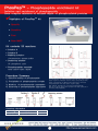
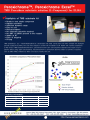
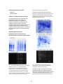
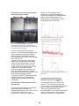
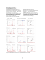
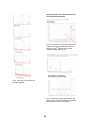
2010 Products Catalog of Genomine,Inc. Thy best research partner in proteomics INDEX Overview, User manual, and Technical bulletin 1. Overview PhosPro TM - Phosphoprotein enrichment kit ................. 1 PhosPepTM - Phosphopeptide enrichment kit ............... 2 Antibody biotin conjugation kit ..................................... 3 Peroxichrom TM - TMB substrate solution ...................... 4 2. User manual & Technical bulletin PhosProTM - User manual ............................................. PhosPro TM - Technical bulletin ..................................... 5 8 PhosPepTM - User manual ............................................ 13 PhosPepTM - Technical bulletin .................................... 15 Antibody biotin conjugation kit ..................................... 19 PeroxichromTM - TMB substrate solution ...................... 21 Phosphoprotein enrichment kit - PhosProTM Highly selective and enrichment of phosphoprotein from complex mixtures of cell lysate ✔Highlights of PhosPro TM kit • Phosphoprotein specific • Sensitive • Convinient • Reproducible • Enrichment from native or denature protein solution Kit contents 10 reactions • • • • • • Lysis buffer 10ml x 1 Dilution buffer 20ml x 1 Solution A 3ml x 1 Solution B 5ml x 1 Dissolving Solution 8ml x 1 Delipidation Solution 10ml x 1 Procedure Summary 1. 2. 3. 4. Evaluation category Specificity Selectivity Sensitivity Yield Native & denatured Protein extraction Phosphoprotein specific complex forming Precipitation of phosphoprotein complex Dedelipidation and recovering of phosphoprotein Specification 100% >87% >93% / 50ng >93% Ordering information Product PhosProTM kit TM PhosPro kit Description 1 box (5 Reaction) 1 box (10 Reaction) Cat. No. P5012-5 P5010-10 Fig. 2-DE analysis of enriched phosphoprotein fraction from total denatured(A,C) & native protein(B,D) stained with CBB(A,B), and stained with ProQ Diamond staining(C,D) 1 PhosPepTM - Phosphopeptide enrichment kit Isolation and enrichment of phosphopeptide from complex mixtures of enzyme digest of phosphorylated proteins ✔Highlights of PhosPep TM kit • Specific • Sensitive • Fast • Non-IMAC Kit contents 50 reactions • Solution A • Solution B • Washing solution: 4×ammonium acetate buffer • Dissolving solution: 1% phosphoric acid • Phosphopeptide standard: beta casein tryptic digest Procedure Summary 1. 2. 3. 4. Selective binding of phosphpeptide Precipitation of phosphopeptide com plex W ashing of phosphopeptide aggregates Dissolving of phosphopeptide aggregates Fig. Identification of phosphopeptide enriched from beta casein trypsin digest by PSD(post source decay) using MALDI-TOF. Panel A represents the MALDI-TOF spectrum of enriched phosphopeptide from beta casein trypsin digest. Blue asterisk represent the enriched phosphopeptides. Rest of the seven spectrums represent the PSD spectrum of enriched phosphopeptide. Ordering information Product TM PhosPep kit Description 1 box (50 Reaction) Cat. No. P5010 2 Antibody biotin conjugation kit One stop labeling of Antibody Purification to Biotinylation ✔Highlights of Biotinylation kit • Quick Only 1 hour to get conjugates • Easy No filtration tube required • Reliable High recovery & consistent • Efficient Applicable for 20ug to 10mg IgG Kit contents 10 reactions • Antibody purification - 1M Sodium Acetate pH4.0 1ml x 1 - Carprylic Acid 0.5ml x 1 - Neutralizing Buffer 1ml x 1 • Desalting & buffer exchange - Solution A 8ml x 1 - Labeling Buffer 6ml x 1 • Biotin labeling - Reactive Biotin x 1 - Stop Solution 0.3ml x 1 Procedure Summary 1. 2. 3. 4. Antibody Purification Desalting and Buffer Exchange Biotin labeling Stopping and Storage Ordering information Product Antibody biotin conjugation kit Description 1 box (10 Reaction) 3 Cat. No. P5014 PeroxichromeTM, Peroxichrome ExcelTM TMB Peroxidase substrate solution (1-Component) for ELISA ✔Highlights of TMB substrate kit • • • • • • • • • • Ready to use single component Highest sensitivity Sufficient dynamic range Easy to use Noncarcinogenic No hydrogen peroxide required No DMF or DMSO present in the reagent Stable at RT Ease of shipping Pricing TMB substrate(3,3´ ,5,5´ -tetramethylbenzidine) is a chromogen th at yields a deep blue color when oxidized with hydrogen peroxide (catalyzed by HRP). The color then changes to yellow w ith the addition of 2N H2SO4 with maximum absorbance at 450 nm.Our TMB Substrates(Peroxichrom TM , peroxichrom Excel TM ) are one-component substrates that require no preparation before using, stable and sensitivity. Also (Peroxichrom TM , peroxichrom Excel TM ) contain no solvents or organics such as DMF, DMSO, methanol so there is no issue of safety with user. Ordering information Product TM Peroxichrom PeroxichromTM TM Peroxichrom Excel TM Peroxichrom Excel Description 1 box (100ml X 4) 100ml 1 box (100ml X 4) 100ml Cat. No. D5015-400 D5015-100 D5016-400 D5016-100 4 Tel: +82-54-223-2463 Fax: +82-54-223-2460 Email: [email protected] http://www.genomine.com Venture Bldg 306 Pohang Techno Park Pohang, Kyungbuk, Korea (ROK) INSTRUCTIONS PhosProTM Phosphoprotein enrichment kit This phosphoprotein enrichment and exclusion of unphosphorylated proteins provides advanced chance in detecting protein phosphorylation in gels with non-radiolabeling method(eg. Staining with fluorescence dye) and enables quantitative comparison between cells. Product Number P 5012 Store at RT INTRODUCTION Protein phosphorylation is one of the most frequently occurred posttranslational modifications and plays a critical role in cellular regulatory events. Most cellular processes are in fact regulated by the reversible phosphorylation of proteins on serine, threonine and tyrosine residues. In fact, phosphorylation of proteins plays a key role in oncogenesis, cell signaling, apoptosis and immune disorders1. Despite the importance and widespread occurrence of this modification, profiling of phosphoproteins in cells is still a challenge, due to the low copy of phosphorylated proteins in cell and the relative amount of phosphoproteins compared to unphosphorylated proteins. Kit contents 10 reactions LYSIS BUFFER DILUTION BUFFER Native Homogenation Buffer SOLUTION A SOLUTION B DISSOLVING SOLUTION DELIPIDATION SOLUTION 10ml x 1 30ml x 1 30ml x 1 3ml x 1 5ml x 1 8ml x 1 10ml x 1 Additional Materials Required Methanol Ultrapure water Radiolabeling by 32P labeling is frequently used conventional method for investigation of phosphoprotein profile in conjunction with 2-DE or 1-D gel electrophoresis and autoradiogram. Alternatively, western blot analysis probed by phosphoprotein-specific antibody is also used for this purpose. Mass spectrometry has been shown to be a reliable and routine tool to identify proteins in a high throughput manner. However, the identification of phosphorylation by mass spectrometry is not a trivial matter and to this day is not routine also due to the low copy of phosphorylated proteins in cells. Detecting Phosphorylated Proteins This phosphoprotein enrichment kit was optimized for the protein solution in denatured condition, for example, the samples prepared for 2-DE and can be applied to native proteins. Enriched phosphorylated proteins could be detected by staining commercially available staining method using fluorescent dye2 or by probing with antibodies, specific for phosphorylated proteins. 5 Procedure Summary vortexing) and vortex for 5 min. 1. Protein extraction 2. Phosphoprotein specific complex forming 3. Precipitation of phosphoprotein complex 4. Dedelipidation and recovering of phosphoprotein 5. Add 750 µl of delipidation soln. (methanol:chloroform=600:150) and vortex vigorously for 5 min and centrifuge at 12,000rpm for 10 min for phase separation of solution. Recover the middle phase protein disk and discard lower and upper phase solution completely. Then wash the protein disk with sufficient (~1ml) methanol for two times. Procedure for phosphoprotein enrichment from cell lysate ( Denatured protein condition) 6. Dry the protein pellet in air or oven completely and dissolve the protein pellet with the solution for 2-DE electrophoresis or 1-D SDS PAGE. 1. Add 300~600µl LYSIS SOLUTION to the cells or tissue and disrupt the cells and tissue by sonication or motor driven homogenation. (Adjust the volume of LYSIS SOLUTION in order the final concentration of extracted protein to be above 4mg/m) Vortex the cell lysate for 15min and centrifuge for 20min. at 12,000 x g and save the supernatant. Assay the protein concentration and dilute 2mg protein with DILUTION SOLUTION to be the final volume of 3ml. (Use 5ml tube) Alternative procedure for phosphoprotein enrichment from cell lysate ( Native protein condition ) If you want to isolate the phosphorylated protein from cell lysate in native conformation of proteins, omit step 1 using LYSIS SOLUTION. Instead, prepare the cell lysate with Native Homogenation Buffer or appropriate buffer solution except solution including phosphate. Alternatively, protein solution prepared for twodimensional gel electrophoresis could be directly used for enrichment by appropriately diluted with DILUTION SOLUTION. 1. Add 1~3ml Native Homogenation Buffer to the cells or tissue and disrupt the cells and tissue by sonication or motor driven homogenation in order the final concentration of extracted protein to be 2~30mg/ml. Centrifuge for 20min. at 12,000 x g and save the supernatant. (Use1 or 5ml tube) 2. Add 240µl of SOLUTION A and rapidly mix by vortex vigorously for a few seconds then incubate for 15min.by inverting or gentle vortexing. After subsequent adding 360µl SOLUTION B and brief mixing, incubate for 15min. by gentle vortexing then stand still for 5~10 min. for the aggregated materials to be settled down. And discard about 4ml of upper clear solution. 2. Add 80µl of SOLUTION A per 1ml protein solution and mix gently for 15min by inverting or gentle vortexing. After subsequent adding 120µl SOLUTION B per 1ml original solution, incubate for 15min by gentle inverting then centrifuge briefly for 5~10 min for the aggregated materials to be settled down. And discard upper clear solution. 3. Transfer the remaining aggregate suspension to 1.5ml microcentrifuge tube and centrifuge the suspension at 12,000rpm for 5min.. Discard the supernatant and save the aggregate in hard pellet. This aggregate can be stored for several days. 3. Add one fourth volume of Native Homogenation Buffer to the pelletted phosphoprotein complex and wash the residual non-phosphorylated protein solution by resuspension the pellet and recover the pellet 4. Add 0.7ml DISSOLVING SOLUTION and dissolve the pellet by pipetting several times(*caution : at this time CO2 gas will be formed. Open lid and degas sufficiently before 6 by brief centrifugation. Repeat this washing one time and save the aggregate in hard pellet. P5010 4. Add 0.7ml DISSOLVING SOLUTION and dissolve the pellet by pipetting several times(*caution : at this time CO2 gas will be formed. Open lid and degas sufficiently) and stand for 10 min. the solution to be clear. This solution contains enriched phosphoprotein in 250mM salt solution containing EDTA. Dialysis the enriched phosphoprotein solution with appropriate buffer solution. Or skip to the next step for preparation of 1D or 2D electrophoresis samples. 5. Add 750 µl of delipidation soln. (methanol:chloroform=600:150) and vortex vigorously for 5 min and centrifuge at 12,000rpm for 10 min for phase separation of solution. Recover the middle phase protein disk and discard lower and upper phase solution completely. Then wash the protein disk with sufficient (~1ml) methanol for two times. 6. Dry the protein pellet in air or oven completely and dissolve the protein pellet with the solution for 2-DE electrophoresis or 1-D SDS PAGE. Optimization of Results When you start with cell lysate at higher concentration and smaller volume of proteins, use SOLUTION A and SOLUTION B, 80µl and 120µl per 1ml protein solution respectively. References 1. Philip Cohen, Eur. J. Biochem., 568, 20012010 (2001) 2. Alein, L. et al. Proteomics, 6, 2157–2173 (2006). Related Products Product Code Phospep Phosphopeptide enrichment kit 7 Technical bulletin Tel: +82-54-223-2463 Fax : +82-54-223-2460 http://www.genomine.com venture Bldg 306 Pohang techno park Pohang, kyungbuk, Korea(ROK) Enrichment and identification of phosphopeptide using PhosProTM Introduction Materials & Methods PhosProTM, phosphoprotein enrichment kit, was developed to fractionate phosphorylated proteins from protein mixtures, such as cell lysate or body fluids and it is efficient to isolate and concentrate low copy phosphorylated proteins in cells. This kit utilized proprietary phosphoprotein precipitation method instead using column or bead such as IMAC column or immobilized anti-phophoprotein antibody beads. Then in addition to its specificity for isolation of phosphoproteins, it provides simple and convenient method for phosphoprotein fractionation and all processes are to be done with multi parallel samples in each one tube. This kit was designed to use denaturant and detergent solution as the starting material of protein extraction in order phosphoprotein isolation not to be prevented by possible steric hindrance of phosphorylated moiety of proteins and not to be omitted by difficulty in solubilization of phosphoproteins embedded in membrane fraction or cell debris. Materials PhosProTM kit contents LYSIS BUFFER DILUTION BUFFER NATIVE HOMOGENATION BUFFER SOLUTION A SOLUTION B DISSOLVING SOLUTION DELIPIDATION SOLUTION Procedure Summary 1. Native & Denatured Protein extraction 2. Phosphoprotein specific complex forming 3. Precipitation of phosphoprotein complex 4. Delipidation and recovering of phosphoprotein Strategy Fig.1. Phosphoprotein enrichment using PhosProTM. Protein phosphorylation could be identified by phosphoprotein specific enrichment in conjunction with phosphoprotein specific staining or MSbased phosphopeptide identification and phosphorylation site determination. 8 Additional Materials Required performed and analysed by SDS-PAGE. Methanol Ultrapure water In order to evaluate the performance of PhosProTM , yeast protein extract was used. The resulting enriched phosphorylated protein was analysed with 1-D(Fig.2) or two-dimensional gel electrophoresis(2-DE, Fig.3) Detection of Phosphorylated Proteins This phosphoprotein enrichment kit was optimized for the protein solution in denatured condition, for example, the samples prepared for 2-DE and can be adapted to native proteins. Enriched phosphorylated proteins could be detected by staining commercially available staining method using fluorescent dye1 or by probing with antibodies specific for phosphorylated proteins. Results and Discussion Phosphoprotein enrichment from Saccharomyces cerevisiae protein extract Fig.3. 2-DE analysis of total protein(A), enriched phosphoprotein fraction stained with CBB(B), and Fig.2. SDS-PAGE analysis of enriched phosphorylated TM stained with ProQ Diamond staining(C) proteins by PhosPro . A: Coomassie Brillant Blue(SIGMA) staing, B:ProQ Diamond(Invitrogen) fraction containing phosphorylated proteins. Independent As shown in Fig.2. and Fig.3. most of the proteins stained with ProQ Diamond was detected in enriched phosphoprotein fraction. This result represent that the PhosProTM is seven trials for phosphoprotein enrichment were highly specific for phosphoprotein fractionation. staining. Lane 1~7 ; The supernatant containing unphosphorylated proteins discarded in step3(See Procedure Summary above), Lane 8~15 ; enriched 9 MALDI-TOF-based peptide mass fingerprinting. Its phosphopeptide was enriched by PhosPepTM and identified with MALDI-PSD by detecting the loss of phosphoprous group(98Da) from mother phosphopeptide(m/z 2900.392). Phosphoprotein enrichment from mouse brain native protein extract Enriched Denatured protein Enriched Native protein A B C D Fig.4. 2-DE analysis of enriched mouse brain phosphoprotein fraction from total denatured(A,C) & native protein(B,D) stained with CBB(A,B), and stained with ProQ Diamond staining(C,D) In order to evaluate the performance of PhosProTM , mouse brain denatured(A,C) and native protein(B,D) extract was used as a starting material. The resulting enriched native phosphorylated protein was analysed using 2-DE (Fig.4) As shown in Fig.4.D most of the proteins stained with ProQ Diamond in denatured protein fraction was detected in enriched phosphoprotein fraction from total native protein. But native phosphoprotein fraction contains more non-phosphorylated proteins(not stained with ProQ Diamond), which is assumed as subunit or proteins interacting with phosphorylated proteins than denatured phosphoprotein fraction. As a result, PhosProTM is applicable to both denatured rather pure phosphoprotein fraction and active phosphylated protein fractionation. Fig.5. Identification of enriched phosphoprotein by mass spectrometry. Phosphoprotein identification by dephosphorylation using λPPase The phosphoproteins were enriched by PhosProTM from cell lysate of h460 lung cancer cell line. The enriched protein fraction which was stained with phosphoprotein staining and presumed to be the phosphoproteins, was confirmed whether the staining was derived from the phosphate moiety on the proteins by examining the Phosphoprotein identification by mass spectrometry The protein spot enriched and stained with ProQ Diamond and identified as a phosphoprotein(spot N. 3405 in Fig.3. B and C) was further confirmed by mass spectrometry. Protein was identified by 10 changes after treatment of phosphatase. The λPPase was used as a phosphatase as previously described2. lines and lung cancer tissues In 2-DE gel analysed using 300ug proteins of total cell lysate of lung cancer cell lines(h460), about 35 protein spots was stained with phosphoprotein staining. When the phosphoproteins(300ug) were enriched from total protein extract of lung cancer cell line, 198 protein spots were detected as a phosphoproteins.(Fig.6.B). Fig.6. Phosphoprotein staining of enriched phosphoproteins and dephosphorylated proteins. Lane1: standard phosphoproteins, lane2, standard phosphoprotein treated with λPPase, lane3: supernatant Fig.7. 2-DE gel analysis of total extract of lung cancer fraction remained from phosphoprotein enrichment, lane4: phosphoprotein fraction enriched by PhosPro lane5:phosphoprotein fraction treated with λPPase TM cell line(A), enriched phosphoprotein pool from the , extract of human lung cancer cell line(B) and enriched phosphoprotein pool from the extract of human lung cancer tissues(c) As shown in Fig.5 the proteins in enriched phosphoprotein fraction was stained with phosphoprotein staining whereas the same proteins treated with phosphatase, λPPase, was not stained with phosphoprotein staining but only with CBB staining. This result represent that the proteins stained with phosphoprotein staining was phosphate group specific and the PhosProTM isolate the phosphoproteins from cell extract in a specific manner. This result was represented in human lung cancer tissues which is composed more complex composition of cell types and body fluids. As a result from the evaluation with phosphoprotein standards, α-casein, βcasein, pepsin, ovalbumin and phosvitin, PhosProTM technology showed phosphoprotein specific, sensitive and high yield effective fractionation capabilities.(Table.1) Enrichment of low abundant phosphoproteins from lung cancer cell 11 Table.1 Phosphoprotein fractionation capability of PhosProTM Evaluation category Specification Specificity Selectivity Sensitivity Yield 100% >87% >93% / 50ng >93% References 1. Alein, L. et al. Proteomics, 6, 2157–2173 (2006). 2. Akira Yamagata, et al., Proteomics, 2, 1267–1276 (2002) 12 Tel: +82-54-223-2463 Fax: +82-54-223-2460 Email: [email protected] http://www.genomine.com Venture Bldg 306 Pohang Techno Park Pohang, Kyungbuk, Korea (ROK) INSTRUCTIONS Phos-PepTM Phospho-peptide enrichment kit phosphopeptide by mass spectrometry. Phosphopeptde identification relies on measuring the loss of mass. Phosphopeptides tend to lose their phosphate group as phosphoric (H3PO4) or phosphorous acid (HPO3) due to metastable decay in MALDITOF, ESI (PSD)2,3 ,ion trap(CID) or as phosphorous acid (HPO3) by phosphatase4 . Product Number P 5010 Storage Temperature 10 to 37 °C (RT) INTRODUCTION (Technical bulletin http:// www.genomine.com) Protein phosphorylation is one of the most frequently occurred posttranslational modification and plays a critical role in cellular regulatory events. Most cellular processes are in fact regulated by the reversible phosphorylation of proteins on serine, threonine, and tyrosine residues. Despite the importance and widespread occurrence of this modification, identification of protein phosphorylation site is still a challenge, due to the low copy of phosphorylated proteins in cell, even when performed on highly purified protein. Kit contents 50 reaction Solution A Solution B Washing solution:4×ammonium acetate buffer Dissolving solution:1% phosphoric acid Phosphopeptide standard:beta casein tryptic digest 10μg Additional Materials Required Mass spectrometry has been shown to be a reliable and routine tool to identify proteins in a high throughput manner. However, the identification of phosphorylation sites by mass spectrometry is not a trivial matter and to this day is not routine. The detection of phosphopeptides by mass spectrometry in a complex mixture, such as a tryptic mass fingerprint, is a rare occurrence. This is thought to be caused by suppression of the ionization of the mainly negatively charged phosphopeptide in the presence of a large excess of nonphosphorylated peptides1. C18 microtip Ultrapure water Binding Capacity In binding assays performed using this product, binding of greater than 90 pmoles of phosphopeptide per 10μl of A solution is observed in one reaction. From 1μg(45 pmole) trypsin digest of b-casein, single enrichment recover over 95% mono(2062 Da) and tetra(3124 Da) phosphopeptide. This phosphopeptide enrichment kit provides highly selective enrichment of phosphopeptide from non-phosphorylated complex trytic digest of proteins and facilitates the identification of Procedure Summary 13 1. Selective binding of phosphpeptide 2. Precipitation of phosphopeptide complex 3. Washing phosphopeptide aggregates 4. Dissolving of phosphopeptide aggregates When peptide solution contains high salt, dilute the solution below 100mM of salt prior to enrichment to obtain better result. References Procedure for phosphopeptide enrichment from trypsin digest 1. Joerg R., et al., Proteomics, 4, 3686–3703 (2004) 1. Prepare trypsin digest 2. Metzger, S. and Hoffmann, R. J Mass Spectrom 35, 1165–1177 (2000). 2. Add 5μl of solution A to 10μl trypsin digest in microcentrifuge tube and vortex briefly for a few seconds then stand about 1~5min. 3. Hoffmann, R. et al. J Mass Spectrom 34, 1195–1204 (1999). 3. Add 5μl of solution B to the previous mixture and vortex well to disperse the aggregate to homogeneous cloudy suspension. Stand at least 30min until the cloudy aggregate settle down and disappear.(When cloudy aggregate was not disappeared within 30 min., stand prolonged time till the aggregate disappear completely.) 4. Akira Yamagata, et al., Proteomics, 2, 1267– 1276 (2002) Related Products Product Code Phos-pro Phosphoprotein enrichment kit P5012 4. Discard solution to the last drop with pipet tip. (Phosphopeptide aggregates was stuck to the wall and remained coated in microcentrifuge tube) This aggregate can be stored for several days. 5. Add 50μl of 1X washing solution(dilute stock solution four fold) and vortex briefly for a few seconds then discard washing solution completely. 6. Add 10μl dissolving solution and stand at least 5min. (You can see sometimes some bubble gas formed along with the surface of microcentrifuge tube) 7. Vortex briefly for a few seconds and,if necessary, dissolve the remaining crystals with pipetting. 8. Desalt or concentrate the solution with C18 microtip column for mass spectrum analysis. Optimization of Results 14 Technical bulletin Tel: +82-54-223-2463 Fax : +82-54-223-2460 http://www.genomine.com venture Bldg 306 Pohang techno park Pohang, kyungbuk, Korea(ROK) Enrichment and identification of phosphopeptide using Phos-PepTM Introduction Procedure Summary Phos-pepTM was designed to ensure the charactrization of phosphopeptides. Investigators to characterize the phosphoproteins by mass spectrometry hampered by the low abundance of phosphoprotein and the suppression of ionization of phosphopeptide in mass spectrometry resulting in failure to obtain TM sufficient signals. Phos-Pep facilitates the isolation and enrichment of phosphopeptide from complex mixtures of trypsin digest of phosphorylated proteins. 1. Selective binding of phosphpeptide 2. Precipitation of phosphopeptide complex 3. Washing phosphopeptide aggregates 4. Dissolving of phosphopeptide aggregates 5. Desalting and concentration 6. Characterization of phosphopeptide Results and Discussion Phosphopeptide enrichment from beta casein Strategy Fig.1. Phosphopeptide enrichment using TM Phos-Pep in conjunction with MALDI-TOFbased CAF sequencing or MS/MS for identification of phosphopeptide and phosphorylation site determination. Materials & Methods Materials Phos-pepTMkit contents Solution A Solution B Washing solution : ammonium acetate stock solution Dissolving solution : 1% phosphoric acid Phosphopeptide standard : beta casein tryptic digest 10μg Fig.2. MALDI-TOF mass spectrum of beta casein trypsin digest(upper) and enriched phosphopeptide from beta casein trypsin digest using Phos-PepTM (lower). Blue asterisk represent the enriched phosphopeptides. 15 Phosphopeptide identification Beta casein was used to examine the capacity of Phos-PepTM , non-IMAC phosphopeptide enrichment kit. Beta casein (purchased from Sigma, Cat.No.C6905) was digested with trypsin and phosphopeptide was enriched using Phos-PepTM and peptide map was measured. Phosphopeptide which was not shown in total digest of beta casein(Fig.2.) was detected in enriched fraction(Fig.2. lower panel, Fig.3. A). Among these phosphopeptide, mono phosphopeptide(2062) and tetraphosphopeptide(3124) was originated from beta casein. Some phosphopeptide, 1660, and 1952, seems to be originated from contaminated alpha casein, because this phosphopeptide is a major component of commercially available alpha casein phosphopeptide(see Fig.4.). Some peptides, ranging 2884 to 3054, seems to be the derivatives of tetra-phophopeptide of 3124, which have differential mass according to the status of the number of phosphorylation site. Some of phosphopeptide was identified by PDS of MALDI-TOF by detecting the mass loss of phosphoric or phosphorous acid (Fig.3.). A Fig.3. Identification of phosphopeptide enriched from beta casein trypsin digest by PSD(post source decay) using MALDI-TOF. Panel A represents the MALDI-TOF spectrum of enriched phosphopeptide from beta casein trypsin digest. Blue asterisk represent the enriched phosphopeptides. Rest of the seven spectrums represent the PSD spectrum of enriched phosphopeptide. 16 Specificity and sensitivity of phosphopeptide enrichment digest of model protein used and characterized as a phgosphopeptide by PSD using MALDI-TOF. The sensitivity of capability of Phos-PepTM was also tested. From Heterogeniety of phosphorylation site in peptide sequence could affect the efficiency of phosphopeptide enrichment. We examined the specificity of Phos-PepTM kit for phosphopeptide from known phosphoprotein as a model peptide, such as alpha casein, ovalbumin, pepsin and phosvitin. As shown in Fig.4., Phos-PepTM isolated the phosphopeptide from most of the tryptic one microgram trypsin digest of beta casein, which is equivalent to 45 pmole of each digested peptide, to 175 fmole of 256-fold diluent, the capacity of enrichment was tested(Fig.5.) Fig.4. Phosphopeptide enrichment from trypsin digest of phosphoprotein 17 Phosphorylation site determination from enriched phosphopeptide Fig.6. Determination of phosphorylation site of beta casein phosphopeptide enriched using Phos-PepTM followed by chemical assisted fragmentation (CAF). Fig.5. Sensitivity of enrichment of phosphopeptides. Fig.7. Enrichment and phosphorylation site determination of phosphopeptide containing phosphotyrosin amino acid residue. 18 Tel: +82-54-223-2463 Fax: +82-54-223-2460 Email: [email protected] http://www.genomine.com Venture Bldg 306 Pohang Techno Park Pohang, Kyungbuk, Korea (ROK) INSTRUCTIONS Antibody Biotinylation Kit Additional Materials Required Ultrapure water 80% Glycerol (Optional) Product Number P 5013 Store at RT Alternatives For Reactive Biotin INTRODUCTION Reactive Biotin, NHS-Biotin, could be replaced by commercially available activated biotin, Biotin is used in two-step detection systems in concert with conjugated avidin. Biotin is typically conjugated to proteins via primary amines (i.e., lysines). Usually, between 3 and 6 biotin molecules are conjugated to each antibody. Procedure Summary 1. Antibody Purification 2. Desalting and Buffer Exchange 3. Biotin labeling 4. Stopping and Storage The entire conjugation can be performed in about an hour. In general, you will need to have a solution of your antibody at a concentration (optimally) of at least 2 mg/ml. The extent of biotin conjugation to the antibody may depend on the concentration of antibody in solution; for consistent conjugations, use a consistent concentration. This product provides convenient and efficient method for removing salt and amine compound interfering biotin coupling reaction from antibody solution. Kit contents 1M Sodium Acetate pH4.0 Carprylic Acid Neutralizing Buffer Solution A Labeling Buffer DMSO Reactive Biotin Stop Solution Solution B Procedure Antibody Purification by Carprylic Acid precipitation 1. Add 500µl of DW to 500µl serum and acidify by adding 60µl of 1M sodium acetate pH 4.0 10 reactions 2. Slowly (drop-wise) add 37µl (20ul for rat or mouse serum) of caprylic acid and continue stirring for 20 min at room temperature. 1ml x 1 0.5ml x 1 1ml x 1 8ml x 1 8ml x 1 0.1ml x 1 1 0.3ml x 1 8ml x 1 3. Centrifuge at 12,000xg for 10min. and carefully remove and save the supernatant. 4. Adjust the pH 7.5~8.5 of supernatant antibody solution by adding 50µl of neutralizing solution and if necessary, centrifuge the solution at 8,000xg for 5min. and discard precipitate. 19 Alternatively start here if you have your own affinity purified IgG. Desalting and Buffer Exchange 5. Add half volume of Solution A (0.5ml) and mix thoroughly by gentle inverting then stand for 10 min 6. Precipitate the immunoglobulin aggregate by centrifugation at 12,000xg for 5min and remove completely the turbid supernatant. 7. Dissolve the precipitated immunoglobulin with 300µl Labeling Buffer (to be approximate 2mg/ml IgG). 8. If there is any insoluble material discard it by high speed centrifugation for 10 min. Biotin Labeling 9. Dissolve the Reactive Biotin with 60 µl DMSO(10mM in DMSO). Aliquote and store at -70oC. Add 2µl Reactive Biotin solution per 100µl IgG solution and incubate 30min at room temperature. 10. At the end of the incubation, add 2µl Stop Solution and subsequently add half volume of Solution B to aggregate the biotin labeled IgG and remove the residual reactive biotin. Stand this suspension at 4°C for 20min. 11. Centrifugation the suspension at 12,000xg for 10min and dissolve the aggregated IgG with equal volume of Labeling Buffer(phosphate buffer). Add 80% glycerol and store at -20℃. 20 Tel : +82-54-223-2464 E-mail : [email protected] Venture Bldg 307 Pohang Techno Park 601 Pohang, Gyeongbuk, Korea PeroxichromTM,Peroxichrom ExcelTM TMB peroxidase substrate solution (1-component) for ELISA TM Cat. # : D5015-100(Peroxichrom : 100ml) D5015-400 (PeroxichromTM : 400ml) D5016-100 (peroxichrom ExcelTM : 100ml) TM D5016-400 (peroxichrom Excel : 400m) Highlights of TMB substrate ‣ Ready to use single component ‣ Highest sensitivity ‣ Sufficient dynamic range ‣ Easy to use ‣ Noncarcinogenic ‣ No DMF or DMSO present in the reagent ‣ Stable at RT ‣ Easy to transport Description : TMB substrate(3,3'.5,5'-tetramethylbenzidine) is a chromogen that yields a deep blue color (maximum absorbance at 605nm or 650nm) when oxidized with hydrogen peroxide(catalyzed by HRP). The color then changes to yellow with the addition of 2N H2SO4 with maximum absorbance at 450 nm. Our TMB Substrates(Peroxichrom TM , Peroxichrom ExcelTM) are one-component substrates that require no preparation before using, stable and sensitivity. Also PeroxichromTM and Peroxichrom ExcelTM contain no organic solvents such as DMF, DMSO, methanol so there is no issue of safety with user. Precautions: TMB substrate is sensitive to contamination from a variety of oxidizing agents. Avoid prolonged exposure to light, contact with metal or air. we recommend using TMB substrate by pouring out required amount into a reservoir and do not return excess TMB to provided bottle. Storage : Stable at 4℃ or Room temperature for 1 year Procedure: 1. Warm to room temperature prior to use if you store TMB solution at 4℃ 2. Dispense 1-Component appropriate TMB solution or 100ul into each well 3. After sufficient color development (5-10minutes at room temperature or at 37℃) add 50ul Stop Solution (2N H2SO4 or 1M H3PO4) to each well. 4. Read plates at 450nm 21