1
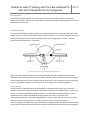
Radiation Safety Training and Procedure Manual for 2012
Safe Use of Analytical X-ray Equipment
Purpose
Analytical X-ray equipment is used for x-ray diffraction analysis, fluorescence or direct x-ray
transmission analysis of materials. These analytical x-ray systems are comprised of components that
utilize X-rays to determine elemental composition, or to examine the microstructure of materials. These
analytical x-ray devices are used for non-medical purposes. The primary objective of this program is to
keep occupational exposures to radiation as low as reasonably achievable (ALARA) while utilizing these
types of equipment. This safety program establishes UVa procedures for the safe operation of analytical
X-ray equipment and the associated potential radiation hazards.
Regulatory Authority
The Ohio Department of Health, Bureau of Radiation Protection is responsible for the following
program areas:
•
•
•
Registration and inspection of radiation-generating equipment.
Licensing radiation-generating equipment operators and nuclear medical technologists.
Certification of radiation experts.
X-ray machines and other ionizing radiation producing devices, such as electron microscopes,
present a potential safety hazard to students, staff, and faculty if the device is not used and/or
stored properly. The Ohio Department of Health ( ODH) will inspect the university’s x-ray
machines and operations on a periodic basis. For research and analytical devices this period is
every 3 years. The ODH has the authority to issue violations for any regulation that an x-ray
user, researcher, or Principal Investigator (PI) is not following current Ohio Law.
This manual is intended to inform non-human/non-medical use x-ray device users of the
regulations with which they are required comply. Also included are the ODH regulations for the
safe operation of x-ray machines and other Radiation Producing Device (RPD) and specific
practices to aid radiation equipment users in minimizing their exposure to ionizing radiation. If
there are any questions concerning the applicability of any regulation, please contact the
Radiation Safety Officer In Environmental Health and Safety 216-687-9306.
Potential hazards of Radiation Generating Devices
Analytical x-ray devices, such as diffract-o-meters, use very high intensity, collimated x-ray
beams to examine the properties of materials. The x-ray dose rate at the beam port can be
several thousand rad per second. Exposure of extremities to the primary x-ray beam can result
in severe radiation burns in a matter of seconds. Radiation burns are the principle hazard
associated with the use of analytical x-ray equipment.
The leakage or scatter of the primary beam through apertures in ill-fitting or defective
equipment can produce very high intensity beams of possibly small and irregular cross section.
Radiation Safety Training and Procedure Manual for 2012
Safe Use of Analytical X-ray Equipment
Dose rates near the machine from scattered radiation can be as high as a few hundred
millirems (mrem) per hour. Although not likely to cause burns, doses from scattered radiation
can exceed regulatory limits if the beam is not properly enclosed or contained. In fluorescence,
the primary beam strikes the sample inside a shielded enclosure, and only scattered radiation
and secondary beam radiation excited in the sample as a result of irradiation emerges from the
machine for analysis. Consequently, external levels are much lower in the fluorescence mode
than in the x-ray diffraction mode.
Modern x-ray diffraction machines incorporate shielding and safety design features to prevent
both acute local accidental exposure and chronic exposure to radiation. Operators should be
especially cognizant of protective devices incorporated into their machines and the possibility
for failure or malfunction. In addition, decreasing time, increasing distance, and shielding
represent the most practical methods a radiation worker can use to minimize radiation
exposure.
X-ray radiography is of even greater potential hazard due to the higher applied voltages and the
longer exposure times. Typical applied voltages of 200 keV or greater are common. The
distances between the primary beam and the target are generally an open area up to a couple
of feet. Exposure times can be of several minutes or even hours in gauging devices.
Responsibilities
The University Radiation Safety Officer has the responsibility to audit and survey all registered
radiation generating equipment at Cleveland State University. RSO is also responsible for
maintaining an accurate registration of all units with ODH.
Principle Investigators (PI) bear the primary authority, responsibility, and accountability for
ensuring regulatory compliance and radiological safety within their work groups as set forth in
this manual and with the Ohio Department of Health regulations. The PI is responsible for
notifying RSO of the acquisition, relocation, transfer, or disposal of any x-ray generating
machine.
Individual users are also responsible for compliance with this manual and the ODH regulations.
Individuals responsible for proposal development are responsible for allocating sufficient
resources during proposal development to allow adequate implementation of the requirements
of the Radiation Safety Program.
Planning
The importance of planning the installation and use of machines cannot be overemphasized.
Adequate lead times must be allowed for review of facilities that require new construction or
remodeling and registration with ODH. Pre-operational evaluation of shielding and operating
procedures shall be also required before routine use of such machines can be authorized.
Radiation Safety Training and Procedure Manual for 2012
Safe Use of Analytical X-ray Equipment
Inspections, Surveys and Inventory
The ODH will inspect all x-ray devices on a periodic basis (every 3 years). ODH has the authority
to issue violations of the regulations. If the x-ray PI does not correct these violations within 30
days, the ODH has the authority to issue a “Cease Operations Order”. Financial penalties may
also be assessed the PI.
The Radiation Safety Officer shall perform inspections, surveys, and inventories with the
appropriate frequencies as indicated in the appropriate section of the ODH regulations. RSO will
also perform an annual inventory of all inoperable units.
Radiation Basics
X-rays are known as ionizing radiation because x-rays posses sufficient energy to remove
electrons from the atoms with which the x-rays interact with. There are three quantities
primarily used for describing the intensity of an x-ray beam:
•
•
•
Exposure
Absorbed Dose
Dose Equivalent
Exposure
Is a measure of the ionization produced in air by x-ray or gamma radiation. It is the sum of the
electrical charges of all of the ions of one sign produced in air when all electrons liberated by
photons in a volume of air are completely stopped in the air, divided by the mass of air in the
volume element. The unit of exposure is the roentgen ( R).
1 roentgen = 2.58 *10^-4 Coulombs of charge produced per Kg of air.
Absorbed Dose
Is the amount of energy imparted to matter by ionizing radiation per unit mass of irradiated
material. The unit of absorbed dose is the rad.
1 rad = 100 ergs of energy deposited in one gram of material.
Limitations: Absorbed dose does not indicate the effectiveness of various types of radiation in
causing biological harm. Different types of radiation may deposit the same amount of absorbed
dose but produce different effects and different levels of damage. For instance, charged
massive alpha particles will interact more intensely and deposit energy over a shorter distance
within a cell than uncharged mass less gamma rays. Consequently, some radiations are more
effective than other radiations at producing biological damage, even though equivalent
amounts of energy are deposited overall. The quantity, dose equivalent, described in the next
paragraph takes into account the abilities of differing radiations to cause damage
Radiation Safety Training and Procedure Manual for 2012
Safe Use of Analytical X-ray Equipment
Dose Equivalent
Is the product of absorbed dose, the quality factor, and any other modifying factors necessary to
express, on a common scale for all ionizing radiation, the dose incurred by exposed persons. The unit of
dose equivalent is the rem.
X-ray Production
X-rays are produced when charged particles, are accelerated by an electrical voltage. Whenever a high
voltage, vacuum, and a source of electrons are present in any device, x-ray can be produced. Most x-rays
devices emit electrons from a cathode, accelerate them with a voltage within a vacuum, and then
impact an anode, which emits x-ray photons.
Figure 1, Coolidge x-ray tube, Oak Ridge Associated Univ.
While x-rays are extremely useful in areas ranging from basic research to trace element analysis to
radiography, the actual production of x-rays is rather inefficient. More than 99% of the kinetic energy of
the electrons impacting a particular target material results only in the production of heat. Heat buildup
in the x-ray production target is a key limiting factor in the design of x-ray producing devices.
Bremsstrahlung
The electron as it strikes the anode, can be stopped by a single dramatic collision or by a series of
collisions. During each collision the electron loses kinetic energy, which is balanced by the creation of a
photon. The photons produced during these collisions are called Bremsstrahlung radiation. And is a
consequence of the electrons deceleration. Most photons are in the x-ray portion of the
electromagnetic spectrum. Bremsstrhlung production in a given target material varies directly as the
square of the target material’s atomic number (Z) and inversely as its atomic weight (A).
Bremsstrahlung is most effectively produced when small charged particles, bombard atoms of a high (Z)
number such as tungsten. Bremsstrahlung can in theory be produced with low atomic weight materials.
Radiation Safety Training and Procedure Manual for 2012
Safe Use of Analytical X-ray Equipment
Photon Energy and Total Power
For radiation protection purposes, it is important to distinguish between the energy of individual
photons in an x-ray beam and total energy of all photons in the beam. It is important to distinguish
between average power and peak power in a pulsed x-ray device.
Normally, the individual photon energy is given in electron volts (eV), whereas the total power of a
beam is given in watts (W). The photon energy may be varied either by changing the voltage or by using
filters that are analogous to the colored filters used in photography. Changing the current may vary the
number of photons emitted.
Voltage
The photon energy produced by an x0ray device on the voltage , which is measured in volts (V). A
voltage of 10 kV will produce up to 10 KeV x-ray photons. Most of the x-ray photons produced by a given
maximum electron acceleration potential will be approximately one third of the maximum electron
acceleration potential.
Current
The total number of photons produced by an x-ray device depends on the current, which is measured in
amperes, or amps (A). The higher the electron current, the more x-ray photons are emitted from the
anode. The x-ray current from many international x-ray devices are measured on the order of milliamps
(mA).
Determining Total Power
Total power equals voltage multiplied by current (W=V x A). an example of this would be a 1kV device
with a current of 1 mA would produce 1 Watt of power.
Biological Effects
X-rays can penetrate deeply into the human body, strip electrons from orbit, and break or modify
chemical bonds within critical molecules that make of the cells of the human body. This process can
cause cell injury and even cell death, depending on the dose , dose rate of exposure. In some cases ,
altered cells are able to repair themselves . In other cases, the effects are passed to daughter cells
through cell division and after several divisions can result in a group of cells with altered characteristics.
The division of these cells may be the first step in tumor or cancer development.
Several factors contribute to the biological effects of x-ray exposure.
•
•
•
•
•
•
Dose rate
Total Dose received
Energy of the radiation
Area of the body exposed
Individual Sensitivity
Cell Sensitivity
Radiation Safety Training and Procedure Manual for 2012
Safe Use of Analytical X-ray Equipment
Dose rate
Depending on the period of time over which it is received, a dose is commonly categorized as acute or
chronic. An acute dose is received in a short period of time (seconds to days); chronic doses are received
over a long period of time (weeks to years). For the same total dose, an acute dose is more damaging
than a chronic dose because the cell does not have adequate time to repair all damage between
exposures, thus resulting in residual enduring cell damage.
Total Dose Received
The higher the total amount of radiation received, the greater the effects observed. The effects of
an acute dose of more than 100 rem are easily observed. However, the signs and symptoms of an
acute dose of amounts less than 10 to 25 rem are not easily observed. Currently effects below 10
rem exposure cannot be reliably quantified.
The effects of a chronic dose are also difficult to observe. Although chronic effects have not been
observed directly, it is assumed under the ALARA philosophy that the higher the total dose, the
greater the risk of contracting cancer or other long-term effects.
Energy of the Radiation
The energy of x-rays can range from less than 1 KeV up to more than 10 MeV, but are typically 40 to
100 KeV. The higher the energy of the x-ray, the greater the penetration into body tissue ,(deep
dose) and the higher the probability of damage to internal organs, bone, or bone marrow, the site
of blood-forming tissue. Lower energy x-rays are absorbed in the first few millimeters of tissue
(shallow dose) and can cause damage to the skin but less damage to the internal organs of the
body.
Area of the Body Exposed
Just as a burn to the majority of the body is more damaging than a burn confined to a small area,
similarly a radiation dose to the whole body, which contains the vital organs and blood-forming
tissue, is much more damaging than a dose delivered only to a hand. In addition, the larger the area
exposed, the more difficult it is for the body to repair the damage.
Individual Sensitivity
Some individuals are more sensitive to radiation than others are. Age, gender, lifestyle, and overall
health can have an effect on how the body responds to radiation dose.
Cell Sensitivity
Some cells are more sensitive to radiation than others. Cells that are more sensitive to radiation are
radiosensitive; cells that are less sensitive to radiation are radioresistant. Cells that are nonspecialized, such as sperm and ovum cells, or cells that are actively dividing, such as hair follicle and
gastrointestinal cells, are the most radiosensitive. Cells that are specialized (mature) or cells that
are less-actively dividing, such as bone, muscle, or brain cells, are more radioresistant.
Radiation Safety Training and Procedure Manual for 2012
Safe Use of Analytical X-ray Equipment
Somatic Effects
Somatic effects are biological effects that occur in the individual exposed to radiation. Somatic effects
may result from acute or chronic doses of radiation.
Early Effects
The most common injury associated with the operation of analytical x-ray equipment occurs when a
part of the body, usually a hand or finger, is exposed to the primary x-ray beam. Both x-ray
diffraction and fluorescence analysis equipment generate high-intensity x-rays that can cause
severe and permanent injury if any part of the body is exposed to the primary beam. The most
common injury associated with the operation of industrial x-ray equipment occurs when an
operator is exposed to the intense primary x-ray beam for even a short time.
X-ray Burns versus thermal burns
Most nerve endings are near the surface of the skin, so they give immediate warning of a surface
burn such as you might receive from touching a high temperature object. In contrast, high-energy xrays readily penetrate the outer layer of skin that contains most of the nerve endings, so you may
not feel an x-ray burn until the damage has been done. X-ray burns do not harm the outer, mature,
non-dividing skin layers. Rather, the x-rays penetrate to the deeper, basal skin layer, damaging or
killing the rapidly dividing germinal cells that were destined to replace the outer layers that slough
off. Following this damage, the outer cells that are naturally sloughed off are not replaced. Lack of a
fully viable basal layer of cells means that x-ray burns are slow to heal, and in some cases, may
never heal. Frequently, such burns require skin grafts. In some cases, severe x-ray burns have
resulted in gangrene and amputation of an extremity.
The important variable is the energy of the radiation. Heat radiation is infrared, typically 1 eV;
sunburn is caused by ultraviolet radiation, typically 4 eV; x-rays are typically 10 to 100 KeV.
Biological Effects of X-Ray Exposure
X-rays produced by X-ray diffraction equipment are normally low in energy potential to high
energy potential deeply penetrating. Most x-ray radiation injuries are acute "local" injuries,
frequently involving the hands. Local injuries seldom cause the classical signs and symptoms of
the acute radiation syndrome. Symptoms may include a skin lesion, erythema, blistering, dry or
wet desquamation, epilation, ulceration. Local injuries to the skin evolve very slowly over time
and symptoms may not manifest for days to weeks after exposure. Typically , there is no
immediate pain, but a sensation of warmth or itching occurs within about a day from exposure.
A reddening or inflammation of the affected area usually appears within a day and fades away a
few days later. A reddening of the affected area may reappear two to three weeks after
exposure. If this occurs then dry scaling or peeling of the irradiated area of the skin is likely to
follow. In sever exposure cases amputation has occurred months to years after the exposure
event has occurred.
Radiation Safety Training and Procedure Manual for 2012
Safe Use of Analytical X-ray Equipment
If you have been working with or around an x-ray device and you notice an unexplained
reddening of your skin, notify your supervisor and the Radiation Safety Officer.
Chronic effects may not appear until several years after exposure to radiation. The higher the
total cumulative dose, the greater the risk of developing a chronic effect. An example of chronic
effect; a dose to the lens of the eye that can result in cataracts or other optic conditions can
occur if the total dose exceeds 500 rem.
Prenatal exposure effects on the embryo/fetus; embryo/ fetuses are most sensitive to the
effects of ionizing radiation during the first trimester of pregnancy when cells are rapidly
dividing and the major organs are forming. If you are planning a pregnancy, you should seek
advice from the Radiation Safety Officer and keep your radiation dose ALARA.
If you become pregnant, you are strongly encouraged to declare your pregnancy in writing to
the RSO and to keep your total accumulated dose ALARA during the nine months of pregnancy.
The dose limit for a declared pregnant worker is 500 mrem during the term of pregnancy, with
no more than 50 mrem per month.
Figure 2, University of Virginia ,X-Ray training
Radiation Safety Training and Procedure Manual for 2012
Safe Use of Analytical X-ray Equipment
Radiation Protection
There are three principles of radiation protection: Time, Distance and Shielding.
Time
Decreasing the amount of time spent in the vicinity of the source of radiation will decrease the amount
of radiation exposure incurred. Radiation doses are approximately directly proportional to the time
spent in the irradiated area. Reducing the time in the irradiated area to reduce exposure is a simple
concept, it is a very effective concept.
Distance
Increasing the distance from the source of radiation will decrease the amount of exposure. Radiation
doses will decrease as the inverse square of the distance from the radioactive source. In more simple
terms the dose will decrease in magnitude to the amount of distance from the source.
Shielding
Increasing the amount of shielding around the source of radiation will decrease the amount of exposure
incurred. Shielding for analytical x-ray units range from leaded glass to lead blocks.
Precautions and guidelines for use of x-ray generating equipment
•
•
•
•
•
•
•
•
•
•
•
•
The operator of the x-ray device shall be responsible for the operations associated with the
equipment, including radiation safety.
All operators must be trained in x-ray safety and have their training document in the log book
for the x-ray device.
The log book for the device will have personnel training records, dates of service, run times of
the device with date and operator names. All run times will be averaged on a weekly basis in the
log book.
Radiation exposure will be in line with As Low As is Reasonably Achievable (ALARA).
Personnel shall not expose any part of their bodies to the primary x-ray beam.
All personnel shall be familiar with safety procedures as they apply to each device.
Wear personnel monitoring devices, if applicable.
All labs must have emergency contact information posted in the lab.
In the event of a known or possible exposure to a beam, Notify the RSO immediately. Once the
RSO has been notified arrangements for medical examination will occur. Be sure to notify the
examining physician that exposure to X-rays may have occurred. The RSO phone number is 216687-3715, Cell phone number: 216-276-4324.
The RSO must be informed if a equipment failure has occurred with an x-ray device.
Only manufacturer trained equipment technicians may repair/ service x-ray devices.
Equipment will be secured so it will not be used or approached by unauthorized personnel.
Radiation Safety Training and Procedure Manual for 2012
Safe Use of Analytical X-ray Equipment
•
•
•
•
Never assume that the x-ray unit was left in a safe working condition by the previous user.
Check the shielding and interlocks before turning the device on.
Do not bypass any safety device on any x-ray unit When any safety system is defective, inform
the lab manager and RSO. Post a sign on device with your name, date, and description of defect.
Know what you are doing and where to expect problems. Be aware of the dangers. Do not work
in a hurry or allow yourself to become distracted.
Open beam configurations are to be avoided at CSU, Any such operation must be approved by
the Radiation Safety Committee.
Radiation Safety Surveys
The following surveys are required for any X-ray device on campus.
1.
2.
3.
4.
Radiation Safety survey will be preformed prior to use of any x-ray new device.
Bi-annual safety Inspections will be conducted on all x-ray devices.
Inspections will occur with any change in initial arrangement, number , or change in equipment.
When maintenance/ servicing of x-ray equipment is scheduled prior and after.
Notifications
• Notify the RSO when the unit is moved or modified before resuming use.
• Notify the RSO of new x-ray purchases
• Notify the RSO if x-ray equipment is removed from university property, becomes inoperable, or
is placed in storage.
• Report any real or suspected radiation exposure to your supervisor and RSO.
• In case of emergency call 911 from a campus phone. If using a cell phone please remember to
be directed to Cleveland State University Police when you respond to the dispatcher. This will
help increase response times to any emergency that occur on Campus.
Radiation Safety Training and Procedure Manual for 2012
Safe Use of Analytical X-ray Equipment
Electron Microscopes
Principle investigators and users of electron microscopes must maintain compliance with OAC
3701:1-66-13. Note that handlers of electron microscopes are exempt from the requirements of
paragraphs (C)(2)(b) to (C)(2)(g) and (C)(3). Requirements include (but are not limited to) the
following items:
•
Equipment is labeled with signs that have the radiation symbol and “Caution – this
equipment produces radiation when energized”.
•
Equipment is labeled with a sign that has a radiation symbol “Caution- X-Ray Radiation”
•
Radiation measured at 5 centimeters from the surface is < 0.25 millirem per hour.
•
Maintain written operating procedures. This may include the manufacturer’s manual, but
may not rely exclusively upon it. A template standard operating procedure (SOP) for
electron microscopes is available from Radiation Safety.
•
Maintain documentation that users have received appropriate training.
•
Maintain Documentation of usage logs with users ID information, date, run time and a
average weekly usage is compiled at the end of the week.
•
Inform Radiation Safety Officer of the repair, acquisition, relocation, transfer, or disposal of
any unit. Radiation Safety must also be notified if an operable unit becomes inoperable or
an inoperable unit is returned to service.
•
ODH “Notice to Employees” is posted.
Radiation safety Officer is responsible for the following:
•
Perform an initial audit and survey of all newly installed equipment or after any
modifications or repairs to existing equipment.
•
Perform a bi-annual audit/ inspection of all operable units.
•
Perform an annual inventory of all inoperable units.
•
Maintain ODH registration of all electron microscopes.
Radiation Safety Training and Procedure Manual for 2012
Safe Use of Analytical X-ray Equipment
Analytical Radiation-Generating Equipment
(Enclosed x-ray systems including gauging units, x-ray diffraction, luminoscopes, and spectrometer
devices.)
Principle investigators and users of analytical radiation-generating equipment must maintain
compliance with OAC 3701:1-66-02, 04, 05 and particular attention should be given to OAC 3701:166-13. Note that handlers of gauging units are exempt from the requirements of paragraphs
(C)(2)(d) to (C)(2)(f).
Requirements include (but are not limited to) the following items:
•
•
•
•
•
•
•
•
•
•
Unit is labeled with signs that have the radiation symbol and “Caution – this equipment
produces radiation when energized”.
Equipment is labeled with a sign that has a radiation symbol “Caution- X-Ray Radiation”
Operable interlocks.
Unused radiation ports on radiation source housings secured in the closed position, or
mechanically blocked.
Written standard operating procedures available (SOPs).
ODH “Notice to Employees” posted.
Maintain documentation that users have received appropriate training.
Maintain Documentation of usage logs with users ID information, date, run time and a
average weekly usage is compiled at the end of the week.
Inform Radiation Safety Officer of the repair, acquisition, relocation, transfer, or disposal of
any unit. Radiation Safety Officer must also be notified if an operable unit becomes
inoperable or an inoperable unit is returned to service.
Leakage radiation at 5 cm from surface <0.25 millirem in one hour.
Radiation safety Officer is responsible for the following:
•
Perform an initial audit and survey of all newly installed equipment or after any
modifications or repairs to existing equipment.
•
Perform bi-annual inspections.
•
Perform an annual survey.
•
Perform an annual inventory of all inoperable units.
•
Maintain ODH registration of all industrial analytical radiation-generating equipment.
Radiation Safety Training and Procedure Manual for 2012
Safe Use of Analytical X-ray Equipment
Radiation Dose limits and ODH notification of overexposures
In order to detect and evaluate exposure to external radiation, individual monitoring devices
will be issued to individuals who are likely to receive, in one year from sources external to the
body, a dose in excess of 10 percent of the applicable limits.
Occupational exposures
ODH Dose Limit (rem/yr)
Total Effective Dose
5
Equivalent
Total Organ Dose equivalent
50
Lens Dose Equivalent
15
Skin/extremities Shallow Dose
50
ODH Dose Limits ( Sv/yr)
0.05
Embryo/ Fetus total effective
dose equivalent
Declared Pregnant Worker
ODH Dose Limit (rem/yr)
ODH Dose Limits ( Sv/yr)
0.5
0.005
Occupational exposures for
Minors
Total Effective Dose
Equivalent
Total Organ Dose equivalent
Lens Dose Equivalent
Skin/extremities Shallow Dose
0.5
0.15
0.5
ODH Dose Limit (rem/yr)
ODH Dose Limits ( Sv/yr)
.5
0.005
5
1.5
5
0.05
0.015
0.05