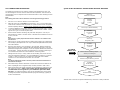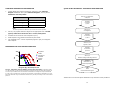
1
Contact Information DRX UM PH ARRESTIN 0812V1 DiscoveRx Corporation (World Wide Headquarters) 42501 Albrae Street Fremont, CA 94538 United States t | 510.979.1415 f | 510.979.1650 toll-free | 866.448.4864 PathHunter® β-Arrestin GPCR Assays For Chemiluminescent Detection of Activated GPCRs User Manual KINOMEscan A division of DiscoveRx 11180 Roselle Street, Suite D San Diego, CA 92121 United States t | 800.644.5687 f | 858.630.4600 DiscoveRx Corporation Ltd. (Europe Headquarters) Faraday Wharf, Holt Street Birmingham Science Park Aston Birmingham, B7 4BB United Kingdom t | +44.121.260.6142 f | +44.121.260.6143 www.discoverx.com © 2012 DiscoveRx Corporation, Fremont, CA 94538. All rights reserved. Simple Solutions for Complex Biology CONTENTS NOTES: LEGAL SECTION PAGE 3 INTENDED USE PAGE 4 TECHNOLOGY PRINCIPLE PAGE 4 ASSAY OVERVIEW PAGE 5 MATERIALS PROVIDED PAGE 5 ADDITIONAL MATERIALS REQUIRED PAGE 6 FROZEN CELL HANDLING PROCEDURE PAGE 6 CELL PLATING REAGENT REQUIREMENTS PAGE 6 SOLVENTS AND PREPARATION OF COMPOUND DILUTIONS PAGE 7 USE OF PLASMA OR SERUM CONTAINING SAMPLES PAGE 7 STORING & REMOVING CRYOVIALS FROM LIQUID NITROGEN PAGE 7 CELL THAWING AND PROPAGATION PAGE 8 CELL FREEZING PROTOCOL PAGE 9 PREPARATION OF ASSAY PLATES PAGE 9 ASSAY PROCEDURE — AGONIST DOSE RESPONSE PROTOCOL QUICK START PROCEDURE PAGE 10 PAGE 13 ASSAY PROCEDURE — ANTAGONIST DOSE RESPONSE PROTOCOL QUICK START PROCEDURE PAGE 14 PAGE 17 ASSAY PROCEDURE — ALLOSTERIC MODULATOR RESPONSE PROTOCOL QUICK START PROCEDURE PAGE 18 PAGE 21 ASSAY PROCEDURE — NEUTRALIZING ANTIBODY RESPONSE PROTOCOL QUICK START PROCEDURE PAGE 22 PAGE 25 TROUBLESHOOTING GUIDE PAGE 26 APPENDIX A: ASSAY FORMATS PAGE 28 APPENDIX B: RELATED PRODUCTS PAGE 28 2 31 LEGAL SECTION NOTES: This product and/or its use is covered by one or more of the following U.S. patents #6,342,345 B1, #7,135,325 B2, #8,101,373 B2 and/or foreign patents, patent applications, and trade secrets that are either owned by or licensed to DiscoveRx® Corporation. This product is for in vitro use only and in no event can this product be used in whole animals. The right to use or practice the inventions in the foregoing patents (including method of use claims) by using or propagating this product is granted solely in connection with the use of appropriate Detection Reagents (protected under trade secret) purchased from DiscoveRx Corporation or its authorized distributors. LIMITED USE LICENSE AGREEMENT The cells and detection reagents (collectively Materials) purchased from DiscoveRx® are expressly restricted in their use. DiscoveRx has developed a Protein:Protein Interaction assay (Assay) that employs genetically modified cells and vectors (collectively, the “Cells”), and related detection reagents (the “Reagents”) (collectively referred to as “Materials”). By purchasing and using the Materials, the Purchaser agrees to comply with the following terms and conditions of this label license and recognizes and agrees to such restrictions: 1. The Materials are not transferable and will be used only at the site for which they were purchased. Transfer to another site owned by Purchaser will be permitted only upon written request by Purchaser followed by subsequent written approval by DiscoveRx. 2. The Reagents contain or are based upon the proprietary and valuable knowhow developed by DiscoveRx, and the Reagents have been optimized by DiscoveRx to function more effectively with the Cells in performing the Assay. Purchaser will not analyze or reverse engineer the Materials nor have them analyzed on Purchaser’s behalf. 3. In performing the Assay, Purchaser will use only Reagents supplied by DiscoveRx or an authorized DiscoveRx distributor for the Materials. If the purchaser is not willing to accept the limitations of this limited use statement and/or has any further questions regarding the rights conferred with purchase of the Materials, please contact: DiscoveRx Corporation Attn: Licensing Department 42501 Albrae Street Fremont, CA 94538 tel | 510.979.1415 x104 [email protected] For some products/cell lines, certain 3rd party gene specific patents may be required to use the cell line. It is the purchaser's responsibility to determine if such patents or other intellectual property rights are required. 30 3 INTENDED USE NOTES: ® PathHunter β-Arrestin GPCR Assays are whole cell, functional assays that directly measure GPCR activity by detecting the interaction of β-Arrestin with the activated GPCR. Because Arrestin recruitment occurs independent of G-protein coupling, PathHunter β-Arrestin assays offer a powerful and universal screening platform that can be used with virtually any Gi-, Gs-, or Gq-coupled receptor. This PathHunter system combines engineered clonal cell lines stably expressing the ProLink™ (PK)tagged GPCR of interest and the Enzyme acceptor (EA)-tagged β-Arrestin fusion proteins with optimized PathHunter® Detection Reagents (Cat. #93-0001, 93-0001L and 93-0001XL). Each cell line has been characterized for appropriate GPCR pharmacology, specificity and stability in cell culture. By combining a simple, one-step addition protocol and standard chemiluminescent detection, these assays are ideally suited for 96-well, 384-well, or 1536-well compound screening. TECHNOLOGY PRINCIPLE PathHunter® β-Arrestin cell lines monitor GPCR activity by detecting the interaction of β-Arrestin with the activated GPCR using β-galactosidase (β-gal) enzyme fragment complementation (EFC, Figure 1). In this system, the GPCR of interest is fused in frame with the small, 42 amino acid fragment of β-gal called ProLink™ and coexpressed in cells stably expressing a fusion protein of β-Arrestin and the larger, N-terminal deletion mutant of β-gal (called enzyme acceptor or EA). Activation of the GPCR stimulates binding of β-Arrestin to the ProLink-tagged GPCR and forces complementation of the two enzyme fragments, resulting in the formation of an active β-gal enzyme. This action leads to an increase in enzyme activity that can be measured using chemiluminescent PathHunter Detection Reagents. Because arrestin recruitment occurs independent of G-protein coupling, these assays provide a direct, universal platform for measuring receptor activation. Figure 1. PathHunter® β-Arrestin Assay Principle. Activation of the ProLink™-tagged GPCR results in β-Arrestin recruitment and formation of a functional enzyme capable of hydrolyzing substrate and generating a chemiluminescent signal. 4 29 APPENDIX A: ASSAY FORMATS ASSAY OVERVIEW PathHunter® Certified Assay Format Plate Format 96-well FV 384-well LV 384-well 1536-well Total Volume 150 μL 40 μL 20 μL 8 μL Cell Numbers 10,000 5,000 2,500 1,250 Cell Plating Reagents* 90 μL 20 μL 10 μL 4 μL Ligand 10 μL 5 μL 2.5 μL 1 μL Detection Reagents 50 μL 12 μL 6 μL 3 μL *Cell Plating Reagent volume used to resuspend cells for assay plates APPENDIX B: RELATED PRODUCTS Description Control Ligands Ordering Information www.discoverx.com/pathway_assays/ control_ligands.php Please read the entire protocol completely before running the assay. The Assay Procedure sections and Quick Start Guides in this booklet contain detailed information about how to run the assays. Refer to the cell-line specific datasheet for additional information on the optimized Cell Plating Reagent and reference ligand recommended for the assay. Assays should be run using a fresh split of low-passage cells that have not been allowed to reach confluency for more than 24 hours. Following treatment of the cells with compound, GPCR activity is detected by adding a working solution of chemiluminescent PathHunter® Detection Reagents using a simple, mix-and-read protocol. The following steps are required to monitor GPCR activity using a PathHunter® β-Arrestin GPCR cell line (Figure 2). 1. Plate cells (p.9). 2. Dilute and add compounds or antibodies. 3. Perform functional assay in agonist (p.10), antagonist (p.14) or allosteric modulator mode (p.18). Plate cells & add compounds Add PathHunter® Detection Reagents Read Luminescence PathHunter® Cell Plating Reagents www.discoverx.com/certified/ cell_plating_reagents.php PathHunter® Certified Cell Culture Reagents www.discoverx.com/certified/PH_cellculture_reagents.php PathHunter®select Cell Culture Kit Revive™ Media Preserve™ Freezing Reagent www.discoverx.com/certified/PH_cellculture_reagents.php Figure 2. Simple chemiluminescent assay protocol for monitoring GPCR activity in response to compound challenge. PathHunter® Detection Reagents www.discoverx.com/certified/ PH_detection_reagents.php MATERIALS PROVIDED Microplates www.discoverx.com/certified/microplates.php PathHunter® eXpress β-Arrestin GPCR Assays www.discoverx.com/gpcrs/express_arrestin.php PathHunter® eXpress β-Arrestin Orphan GPCR Assays www.discoverx.com/gpcrs/express_orphan.php PathHunter® eXpress β-Arrestin Ortholog GPCR Assays www.discoverx.com/gpcrs/express_ortholog.php 28 Description Contents ® PathHunter β-Arrestin GPCR Cells* 2 vials Storage Liquid N2 (vapor phase) *Please refer to the cell line specific datasheet for detailed information on the PathHunter® β-Arrestin cell line you are testing. 5 ADDITIONAL MATERIALS REQUIRED TROUBLESHOOTING GUIDE (CONTINUED) The following additional materials are required to perform PathHunter® β-Arrestin GPCR Assays: Equipment Materials Green V-Bottom PP Ligand Dilution Plates, 10 plates/pack (DiscoveRx, Cat. #92-0011) 96-well Clear Bottom TC treated, Sterile WCB, FB w/lid, 10 plates/pack (DiscoveRx, Cat. #92-0014) 384-well Clear Bottom TC treated, Sterile WCB, FB w/lid, 10 plates/pack (DiscoveRx, Cat. #92-0013) 384-well White Bottom TC treated, Sterile w/lid, 10 plates/pack (DiscoveRx, Cat. #92-0015) Disposable Reagent Reservoir (Thermo Scientific, Cat. #8094 or similar) Hemocytometer Cryogenic Freezing Container (Nalgene, Cat. #5100-0001 or similar) Cryogenic Freezer Vials (Fisher Scientific, Cat. #375418 or similar) Multimode or luminescence plate reader* Single and multi-channel pipettors and pipette tips Tissue culture disposables and plasticware (T25 and T75 flasks, etc.) PathHunter® Detection Kit (DiscoveRx, Cat. #93-0001, #93-0001L or #93-0001XL) Revive™ Media (DiscoveRx, Cat. #92-0016RM Series) PathHunter®select Cell Culture Kits (DiscoveRx, Cat. #92-0018G Series) Preserve™ Freezing Reagent (DiscoveRx, Cat. #92-0017FR Series) Cell Detachment Reagent (DiscoveRx, Cat. #92-0009) PathHunter® Cell Plating (CP) Reagent (DiscoveRx, Cat. #93-0563R Series) Phosphate buffered saline (PBS) GPCR control agonist GPCR test compound(s) and/or antagonists *For 96-well analysis, we recommend the LumiLITE™ Microplate Reader (DiscoveRx, Cat. #75-0001) ±Please refer to the cell line specific datasheet to determine catalog numbers for the media and reagent requirements for the PathHunter β-Arrestin cell line you are testing. PROBLEM Cells growing slowly EC50 is right-shifted CAUSE U2OS grows slower than CHO-K1 or HEK 293 Average doubling time is 3 days, so please observe cells under microscope and monitor cell health Slow growing clones Use of DiscoveRx functionally validated and optimized media and reagents improves assay performance Improper ligand handling or storage Check ligand handling requirements Difference in agonist binding affinity Confirm that the ligand used is comparable to the ligand in the Product Insert Problems with plate type and compound stability Hydrophobic compounds should be tested for solubility and may be diluted in buffer containing 0.1% BSA Non-binding surface plates may be necessary for hydrophobic compounds High well-to-well variability in Z’ study Problems with plate type and compound solubility Z’ studies should be performed with automation It may be necessary to test plate types and compound stability For additional information or technical support, please call 1.866.448.4864 (US) +44.121.260.6142 (Europe) or email [email protected] FROZEN CELL HANDLING PROCEDURE To ensure maximum cell viability, thaw the vial and initiate the culture as soon as possible upon receipt. If continued storage of the frozen vials is necessary, store vials in the vapor phase of liquid nitrogen (N2). DO NOT store at –80°C for extended periods as this could result in significant loss in cell viability. CELL PLATING REAGENT REQUIREMENTS Each PathHunter® β-Arrestin GPCR cell line has been validated for optimal assay performance using the recommended Cell Plating (CP) Reagent and control ligand as indicated in the cell line specific datasheet. For optimal performance using this PathHunter® Certified System, always use the CP Reagent recommended for the cell line and DO NOT substitute at any time. 6 SOLUTION 27 TROUBLESHOOTING GUIDE PROBLEM No Response Decreased Response Low or No Signal Experimental S:B does not match datasheet value SOLVENTS AND PREPARATION OF COMPOUND DILUTIONS CAUSE SOLUTION Improper cell growth conditions See datasheet for cell culture conditions High DMSO/solvent concentration Maintain DMSO/solvent at <1% in serial dilutions of compounds. Improper ligand used or improper ligand incubation time See datasheet for recommended ligand and assay conditions Improper preparation of ligand (agonist or antagonist) Refer to vendor specific datasheet to ensure proper handling, dilution and storage of ligand Improper time course for induction Optimize time course of induction with agonist and antagonist. Higher passages give reduced performance PathHunter cells are stable up to 10 passages. Use low passage cells whenever possible Cells are not adherent and exhibit incorrect morphology Confirm adherence of cells using microscopy Improper preparation of detection reagents Detection reagents should be prepared just prior to use and are sensitive to light. Problem with cell growth, cell viability, cell adherence or cell density See datasheet for cell culture conditions. Problem with microplate reader Microplate reader should be in luminescence mode. Read at 1 sec/well. For cell pools, S:B may vary greatly from passage to passage or day to day Prepare a clonal cell line or use lower passage number cells. Repeat the assay PathHunter β-Arrestin GPCR assays are routinely carried out in the presence of ≤ 1% solvent (i.e. DMSO, ethanol, PBS or other). As solvents can affect assay performance, optimize the assay conditions accordingly if other solvents or solvent concentrations are required. To validate each PathHunter β-Arrestin GPCR Assay, reference ligand was diluted using the Cell Plating (CP) Reagent recommended for the cell line (containing the appropriate solvent). For antibodies or other compounds that may be sensitive to serum and/or other assay components, dilutions can be prepared in either Hanks Buffered Salt Solution (HBSS) + 10 mM HEPES + 0.1% Bovine Serum Albumin (BSA) or OptiMEM® + 0.1% BSA without affecting assay performance. USE OF PLASMA OR SERUM CONTAINING SAMPLES PathHunter β-Arrestin GPCR Assays can be run in the presence of high levels of serum or plasma without negatively impacting assay performance. Standard curves of control ligand can be prepared in neat, heparinized plasma and added directly to the cells (without further dilution, ie. 100% plasma in the well). After ligand stimulation, the samples should be removed and replaced with fresh CP Reagent before the addition of the PathHunter Detection Reagents. Refer to p.22 for more information. NOTE: EDTA anti-coagulated plasma samples do not give a positive response in the assay. Therefore, the choice of anti-coagulant treatment is very important. STORING & REMOVING CRYOVIALS FROM LIQUID NITROGEN Cells are shipped in 2 vials on dry ice and contain approximately 1 x 10⁶ cells per vial in 1 mL of Preserve™ Freezing Reagent. The following procedures are for safely storing and removing cryovials from liquid nitrogen storage. 1. PathHunter cells must arrive in a frozen state on dry ice. If cells arrive thawed, do not proceed, contact technical support. 2. Frozen cells must be immediately transferred to liquid N2 storage or thawed and put into culture upon arrival. 3. When removing cryovials from liquid N2 storage, use tongs and place immediately on dry ice in a covered container. Wait at least one minute for any liquid N2 inside the vial to evaporate. 4. Proceed with the thawing protocol in the following section. Confirm assay conditions Improper preparation of ligand (agonist or antagonist) 26 Some ligands are difficult to handle. Confirm the final concentration of ligands SAFETY WARNING: A face shield, gloves and lab coat should be worn at all times when handling frozen vials. Some cryovials can leak when submerged in liquid N2. Upon thawing, the liquid N2 present in the cryovial converts back to its gas phase which can result in the vessel exploding. 7 CELL THAWING AND PROPAGATION QUICK-START PROCEDURE: NEUTRALIZING ANTIBODY RESPONSE The following procedures are for thawing, seeding and expanding the cells, and for maintaining the cultures once the cells have been expanded. Cells are free of contamination prior to shipment and care should be taken in their handling to avoid contamination. Plate 20 µL PathHunter cells/well NOTE: Face shield, gloves and a lab coat should be worn during the thawing procedure. 1. Pre-warm 15 mL Revive™ Media in a 37°C water bath. 2. Place the frozen cell vials briefly (10 seconds to 1 min) in a 37°C water bath under sterile conditions until only small ice crystals remain and the cell pellet is almost completely thawed. Caution: Longer incubation may result in cell death. 3. To remove DMSO from the media, carefully transfer the thawed cells to a sterile 15 mL tube and then fill tube with 10 mL pre-warmed Revive™ Media. Centrifuge at 300 x g for 4 minutes to pellet cells. 4. Remove media without disturbing cell pellet and resuspend in 5 mL of prewarmed Revive™ Media. Transfer cells to a T25 flask and incubate for 24 hours at 37°C, 5% CO2. Incubate overnight @ 37C Add 2.5 µL of Diluted Antibody Incubate 30 minutes @ 37°C NOTE: Cell recovery is greatly improved when selection antibiotics are omitted for the first 24 hours. 5. After 24 hours, gently remove Revive™ Media (being careful not to disturb the cell monolayer) and replace with 5 mL of pre-warmed complete PathHunter®select Cell Culture Media. 6. Once the cells become >70% confluent in the T25 flask, aspirate media and wash cells with 5 mL PBS. Aspirate PBS and dissociate cells with 0.5 mL Cell Detachment Reagent and resuspend in 5 mL of complete PathHunter®select Cell Culture Media. Transfer the entire cell suspension to a T75 flask containing 15 mL of PathHunter®select Cell Culture Media for continued growth. 7. Passage the cells every 2-3 days, based on the doubling time of the cell line, using cell Detachment Reagent. For routine passaging, prepare a 1:3 dilution of cells in a total volume of 15 mL PathHunter®select Cell Culture Media. Transfer 5 mL of the diluted cells to each new T75 flask. NOTE: To maintain logarithmic growth of the cells, cultures should be maintained in a subconfluent monolayer. 8. Each PathHunter β-Arrestin GPCR Cell Line has been found to be stable for at least 10 passages with no significant drop in assay window or shift in EC50. 9. Assay performance and cellular response can be assessed by treating the cells with reference agonist. Refer to the cell line specific datasheet for the recommended control agonist for your PathHunter β-Arrestin GPCR Cell Line. For antagonist assays, cells can be pretreated with varying doses of antagonist/inhibitor compounds followed by agonist challenge, typically at an EC80 concentration. Add 2.5 µL of Agonist @ EC80 Incubate 90 minutes @ 37°C* FOR SERUM SAMPLES ONLY! Remove serum-containing sample. Add 25 µL CP Reagent Add 12 µL Detection Reagent Working Solution Incubate 60 Minutes @ Room Temperature Read Chemiluminescent Signal *Please refer to the cell line specific datasheet any variations in assay conditions. 8 25 ATTENTION! PLASMA OR SERUM-CONTAINING SAMPLES ONLY CELL FREEZING PROTOCOL 5. After incubation is complete, gently aspirate the plasma or serum-containing samples from the well. Be careful to remove as much sample as possible without disturbing the cell monolayer. The following procedures are for freezing cells from confluent T225 flasks. If smaller flasks are used, adjust the volumes accordingly. Care should be taken in handling to avoid contamination. 6. Immediately add 25 μL of fresh CP Reagent to each well. Proceed with substrate preparation and addition. 1. Remove T225 flasks from incubator and place in the tissue culture hood. Aspirate the media from the flasks. 2. Add 10 mL PBS into each T225 flask and swirl to rinse the cells. Aspirate PBS from flask. 3. Add 5 mL of Cell Detachment Reagent to the flask. Rock the flask back and forth gently to ensure the surface of the flask is covered. Incubate at 37°C, 5% CO2 for 2–5 minutes or until the cells have detached. 4. Remove the flask from the incubator and view under a microscope to confirm that the cells have detached. If necessary, tap the edge of the flask to detach cells from the surface. 5. Add 8–10 mL of Revive™ Media to each T225 flask. Rinse the cells from the surface of the flask using the added media. Remove the cells from the flask and transfer to a 50 mL conical tube. (If necessary, add an additional 5 mL of media to the flask and rinse to collect the remaining cells and transfer the additional volume to the 50 mL conical tube). Remove 0.5 mL of the resuspended cells and count the cells using a hemocytometer. 6. Centrifuge the collected cells at 300 x g for 4 minutes. 7. After centrifugation, discard the supernatant. Resuspend the cell pellet in Preserve™ Freezing Reagent. Based on the cell number obtained from Step 5, dilute the resuspended cells to a concentration of 1.2 x 10⁶ cells/mL using Preserve™ Freezing Reagent. 8. Transfer 1 mL cells to each 2 mL cryogenic tube. (Keep cells on ice during this process and transfer to a cryogenic container pre-chilled at 4°C). 9. Transfer tubes to –80°C and store overnight. Transfer tubes into the vapor phase of a liquid N2 tank for long-term storage. SUBSTRATE PREPARATION AND ADDITION 1. Prepare PathHunter Detection Reagent by combining 1 part Substrate Reagent 2 Substrate with 5 parts Substrate Reagent 1, and 19 parts of PathHunter Cell Assay Buffer. Component Entire Plate (384 wells) Cell Assay Buffer 4.75 mL Substrate Reagent 1 1.25 mL Substrate Reagent 2 0.25 mL NOTE: The working solution is stable for up to 8 hours at room temperature. 2. Add 12 μL of prepared detection reagent to the appropriate wells and incubate for 60 minutes at room temperature (23°C). DO NOT pipette up and down in the well to mix or vortex/shake plates. 3. Incubate for 60 minutes at room temperature (23ºC). 4. Read samples on any standard luminescence plate reader. 5. Use GraphPad Prism® or other comparable program to plot your dose response. PREPARATION OF ASSAY PLATES Each PathHunter β-Arrestin GPCR Assay has been validated for optimal assay performance using the specific PathHunter Cell Plating Reagent. Always use the CP Reagent recommended for the cell line and DO NOT substitute at any time. 1. 24 Harvest the cells as follows from a confluent T25 or T75 flask using Cell Detachment Reagent. Do not use Trypsin. a) Remove PathHunter®select Cell Culture Media. b) Gently wash cells with 5 mL PBS and aspirate. c) Add 0.5 mL Cell Detachment Reagent to each T25 flask, or 1 mL to each T75 flask. d) Place the flask in the incubator for 5 minutes or until cells have detached. e) Add 3 mL of CP Reagent and transfer to a 15 mL conical tube. 9 2. Determine the cell density using a hemocytometer. Centrifuge the cells at 300 x g for 4 minutes to pellet cells. Remove supernatant. 3. Resuspend cells in CP Reagent at a concentration of 250,000 cells/mL (5,000 cells/20 μL). Transfer 20 μL of the cell suspension to each well of a 384-well microplate. Please refer to Appendix A for cell numbers and volumes for alternate formats. 4. Incubate the plate overnight at 37°C, 5% CO2. To prepare the 11-point dose curve serial dilutions, we recommend starting with a concentration that is 50X the expected IC50 value for the compound (e.g. 500X IC50 would be the final working concentration). Example: If the expected IC50 is 10 nM, prepare the highest starting concentration of the corresponding dilution at 5 µM. This is the working concentration. a. For each antibody tested, label the wells of a 384-well dilution plate #1 through #12. b. Add 20 µL of CP Reagent containing appropriate solvent to wells #1-11. c. Prepare a working concentration of antibody in the appropriate CP Reagent. d. Add 30 µL of the working concentration of antibody to well #12. e. Remove 10 µL of antibody from well #12, add it to well #11 and mix gently by pipetting up and down. Discard pipet tip. f. With a clean pipet tip, remove 10 µL of diluted antibody from well #11, add it to well #10 and mix gently by pipetting up and down. Discard the pipet tip. g. Repeat this process 7 more times in succession to prepare serial dilutions for the remaining wells, from right to left across the plate. DO NOT add antibody to wells #1 and 2. This sample serves as the no antibody control and completes the dose curve. h. Repeat this process for each additional antibody to be tested. i. Set antibodies aside until they are ready to be added. ASSAY PROCEDURE — AGONIST DOSE RESPONSE CURVE 1 Compound 1 Compound 3 Compound 5 Compound 7 Compound 9 Compound 11 Compound 13 Compound 15 3-fold serial dilutions of agonist Low 2 3 4 5 6 7 8 High No agonist No agonist The steps outlined below provide the assay volumes and procedures for performing GPCR agonist assays using the PathHunter β-Arrestin GPCR Cell Lines and PathHunter Detection Reagents in a 384-well format. Refer to Appendix A for cell numbers and volumes for alternate formats. Although plate layouts and experimental designs may vary, we recommend performing a 12-point dose curve for each compound using at least duplicate wells for each dilution. Low 3-fold serial dilutions of agonist High 9 10 11 12 13 14 15 16 17 18 19 20 21 22 23 24 A B C D E F G H I J K L M N O P Compound 2 3. Remove PathHunter cells from the incubator (previously plated on day 1). 4. Transfer 2.5 µL from wells #1–12 to duplicate wells according to the plate map shown on p.22. 5. Incubate for 30 minutes @ 37°C. Compound 4 Compound 6 Compound 8 Compound 10 AGONIST COMPOUND PREPARATION AND ADDITION Compound 12 1. During the antibody incubation, determine the EC80 concentration of the agonist to be used in the assay. Prepare a 10X EC80 concentration of agonist compound as shown below: Example: If the expected EC80 of the agonist compound is 10 nM, prepare a stock at 100 nM. 2. Add 2.5 µL of agonist to each well. Add 2.5 µL of CP Reagent containing appropriate solvent to the no agonist wells (columns 1 & 13 in figure 8). 3. Incubate for 90 minutes @ 37°C. 4. If samples do not contain plasma or serum, omit steps 5 and 6 and proceed directly to the substrate preparation and addition. Compound 14 Compound 16 Figure 3. This plate map shows 12-point dose curves with 2 data points at each concentration. Plate map allows 16 compounds to be tested in duplicate per 384-well plate. DAY 1: PREPARATION OF ASSAY PLATES Plate PathHunter cells in the appropriate number of wells in a 384-well plate as described in the “Preparation of Assay Plates” section on p.9. Allow cells to incubate overnight. 10 23 ASSAY PROCEDURE — NEUTRALIZING ANTIBODY DOSE RESPONSE DAY 2: AGONIST COMPOUND PREPARATION AND ADDITION The steps outlined below provide the assay volumes and procedure for performing detection of anti-GPCR neutralizing antibodies using the PathHunter β-Arrestin GPCR Cell Lines and PathHunter Detection Reagents in a 384-well format. Refer to Appendix A for cell numbers and volumes for alternate formats. Although plate layouts and experimental designs may vary, we recommend performing an 11-point dose curve for each compound using at least duplicate wells for each dilution. The protocol and volumes described below are designed for a complete 384-well plate. 1. Dissolve agonist compound in the vehicle of choice (DMSO, ethanol, PBS or other) at the desired stock concentration. 2. Prepare a series of twelve 3-fold serial dilutions of agonist compound in CP Reagent containing the appropriate solvent (DMSO, ethanol, PBS or other) as described below. The concentration of each dilution should be prepared at 5X of the final screening concentration (i.e. 5 µL compound + 20 µL of cells). For each dilution, the final concentration of solvent should remain constant. To prepare the 12-point dose curve serial dilutions, we recommend starting with a concentration that is 50X the expected EC50 value for the compound (e.g. 250X EC50 would be the final working concentration). Example: If the expected EC50 is 10 nM, prepare the highest starting concentration of the corresponding dilution at 2.5 µM. This is the working concentration. a. For each compound tested, label the wells of a 384-well dilution plate #1 through #12. b. Add 20 µL of CP Reagent containing appropriate solvent to wells #1-11. c. Prepare a working concentration of agonist compound in the appropriate CP Reagent. d. Add 30 µL of the working concentration of agonist compound to well #12. e. Remove 10 µL of compound from well #12, add it to well #11 and mix gently by pipetting up and down. Discard pipet tip. With a clean pipet tip, remove 10 µL of diluted compound from well #11, add it to well #10 and mix gently by pipetting up and down. Discard the pipet tip. g. Repeat this process 8 more times in succession to prepare serial dilutions for the remaining wells, from right to left across the plate. DO NOT add agonist compound to well #1. This sample serves as the no agonist control and completes the dose curve. h. Repeat this process for each additional agonist compound to be tested. i. Set compounds aside until agonist compounds are ready to be added. f. Figure 8. This plate map shows a 11-point dose curves with 2 data points at each concentration. Plate layout allows 16 antibodies to be tested in duplicate per 384-well plate. DAY 1: PREPARATION OF ASSAY PLATES Plate PathHunter cells in the appropriate number of wells in a 384-well plate as described in the “Preparation of Assay Plates” section on p. 9. Allow cells to incubate overnight. DAY 2: ANTIBODY PREPARATION AND ADDITION 1. Dissolve antibody in the vehicle of choice (PBS, water or other) at the desired stock concentration. 2. Prepare a series of eleven 3-fold serial dilutions of antibody in Cell Plating Reagent containing the appropriate solvent (PBS, water or other). The concentration of each dilution should be prepared at 10X of the final screening concentration (i.e. 2.5 µL antibody will be used in a final volume of 25 µL). For each dilution, the final concentration of solvent should remain constant. 22 3. Remove PathHunter cells from the incubator (previously plated on day 1). NOTE: 93-0203C7 PathHunter® C2C12 CXCR4 β-Arrestin cell line uses an additional media exchange step. Please refer to cell line specific datasheet. 4. Transfer 5 µL from wells #1–12 to duplicate wells according to the plate map shown on p.10. 5. Incubate for 90 minutes @ 37°C. 11 SUBSTRATE PREPARATION AND ADDITION 1. QUICK-START PROCEDURE: ALLOSTERIC MODULATOR DOSE RESPONSE Prepare PathHunter Detection Reagent by combining 1 part Substrate Reagent 2 Substrate with 5 parts Substrate Reagent 1, and 19 parts of PathHunter® Cell Assay Buffer. Component Entire Plate (384 wells) Cell Assay Buffer 4.75 mL Substrate Reagent 1 1.25 mL Substrate Reagent 2 0.25 mL NOTE: The working solution is stable for up to 8 hours at room temperature. 2. Add 12 μL of prepared detection reagent to the appropriate wells. DO NOT pipet up and down in the well to mix or vortex/shake plates. 3. Incubate for 60 minutes at room temperature (23°C). 4. Read samples on any standard luminescence plate reader. 5. Use GraphPad Prism® or other comparable program to plot your agonist dose response. See the example shown in Figure 4. Plate 20 µL PathHunter® cells/well Incubate overnight @ 37°C Add 2.5 µL of Allosteric Modulator Compound Incubate 30 minutes @ 37°C Add 2.5 µL of Agonist REPRESENTATIVE DATA AND DATA ANALYSIS Incubate 90 minutes @ 37°C* 20000 17500 RLU 15000 Add 12 µL Detection Reagent Working Solution 12500 10000 7500 5000 2500 0 10 - 1 3 10 - 1 2 10 - 1 1 10 - 1 0 10 - 9 10 - 8 10 - 7 10 - 6 Somatostatin 28 [M] Figure 4. PathHunter® CHO-K1 SSTR2 β-Arrestin Cells (93-0181C2). Cells were plated in a 384-well plate at 5,000 cells/well and stimulated with the known agonist Somatostatin 28 (DiscoveRx, 92-1068) for 90 minutes. Signal was detected using the PathHunter Detection Kit (93-0001) according to the recommended protocol. An assay window of 52.4-fold S:B was achieved in this example, and the EC50 for agonist was estimated at 1.8 nM. Incubate 60 Minutes @ Room Temperature Read Chemiluminescent Signal *Please refer to the cell line specific datasheet for any variations in assay conditions. 12 21 SUBSTRATE PREPARATION AND ADDITION 1. QUICK-START PROCEDURE: AGONIST DOSE RESPONSE Prepare PathHunter Detection Reagent by combining 1 part Substrate Reagent 2 Substrate with 5 parts Substrate Reagnet 1, and 19 parts of PathHunter Cell Assay Buffer. Plate 20 µL PathHunter cells/well Component Entire Plate (384 wells) Cell Assay Buffer 4.75 mL Substrate Reagent 1 1.25 mL Substrate Reagent 2 0.25 mL Incubate overnight @ 37°C NOTE: The working solution is stable for up to 8 hours at room temperature. 2. Add 12 μL of prepared detection reagent to the appropriate wells and incubate for 60 minutes at room temperature (23°C). DO NOT pipette up and down in the well to mix or vortex/shake plates. 3. Incubate for 60 minutes at room temperature (23ºC). 4. Read samples on any standard luminescence plate reader. 5. ® Use GraphPad Prism or other comparable program to plot your allosteric modulator dose response. Add 5 µL of Agonist Incubate 90 minutes @ 37°C* Add 12 µL Detection Reagent Working Solution Incubate 60 Minutes @ Room Temperature Read Chemiluminescent Signal *Please refer to the cell line specific datasheet for any variations in assay conditions. 20 13 ASSAY PROCEDURE — ANTAGONIST DOSE RESPONSE CURVE for the compound (e.g. 500X IC50 would be the final working concentration). Example: If the expected IC50 is 10 nM, prepare the highest starting concentration of the corresponding dilution at 5 µM. This is the working concentration. a. For each compound tested, label the wells of a 384-well dilution plate #1 through #12. b. Add 20 µL of CP Reagent containing appropriate solvent to wells #1-11. c. Prepare a working concentration of modulator compound in the appropriate CP Reagent. d. Add 30 µL of the working concentration of modulator compound to well #12. The steps outlined below provide the assay volumes and procedures for performing GPCR antagonist assays using the PathHunter β-Arrestin GPCR Cell Lines and PathHunter Detection Reagents in a 384-well format. Refer to Appendix A for cell numbers and volumes for alternate formats. Although plate layouts and experimental designs may vary, we recommend performing a 11-point dose curve for each compound using at least duplicate wells for each dilution. The protocol and volumes described below are designed for a complete 384-well plate. e. Remove 10 µL of compound from well #12, add it to well #11 and mix gently by pipetting up and down. Discard the pipet tip. f. With a clean pipet tip, remove 10 µL of diluted compound from well #11, add it to well #10 and mix gently by pipetting up and down. Discard the pipet tip. g. Repeat this process 7 more times in succession to prepare serial dilutions for the remaining wells, or from right to left across the plate. DO NOT add modulator compound to wells #1 and 2. These samples serve as the no modulator controls and complete the dose curve. h. Repeat this process for any additional modulator compounds to be tested. i. Set compounds aside until you are ready to add them to the cells. Figure 5. This plate map shows 11-point dose curves with 2 data points at each concentration. Plate map allows 16 compounds to be tested in duplicate per 384-well plate. DAY 1: PREPARATION OF ASSAY PLATES 3. Remove PathHunter cells from the incubator (previously plated on day 1). 4. Transfer 2.5 µL from wells #1–12 to duplicate wells according to the plate map on p.18. 5. Incubate cells with modulator compounds for 30 minutes @ 37°C. AGONIST COMPOUND PREPARATION AND ADDITION 1. During the modulator compound incubation, determine the EC10/EC90 concentration of the agonist from the agonist dose response curve (described on p.10-13). Prepare a 10X EC10 concentration (PAM) or 10X EC90 concentration (NAM) of agonist compound in the appropriate CP Reagent/solvent as shown below: Example: If the expected EC10/EC90 of the agonist compound is 10 nM, prepare a stock at 100 nM. 2. When the modulator incubation is complete, add 2.5 µL of agonist compound to well #2-12. Add 2.5 µL of CP Reagent containing appropriate solvent to the “No modulator/No agonist” wells (columns 1 & 13 in Figure 7). 3. Incubate for 90 minutes @ 37ºC. Plate PathHunter cells in the appropriate number of wells in a 384-well plate as described in the “Preparation of Assay Plates” section on p.9. Allow cells to incubate overnight. DAY 2: ANTAGONIST COMPOUND PREPARATION AND ADDITION 1. Dissolve your antagonist compound in the vehicle of choice (DMSO, ethanol, PBS or other) at the desired stock concentration. 2. Prepare a series of eleven 3-fold serial dilutions of antagonist compound in CP Reagent containing the appropriate solvent (DMSO, ethanol, PBS or other) as described below. The concentration of each dilution should be prepared at 10X the final screening concentration (i.e. 2.5 µL antagonist compound will be used in a final volume of 25 µL). For each dilution, the final concentration of 14 19 ASSAY PROCEDURE — ALLOSTERIC MODULATOR DOSE RESPONSE solvent should remain constant. To prepare the 11-point dose curve serial dilutions, we recommend starting with a concentration that is 50X the expected IC50 value for the compound (e.g. 500X IC50 would be the final working concentration). Example: If the expected IC50 is 10 nM, prepare the highest starting concentration of the corresponding dilution at 5 µM. This is the working concentration. a. For each compound tested, label the wells of a 384-well dilution plate #1 through #12. b. Add 20 µL of CP Reagent containing appropriate solvent to wells #1-11. c. Prepare a working concentration of antagonist compound in the appropriate CP Reagent. d. Add 30 µL of the working concentration of antagonist compound to well #12. e. Remove 10 µL of compound from well #12, add it to well #11 and mix gently by pipetting up and down. Discard the pipet tip. f. With a clean pipet tip, remove 10 µL of diluted compound from well #11, add it to well #10 and mix gently by pipetting up and down. Discard the pipet tip. g. Repeat this process 7 more times in succession to prepare serial dilutions for the remaining wells, or from right to left across the plate. DO NOT add antagonist compound to tubes #1 and 2. These samples serve as the no antagonist controls and complete the dose curve. h. Repeat process for any additional antagonist compounds to be tested. i. Set compounds aside until you are ready to add them to the cells. The steps outlined below provide the assay volumes and procedure for performing allosteric modulator assays using PathHunter β-Arrestin GPCR Cell Lines and PathHunter Detection Reagents in a 384-well format. Refer to Appendix A for cell numbers and volumes for alternate formats. Although plate layouts and experimental designs may vary, we recommend performing a 11-point dose curve for each compound using at least duplicate wells for each dilution. The protocol and volumes described below are designed for a complete 384-well plate. Figure 7. This plate map shows 11-point dose curves with 2 data points at each concentration. Plate map allows 16 modulator compounds to be tested in duplicate per 384-well plate. DAY 1: PREPARATION OF ASSAY PLATES Plate PathHunter cells in the appropriate number of wells in a 384-well plate as described in the “Preparation of Assay Plates” section on p.9. Allow cells to incubate overnight. 3. Remove PathHunter cells from the incubator (previously plated on day 1). 4. Transfer 2.5 µL from wells #1–12 to duplicate wells according to the plate map on p.14. 5. Incubate cells with antagonist compounds for 30 minutes @ 37°C. AGONIST COMPOUND PREPARATION AND ADDITION 1. During the antagonist incubation, determine the EC80 concentration of the agonist from the agonist dose response curve (described on p.10-13). Prepare a 10X EC80 concentration of agonist compound in the appropriate CP Reagent/solvent as shown below: Example: If the expected EC80 of the agonist compound is 10 nM, prepare a stock at 100 nM. 2. When the antagonist incubation is complete, add 2.5 µL of agonist compound to wells #2–12. Add 2.5 µL of CP Reagent containing appropriate solvent to the “No antagonist/No agonist” wells (columns 1 & 13 in Figure 5). 3. Incubate for 90 minutes @ 37°C. DAY 2: MODULATOR COMPOUND PREPARATION AND ADDITION 1. Dissolve your allosteric modulator compound in the vehicle of choice (DMSO, ethanol, PBS or other) at the desired stock concentration. 2. Prepare a series of eleven 3-fold serial dilutions of modulator compound in CP Reagent containing the appropriate solvent (DMSO, ethanol, PBS or other) as described below. The concentration of each dilution should be prepared at 10X the final screening concentration (i.e. 2.5 μL modulator compound will be used in a final volume of 25 μL). For each dilution, the final concentration of solvent should remain constant. To prepare the 11-point dose curve serial dilutions, we recommend starting with a concentration that is 50X the expected IC50 value 18 15 SUBSTRATE PREPARATION AND ADDITION 1. QUICK-START PROCEDURE: ANTAGONIST DOSE RESPONSE Prepare PathHunter Detection Reagent by combining 1 part Substrate Reagent 2 Substrate with 5 parts Substrate Reagent 1, and 19 parts of PathHunter Cell Assay Buffer. Component Entire Plate (384 wells) Cell Assay Buffer 4.75 mL Substrate Reagent 1 1.25 mL Substrate Reagent 2 0.25 mL NOTE: The working solution is stable for up to 8 hours at room temperature. 2. Add 12 μL of prepared detection reagent to the appropriate wells. DO NOT pipet up and down in the well to mix or vortex/shake plates. 3. Incubate for 60 minutes at room temperature (23°C). 4. Read samples on any standard luminescence plate reader. 5. Use GraphPad Prism® or other comparable program to plot your antagonist dose response. Plate 20 µL PathHunter cells/well Incubate overnight @ 37°C Add 2.5 µL of Antagonist Incubate 30 minutes @ 37°C Add 2.5 µL of Agonist @ EC80 REPRESENTATIVE DATA AND DATA ANALYSIS Incubate 90 minutes @ 37°C* 30000 Propanolol Timolol CGP12177a Carvedilol Atenolol Nadolol RLU 20000 10000 Add 12 µL Detection Reagent Working Solution Incubate 60 Minutes @ Room Temperature 0 10 - 1 1 10 - 1 0 10 - 9 10 - 8 10 - 7 10 - 6 10 - 5 10 - 4 10 - 3 Antagonist [M] + 500 nM Isoproterenol ® Figure 6. PathHunter CHO-K1 ADRB2 β-Arrestin Cells (93-0182C2). Cells were plated in a 384-well plate at 5,000 cells/well and levels of β-Arrestin recruitment was measured after 30 minutes of pre-incubation with the indicated concentrations of antagonist compounds followed by a 90 minute incubation with a single EC80 concentration of isoproterenol (DiscoveRx; 92-1119). Signal was detected using the PathHunter Detection Kit (93-0001) according to the recommended protocol. Read Chemiluminescent Signal *Please refer to the cell line specific datasheet for any variations in assay conditions. 16 17 SUBSTRATE PREPARATION AND ADDITION 1. QUICK-START PROCEDURE: ANTAGONIST DOSE RESPONSE Prepare PathHunter Detection Reagent by combining 1 part Substrate Reagent 2 Substrate with 5 parts Substrate Reagent 1, and 19 parts of PathHunter Cell Assay Buffer. Component Entire Plate (384 wells) Cell Assay Buffer 4.75 mL Substrate Reagent 1 1.25 mL Substrate Reagent 2 0.25 mL NOTE: The working solution is stable for up to 8 hours at room temperature. 2. Add 12 μL of prepared detection reagent to the appropriate wells. DO NOT pipet up and down in the well to mix or vortex/shake plates. 3. Incubate for 60 minutes at room temperature (23°C). 4. Read samples on any standard luminescence plate reader. 5. Use GraphPad Prism® or other comparable program to plot your antagonist dose response. Plate 20 µL PathHunter cells/well Incubate overnight @ 37°C Add 2.5 µL of Antagonist Incubate 30 minutes @ 37°C Add 2.5 µL of Agonist @ EC80 REPRESENTATIVE DATA AND DATA ANALYSIS Incubate 90 minutes @ 37°C* 30000 Propanolol Timolol CGP12177a Carvedilol Atenolol Nadolol RLU 20000 10000 Add 12 µL Detection Reagent Working Solution Incubate 60 Minutes @ Room Temperature 0 10 - 1 1 10 - 1 0 10 - 9 10 - 8 10 - 7 10 - 6 10 - 5 10 - 4 10 - 3 Antagonist [M] + 500 nM Isoproterenol ® Figure 6. PathHunter CHO-K1 ADRB2 β-Arrestin Cells (93-0182C2). Cells were plated in a 384-well plate at 5,000 cells/well and levels of β-Arrestin recruitment was measured after 30 minutes of pre-incubation with the indicated concentrations of antagonist compounds followed by a 90 minute incubation with a single EC80 concentration of isoproterenol (DiscoveRx; 92-1119). Signal was detected using the PathHunter Detection Kit (93-0001) according to the recommended protocol. Read Chemiluminescent Signal *Please refer to the cell line specific datasheet for any variations in assay conditions. 16 17 ASSAY PROCEDURE — ALLOSTERIC MODULATOR DOSE RESPONSE solvent should remain constant. To prepare the 11-point dose curve serial dilutions, we recommend starting with a concentration that is 50X the expected IC50 value for the compound (e.g. 500X IC50 would be the final working concentration). Example: If the expected IC50 is 10 nM, prepare the highest starting concentration of the corresponding dilution at 5 µM. This is the working concentration. a. For each compound tested, label the wells of a 384-well dilution plate #1 through #12. b. Add 20 µL of CP Reagent containing appropriate solvent to wells #1-11. c. Prepare a working concentration of antagonist compound in the appropriate CP Reagent. d. Add 30 µL of the working concentration of antagonist compound to well #12. e. Remove 10 µL of compound from well #12, add it to well #11 and mix gently by pipetting up and down. Discard the pipet tip. f. With a clean pipet tip, remove 10 µL of diluted compound from well #11, add it to well #10 and mix gently by pipetting up and down. Discard the pipet tip. g. Repeat this process 7 more times in succession to prepare serial dilutions for the remaining wells, or from right to left across the plate. DO NOT add antagonist compound to tubes #1 and 2. These samples serve as the no antagonist controls and complete the dose curve. h. Repeat process for any additional antagonist compounds to be tested. i. Set compounds aside until you are ready to add them to the cells. The steps outlined below provide the assay volumes and procedure for performing allosteric modulator assays using PathHunter β-Arrestin GPCR Cell Lines and PathHunter Detection Reagents in a 384-well format. Refer to Appendix A for cell numbers and volumes for alternate formats. Although plate layouts and experimental designs may vary, we recommend performing a 11-point dose curve for each compound using at least duplicate wells for each dilution. The protocol and volumes described below are designed for a complete 384-well plate. Figure 7. This plate map shows 11-point dose curves with 2 data points at each concentration. Plate map allows 16 modulator compounds to be tested in duplicate per 384-well plate. DAY 1: PREPARATION OF ASSAY PLATES Plate PathHunter cells in the appropriate number of wells in a 384-well plate as described in the “Preparation of Assay Plates” section on p.9. Allow cells to incubate overnight. 3. Remove PathHunter cells from the incubator (previously plated on day 1). 4. Transfer 2.5 µL from wells #1–12 to duplicate wells according to the plate map on p.14. 5. Incubate cells with antagonist compounds for 30 minutes @ 37°C. AGONIST COMPOUND PREPARATION AND ADDITION 1. During the antagonist incubation, determine the EC80 concentration of the agonist from the agonist dose response curve (described on p.10-13). Prepare a 10X EC80 concentration of agonist compound in the appropriate CP Reagent/solvent as shown below: Example: If the expected EC80 of the agonist compound is 10 nM, prepare a stock at 100 nM. 2. When the antagonist incubation is complete, add 2.5 µL of agonist compound to wells #2–12. Add 2.5 µL of CP Reagent containing appropriate solvent to the “No antagonist/No agonist” wells (columns 1 & 13 in Figure 5). 3. Incubate for 90 minutes @ 37°C. DAY 2: MODULATOR COMPOUND PREPARATION AND ADDITION 1. Dissolve your allosteric modulator compound in the vehicle of choice (DMSO, ethanol, PBS or other) at the desired stock concentration. 2. Prepare a series of eleven 3-fold serial dilutions of modulator compound in CP Reagent containing the appropriate solvent (DMSO, ethanol, PBS or other) as described below. The concentration of each dilution should be prepared at 10X the final screening concentration (i.e. 2.5 μL modulator compound will be used in a final volume of 25 μL). For each dilution, the final concentration of solvent should remain constant. To prepare the 11-point dose curve serial dilutions, we recommend starting with a concentration that is 50X the expected IC50 value 18 15 ASSAY PROCEDURE — ANTAGONIST DOSE RESPONSE CURVE for the compound (e.g. 500X IC50 would be the final working concentration). Example: If the expected IC50 is 10 nM, prepare the highest starting concentration of the corresponding dilution at 5 µM. This is the working concentration. a. For each compound tested, label the wells of a 384-well dilution plate #1 through #12. b. Add 20 µL of CP Reagent containing appropriate solvent to wells #1-11. c. Prepare a working concentration of modulator compound in the appropriate CP Reagent. d. Add 30 µL of the working concentration of modulator compound to well #12. The steps outlined below provide the assay volumes and procedures for performing GPCR antagonist assays using the PathHunter β-Arrestin GPCR Cell Lines and PathHunter Detection Reagents in a 384-well format. Refer to Appendix A for cell numbers and volumes for alternate formats. Although plate layouts and experimental designs may vary, we recommend performing a 11-point dose curve for each compound using at least duplicate wells for each dilution. The protocol and volumes described below are designed for a complete 384-well plate. e. Remove 10 µL of compound from well #12, add it to well #11 and mix gently by pipetting up and down. Discard the pipet tip. f. With a clean pipet tip, remove 10 µL of diluted compound from well #11, add it to well #10 and mix gently by pipetting up and down. Discard the pipet tip. g. Repeat this process 7 more times in succession to prepare serial dilutions for the remaining wells, or from right to left across the plate. DO NOT add modulator compound to wells #1 and 2. These samples serve as the no modulator controls and complete the dose curve. h. Repeat this process for any additional modulator compounds to be tested. i. Set compounds aside until you are ready to add them to the cells. Figure 5. This plate map shows 11-point dose curves with 2 data points at each concentration. Plate map allows 16 compounds to be tested in duplicate per 384-well plate. DAY 1: PREPARATION OF ASSAY PLATES 3. Remove PathHunter cells from the incubator (previously plated on day 1). 4. Transfer 2.5 µL from wells #1–12 to duplicate wells according to the plate map on p.18. 5. Incubate cells with modulator compounds for 30 minutes @ 37°C. AGONIST COMPOUND PREPARATION AND ADDITION 1. During the modulator compound incubation, determine the EC10/EC90 concentration of the agonist from the agonist dose response curve (described on p.10-13). Prepare a 10X EC10 concentration (PAM) or 10X EC90 concentration (NAM) of agonist compound in the appropriate CP Reagent/solvent as shown below: Example: If the expected EC10/EC90 of the agonist compound is 10 nM, prepare a stock at 100 nM. 2. When the modulator incubation is complete, add 2.5 µL of agonist compound to well #2-12. Add 2.5 µL of CP Reagent containing appropriate solvent to the “No modulator/No agonist” wells (columns 1 & 13 in Figure 7). 3. Incubate for 90 minutes @ 37ºC. Plate PathHunter cells in the appropriate number of wells in a 384-well plate as described in the “Preparation of Assay Plates” section on p.9. Allow cells to incubate overnight. DAY 2: ANTAGONIST COMPOUND PREPARATION AND ADDITION 1. Dissolve your antagonist compound in the vehicle of choice (DMSO, ethanol, PBS or other) at the desired stock concentration. 2. Prepare a series of eleven 3-fold serial dilutions of antagonist compound in CP Reagent containing the appropriate solvent (DMSO, ethanol, PBS or other) as described below. The concentration of each dilution should be prepared at 10X the final screening concentration (i.e. 2.5 µL antagonist compound will be used in a final volume of 25 µL). For each dilution, the final concentration of 14 19 SUBSTRATE PREPARATION AND ADDITION 1. QUICK-START PROCEDURE: AGONIST DOSE RESPONSE Prepare PathHunter Detection Reagent by combining 1 part Substrate Reagent 2 Substrate with 5 parts Substrate Reagnet 1, and 19 parts of PathHunter Cell Assay Buffer. Plate 20 µL PathHunter cells/well Component Entire Plate (384 wells) Cell Assay Buffer 4.75 mL Substrate Reagent 1 1.25 mL Substrate Reagent 2 0.25 mL Incubate overnight @ 37°C NOTE: The working solution is stable for up to 8 hours at room temperature. 2. Add 12 μL of prepared detection reagent to the appropriate wells and incubate for 60 minutes at room temperature (23°C). DO NOT pipette up and down in the well to mix or vortex/shake plates. 3. Incubate for 60 minutes at room temperature (23ºC). 4. Read samples on any standard luminescence plate reader. 5. ® Use GraphPad Prism or other comparable program to plot your allosteric modulator dose response. Add 5 µL of Agonist Incubate 90 minutes @ 37°C* Add 12 µL Detection Reagent Working Solution Incubate 60 Minutes @ Room Temperature Read Chemiluminescent Signal *Please refer to the cell line specific datasheet for any variations in assay conditions. 20 13 SUBSTRATE PREPARATION AND ADDITION 1. QUICK-START PROCEDURE: ALLOSTERIC MODULATOR DOSE RESPONSE Prepare PathHunter Detection Reagent by combining 1 part Substrate Reagent 2 Substrate with 5 parts Substrate Reagent 1, and 19 parts of PathHunter® Cell Assay Buffer. Component Entire Plate (384 wells) Cell Assay Buffer 4.75 mL Substrate Reagent 1 1.25 mL Substrate Reagent 2 0.25 mL NOTE: The working solution is stable for up to 8 hours at room temperature. 2. Add 12 μL of prepared detection reagent to the appropriate wells. DO NOT pipet up and down in the well to mix or vortex/shake plates. 3. Incubate for 60 minutes at room temperature (23°C). 4. Read samples on any standard luminescence plate reader. 5. Use GraphPad Prism® or other comparable program to plot your agonist dose response. See the example shown in Figure 4. Plate 20 µL PathHunter® cells/well Incubate overnight @ 37°C Add 2.5 µL of Allosteric Modulator Compound Incubate 30 minutes @ 37°C Add 2.5 µL of Agonist REPRESENTATIVE DATA AND DATA ANALYSIS Incubate 90 minutes @ 37°C* 20000 17500 RLU 15000 Add 12 µL Detection Reagent Working Solution 12500 10000 7500 5000 2500 0 10 - 1 3 10 - 1 2 10 - 1 1 10 - 1 0 10 - 9 10 - 8 10 - 7 10 - 6 Somatostatin 28 [M] Figure 4. PathHunter® CHO-K1 SSTR2 β-Arrestin Cells (93-0181C2). Cells were plated in a 384-well plate at 5,000 cells/well and stimulated with the known agonist Somatostatin 28 (DiscoveRx, 92-1068) for 90 minutes. Signal was detected using the PathHunter Detection Kit (93-0001) according to the recommended protocol. An assay window of 52.4-fold S:B was achieved in this example, and the EC50 for agonist was estimated at 1.8 nM. Incubate 60 Minutes @ Room Temperature Read Chemiluminescent Signal *Please refer to the cell line specific datasheet for any variations in assay conditions. 12 21 ASSAY PROCEDURE — NEUTRALIZING ANTIBODY DOSE RESPONSE DAY 2: AGONIST COMPOUND PREPARATION AND ADDITION The steps outlined below provide the assay volumes and procedure for performing detection of anti-GPCR neutralizing antibodies using the PathHunter β-Arrestin GPCR Cell Lines and PathHunter Detection Reagents in a 384-well format. Refer to Appendix A for cell numbers and volumes for alternate formats. Although plate layouts and experimental designs may vary, we recommend performing an 11-point dose curve for each compound using at least duplicate wells for each dilution. The protocol and volumes described below are designed for a complete 384-well plate. 1. Dissolve agonist compound in the vehicle of choice (DMSO, ethanol, PBS or other) at the desired stock concentration. 2. Prepare a series of twelve 3-fold serial dilutions of agonist compound in CP Reagent containing the appropriate solvent (DMSO, ethanol, PBS or other) as described below. The concentration of each dilution should be prepared at 5X of the final screening concentration (i.e. 5 µL compound + 20 µL of cells). For each dilution, the final concentration of solvent should remain constant. To prepare the 12-point dose curve serial dilutions, we recommend starting with a concentration that is 50X the expected EC50 value for the compound (e.g. 250X EC50 would be the final working concentration). Example: If the expected EC50 is 10 nM, prepare the highest starting concentration of the corresponding dilution at 2.5 µM. This is the working concentration. a. For each compound tested, label the wells of a 384-well dilution plate #1 through #12. b. Add 20 µL of CP Reagent containing appropriate solvent to wells #1-11. c. Prepare a working concentration of agonist compound in the appropriate CP Reagent. d. Add 30 µL of the working concentration of agonist compound to well #12. e. Remove 10 µL of compound from well #12, add it to well #11 and mix gently by pipetting up and down. Discard pipet tip. With a clean pipet tip, remove 10 µL of diluted compound from well #11, add it to well #10 and mix gently by pipetting up and down. Discard the pipet tip. g. Repeat this process 8 more times in succession to prepare serial dilutions for the remaining wells, from right to left across the plate. DO NOT add agonist compound to well #1. This sample serves as the no agonist control and completes the dose curve. h. Repeat this process for each additional agonist compound to be tested. i. Set compounds aside until agonist compounds are ready to be added. f. Figure 8. This plate map shows a 11-point dose curves with 2 data points at each concentration. Plate layout allows 16 antibodies to be tested in duplicate per 384-well plate. DAY 1: PREPARATION OF ASSAY PLATES Plate PathHunter cells in the appropriate number of wells in a 384-well plate as described in the “Preparation of Assay Plates” section on p. 9. Allow cells to incubate overnight. DAY 2: ANTIBODY PREPARATION AND ADDITION 1. Dissolve antibody in the vehicle of choice (PBS, water or other) at the desired stock concentration. 2. Prepare a series of eleven 3-fold serial dilutions of antibody in Cell Plating Reagent containing the appropriate solvent (PBS, water or other). The concentration of each dilution should be prepared at 10X of the final screening concentration (i.e. 2.5 µL antibody will be used in a final volume of 25 µL). For each dilution, the final concentration of solvent should remain constant. 22 3. Remove PathHunter cells from the incubator (previously plated on day 1). NOTE: 93-0203C7 PathHunter® C2C12 CXCR4 β-Arrestin cell line uses an additional media exchange step. Please refer to cell line specific datasheet. 4. Transfer 5 µL from wells #1–12 to duplicate wells according to the plate map shown on p.10. 5. Incubate for 90 minutes @ 37°C. 11 2. Determine the cell density using a hemocytometer. Centrifuge the cells at 300 x g for 4 minutes to pellet cells. Remove supernatant. 3. Resuspend cells in CP Reagent at a concentration of 250,000 cells/mL (5,000 cells/20 μL). Transfer 20 μL of the cell suspension to each well of a 384-well microplate. Please refer to Appendix A for cell numbers and volumes for alternate formats. 4. Incubate the plate overnight at 37°C, 5% CO2. To prepare the 11-point dose curve serial dilutions, we recommend starting with a concentration that is 50X the expected IC50 value for the compound (e.g. 500X IC50 would be the final working concentration). Example: If the expected IC50 is 10 nM, prepare the highest starting concentration of the corresponding dilution at 5 µM. This is the working concentration. a. For each antibody tested, label the wells of a 384-well dilution plate #1 through #12. b. Add 20 µL of CP Reagent containing appropriate solvent to wells #1-11. c. Prepare a working concentration of antibody in the appropriate CP Reagent. d. Add 30 µL of the working concentration of antibody to well #12. e. Remove 10 µL of antibody from well #12, add it to well #11 and mix gently by pipetting up and down. Discard pipet tip. f. With a clean pipet tip, remove 10 µL of diluted antibody from well #11, add it to well #10 and mix gently by pipetting up and down. Discard the pipet tip. g. Repeat this process 7 more times in succession to prepare serial dilutions for the remaining wells, from right to left across the plate. DO NOT add antibody to wells #1 and 2. This sample serves as the no antibody control and completes the dose curve. h. Repeat this process for each additional antibody to be tested. i. Set antibodies aside until they are ready to be added. ASSAY PROCEDURE — AGONIST DOSE RESPONSE CURVE 1 Compound 1 Compound 3 Compound 5 Compound 7 Compound 9 Compound 11 Compound 13 Compound 15 3-fold serial dilutions of agonist Low 2 3 4 5 6 7 8 High No agonist No agonist The steps outlined below provide the assay volumes and procedures for performing GPCR agonist assays using the PathHunter β-Arrestin GPCR Cell Lines and PathHunter Detection Reagents in a 384-well format. Refer to Appendix A for cell numbers and volumes for alternate formats. Although plate layouts and experimental designs may vary, we recommend performing a 12-point dose curve for each compound using at least duplicate wells for each dilution. Low 3-fold serial dilutions of agonist High 9 10 11 12 13 14 15 16 17 18 19 20 21 22 23 24 A B C D E F G H I J K L M N O P Compound 2 3. Remove PathHunter cells from the incubator (previously plated on day 1). 4. Transfer 2.5 µL from wells #1–12 to duplicate wells according to the plate map shown on p.22. 5. Incubate for 30 minutes @ 37°C. Compound 4 Compound 6 Compound 8 Compound 10 AGONIST COMPOUND PREPARATION AND ADDITION Compound 12 1. During the antibody incubation, determine the EC80 concentration of the agonist to be used in the assay. Prepare a 10X EC80 concentration of agonist compound as shown below: Example: If the expected EC80 of the agonist compound is 10 nM, prepare a stock at 100 nM. 2. Add 2.5 µL of agonist to each well. Add 2.5 µL of CP Reagent containing appropriate solvent to the no agonist wells (columns 1 & 13 in figure 8). 3. Incubate for 90 minutes @ 37°C. 4. If samples do not contain plasma or serum, omit steps 5 and 6 and proceed directly to the substrate preparation and addition. Compound 14 Compound 16 Figure 3. This plate map shows 12-point dose curves with 2 data points at each concentration. Plate map allows 16 compounds to be tested in duplicate per 384-well plate. DAY 1: PREPARATION OF ASSAY PLATES Plate PathHunter cells in the appropriate number of wells in a 384-well plate as described in the “Preparation of Assay Plates” section on p.9. Allow cells to incubate overnight. 10 23 ATTENTION! PLASMA OR SERUM-CONTAINING SAMPLES ONLY CELL FREEZING PROTOCOL 5. After incubation is complete, gently aspirate the plasma or serum-containing samples from the well. Be careful to remove as much sample as possible without disturbing the cell monolayer. The following procedures are for freezing cells from confluent T225 flasks. If smaller flasks are used, adjust the volumes accordingly. Care should be taken in handling to avoid contamination. 6. Immediately add 25 μL of fresh CP Reagent to each well. Proceed with substrate preparation and addition. 1. Remove T225 flasks from incubator and place in the tissue culture hood. Aspirate the media from the flasks. 2. Add 10 mL PBS into each T225 flask and swirl to rinse the cells. Aspirate PBS from flask. 3. Add 5 mL of Cell Detachment Reagent to the flask. Rock the flask back and forth gently to ensure the surface of the flask is covered. Incubate at 37°C, 5% CO2 for 2–5 minutes or until the cells have detached. 4. Remove the flask from the incubator and view under a microscope to confirm that the cells have detached. If necessary, tap the edge of the flask to detach cells from the surface. 5. Add 8–10 mL of Revive™ Media to each T225 flask. Rinse the cells from the surface of the flask using the added media. Remove the cells from the flask and transfer to a 50 mL conical tube. (If necessary, add an additional 5 mL of media to the flask and rinse to collect the remaining cells and transfer the additional volume to the 50 mL conical tube). Remove 0.5 mL of the resuspended cells and count the cells using a hemocytometer. 6. Centrifuge the collected cells at 300 x g for 4 minutes. 7. After centrifugation, discard the supernatant. Resuspend the cell pellet in Preserve™ Freezing Reagent. Based on the cell number obtained from Step 5, dilute the resuspended cells to a concentration of 1.2 x 10⁶ cells/mL using Preserve™ Freezing Reagent. 8. Transfer 1 mL cells to each 2 mL cryogenic tube. (Keep cells on ice during this process and transfer to a cryogenic container pre-chilled at 4°C). 9. Transfer tubes to –80°C and store overnight. Transfer tubes into the vapor phase of a liquid N2 tank for long-term storage. SUBSTRATE PREPARATION AND ADDITION 1. Prepare PathHunter Detection Reagent by combining 1 part Substrate Reagent 2 Substrate with 5 parts Substrate Reagent 1, and 19 parts of PathHunter Cell Assay Buffer. Component Entire Plate (384 wells) Cell Assay Buffer 4.75 mL Substrate Reagent 1 1.25 mL Substrate Reagent 2 0.25 mL NOTE: The working solution is stable for up to 8 hours at room temperature. 2. Add 12 μL of prepared detection reagent to the appropriate wells and incubate for 60 minutes at room temperature (23°C). DO NOT pipette up and down in the well to mix or vortex/shake plates. 3. Incubate for 60 minutes at room temperature (23ºC). 4. Read samples on any standard luminescence plate reader. 5. Use GraphPad Prism® or other comparable program to plot your dose response. PREPARATION OF ASSAY PLATES Each PathHunter β-Arrestin GPCR Assay has been validated for optimal assay performance using the specific PathHunter Cell Plating Reagent. Always use the CP Reagent recommended for the cell line and DO NOT substitute at any time. 1. 24 Harvest the cells as follows from a confluent T25 or T75 flask using Cell Detachment Reagent. Do not use Trypsin. a) Remove PathHunter®select Cell Culture Media. b) Gently wash cells with 5 mL PBS and aspirate. c) Add 0.5 mL Cell Detachment Reagent to each T25 flask, or 1 mL to each T75 flask. d) Place the flask in the incubator for 5 minutes or until cells have detached. e) Add 3 mL of CP Reagent and transfer to a 15 mL conical tube. 9 CELL THAWING AND PROPAGATION QUICK-START PROCEDURE: NEUTRALIZING ANTIBODY RESPONSE The following procedures are for thawing, seeding and expanding the cells, and for maintaining the cultures once the cells have been expanded. Cells are free of contamination prior to shipment and care should be taken in their handling to avoid contamination. Plate 20 µL PathHunter cells/well NOTE: Face shield, gloves and a lab coat should be worn during the thawing procedure. 1. Pre-warm 15 mL Revive™ Media in a 37°C water bath. 2. Place the frozen cell vials briefly (10 seconds to 1 min) in a 37°C water bath under sterile conditions until only small ice crystals remain and the cell pellet is almost completely thawed. Caution: Longer incubation may result in cell death. 3. To remove DMSO from the media, carefully transfer the thawed cells to a sterile 15 mL tube and then fill tube with 10 mL pre-warmed Revive™ Media. Centrifuge at 300 x g for 4 minutes to pellet cells. 4. Remove media without disturbing cell pellet and resuspend in 5 mL of prewarmed Revive™ Media. Transfer cells to a T25 flask and incubate for 24 hours at 37°C, 5% CO2. Incubate overnight @ 37C Add 2.5 µL of Diluted Antibody Incubate 30 minutes @ 37°C NOTE: Cell recovery is greatly improved when selection antibiotics are omitted for the first 24 hours. 5. After 24 hours, gently remove Revive™ Media (being careful not to disturb the cell monolayer) and replace with 5 mL of pre-warmed complete PathHunter®select Cell Culture Media. 6. Once the cells become >70% confluent in the T25 flask, aspirate media and wash cells with 5 mL PBS. Aspirate PBS and dissociate cells with 0.5 mL Cell Detachment Reagent and resuspend in 5 mL of complete PathHunter®select Cell Culture Media. Transfer the entire cell suspension to a T75 flask containing 15 mL of PathHunter®select Cell Culture Media for continued growth. 7. Passage the cells every 2-3 days, based on the doubling time of the cell line, using cell Detachment Reagent. For routine passaging, prepare a 1:3 dilution of cells in a total volume of 15 mL PathHunter®select Cell Culture Media. Transfer 5 mL of the diluted cells to each new T75 flask. NOTE: To maintain logarithmic growth of the cells, cultures should be maintained in a subconfluent monolayer. 8. Each PathHunter β-Arrestin GPCR Cell Line has been found to be stable for at least 10 passages with no significant drop in assay window or shift in EC50. 9. Assay performance and cellular response can be assessed by treating the cells with reference agonist. Refer to the cell line specific datasheet for the recommended control agonist for your PathHunter β-Arrestin GPCR Cell Line. For antagonist assays, cells can be pretreated with varying doses of antagonist/inhibitor compounds followed by agonist challenge, typically at an EC80 concentration. Add 2.5 µL of Agonist @ EC80 Incubate 90 minutes @ 37°C* FOR SERUM SAMPLES ONLY! Remove serum-containing sample. Add 25 µL CP Reagent Add 12 µL Detection Reagent Working Solution Incubate 60 Minutes @ Room Temperature Read Chemiluminescent Signal *Please refer to the cell line specific datasheet any variations in assay conditions. 8 25 TROUBLESHOOTING GUIDE PROBLEM No Response Decreased Response Low or No Signal Experimental S:B does not match datasheet value SOLVENTS AND PREPARATION OF COMPOUND DILUTIONS CAUSE SOLUTION Improper cell growth conditions See datasheet for cell culture conditions High DMSO/solvent concentration Maintain DMSO/solvent at <1% in serial dilutions of compounds. Improper ligand used or improper ligand incubation time See datasheet for recommended ligand and assay conditions Improper preparation of ligand (agonist or antagonist) Refer to vendor specific datasheet to ensure proper handling, dilution and storage of ligand Improper time course for induction Optimize time course of induction with agonist and antagonist. Higher passages give reduced performance PathHunter cells are stable up to 10 passages. Use low passage cells whenever possible Cells are not adherent and exhibit incorrect morphology Confirm adherence of cells using microscopy Improper preparation of detection reagents Detection reagents should be prepared just prior to use and are sensitive to light. Problem with cell growth, cell viability, cell adherence or cell density See datasheet for cell culture conditions. Problem with microplate reader Microplate reader should be in luminescence mode. Read at 1 sec/well. For cell pools, S:B may vary greatly from passage to passage or day to day Prepare a clonal cell line or use lower passage number cells. Repeat the assay PathHunter β-Arrestin GPCR assays are routinely carried out in the presence of ≤ 1% solvent (i.e. DMSO, ethanol, PBS or other). As solvents can affect assay performance, optimize the assay conditions accordingly if other solvents or solvent concentrations are required. To validate each PathHunter β-Arrestin GPCR Assay, reference ligand was diluted using the Cell Plating (CP) Reagent recommended for the cell line (containing the appropriate solvent). For antibodies or other compounds that may be sensitive to serum and/or other assay components, dilutions can be prepared in either Hanks Buffered Salt Solution (HBSS) + 10 mM HEPES + 0.1% Bovine Serum Albumin (BSA) or OptiMEM® + 0.1% BSA without affecting assay performance. USE OF PLASMA OR SERUM CONTAINING SAMPLES PathHunter β-Arrestin GPCR Assays can be run in the presence of high levels of serum or plasma without negatively impacting assay performance. Standard curves of control ligand can be prepared in neat, heparinized plasma and added directly to the cells (without further dilution, ie. 100% plasma in the well). After ligand stimulation, the samples should be removed and replaced with fresh CP Reagent before the addition of the PathHunter Detection Reagents. Refer to p.22 for more information. NOTE: EDTA anti-coagulated plasma samples do not give a positive response in the assay. Therefore, the choice of anti-coagulant treatment is very important. STORING & REMOVING CRYOVIALS FROM LIQUID NITROGEN Cells are shipped in 2 vials on dry ice and contain approximately 1 x 10⁶ cells per vial in 1 mL of Preserve™ Freezing Reagent. The following procedures are for safely storing and removing cryovials from liquid nitrogen storage. 1. PathHunter cells must arrive in a frozen state on dry ice. If cells arrive thawed, do not proceed, contact technical support. 2. Frozen cells must be immediately transferred to liquid N2 storage or thawed and put into culture upon arrival. 3. When removing cryovials from liquid N2 storage, use tongs and place immediately on dry ice in a covered container. Wait at least one minute for any liquid N2 inside the vial to evaporate. 4. Proceed with the thawing protocol in the following section. Confirm assay conditions Improper preparation of ligand (agonist or antagonist) 26 Some ligands are difficult to handle. Confirm the final concentration of ligands SAFETY WARNING: A face shield, gloves and lab coat should be worn at all times when handling frozen vials. Some cryovials can leak when submerged in liquid N2. Upon thawing, the liquid N2 present in the cryovial converts back to its gas phase which can result in the vessel exploding. 7 ADDITIONAL MATERIALS REQUIRED TROUBLESHOOTING GUIDE (CONTINUED) The following additional materials are required to perform PathHunter® β-Arrestin GPCR Assays: Equipment Materials Green V-Bottom PP Ligand Dilution Plates, 10 plates/pack (DiscoveRx, Cat. #92-0011) 96-well Clear Bottom TC treated, Sterile WCB, FB w/lid, 10 plates/pack (DiscoveRx, Cat. #92-0014) 384-well Clear Bottom TC treated, Sterile WCB, FB w/lid, 10 plates/pack (DiscoveRx, Cat. #92-0013) 384-well White Bottom TC treated, Sterile w/lid, 10 plates/pack (DiscoveRx, Cat. #92-0015) Disposable Reagent Reservoir (Thermo Scientific, Cat. #8094 or similar) Hemocytometer Cryogenic Freezing Container (Nalgene, Cat. #5100-0001 or similar) Cryogenic Freezer Vials (Fisher Scientific, Cat. #375418 or similar) Multimode or luminescence plate reader* Single and multi-channel pipettors and pipette tips Tissue culture disposables and plasticware (T25 and T75 flasks, etc.) PathHunter® Detection Kit (DiscoveRx, Cat. #93-0001, #93-0001L or #93-0001XL) Revive™ Media (DiscoveRx, Cat. #92-0016RM Series) PathHunter®select Cell Culture Kits (DiscoveRx, Cat. #92-0018G Series) Preserve™ Freezing Reagent (DiscoveRx, Cat. #92-0017FR Series) Cell Detachment Reagent (DiscoveRx, Cat. #92-0009) PathHunter® Cell Plating (CP) Reagent (DiscoveRx, Cat. #93-0563R Series) Phosphate buffered saline (PBS) GPCR control agonist GPCR test compound(s) and/or antagonists *For 96-well analysis, we recommend the LumiLITE™ Microplate Reader (DiscoveRx, Cat. #75-0001) ±Please refer to the cell line specific datasheet to determine catalog numbers for the media and reagent requirements for the PathHunter β-Arrestin cell line you are testing. PROBLEM Cells growing slowly EC50 is right-shifted CAUSE U2OS grows slower than CHO-K1 or HEK 293 Average doubling time is 3 days, so please observe cells under microscope and monitor cell health Slow growing clones Use of DiscoveRx functionally validated and optimized media and reagents improves assay performance Improper ligand handling or storage Check ligand handling requirements Difference in agonist binding affinity Confirm that the ligand used is comparable to the ligand in the Product Insert Problems with plate type and compound stability Hydrophobic compounds should be tested for solubility and may be diluted in buffer containing 0.1% BSA Non-binding surface plates may be necessary for hydrophobic compounds High well-to-well variability in Z’ study Problems with plate type and compound solubility Z’ studies should be performed with automation It may be necessary to test plate types and compound stability For additional information or technical support, please call 1.866.448.4864 (US) +44.121.260.6142 (Europe) or email [email protected] FROZEN CELL HANDLING PROCEDURE To ensure maximum cell viability, thaw the vial and initiate the culture as soon as possible upon receipt. If continued storage of the frozen vials is necessary, store vials in the vapor phase of liquid nitrogen (N2). DO NOT store at –80°C for extended periods as this could result in significant loss in cell viability. CELL PLATING REAGENT REQUIREMENTS Each PathHunter® β-Arrestin GPCR cell line has been validated for optimal assay performance using the recommended Cell Plating (CP) Reagent and control ligand as indicated in the cell line specific datasheet. For optimal performance using this PathHunter® Certified System, always use the CP Reagent recommended for the cell line and DO NOT substitute at any time. 6 SOLUTION 27 APPENDIX A: ASSAY FORMATS ASSAY OVERVIEW PathHunter® Certified Assay Format Plate Format 96-well FV 384-well LV 384-well 1536-well Total Volume 150 μL 40 μL 20 μL 8 μL Cell Numbers 10,000 5,000 2,500 1,250 Cell Plating Reagents* 90 μL 20 μL 10 μL 4 μL Ligand 10 μL 5 μL 2.5 μL 1 μL Detection Reagents 50 μL 12 μL 6 μL 3 μL *Cell Plating Reagent volume used to resuspend cells for assay plates APPENDIX B: RELATED PRODUCTS Description Control Ligands Ordering Information www.discoverx.com/pathway_assays/ control_ligands.php Please read the entire protocol completely before running the assay. The Assay Procedure sections and Quick Start Guides in this booklet contain detailed information about how to run the assays. Refer to the cell-line specific datasheet for additional information on the optimized Cell Plating Reagent and reference ligand recommended for the assay. Assays should be run using a fresh split of low-passage cells that have not been allowed to reach confluency for more than 24 hours. Following treatment of the cells with compound, GPCR activity is detected by adding a working solution of chemiluminescent PathHunter® Detection Reagents using a simple, mix-and-read protocol. The following steps are required to monitor GPCR activity using a PathHunter® β-Arrestin GPCR cell line (Figure 2). 1. Plate cells (p.9). 2. Dilute and add compounds or antibodies. 3. Perform functional assay in agonist (p.10), antagonist (p.14) or allosteric modulator mode (p.18). Plate cells & add compounds Add PathHunter® Detection Reagents Read Luminescence PathHunter® Cell Plating Reagents www.discoverx.com/certified/ cell_plating_reagents.php PathHunter® Certified Cell Culture Reagents www.discoverx.com/certified/PH_cellculture_reagents.php PathHunter®select Cell Culture Kit Revive™ Media Preserve™ Freezing Reagent www.discoverx.com/certified/PH_cellculture_reagents.php Figure 2. Simple chemiluminescent assay protocol for monitoring GPCR activity in response to compound challenge. PathHunter® Detection Reagents www.discoverx.com/certified/ PH_detection_reagents.php MATERIALS PROVIDED Microplates www.discoverx.com/certified/microplates.php PathHunter® eXpress β-Arrestin GPCR Assays www.discoverx.com/gpcrs/express_arrestin.php PathHunter® eXpress β-Arrestin Orphan GPCR Assays www.discoverx.com/gpcrs/express_orphan.php PathHunter® eXpress β-Arrestin Ortholog GPCR Assays www.discoverx.com/gpcrs/express_ortholog.php 28 Description Contents ® PathHunter β-Arrestin GPCR Cells* 2 vials Storage Liquid N2 (vapor phase) *Please refer to the cell line specific datasheet for detailed information on the PathHunter® β-Arrestin cell line you are testing. 5 INTENDED USE NOTES: ® PathHunter β-Arrestin GPCR Assays are whole cell, functional assays that directly measure GPCR activity by detecting the interaction of β-Arrestin with the activated GPCR. Because Arrestin recruitment occurs independent of G-protein coupling, PathHunter β-Arrestin assays offer a powerful and universal screening platform that can be used with virtually any Gi-, Gs-, or Gq-coupled receptor. This PathHunter system combines engineered clonal cell lines stably expressing the ProLink™ (PK)tagged GPCR of interest and the Enzyme acceptor (EA)-tagged β-Arrestin fusion proteins with optimized PathHunter® Detection Reagents (Cat. #93-0001, 93-0001L and 93-0001XL). Each cell line has been characterized for appropriate GPCR pharmacology, specificity and stability in cell culture. By combining a simple, one-step addition protocol and standard chemiluminescent detection, these assays are ideally suited for 96-well, 384-well, or 1536-well compound screening. TECHNOLOGY PRINCIPLE PathHunter® β-Arrestin cell lines monitor GPCR activity by detecting the interaction of β-Arrestin with the activated GPCR using β-galactosidase (β-gal) enzyme fragment complementation (EFC, Figure 1). In this system, the GPCR of interest is fused in frame with the small, 42 amino acid fragment of β-gal called ProLink™ and coexpressed in cells stably expressing a fusion protein of β-Arrestin and the larger, N-terminal deletion mutant of β-gal (called enzyme acceptor or EA). Activation of the GPCR stimulates binding of β-Arrestin to the ProLink-tagged GPCR and forces complementation of the two enzyme fragments, resulting in the formation of an active β-gal enzyme. This action leads to an increase in enzyme activity that can be measured using chemiluminescent PathHunter Detection Reagents. Because arrestin recruitment occurs independent of G-protein coupling, these assays provide a direct, universal platform for measuring receptor activation. Figure 1. PathHunter® β-Arrestin Assay Principle. Activation of the ProLink™-tagged GPCR results in β-Arrestin recruitment and formation of a functional enzyme capable of hydrolyzing substrate and generating a chemiluminescent signal. 4 29 LEGAL SECTION NOTES: This product and/or its use is covered by one or more of the following U.S. patents #6,342,345 B1, #7,135,325 B2, #8,101,373 B2 and/or foreign patents, patent applications, and trade secrets that are either owned by or licensed to DiscoveRx® Corporation. This product is for in vitro use only and in no event can this product be used in whole animals. The right to use or practice the inventions in the foregoing patents (including method of use claims) by using or propagating this product is granted solely in connection with the use of appropriate Detection Reagents (protected under trade secret) purchased from DiscoveRx Corporation or its authorized distributors. LIMITED USE LICENSE AGREEMENT The cells and detection reagents (collectively Materials) purchased from DiscoveRx® are expressly restricted in their use. DiscoveRx has developed a Protein:Protein Interaction assay (Assay) that employs genetically modified cells and vectors (collectively, the “Cells”), and related detection reagents (the “Reagents”) (collectively referred to as “Materials”). By purchasing and using the Materials, the Purchaser agrees to comply with the following terms and conditions of this label license and recognizes and agrees to such restrictions: 1. The Materials are not transferable and will be used only at the site for which they were purchased. Transfer to another site owned by Purchaser will be permitted only upon written request by Purchaser followed by subsequent written approval by DiscoveRx. 2. The Reagents contain or are based upon the proprietary and valuable knowhow developed by DiscoveRx, and the Reagents have been optimized by DiscoveRx to function more effectively with the Cells in performing the Assay. Purchaser will not analyze or reverse engineer the Materials nor have them analyzed on Purchaser’s behalf. 3. In performing the Assay, Purchaser will use only Reagents supplied by DiscoveRx or an authorized DiscoveRx distributor for the Materials. If the purchaser is not willing to accept the limitations of this limited use statement and/or has any further questions regarding the rights conferred with purchase of the Materials, please contact: DiscoveRx Corporation Attn: Licensing Department 42501 Albrae Street Fremont, CA 94538 tel | 510.979.1415 x104 [email protected] For some products/cell lines, certain 3rd party gene specific patents may be required to use the cell line. It is the purchaser's responsibility to determine if such patents or other intellectual property rights are required. 30 3 CONTENTS NOTES: LEGAL SECTION PAGE 3 INTENDED USE PAGE 4 TECHNOLOGY PRINCIPLE PAGE 4 ASSAY OVERVIEW PAGE 5 MATERIALS PROVIDED PAGE 5 ADDITIONAL MATERIALS REQUIRED PAGE 6 FROZEN CELL HANDLING PROCEDURE PAGE 6 CELL PLATING REAGENT REQUIREMENTS PAGE 6 SOLVENTS AND PREPARATION OF COMPOUND DILUTIONS PAGE 7 USE OF PLASMA OR SERUM CONTAINING SAMPLES PAGE 7 STORING & REMOVING CRYOVIALS FROM LIQUID NITROGEN PAGE 7 CELL THAWING AND PROPAGATION PAGE 8 CELL FREEZING PROTOCOL PAGE 9 PREPARATION OF ASSAY PLATES PAGE 9 ASSAY PROCEDURE — AGONIST DOSE RESPONSE PROTOCOL QUICK START PROCEDURE PAGE 10 PAGE 13 ASSAY PROCEDURE — ANTAGONIST DOSE RESPONSE PROTOCOL QUICK START PROCEDURE PAGE 14 PAGE 17 ASSAY PROCEDURE — ALLOSTERIC MODULATOR RESPONSE PROTOCOL QUICK START PROCEDURE PAGE 18 PAGE 21 ASSAY PROCEDURE — NEUTRALIZING ANTIBODY RESPONSE PROTOCOL QUICK START PROCEDURE PAGE 22 PAGE 25 TROUBLESHOOTING GUIDE PAGE 26 APPENDIX A: ASSAY FORMATS PAGE 28 APPENDIX B: RELATED PRODUCTS PAGE 28 2 31 Contact Information DRX UM PH ARRESTIN 0812V1 DiscoveRx Corporation (World Wide Headquarters) 42501 Albrae Street Fremont, CA 94538 United States t | 510.979.1415 f | 510.979.1650 toll-free | 866.448.4864 PathHunter® β-Arrestin GPCR Assays For Chemiluminescent Detection of Activated GPCRs User Manual KINOMEscan A division of DiscoveRx 11180 Roselle Street, Suite D San Diego, CA 92121 United States t | 800.644.5687 f | 858.630.4600 DiscoveRx Corporation Ltd. (Europe Headquarters) Faraday Wharf, Holt Street Birmingham Science Park Aston Birmingham, B7 4BB United Kingdom t | +44.121.260.6142 f | +44.121.260.6143 www.discoverx.com © 2012 DiscoveRx Corporation, Fremont, CA 94538. All rights reserved. Simple Solutions for Complex Biology