1
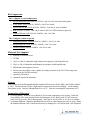
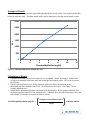
Product Manual Phosphatidylcholine Assay Kit Catalog Number STA-600 96 assays FOR RESEARCH USE ONLY Not for use in diagnostic procedures Introduction Phospholipids are important structural lipids that are the major component of cell membranes and lipid bilayers. Phospholipids contain a hydrophilic head and a hydrophobic tail which give the molecules their unique characteristics. Most phospholipids contain one diglyceride, a phosphate group, and one choline group. Phosphatidylcholine is the foremost membrane phospholipid in eukaryotic cell membranes and is present in every cell in the body. It serves as a pool for many lipid messengers and is a source for bioactive lipids such as phosphatidic acid, diacylglycerol, lysophosphatidylcholine, and others. Regulation of phosphatidylcholine biosynthesis and metabolism is important to membrane structure maintenance and function. Phosphatidylcholine is the chief source of choline in the body, which itself and its derivative compounds form cell signaling molecules such as acetylcholine, platelet activating factor and sphingophosphorylcholine. As one of the most prevalent phospholipids in lipoproteins, phosphatidylcholine exerts considerable influence on lipid homeostasis. Since phosphatidylcholine comprises about 70% of the total phospholipids in plasma lipoproteins, it is very important to lipid homeostasis. Determining circulatory levels of phospholipids and lipoproteins is critical to the diagnosis of lipid transport disorders. Fatty acid residues from phosphatidylcholine are transferred via the enzyme lecithin cholesterol acyltransferase (LCAT) to cholesterol to create cholesterol esters. High levels of cholesterol and cholesterol esters (hypercholesterolemia) have been associated with cardiovascular disease such as atherosclerosis and heart disease. In addition, phosphatidylcholine synthesis is altered in many neurological, cardiovascular, and pulmonary disease states. Cell Biolabs’ Phosphatidylcholine Assay Kit is a simple fluorometric assay that measures the amount of phosphatidylcholine present in plasma or serum, tissue homogenates, or cell suspensions in a 96-well microtiter plate format. Each kit provides sufficient reagents to perform up to 96 assays, including blanks, phosphatidylcholine standards and unknown samples. Sample phosphatidylcholine concentrations are determined by comparison with a known phosphatidylcholine standard. Assay Principle Cell Biolabs’ Phosphatidylcholine Assay Kit measures the phosphatidylcholine present within serum, plasma, or tissue samples. The assay is based on the enzyme driven reaction that will detect phosphatidylcholine via phosphatidylcholine-specific phospholipase D enzyme and choline oxidase. First, phospholipase D hydrolyzes phosphatidylcholine into choline and phosphatidic acid. Choline is then oxidized by choline oxidase to produce hydrogen peroxide. The hydrogen peroxide is then detected with a highly specific fluorescence probe. Horseradish peroxidase catalyzes the reaction between the probe and hydrogen peroxide, which bind in a 1:1 ratio. Samples are compared to a known concentration of phosphatidylcholine standard within the 96-well microtiter plate format. Samples and standards are incubated for 60 minutes and then read with a standard 96-well fluorometric plate reader (Figure 1). 2 Figure 1. Phosphatidylcholine Assay Principle Related Products 1. STA-361: Human ApoAI and ApoB Duplex ELISA Kit 2. STA-368: Human ApoB-100 ELISA Kit 3. STA-369: OxiSelect™ Human Oxidized LDL ELISA Kit (MDA-LDL Quantitation) 4. STA-384: Total Cholesterol Assay Kit (Colorimetric) 5. STA-391: HDL and LDL/VLDL Cholesterol Assay Kit 6. STA-394: HDL Cholesterol Assay Kit 7. STA-396: Serum Triglyceride Quantification Kit (Colorimetric) 8. STA-398: Free Glycerol Assay Kit (Colorimetric) 9. STA-601: Sphingomyelin Assay Kit 3 Kit Components Box 1 (shipped at room temperature) 1. 96-well Microtiter Plate (Part No. 234501): One 96-well clear bottom black plate. 2. Assay Buffer (10X) (Part No. 260002): One 25 mL bottle. 3. Fluorescence Probe (100X) (Part No. 260003): One 100 µL tube in DMSO. 4. HRP (Part No. 234402): One 100 μL tube of 100 U/mL HRP solution in glycerol. 5. Standard Diluent (10X) (Part No. 260006): One 1 mL tube. Box 2 (shipped on blue ice packs) 1. Phosphatidylcholine Standard (Part No. 260001): One 25 µL tube. 2. Phospholipase D (350X) (Part No. 260004): One 30 µL tube. 3. Choline Oxidase (Part No. 260005): One 50 µL tube. Materials Not Supplied 1. Distilled or deionized water 2. 1X PBS 3. 10 μL to 1000 μL adjustable single channel micropipettes with disposable tips 4. 50 μL to 300 μL adjustable multichannel micropipette with disposable tips 5. Multichannel micropipette reservoir 6. Fluorescence microplate reader capable of reading excitation in the 530-570 nm range and emission in the 590-600 nm range. 7. (optional) Chloroform 8. (optional) Superoxide dismutase Storage Upon receipt, store the Phosphatidylcholine Standard, Fluorescence Probe, HRP, and Choline Oxidase at -20ºC. The Fluorescence Probe is light sensitive and must be stored accordingly. Avoid multiple freeze/thaw cycles. Store the Phospholipase D at -80 ºC. Store the remaining kit components at 4ºC. Preparation of Reagents • • 1X Assay Buffer: Warm the Assay Buffer (10X) to room temperature prior to using. Dilute the Assay Buffer (10X) with deionized water by diluting the 25 mL Buffer with 225 mL deionized water for 250 mL total. Mix to homogeneity. Store the 1X Assay Buffer at 4ºC up to six months. 1X Standard Diluent: Warm the Standard Diluent (10X) to room temperature prior to using. Dilute the Standard Diluent (10X) with deionized water by diluting the 1 mL Diluent with 9 mL deionized 4 • water for 10 mL total. Vortex to homogeneity. Store the 1X Standard Diluent at 4ºC up to six months. Reaction Reagent: Prepare a Reaction Reagent by diluting the Choline Oxidase 1:200, HRP 1:500, Fluorescence Probe 1:100, and Phospholipase D 1:350 in 1X Assay Buffer. (e.g. for 50 assays, combine 25 μL of Choline Oxidase, 10 μL of HRP, 50 μL Fluorescence Probe, and 15 μL Phospholipase D with 1X Assay Buffer to 5 mL total solution). Mix thoroughly and protect the solution from light. For best results, place the Reaction Reagent on ice and use within 30 minutes of preparation. Do not store the Reaction Reagent solution. Note: The Fluorescence Probe is light sensitive and must be stored accordingly. Preparation of Samples Samples should be assayed immediately or stored at -80ºC prior to performing the assay. Optimal experimental conditions for samples must be determined by the investigator. The following recommendations are only guidelines and may be altered to optimize or complement the user’s experimental design. A set of serial dilutions is recommended for samples to achieve optimal assay results and minimize possible interfering compounds. Run proper controls as necessary. Always run a standard curve with samples. • Tissues or Cell Suspensions: Homogenize 250 mg of sample (wet tissue or cell pellet) in 4.5 mL of chloroform/methanol (2:1, v/v). Centrifuge to remove debris. After centrifugation, incubate the homogenate at room temperature for 1 hour on an orbital shaker. Induce phase separation by adding 1.25 mL dH2O. Incubate 10 minutes at room temperature and centrifuge at 1000 x g for 10 minutes. Collect the lower (chloroform) organic phase and re-extract the upper phase with 2 mL of solvent mixture whose composition is CHCl3/MeOH/water (86:14:1, v/v/v). Combine organic phases and dry in a vacuum centrifuge. Dissolve in 200 µL CHCl3/MeOH/water (60:30:4.5, v/v/v) for storage. Before phosphatidylcholine assay, samples must be diluted at least 1:50 to 1:400 with Assay Buffer. • Serum: Collect blood without using an anticoagulant. Allow blood to clot for 30 minutes at room temperature. Centrifuge at 2000 x g and 4ºC for 10 minutes. Remove the serum layer and store on ice. Take care to avoid disturbing the white buffy layer. Aliquot samples for testing and store remaining solution at -80ºC. Perform serum dilutions in 1X Assay Buffer. Serum samples must be diluted at least 1:50 to 1:400 with Assay Buffer. This will provide values within the range of the standard curve. • Plasma: Collect blood with heparin or citrate and centrifuge at 1000 x g and 4ºC for 10 minutes. Remove the plasma layer and store on ice. Take care to avoid disturbing the white buffy layer. Aliquot samples for testing and store remaining solution at -80ºC. Perform plasma dilutions in 1X Assay Buffer. Plasma samples must be diluted at least 1:50 to 1:400 with Assay Buffer. This will provide values within the range of the standard curve. Notes: 1. Samples with NADH concentrations above 10 μM and glutathione concentrations above 50 μM will oxidize the probe and could result in erroneous readings. To minimize this interference, it is recommended that superoxide dismutase (SOD) be added to the reaction at a final concentration of 40 U/mL. 5 2. Avoid samples containing DTT or β-mercaptoethanol since the fluorescence probe is not stable in the presence of thiols (above 10 μM). 3. Choline can generate high background if present in samples. If choline may be present, run a background control without Phospholipase D. Subtract this value from sample reading values. Preparation of Phosphatidylcholine Standard Curve 1. Prepare fresh phosphatidylcholine standards by first diluting a portion of the 100 mg/mL Phosphatidylcholine Standard stock solution 1:100 in 1X Standard Diluent. (eg. Add 5 µL of Phosphatidylcholine Standard in 495 µL Standard Diluent). Vortex thoroughly. This provides a 100 mg/dL concentration. Use this 100 mg/dL solution to prepare a series of the remaining phosphatidylcholine standards according to Table 1 below. Tubes 1 2 3 4 5 6 7 8 100 mg/dL Phosphatidylcholine Standard (µL) 50 250 of Tube #1 250 of Tube #2 250 of Tube #3 250 of Tube #4 250 of Tube #5 250 of Tube #6 0 1X Assay Buffer (µL) 450 250 250 250 250 250 250 500 Resulting Phosphatidylcholine Concentration (mg/dL) 10 5 2.5 1.25 0.625 0.313 0.156 0 Table 1. Preparation of Phosphatidylcholine Standards. Note: Do not store diluted phosphatidylcholine standard solutions. Assay Protocol Each phosphatidylcholine standard and sample should be assayed in duplicate or triplicate. A freshly prepared standard curve should be used each time the assay is performed. 1. Add 10 µL of the diluted phosphatidylcholine standards or samples to the 96-well microtiter plate. 2. Add 100 µL of the prepared Reaction Reagent to each well and mix the well contents thoroughly. 3. Cover the plate wells to protect the reaction from light. Incubate the plate for 60 minutes at 37ºC. 4. Read the plate with a fluorescence microplate reader equipped for excitation in the 530-570 nm range and for emission in the 590-600 nm range. 5. Calculate the concentration of phosphatidylcholine within samples by comparing the sample RFU to the phosphatidylcholine standard curve. 6 Example of Results The following figures demonstrate typical Phosphatidylcholine Assay results. One should use the data below for reference only. This data should not be used to interpret or calculate actual sample results. 3000 2500 RFU 2000 1500 1000 500 0 0 2 4 6 8 Phosphatidylcholine (mg/dL) 10 12 Figure 2: Phosphatidylcholine Standard Curve. Calculation of Results 1. Calculate the average fluorescence values for every standard, control, and sample. Subtract the average zero standard value from itself and all standard and sample values. This is the corrected fluorescence. 2. Plot the corrected fluorescence for the standards against the final concentration of the phosphatidylcholine standards from Table 1 to determine the best curve. See Figure 2 for an example standard curve. 3. Determine the phosphatidylcholine concentration of the samples with the equation obtained from the linear regression analysis of the standard curve. Substitute the corrected fluorescence values for each sample. Remember to account for dilution factors. Sample corrected fluorescence Total Phosphatidylcholine (mg/dL) = x Sample dilution Slope 7 References 1. Chapman, D., et al. (1967) Chem. Phys. Lipids 1: 445-475. 2. Hojjati, M.R., et al. (2006) J. Lipid Res. 47(3): 673-676. 3. Linsel-Nitchke, P., et al. (2005) Nature Reviews Drug Discovery 4: 193-205. 4. Ohta-Fukuyama, M. (1980) J. Biochem. 88: 197-203. 5. Phillips, G.B., et al. (1967) J. Lipid Res. 8: 676-681. Recent Product Citation Park, E. S. et al. (2014). Phosphatidylcholine alteration identified using MALDI imaging MS in HBVinfected mouse livers and virus-mediated regeneration defects. PLoS One. 9:e103955. Warranty These products are warranted to perform as described in their labeling and in Cell Biolabs literature when used in accordance with their instructions. THERE ARE NO WARRANTIES THAT EXTEND BEYOND THIS EXPRESSED WARRANTY AND CELL BIOLABS DISCLAIMS ANY IMPLIED WARRANTY OF MERCHANTABILITY OR WARRANTY OF FITNESS FOR PARTICULAR PURPOSE. CELL BIOLABS’s sole obligation and purchaser’s exclusive remedy for breach of this warranty shall be, at the option of CELL BIOLABS, to repair or replace the products. In no event shall CELL BIOLABS be liable for any proximate, incidental or consequential damages in connection with the products. Contact Information Cell Biolabs, Inc. 7758 Arjons Drive San Diego, CA 92126 Worldwide: +1 858 271-6500 USA Toll-Free: 1-888-CBL-0505 E-mail: [email protected] www.cellbiolabs.com 2012-2015: Cell Biolabs, Inc. - All rights reserved. No part of these works may be reproduced in any form without permissions in writing. 8