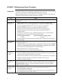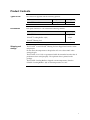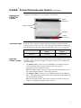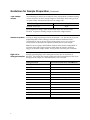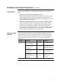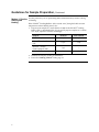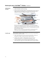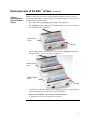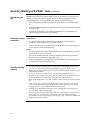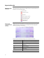
1
USER GUIDE E-PAGE™ 48 Protein Electrophoresis System For electrophoresis of 48 protein samples Catalog Number EP048-08 Publication Part Number 25-0735 Publication Number MAN0000374 Revision A.0 For Research Use Only. Not for use in diagnostic procedures. For Research Use Only. Not for use in diagnostic procedures. Information in this document is subject to change without notice. DISCLAIMER LIFE TECHNOLOGIES CORPORATION AND/OR ITS AFFILIATE(S) DISCLAIM ALL WARRANTIES WITH RESPECT TO THIS DOCUMENT, EXPRESSED OR IMPLIED, INCLUDING BUT NOT LIMITED TO THOSE OF MERCHANTABILITY, FITNESS FOR A PARTICULAR PURPOSE, OR NON-INFRINGEMENT. TO THE EXTENT ALLOWED BY LAW, IN NO EVENT SHALL LIFE TECHNOLOGIES AND/OR ITS AFFILIATE(S) BE LIABLE, WHETHER IN CONTRACT, TORT, WARRANTY, OR UNDER ANY STATUTE OR ON ANY OTHER BASIS FOR SPECIAL, INCIDENTAL, INDIRECT, PUNITIVE, MULTIPLE OR CONSEQUENTIAL DAMAGES IN CONNECTION WITH OR ARISING FROM THIS DOCUMENT, INCLUDING BUT NOT LIMITED TO THE USE THEREOF. IMPORTANT LICENSING INFORMATION These products may be covered by one or more Limited Use Label Licenses. By use of these products, you accept the terms and conditions of all applicable Limited Use Label Licenses. TRADEMARKS © 2014 Thermo Fisher Scientific Inc. All rights reserved. All trademarks are the property of Thermo Fisher Scientific and its subsidiaries unless otherwise specified. ii Contents E-PAGE™ 48 Experienced Users Procedure ............................................................................................ iv Product Contents ..........................................................................................................................................1 Introduction .............................................................................................................................. 2 E-PAGE™ Protein Electrophoresis System ................................................................................................2 Methods .................................................................................................................................... 5 Guidelines for Sample Preparation ............................................................................................................5 Electrophoresis of E-PAGE™ 48 Gels ....................................................................................................... 10 Guidelines for Loading E-PAGE™ Gels by Automated Liquid Handling .......................................... 15 Maintenance of the E-PAGE™ Device ...................................................................................................... 16 Opening the E-PAGE™ Cassette ............................................................................................................... 17 Visualizing and Staining of E-PAGE™ Gels .......................................................................18 Visualizing Lumio Fusion Proteins ....................................................................................................... 18 Coomassie Staining .................................................................................................................................... 19 Blotting of E-PAGE™ Gels..................................................................................................21 Semi-Dry Blotting of E-PAGE™ Gels ........................................................................................................ 21 Expected Results ......................................................................................................................................... 24 Using E-Editor™ 2.02 Software.................................................................................................................. 26 Troubleshooting.......................................................................................................................................... 27 Appendix .................................................................................................................................29 Product Specifications................................................................................................................................ 29 Technical Support ....................................................................................................................................... 32 iii E-PAGE™ 48 Experienced Users Procedure Introduction This quick reference sheet is included for experienced users of E-PAGE™ 48 Gels. If you are a first time user, follow the detailed protocols provided in this manual. Note: For optimal results, load each E-PAGE™ 48 Gel within 20 minutes of removing the gel from the foil pouch and run within 10 minutes of loading. Step Action Prepare Sample Use up to 20 µg protein per lane of the E-PAGE™ 48 Gel. See page 5 for sample preparation. Select Program and Load 1. 2. 3. 4. 5. 6. 7. Electrophoresis with E-Base™ 1. 2. 3. 4. 5. Plug the Mother E-Base™ into an electrical outlet. Connect the Daughter E-Base™ to a Mother E-Base™ or to another Daughter E-Base™ connected to a Mother E-Base™. Select the program EP for E-PAGE™ 48 Gels using the pwr/prg button . Manually change the run time to 23 minutes using the time button. Remove gel from the package and remove the plastic comb from the gel. Slide the gel into the two electrode connections on the Mother E-Base™ or Daughter E-Base™. Load samples into the gels using a multichannel pipetter or an automated liquid handling system. Do not exceed 15 µL of total well volume. First Load Deionized Water Then Load Sample in Loading Buffer 5–10 µL 10–5 µL Load the appropriate protein molecular weight marker (5–10 µL) in the four marker wells of the gel. Press and release the pwr/prg button located on the lower right corner of the base to begin electrophoresis. The Mother E-Base™ and Daughter E-Base™ signals the end of the run with a flashing red light and rapid beeping for 2 minutes followed by a single beep every minute. Press and release the pwr/prg button to stop the beeping. Remove the E-PAGE™ 48 cassette from the base. Open the gel cassette for staining or blotting applications. Opening the E-PAGE™ 48 Cassette Open the E-PAGE™ 48 cassette with a Gel Knife to remove the gel. 1. Insert the Gel Knife in the gap between the two plastic plates of the cassette and lever at each of the four edges to separate the two halves. 2. Pull apart the cassette halves with your hands until the cassette halves are separated. 3. Using the Gel Knife, trim the top and bottom electrode areas of the gel. 4. Proceed to staining or blotting. Staining E-PAGE™ 48 Gels Stain E-PAGE™ 48 Gels using Coomassie R-250 stain (page 19), or other staining methods (detailed information in the E-PAGE™ Technical Guide available for downloading from www.lifetechnolgies.com or by contacting Technical Support, page 32). For blotting E-PAGE™ 48 Gels, see page 21. Using E-Editor™ 2.02 Software 1. Use an appropriate digital documentation system to capture a digital image of the gel. 2. Download E-Editor™ 2.02 software and the instruction manual for free at www.lifetechnolgies.com/epage. 3. Use the E-Editor™ 2.02 software to align and arrange the lanes in the image and save the reconfigured image for further analysis. iv Product Contents Types of kits This manual is supplied with the following product: Product ™ E-PAGE 48 8% Gels Kit contents Catalog no. 8-pack EP048-08 Each pack of E-PAGE™ Gels contain the following contents: E-PAGE™ Gel 8-Pack Contents E-PAGE Gels E-PAGE Loading Buffer 1 (4X) E-PAGE Blotting Pad Shipping and storage Quantity Quantity 8 gels 4.5 mL 1 The E-PAGE™ 48 and E-PAGE Blotting Pad are shipped and stored at room temperature. Do not allow the temperature to drop below 4°C or rise above 40°C when storing the gels. The E-PAGE™ 48 8% Gels are guaranteed stable for 6 months from the date of production when stored properly. The expiration date is printed on the package. The E-PAGE Loading Buffer is shipped at room temperature. Store the E-PAGE™ Loading Buffer 1 (4X) at room temperature or at 4°C. 1 Introduction E-PAGE™ Protein Electrophoresis System About the E-PAGE™ Protein Electrophoresis System Applications E-PAGE 48 Gels The E-PAGE™ Protein Electrophoresis System is designed for fast, medium-tohigh-throughput protein electrophoresis in a horizontal format. The E-PAGE™ 48 System consists of the following components: • E-PAGE™ 48 8% Pre-cast Gels • E-Base™ Electrophoresis Device • E-PAGE™ Loading Buffer 1 (4X) • E-PAGE™ Blotting Pad • E-Editor™ 2.02 Software The E-PAGE™ 48 Protein Electrophoresis System is ideal for protein analysis and screening including: • Coomassie R-250 staining (page 19) • In-gel staining with Lumio™ Green Reagent (page 18) • Silver staining and SYPRO® Ruby staining techniques (protocols available in the E-PAGE™ Technical Guide) • Western blotting • Functional assays • IMPORTANT! E-PAGE™ 48 Gel are not compatible with the E-Gel® 96 base (Cat nos. G7100-01/G7200-01, previously available from Invitrogen). The older E-Gel® 96 bases do not have the ‘E-Base™’ inscription on the platform. • E-PAGE™ 48 Gels are self-contained, pre-cast gels that include a buffered gel matrix and electrodes packaged inside a disposable, UV-transparent cassette. • Each E-PAGE™ 48 Gel contains 48 sample lanes and 4 marker lanes. This configuration provides a 3.2-cm run length. The wells of E-PAGE™ 48 Gels are compatible for loading with a multichannel pipetter in alternating lanes or with an automated liquid handling system (see page 15 for automation specifications). • After electrophoresis, the E-PAGE™ 48 cassette is easily opened with a Gel Knife to remove the gel for staining or blotting applications. • Each E-PAGE™ 48 cassette is labeled with an individual barcode to facilitate identification of the gel using commercial barcode readers (page 10). See page 29 for E-PAGE™ 48 8% Gel specifications. 2 E-PAGE™ Protein Electrophoresis System, Continued Diagram of an E-PAGE 48 Cassette A diagram of the E-PAGE™ 48 Gel cassette is shown below. Copper electrode (–) Plastic combs Barcode Copper electrode (+) Separation range About the E-Base™ System E-PAGE™ 48 8% Gels have a unique separation profile, which gives protein resolution similar to that of a 4-12% Tris-Glycine gel. Specifically, the following table describes the separation range of the E-PAGE™ 48 8% Gels (see page 29): 10% Resolution Range 30% Lower Resolution Range 30% Upper Resolution Range 50-80 kDa 10-50 kDa 80-200 kDa E-PAGE™ 48 Gels are used with a specially designed electrophoresis device that combines a base and a power supply. Two types of devices are available from Life Technologies: • The Mother E-Base™ Device (Cat. no. EB-M03) has an electrical plug that can be connected directly to an electrical outlet and is used for electrophoresis of one E-PAGE™ 48 Gel. Note: The Mother E-Base™ Device has been tested for electrophoresis with up to three Daughter E-Bases™ connected at one time. • The Daughter E-Base™ Device (Cat. no. EB-D03) connects to the Mother EBase™, and together they can be used for the independent electrophoresis of 2 or more E-PAGE™ 48 Gels. Note: The Daughter E-Base™ Device does not have an electrical plug and cannot be used without a Mother E-Base™ Device. 3 E-PAGE™ Protein Electrophoresis System, Continued E-PAGE™ Loading Buffer E-PAGE™ Gels are supplied with E-PAGE™ Loading Buffer 1 (4X), which is optimized for E-PAGE™ Gels when performing routine SDS-PAGE and staining or blotting applications. Do not use any other SDS-PAGE sample buffer. See page 7 for more information about loading buffers. E-PAGE™ Blotting Pad E-PAGE™ Blotting Pads are supplied with the E-PAGE™ 48 Gels. It is necessary to use the pad during semi-dry blotting to ensure a good transfer. The pad is reusable as long as the pad retains porosity and liquid retaining capacity. E-Editor™ 2.02 Software The E-Editor™ 2.02 software allows you to quickly reconfigure digital images of E-PAGE Gel results for analysis and documentation. Capture an image of the gel and use the E-Editor™ 2.02 software to: • Align and arrange the lanes in the image. • Save the reconfigured image for further analysis. • Copy and paste selected lanes or the entire image into other applications for printing, saving, emailing, and/or publishing. The E-Editor™ 2.02 software can be downloaded FREE at www.lifetechnologies/epage. Follow the instructions to download the software and user manual. 4 Methods Guidelines for Sample Preparation Introduction For optimal results using the E-PAGE™ System, follow the guidelines for preparing your protein samples as described in this section. Prepare your protein samples as described in this section for electrophoresis on E-PAGE™ 48 Gels. We recommend that you read this section before preparing your samples. Use the sample preparation method and loading buffer appropriate for your detection method: Materials needed Application Method Loading Buffer Routine staining and western blotting 1 (page 8) Loading Buffer 1 Lumio Green Detection 2 (page 9) Lumio™ Gel Sample Buffer The following items are needed for sample preparation. See page 31 for ordering information. • Protein sample • NuPAGE® Sample Reducing Agent (10X) • E-PAGE™ Loading Buffer 1 (4X) included in the kit • Deionized water • Heating block set at 70°C • Molecular weight markers (page 7) • (Optional) Lumio™ Green Detection Kit for detection of Lumio™ fusion proteins (page 9) E-PAGE™ Gels contain SDS and are designed for performing electrophoresis under denaturing conditions. To obtain the best results, perform SDS-PAGE under reducing conditions. If you need to perform SDS-PAGE under non-reducing conditions, do not add NuPAGE® Sample Reducing Agent (10X) during sample preparation. 5 Guidelines for Sample Preparation, Continued Total sample volume Avoid loading less than 5 µL of sample in wells, and maintain a uniform loading volume. If you do not have enough samples to load all the wells of the gel, load an equal volume of deionized water into any empty wells. E-PAGE™ Gel Type Recommended Loading Volume ™ E-PAGE 48 8% Gel 20 µL We recommend loading 5–10 µL of deionized water into all wells of the E-PAGE™ Gel prior to loading samples or molecular weight markers. Amount of protein Load up to 20 µg of protein per well of the E-PAGE™ Gel. The amount of protein required depends on the staining or western detection method used for visualization. If you are unsure of how much protein to use, test a range of concentrations to determine the optimal concentration for your sample. Note: To ensure a proper LDS (lithium dodecyl sulfate from Loading Buffer 1) to protein ratio, limit sample protein or lipid (from the sample) amount to 2 µg/µL of the final sample volume. Excess protein will cause poor resolution. Samples containing high salt or detergents result in loss of resolution on High salt or detergent samples E-PAGE™ Gels. Dilute the samples such that the final concentration of the salt or detergent in the sample is as described below. NT= not tested. 6 Detergent or Salt Final Concentration for E-PAGE 48Gels Triton® X-100 <0.3% ® Tween 20 <0.3% CHAPS <0.3% NP-40 <0.3% RIPA <0.25X SDS <2% (already in loading buffer) Tris <300 mM NaCl <300 mM Ammonium sulfate <100 mM Sodium acetate <200 mM EDTA <20 mM MES Not recommended DTT, Glycine, Urea, Imidazole No effect seen up to 500 mM Guidelines for Sample Preparation, Continued Loading buffer Based on your application, use the appropriate loading buffer as described below: • SDS-PAGE and routine staining (Method 1, page 8) For SDS-PAGE and routine staining or blotting, use the E-PAGE™ Loading Buffer 1 (4X) included in the kit for preparing samples. The E-PAGE™ Loading Buffer 1 (4X) is optimized for E-PAGE™ Gels. Do not use any other types of SDS-PAGE sample buffer. • SDS-PAGE and detection of Lumio™ Fusion Proteins (Method 2, page 9) For in-gel detection of Lumio™ fusion proteins with the Lumio™ Green Detection Kit, use the Lumio™ Gel Sample Buffer (4X) included with the Lumio™ Green Detection Kit. This buffer is specifically formulated to provide optimal results with the Lumio™ Green Detection Reagent. Do not use the E-PAGE™ Loading Buffer 1 (4X) or any other type of SDS-PAGE buffer to prepare samples for Lumio™ Green detection. Molecular weight standards The following protein molecular weight standards and loading volumes are recommended for E-PAGE™ Gels. Because molecular weight standards have different migration patterns in different gel systems, see page 24 for apparent molecular weight calibration on E-PAGE™ Gels. See page 31 for ordering information. Gel Type Standard E-PAGE 48 8% Gel SeeBlue Plus2 Prestained Standard 5 µL Electrophoresis MagicMark™ XP Western Protein Standard 10 µL Electrophoresis, followed by staining MagicMark™ XP Western Protein Standard 5 µL Western blotting BenchMark™ Fluorescent Protein Standard 5 µL Fluorescent Detection ™ ® Amount Application 7 Guidelines for Sample Preparation, Continued Method 1: Routine Use this protocol if you are performing SDS-PAGE followed by routine staining or blotting. staining and blotting If the E-PAGE™ Loading Buffer 1 (4X) is stored at 4°C, bring the buffer to room temperature and mix briefly prior to use. 1. Prepare your samples in a total volume of 10 µL in the E-PAGE™ Loading Buffer 1 (4X) as described below. If you need to prepare samples in a volume of 5–15 µL, adjust the volumes accordingly. Reagent Protein Sample E-PAGE Loading Buffer 1 (4X) ™ NuPAGE® Sample Reducing Agent (10X) Deionized Water 8 Reduced Non-reduced x µL x µL 2.5 µL 2.5 µL 1 µL — to 10 µL to 10 µL 2. Incubate the samples at 70°C for 10 minutes. 3. Proceed to Loading E-PAGE™ Gels, page 12. Guidelines for Sample Preparation, Continued Method 2: Lumio™ detection A brief protocol to prepare samples for specific detection of Lumio™ fusion proteins using the Lumio™ Green Detection Kit is described below. For details on the Lumio™ Green Detection Kit, refer to the manual available at www.lifetechnologies.com or contact Technical Support (page 32). 1. Refer to the Lumio Detection manual for details on each type of protein. Prepare protein samples as follows: Protein Sample Sample Volume Lumio™ Gel Sample Buffer (4X) Volume Bacterial samples 7.5 µL 2.5 µL Mammalianlysate 7.5 µL 2.5 µL Partially purified sample 7.5 µL 2.5 µL Purified sample 7.5 µL 2.5 µL In vitro expressed 10 µL Not needed* ™ *There is no need to add Lumio Gel Sample Buffer (4X), as the sample is already prepared in this buffer. 2. Thaw the Lumio™ Green Detection Reagent and mix well. 3. Add 0.1 µL Lumio™ Green Detection Reagent to the protein samples from Step 1 in a fume hood. Mix well. Return the Lumio™ Green Detection Reagent to –20°C immediately after use. 4. Incubate the samples at 70°C for 10 minutes. 5. Allow samples to cool for 1–2 minutes and centrifuge briefly at maximum speed in a microcentrifuge. 6. Thaw the Lumio™ In-Gel Detection Enhancer and mix well. Add 1 µL of Lumio™ In-Gel Detection Enhancer to the samples. 7. Mix well and incubate the samples at room temperature for 5 minutes. Return the Lumio™ In-Gel Detection Enhancer to –20°C immediately after use. 8. Proceed to Loading E-PAGE Gels, page 10. When performing electrophoresis of Lumio™ fusion proteins, extending the run time of the gel for an additional 2 minutes can prevent the formation of a fluorescent dye front. 9 Electrophoresis of E-PAGE™ 48 Gels Introduction After preparing your samples, you are ready to load E-PAGE™ Gels. This section describes the procedure for loading protein samples and molecular weight standards. The Mother E-Base™ and Daughter E-Base™ Electrophoresis Devices are designed to fit most robotic platforms allowing you to load and run E-PAGE™ Gels directly on the automated liquid handling system. If you are using an automated liquid handling device, it is important to align the robotic tip loading assembly to the proper setting prior to loading samples on the E-PAGE™ 48 Gel. This ensures proper loading of samples into the wells. See page 15 for automation guidelines. If you need to load multiple gels on a robotic platform while other gels are running on the E-Base™ Electrophoresis Devices, use an E-Holder™ Platform (page 15). General guidelines Using the barcode • Perform manual loading with a pipette or multichannel pipetter (load samples into alternate wells of the gel followed by a second round of loading into the remaining wells). • Use short, rigid tips for loading E-PAGE™ Gels. • Always load 5–10 µL deionized water first into all wells prior to loading sample or molecular weight standard. • E-PAGE™ Gels can only be used once. Do not re-use. • To obtain the best results, run the E-PAGE™ Gel immediately after removal from the pouch and loading. • Store and run E-PAGE™ Gels at room temperature. • For optimal results, do not run reduced and non-reduced samples on the same gel. If you do choose to run these samples on the same gel, avoid running reduced and non-reduced samples in adjacent lanes as the reducing agent may have a carry-over effect on the non-reduced samples. • Avoid running samples containing different salt or protein concentrations in adjacent lanes. • Avoid touching the gel during electrophoresis. Each E-PAGE™ 48 Gel cassette is labeled with an individual barcode. The barcode facilitates the identification of each gel cassette during electrophoresis of multiple gels. Each E-PAGE™ cassette contains an EAN 39 type of barcode, which is recognized by the majority of commercially available barcode readers. Refer to the manufacturer’s instructions to set up the barcode reader. Note: When capturing an image of an E-PAGE™ Gel, note that the barcode label is easily overexposed. To ensure that the barcode label is distinct and readable in the image, experiment with different shutter settings for your particular camera. 10 Electrophoresis of E-PAGE™ 48 Gels, Continued Select a program on the E-Base™ Device Use program EP for running E-PAGE™ 48 Gels. Select the program prior to inserting a gel into the base. Brief instructions for using the E-Base™ Device are included in this manual. For details, refer to the E-Base™ manual. 1. Plug Mother E-Base™ Device into an electrical outlet using the electrical plug on the base. Connect a Daughter E-Base™ Device to a Mother E-Base™ Device or another Daughter E-Base™ Device connected to a Mother E-Base™ Device. 2. The display shows EP or last program used (EG or EP). 3. Press and release the pwr/prg button to select program EP. 4. Press and release the time button to view the time setting. The default run time for program EP is 14 minutes. 5. Press and hold the time button to increase the time to 25 minutes. If the time button is not released, the time setting increases until it reaches 00. To begin cycling through the numbers again, starting from 00, press the time button again. Do not run E-PAGE 48 Gels for more than 30 minutes. • When performing electrophoresis of Lumio™ fusion proteins, extend the run time of the gel for an additional 2 minutes to prevent the formation of a fluorescent dye front. • If your sample contains high salt or detergent concentrations, you may need to manually increase the run time. • To increase the run time when a cassette is inserted, press and release the time button to increase the time setting by 1-minute intervals or press and hold the time button to increase the time continuously. • To increase the run time while a run is in progress, see next page. To manually interrupt or stop a run, see page 14. 11 Electrophoresis of E-PAGE™ 48 Gels, Continued Insert the gel cassette 1. Open the package and remove the gel. 2. Remove the plastic combs from the gel. 3. Slide the gel into the Mother E-Base™ or Daughter E-Base™ Device. The two copper electrodes on the right side of the gel cassette must be in contact with the two electrode connections on the base, as shown below. Cathode (–) Electrode Anode (+) Electrode Digital display Time Pwr/prg When the gel is properly inserted into the base, a fan in the base begins running, and a red light illuminates at the lower-left corner of the base. The digital display shows the appropriate time for a selected program or the last time setting (Ready Mode). Note: If a cassette is inserted into the base before selecting program EP, remove the cassette, select program EP, and reinsert the cassette. Load the gel 12 There should be a uniform volume of liquid in each well. The recommended total volume for each well is 20 µL. 1. Load 5–10 µL deionized water to each well of the E-PAGE™ Gel prior to loading your samples or protein molecular weight standard. 2. Load 10–5 µL of samples in loading buffer into the wells using a multichannel pipetter or an automated liquid handling system. 3. Load the appropriate protein molecular weight standards in the marker wells of the gel. See page 7 for recommended molecular weight standards. Electrophoresis of E-PAGE™ 48 Gels, Continued Perform electrophoresis with the E-Base™ Device Note: It is not necessary to have a gel in the Mother E-Base™ Device if you are using a Daughter E-Base™ Device. However, the Mother E-Base™ Device must be plugged into an electrical outlet. 1. Press and release the pwr/prg button to begin electrophoresis. The red light changes to green and the digital display shows the count down time while the run is in progress. Mother E-Base™ Device Pwr/prg On a Daughter E-Base™ Device, press and release the pwr/prg button on the Daughter E-Base™ Device to begin electrophoresis. Mother E-Base™ Device Daughter E-Base™ Device Pwr/prg To increase the run time while the run is in progress, press the time button to select the desired time and then release the time button. Do not run an E-PAGE™ 48 Gel for more than 30 minutes. To interrupt or stop a run in progress, see page 14. 13 Electrophoresis of E-PAGE™ 48 Gels, Continued Perform electrophoresis with the E-Base™ Device, continued 2. The Mother E-Base™ or Daughter E-Base™ Device signals the end of the run with a flashing red light and an audible alarm that beeps rapidly for 2 minutes, followed by a single beep every minute. The digital display shows the original time setting (it does not indicate time changes that were made during electrophoresis). The digital display also shows the elapsed time since the end of the run (up to 19 minutes). 3. Press and release the pwr/prg button to stop the beeping. The light turns to a steady red and the digital display shows the last time setting. 4. Remove the gel cassette from the Mother E-Base™ or Daughter E-Base™ Device. 5. For visualizing Lumio™ fusion proteins, see page 18. For opening the E-PAGE™ cassette, see page 17. Note: The bands in the gel will diffuse within 40 minutes. Interrupting electrophoresis You can interrupt an electrophoresis run at any time by pressing and releasing the pwr/prg button to stop the current. The stopped current is indicated by a steady red light and the digital display will flash to indicate that the run was interrupted. You can remove the gel from the E-Base™ Device to check the progress of the run. Then: • To continue the run from the point at which it was stopped, reinsert the gel and press and release the pwr/prg button. The light changes to steady green and the digital display shows the count down time. • To cancel the rest of the interrupted run, press and hold the pwr/prg button for a few seconds. The digital display will reset and the base will return to Ready Mode. If desired, you can then program a new run time as described on page 11 and rerun the gel. In case of an external power failure (loss of electricity or the electrical cord is accidentally removed from the outlet), the run will continue when the power resumes. The Mother E-Base™ or Daughter E-Base™ Device signals the end of the run as described on the previous page, except the light will be an alternating red/green to indicate that an external power failure had occurred during the run. 14 Guidelines for Loading E-PAGE™ Gels by Automated Liquid Handling Automated loading of E-PAGE™ 48 Gels E-PAGE 48 Gels are compatible with any automated liquid handling system that has an 8-span loading head with either fixed (9-mm) or variable distance between the loading heads. These loading patterns are described below. To download programming scripts for your automated liquid handling system, go to www.lifetechnologies.com/epage. For automated loading of E-PAGE™ 48 Gels, position the plate in the “Portrait” orientation rather than the “Landscape” position, as shown below: “Portrait” “Landscape” This orientation is available for some automation systems with the use of a 90° adapter. If your system does not have an adapter that allows the “Portrait” configuration, please contact Technical Support (page 32) to obtain a 90° adapter. Loading patterns for E-PAGE 48 Gels Loading E-PAGE 48 Gels requires one of the following loading patterns, depending on your machine: Fixed Tip (4 movements per row: 8+8+4+4) Variable Tip (3 movements per row: 8+8+8) Continued on next page 15 Maintenance of the E-PAGE™ Device Maintaining the E-Base™ Device Keep the surfaces of the Mother E-Base™ Device and Daughter E-Base™ Device free of contaminants. To clean, disconnect bases from power source and wipe with a dry cloth. Do not attempt to open or service the bases. To honor the warranty, bases should only be opened and serviced by Life Technologies. Disconnect the Mother E-Base™Device from the outlet when not in use for a prolonged period of time. 16 Opening the E-PAGE™ Cassette Opening the E-PAGE™ 48 cassette with the Gel Knife To remove the E-PAGE™ 48 Gel from the cassette for blotting or staining, use the Gel Knife to open the cassette. 1. Separate each of the bonded sides of the E-PAGE™ 48 cassette by inserting the Gel Knife into the gap between the two plastic plates that make up the cassette. The side of the cassette with the wells should face up. 2. Lever the handle of the knife gently to separate the plates. Repeat on each side of the cassette until the plates are completely separated. Caution: Use caution while inserting the Gel Knife between the two plates to avoid excessive pressure on the gel. 3. Gently pull apart the cassette halves with your hands until the cassette halves are completely separated and the gel is exposed. 4. Carefully remove the gel from the cassette. 5. Use the Gel Knife to trim the top and bottom electrode areas of the gel. 6. Proceed to blotting (page 21) or staining (pages 18). Note: Small pieces of gel material may remain in the wells of an E-PAGE™ Gel after removal of the gel from the cassette. To obtain the best staining or blotting results, remove any small pieces of gel material in the wells of the gel by gently rubbing a gloved hand over the well side of the gel. 17 Visualizing and Staining of E-PAGE™ Gels Visualizing Lumio Fusion Proteins Introduction The steps involved in detecting Lumio™ fusion proteins run on an E-PAGE™ Gel are described below. To visualize Lumio™ fusion protein bands after electrophoresis, you will need a UV transilluminator or a laser-based scanner (see below). For further details on imaging Lumio™ fusion proteins, refer to the product manual available at www.lifetechnologies.com or contact Technical Support (page 32) Visualizing Lumio™ fusion proteins After electrophoresis is complete, immediately visualize and image the gel as described below. There is no need to remove the E-PAGE™ Gel from the cassette to visualize Lumio™ fusion proteins. 1. Place the gel cassette on a UV transilluminator equipped with a camera and select the ethidium bromide or SYBR® Green filter on the camera. You may also use a laser-based scanner with a laser line that falls within the excitation maxima of the stain (500 nm), and a 535 nm long pass filter or a band pass filter centered near the emission maxima of 535 nm. Note: Adjust the settings on the camera prior to turning on the UV transilluminator. Avoid exposing the gel to UV light for long periods of time. 2. Image the gel with a suitable camera with the appropriate filters using a 4– 10 second exposure. You may need to adjust the brightness and contrast to reduce any faint non-specific bands. You should see fluorescent bands of Lumio™ fusion proteins and the gel should have minimal background, as shown on page 25. 18 Coomassie Staining Introduction Instructions for staining E-PAGE™ Gels using Coomassie R-250 are described in this section. To obtain maximum sensitivity, the total staining time for Coomassie R-250 staining is 1.5 h plus overnight destaining. The use of the Coomassie R-250 microwave protocol reduces the amount of time needed for staining and destaining, however for minimal background, overnight destaining is recommended. Materials needed You will need the following items for staining one E-PAGE™ Gel: • Clean staining containers or incubation trays (if using the Coomassie R-250 microwave protocol, make sure the container is microwave safe. Do NOT use the Incubation Tray cat no. LC2102) • Rotary shaker For Coomassie R-250 Staining • Coomassie R-250 Stain (see note below) • Destaining solution (8% acetic acid in deionized water, see note below) • Methanol (regular protocol only) • Ethanol (microwave protocol only) • 2 pieces of nylon membrane (microwave protocol only) The volume of fixing, staining and destaining solutions will depend on the volume of your staining container. To obtain good results, the volume of solution must be sufficient to cover the gel completely and to allow the gel to move freely during all of the steps. When using the Coomassie R-250 microwave staining protocol, warm the Coomassie R-250 stain and destaining solution to 50°C without boiling. It is important NOT to boil the solutions. Since microwave ovens differ significantly, we recommend testing various times (10 second intervals) and power settings of your microwave oven to achieve a temperature of 50°C in the volume of solution required for your particular staining container. Perform these steps without the gel. Once you have optimized the time and settings for your microwave, use these settings for staining. 19 Coomassie Staining, Continued The staining solutions for the Coomassie R-250 staining protocol and the microwave staining protocol are different. Be sure to use the correct stain for the correct protocol. Coomassie R-250 staining protocol Coomassie R-250 staining microwave protocol For all staining and destaining steps described below, be sure to use sufficient reagents to completely cover the gel using a suitable container such that the gel moves freely during the staining and destaining steps. 1. After electrophoresis, remove the gel from the cassette (page 17) and place the gel in a clean incubation tray. 2. Prepare Coomassie stain (0.03% Coomassie R-250 in 30% methanol and 10% acetic acid). See note on previous page. 3. Stain the gel in the prepared stain for 1.5 hours at room temperature with gentle shaking. 4. Destain the gel in destaining solution (see previous page) at room temperature with gentle shaking with intermittent changes of solution until the bands are visible or overnight for maximum sensitivity and clear background. For all staining and destaining steps described below, be sure to use sufficient reagents to completely cover the gel in a microwave safe container such that the gel moves freely during the staining and destaining steps. 1. After electrophoresis, remove the gel from the cassette (page 17) and place the gel in a clean microwave safe container. 2. Prepare Coomassie stain (0.015% Coomassie R-250 in 30% ethanol and 10% acetic acid). See note on previous page. 3. Add enough stain to completely cover the gel in the microwave safe container. 4. Warm the gel and solution to about 50°C in a microwave oven (see page 19). Note: Do NOT boil the solution. 5. Incubate the gel in the warmed staining solution for 30 minutes on an orbital shaker at room temperature. 6. Discard the stain, rinse the gel briefly with water and discard the water. 7. Add enough destaining solution (see previous page) to cover the gel during incubation 8. Place two pieces of positively charged nylon membrane on top of the destaining solution to speed up the destaining process. 9. Warm the destaining solution, gel and nylon membrane to 50°C in a microwave oven (see previous page). Note: Do NOT boil the solution. 10. Incubate the gel in the warm destaining solution on an orbital shaker at room temperature until the desired background is achieved. Note: To obtain a clear background, perform destaining overnight. Results obtained with Coomassie stain are shown on page 24. 20 Blotting of E-PAGE™ Gels Semi-Dry Blotting of E-PAGE™ Gels Introduction A semi-dry blotting procedure for blotting E-PAGE™ 48 Gels is described in this section. You will need a semi-dry apparatus that can accommodate the dimensions of an E-PAGE™ Gel. Additional blotting protocols Guidelines for semi-wet and other blotting methods can be found in the E-PAGE™ Technical Guide available at www.invitrogen.com or contact Technical Support (page 32). Materials needed For ordering information, see page 31. • Semi-dry blotter • Methanol • NuPAGE® Transfer Buffer (20X) • NuPAGE® Antioxidant • Blotting membranes: Invitrolon™/Filter Paper Sandwich or Nitrocellulose/Filter Paper Sandwiches • E-PAGE™ Blotting Pad (supplied with E-PAGE™ Gels or available separately) • • • 4 pieces of 2.5 mm Blotting Filter Paper Blotting Roller Incubation Tray IMPORTANT! To ensure a good transfer, it is necessary to use the E-PAGE™ Blotting Pad and 2.5 mm Blotting Filter Paper. Preparing 2X transfer buffer We recommend using 2X NuPAGE® Transfer Buffer with 15% methanol and NuPAGE® Antioxidant for optimal transfer of most proteins from E-PAGE™ Gels. When the complete transfer of higher molecular weight proteins (>150 kDa) is desired, reduce methanol to 10%. For one gel, prepare 500 mL of 2X NuPAGE® Transfer Buffer: 2X NuPAGE® Transfer Buffer with 15% Methanol 2X NuPAGE® Transfer Buffer with 10% Methanol NuPAGE® Transfer Buffer (20X) 50 mL 50 mL NuPAGE® Antioxidant 0.5 mL 0.5 mL Methanol 75 mL 50 mL to 500 mL to 500 mL Buffer Component Deionized Water 21 Semi-Dry Blotting of E-PAGE™ Gels, Continued Equilibrating the gel Preparing blotting membrane Equilibration of the gel in transfer buffer results in the removal of salts that may increase conductivity and heat during transfer. Perform equilibration for the recommended time, as longer equilibration results in protein diffusion. 1. After electrophoresis, remove the gel from the cassette as described on page 17. 2. Using the Butterfly Opener or a gel knife, trim off the top and bottom electrode areas of the gel. 3. Equilibrate the E-PAGE™ Gel in 200 mL 2X NuPAGE® Transfer Buffer (see previous page) for 30 minutes with shaking. Nitrocellulose 1. Use pre-cut Nitrocellulose/Filter Paper Sandwich or cut nitrocellulose membrane to the appropriate size (8.6 cm x 13.5 cm). 2. Soak the membrane in a 2X NuPAGE® Transfer Buffer (see previous page) for several minutes in the Incubation Tray. PVDF Semi-Dry blotting protocol 1. Use pre-cut Invitrolon™/Filter Paper Sandwich or cut PVDF membrane to the appropriate size (8.6 cm x 13.5 cm). 2. Pre-wet the membrane for 30 seconds in methanol, ethanol, or isopropanol. Briefly rinse the membrane in deionized water. 3. Soak the membrane in a 2X NuPAGE® Transfer Buffer (see previous page) for several minutes in the Incubation Tray. 1. In a clean container or Incubation Tray, soak 4 pieces of 2.5 mm Blotting Filter Paper (8.6 cm x 13.5 cm) in 2X NuPAGE® Transfer Buffer (see page 21). Remove any air bubbles trapped between filter paper sheets using the Blotting Roller while the paper is still submerged in buffer. 2. In a clean container or Incubation Tray, soak the E-PAGE™ Blotting Pad in 2X NuPAGE® Transfer Buffer (see page 21). Press the pad to ensure the elimination of any visible air bubbles. Inspect both sides of the pad for air bubbles before use. Note: The Blotting Pad has no specific orientation (either side may be facing toward the gel). 22 3. Place 2 pieces of pre-soaked 2.5 mm Blotting Filter Paper from Step 1 on the anode plate of a semi-dry blotting apparatus. Ensure that all filter paper sheets are aligned properly and remove any air bubbles with the Blotting Roller. 4. Place the pre-soaked blotting membrane on top of the filter paper stack and remove any air bubbles with the Blotting Roller. 5. Remove the gel from the transfer buffer. Gently rub a gloved finger over the well side of the gel to remove small gel pieces from the gel surface. Re-submerge the gel in transfer buffer to remove any gel pieces from the gel, as they can cause air bubbles and field distortion during transfer. Semi-Dry Blotting of E-PAGE™ Gels, Continued Semi-Dry blotting protocol, continued 6. Place the flat side of the gel on top of the blotting membrane such that the well side of the gel is facing up and remove any air bubbles with the Blotting Roller. Fill the wells of the gel with 2X NuPAGE® Transfer Buffer (see page 21). 7. Place the pre-soaked E-PAGE™ Blotting Pad on the gel and gently roll out air bubbles using the Blotting Roller. 8. Place 2 pieces of 2.5 mm Blotting Filter Paper from Step 1 on top of the Blotting Pad. Ensure that all filter paper sheets are aligned properly and flush with the gel/membrane sandwich. Remove any air bubbles with the Blotting Roller. 9. Place the cathode plate on the stack without disturbing the blot sandwich. Follow the manufacturer’s instructions to further assemble the semi-dry blotting apparatus. 10. Transfer at 25 V for 1 h (~19 V/cm). You may need to optimize the transfer conditions for your specific proteins or semi-dry blot apparatus. 23 Expected Results Molecular weight calibration Apparent molecular weight values for SeeBlue® Plus2 Pre-stained Standard on an E-PAGE™ 48 8% Gel are shown below: Myosin (215 kDa) Phosphorylase (146 kDa) BSA (78 kDa) Glutamic Dehydrogenase (56 kDa) Alcohol Dehydrogenase (39 kDa) Carbonic Anhydrase (25 kDa) Myoglobin Red (19 kDa) Lysozyme (16 kDa) Results with Coomassie R-250 staining Samples were run on an E-PAGE™ 48 8% Gel and stained with Coomassie R-250 using the microwave staining protocol as described in this manual (page 20). Results after staining are shown below. M 1 2 3 4 5 6 7 8 9 10 11 12 13 14 15 16 17 18 19 20 21 22 23 24 M M 25 26 27 28 29 30 31 32 33 34 35 36 37 38 39 40 41 42 43 44 45 46 47 48 M The gel contains the following samples (rows not indicated are blank): 24 Lane Sample M SeeBlue® Plus2 Pre-stained Standard (5 µL) 2, 4, 21, 23, 26, 28, 45, 47 MagicMark™ XP Western Protein Standard (10 µL) 6, 19, 30, 45 Benchmark™ His-tagged Protein Standard (10 µL, diluted 1:5) 8, 32 Lysozyme (200 ng) 10, 34 Carbonic Anhydrase (200 ng) 12, 36 BSA (100 ng) 15, 17, 39, 41 E. coli lysate (5 µL) Expected Results, Continued Lumio™ Green detection Results obtained with an E-PAGE™ 48 8% Gel using the Lumio™ Green Detection Kit are shown below. Various concentrations of a Lumio™ fusion protein were labeled with Lumio™ Green Detection Kit and run on an E-PAGE™ 48 8% Gel as described in this manual. The gel contains the following samples (lanes not indicated are blank): Lane Sample M BenchMark Fluorescent Marker (5 µL) 2, 8, 13, 19, 26, 32, 37, 43 Human kinase Lumio™ fusion protein (10 µL) 3, 9, 14, 20, 27, 33, 38, 44 E. coli CAT Lumio™ fusion protein (10 µL) 4, 10, 15, 21, 28, 34, 39, 45 E. coli GUS Lumio™ fusion protein (10 µL) 5, 11, 16, 22, 29, 35, 40, 46 E. coli calmodulin Lumio™ fusion protein (10 µL) 6, 12, 17, 23, 30, 36, 41, 47 E. coli kinase D Lumio™ fusion protein (10 µL) 25 Using E-Editor™ 2.02 Software Introduction The E-Editor™ 2.02 software for Windows® allows you to reconfigure digital images of E-PAGE Gels for analysis and documentation. E-Editor™ 2.02 software reconfigures the wells of the E-PAGE Gels into a side-by-side format for easy comparison and analysis. You can reconfigure gels that were scanned in the original gel cassette, or gels that were removed from the cassette. You can also group the images of multiple gels loaded from a 384-well microtiter plate into a single image with a layout corresponding to that of the original plate. Capture an image of the gel as described below and then use the E-Editor™ 2.02 software to: • Align and arrange the lanes in the image • Save the reconfigured image for further analysis • Copy and paste selected lanes or the entire reconfigured image into other applications for printing, saving, emailing, and/or publishing Imaging the gel Use an appropriate gel documentation system to capture a digital image of the gel. When imaging, the gel should be properly aligned (i.e., not at an angle) and gel features should be clear and distinct. Proceed to Downloading Software. Downloading software E-Editor™ 2.02 software can be downloaded FREE from our website. Go to www.lifetechnologies.com/epage and follow the instructions to download the software and user manual. 26 Troubleshooting Troubleshooting Observation The table below provides some solutions to possible problems you might encounter during the electrophoresis of E-PAGE™ Gels. Cause ™ Solution No current Daughter E-Base Device used without a Mother E-Base™ Device Do not use the Daughter E-Base™ Device without a Mother E-Base™ Device. The Daughter E-Base™ Device does not have an electrical plug to connect to an electrical outlet. No electric contact (no red light when cassette is inserted) or run does not start (no green light) Copper contacts in the base are damaged due to improper use Make sure that the copper contacts in the base are intact. Expired or defective gel cassette used Use properly stored gels before the specified expiration date. E-PAGE™ cassette is not correctly inserted into base Remove cassette and reinsert. When the cassette is correctly inserted and power is on, a fan in the base begins to run and a steady red light illuminates on the base (page 13). Sample leaking from the wells Sample is overloaded or wells are damaged Be sure to load the recommended volume of sample per well (page 12). Remove the comb carefully without damaging the wells. Poor resolution or smearing of bands Sample is overloaded Do not load more than 20 µg of protein sample per well. Very low volumes of sample were loaded Do not load less than 5 µL of sample. Always load 10–20 µL deionized water in all wells prior to sample loading. For proper band separation, we recommend keeping sample volumes uniform and loading deionized water into empty wells. Incorrect loading buffer used Make sure that protein sample is in one of Use the recommended loading buffers as described on page 7. Electrophoresis was not started immediately after sample loading For best results, the gel should be run within 15 minutes of sample loading. Continued on next page 27 Troubleshooting, Continued Observation Poor resolution or smearing of bands Cause Solution High salt or detergent concentration in samples Be sure the final concentration of salt or detergent in the sample is as described on page 6. You may need to manually increase the run time for high salt or detergent samples to obtain optimal results. Expired gel used Use properly stored gels before the specified expiration date. Over-run the gel or need more time to run gel Accidentally selected an incorrect program Select program EP for E-PAGE™ Gels. If you accidentally selected an incorrect program and are at the beginning of the run, stop the run and select the desired program. If you are well into the run, check the gel to see where the loading dye is running. Estimate the amount of time remaining and then manually stop the run. Protein bands distorted on membrane after semidry blotting Non-uniform electric field created around wells Be sure that the E-PAGE™ Blotting Pad is used correctly. Incorrect gel orientation Be sure that the well side of the gel is not facing the membrane. Weak transfer of high molecular weight samples during semidry blotting Not enough SDS in sample Reduce methanol concentration in transfer buffer from 15% to 10% if transferring E-PAGE 48. Weak transfer of low molecular weight samples Use of large pore membranes allow small proteins to “blow through” Use 0.2 μm nitrocellulose membrane for optimal capture of small proteins. Uneven transfer of proteins and edge lanes during semi-dry blotting of E-PAGE 48 Gel No methanol in transfer buffer Use 10–15% methanol in the transfer buffer. Weak transfer of proteins during semiwet blotting No methanol in transfer buffer Use 10% methanol in the transfer buffer. 28 Appendix Product Specifications E-PAGE™ gel separation range The migration and resolution range of proteins run on E-PAGE 48 8% Gel. E-PAGE™ 48 Gel specifications Each E-PAGE™ 48 gel contains 48 sample wells and 4 marker wells (M). Cassette Size: 13.5 cm (l) × 10.8 cm (w) × 0.67 cm (thick) Gel Thickness: 3.7 mm Gel Volume: 50 mL Gel Formulation: Proprietary, operating at a neutral pH Well Depth: 3 mm Well Volume: 15 µL Well Opening: 3.6 mm (l) x 2.2 mm (w) Running Distance: (one well to the next) 3.2 cm Space between Wells: 4.5 mm Note: E-PAGE 48 8% Gels have a unique separation profile, which gives protein resolution similar to that of a 4–12% Tris-Glycine gel. 29 Accessory Products E-PAGE™ Gels The following E-PAGE™ Gel kits are available from Life Technologies. Product Quantity Catalog no. E-PAGE 48 8% Gels 8-pack EP048-08 E-PAGE™ 96 6% Gels 8-pack EP096-06 ™ Electrophoresis Bases The following electrophoresis bases are available from Life Technologies for running E-PAGE™ gels. • The Mother E-Base™ Device (Cat. no. EB-M03) is used for electrophoresis of one E-PAGE™ Gel. • The Daughter E-Base™ Device (Cat. no. EB-D03) attaches to the Mother E-Base™ Device and together are used for the independent electrophoresis of two or more E-PAGE™ Gels. E-Holder™ The E-Holder™ Platform is used to hold an E-PAGE™ Gel in place while loading. Ordering information can be found on the following page. E-Editor™ 2.02 Software The E-Editor™ 2.02 software is available FREE with the purchase of any E-PAGE™ Gels or related equipment. The software may be downloaded from www.lifetechnologies.com/epage. iBlot® Gel Transfer Device The iBlot® Gel Transfer Device is available from Life Technologies for transfer of proteins from E-PAGE™ gels to nitrocellulose or PVDF membranes. Product Quantity Catalog no. iBlot Gel Transfer Device 1 unit IB1001, IB1001UK, IB1001EU iBlot® Gel Transfer Stack, Nitrocellulose, Regular 10-pack IB3010-01 10-pack IB4010-01 ® ® iBlot Gel Transfer Stack, PVDF, Regular Continued on next page 30 Accessory Products, Continued The following products for use with E-PAGE™ gels are available separately from Life Technologies: Additional products Product ® SeeBlue Plus2 Pre-Stained Standard ™ ® E-PAGE SeeBlue Pre-stained Protein Standard ™ MagicMark XP Western Standard ™ ™ E-PAGE MagicMark Unstained Protein Standard ™ BenchMark Fluorescent Protein Standard ™ Lumio Green Detection Kit ® SYPRO Ruby Protein Gel Stain Quantity Catalog no. 500 µL LC5925 500 µL LC5700 250 µL LC5602 250 µL LC5701 125 µL LC5928 1 kit LC6090 1L S-12000 ™ 1L LC6060 ™ 1 kit LC6070 1 kit LC6100 SimplyBlue SafeStain SilverQuest Silver Staining Kit ® SilverXpress Silver Staining Kit ™ InVision His-tag In-gel Stain 500 mL LC6030 ® 125 mL NP0006 ® 15 mL NP0005 ® NuPAGE Sample Reducing Agent (10X) 10 mL NP0009 Nitrocellulose/Filter Paper Sandwich 0.45 µm 16/pk LC2006 Nitrocellulose/Filter Paper Sandwich 0.2 µm 16/pk LC2009 Invitrolon PVDF/Filter Paper Sandwich 0.45 µm 16/pk LC2007 Blotting Filter Paper (2.5 mm) 50/pk LC2008 E-PAGE Blotting Pad 4/pk LC2101 Blotting Roller 1 LC2100 Incubation Tray 8/pk LC2102 Gel Knife 1 EI9010 Large Gel Drying Kit 1 kit NI2207 500 mL LC4025 WesternBreeze Chromogenic Kit Anti-Mouse Anti-Rabbit Anti-Goat 1 kit 1 kit 1 kit WB7103 WB7105 WB7107 WesternBreeze® Chemiluminescent Kit Anti-Mouse Anti-Rabbit Anti-Goat 1 kit 1 kit 1 kit WB7104 WB7106 WB7108 Pro-Q® Diamond Phosphoprotein Gel Stain NuPAGE Transfer Buffer (20X) NuPAGE Antioxidant ™ ™ ™ Gel-Dry Drying Solution (1X) ® 1L P-33300 ® 1L P-33310 ® 500 mL P-33354 Pro-Q Diamond Phosphoprotein Gel Destain Solution Pro-Q Sapphire 532 Oligohistidine Gel Stain 31 Technical Support Obtaining support For the latest services and support information for all locations, go to www.lifetechnologies.com At the website, you can: • Access worldwide telephone and fax numbers to contact Technical Support and Sales facilities • Search through frequently asked questions (FAQs) • Submit a question directly to Technical Support ([email protected]) • Search for user documents, SDSs, vector maps and sequences, application notes, formulations, handbooks, certificates of analysis, citations, and other product support documents Obtain information about customer training Download software updates and patches • • Safety Data Sheets (SDS) Safety Data Sheets (SDSs) are available at www.lifetechnologies.com/support. Certificate of Analysis The Certificate of Analysis provides detailed quality control and product qualification information for each product. Certificates of Analysis are available on our website. Go to www.lifetechnologies.com/support and search for the Certificate of Analysis by product lot number, which is printed on the box. Limited warranty Life Technologies and/or its affiliate(s) warrant their products as set forth in the Life Technologies General Terms and Conditions of Sale found on the Life Technologies web site at http://www.lifetechnologies.com/termsandconditions. If you have any questions, please contact Life Technologies. 32 For support visit www.lifetechnologies.com/support or email [email protected] www.lifetechnologies.com 29 October 2014