1
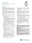
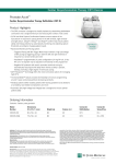
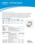
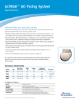
Accent DR
™
Dual-Chamber Pacemaker
MODEL PM2112
FOR POSITION
ONLY
Specifications
n
Upon interrogation, the device displays the last automatically measured capture
threshold results from the atrium and ventricle. In addition, the pacemaker
automatically measures intrinsic P- and R-wave activity daily and displays
the last test results in combination with a weekly P- or R-wave trend.
Results are displayed with follow-up EGMs for quick verification.
AutoCapture™ Pacing System offers the maximum in threshold adaptability
and patient safety with ventricular Beat-by-Beat™ capture confirmation. The
AutoCapture Pacing System automatically delivers a 5,0 V backup safety pulse
when noncapture is detected, and it may be programmed to either a bipolar or
unipolar configuration.
n n
ACap™ Confirm pacing system periodically completes a threshold search and
automatically adjusts amplitude to address patients’ changing atrial thresholds.
Exclusive SenseAbility™ feature, with Decay Delay and Threshold Start, provides
the flexibility to fine-tune sensing to individual patient needs and help eliminate
oversensing of T waves, fractionated QRS complexes, and other extraneous signals.
n
n A two-tone audible alert allows programming for the patient to be alerted to
changes in device performance, or information can be sent directly to the
clinician through the Merlin.net™ Patient Care Network (PCN).
Exclusive AF Suppression™ algorithm is clinically proven to suppress episodes
of paroxysmal and persistent AF.
n
– Studies
show a 25% decrease in symptomatic AF burden.1
n AT/AF Alerts can be programmed to notify patients and their clinics when a
programmed AT/AF threshold or continuous episode duration has been exceeded,
or when a high ventricular rate accompanies the AT/AF episode.
n Clinically proven to reduce unnecessary right ventricular pacing, the Ventricular Intrinsic Preference (VIP™) algorithm allows intrinsic conduction when possible and provides optimised ventricular support when needed.
– Studies
show an 81% decrease in unnecessary RV pacing.2
n QuickOpt™ Timing Cycle Optimisation provides quick and effective optimisation
for more patients at the touch of a button.3
– IEGM-based AV optimisation allows optimised timing without need for
echo-guided optimisation.
n Real-time EGM waveform, as well as the associated event markers that precede
and follow a specific triggering event, can be programmed to automatically record
up to 14 minutes of stored EGMs when encountering one or more programmable
trigger options.
– Preferential EGM storage capability allows prioritisation of episode storage.
Weekly Lead Impedance Trend displays the current measurement, historical
test results, pacing polarity and any polarity switches.
n
1 Carlson MD et al. A new pacemaker algorithm for the treatment of atrial fibrillation: results of the
Atrial Dynamic Overdrive Pacing Trial (ADOPT). JACC 2003; 42:627-633.
2 Hannah G et al. Reduction of ventricular pacing in pacemaker patients using Ventricular Intrinsic
Preference: preliminary results from the VIP trial. Europace Supplement, July 2008.
3 Baker et al. Acute evaluation of programmer-guided AV/PV and VV delay optimization comparing
an IEGM method and echocardiogram for cardiac resynchronization therapy in heart failure patients
and dual-chamber ICD implants. Journal of Cardiovascular Electrophysiology 2007; 18:185-191.
Indications: Implantation is indicated in one or more of the following permanent conditions: syncope,
presyncope, fatigue, disorientation due to arrhythmia/bradycardia, or any combination of those symptoms.
Rate-Modulated Pacing is indicated for patients with chronotropic incompetence, and for those who would
benefit from increased stimulation rates concurrent with physical activity. Dual-Chamber Pacing is indicated
for those patients exhibiting: sick sinus syndrome, chronic, symptomatic second- and third-degree AV block,
recurrent Adams-Stokes syndrome, symptomatic bilateral bundle branch block when tachyarrhythmia and other
causes have been ruled out. Atrial Pacing is indicated for patients with sinus node dysfunction and normal
AV and intraventricular conduction systems. Ventricular Pacing is indicated for patients with significant
bradycardia and normal sinus rhythm with only rare episodes of A-V block or sinus arrest, chronic atrial
fibrillation, severe physical disability. AF Suppression algorithm is indicated for suppression of paroxysmal
or persistent atrial fibrillation episodes in patients with one or more of the above pacing indications.
Contraindications: Implanted Cardioverter-Defibrillator (ICD). Devices are contraindicated in patients
with an implanted cardioverter-defibrillator. Rate-Adaptive Pacing may be inappropriate for patients who
experience angina or other symptoms of myocardial dysfunction at higher sensor-driven rates. An appropriate
Maximum Sensor Rate should be selected based on assessment of the highest stimulation rate tolerated by
the patient. AF Suppression stimulation is not recommended in patients who cannot tolerate high atrial-rate
stimulation. Dual-Chamber Pacing, though not contraindicated for patients with chronic atrial flutter, chronic
atrial fibrillation, or silent atria, may provide no benefit beyond that of single-chamber pacing in such patients.
Single-Chamber Ventricular Demand Pacing is relatively contraindicated in patients who have demonstrated
pacemaker syndrome, have retrograde VA conduction, or suffer a drop in arterial blood pressure with the
onset of ventricular pacing. Single-Chamber Atrial Pacing is relatively contraindicated in patients who have
demonstrated compromise of AV conduction.
Warnings and Precautions: To prevent permanent damage to the device and tissue damage at the
electrode/ tissue interface:
- Electrosurgery. Do not use electrosurgical devices in the vicinity of an implanted device. If electrocautery
is necessary, use a bipolar cauteriser or place the indifferent electrode as far from the device as possible.
- Lithotripsy. Do not focus a lithotripsy beam within 16 cm of the device. Program the device to Sensor Off
prior to lithotripsy to prevent inappropriate increases in pacing rate. A thorough assessment of device
function with special attention to the sensor should be performed following exposure to lithotripsy.
- Therapeutic Radiation. Do not use ionising radiation in the vicinity of an implanted device. Radiation
therapy may damage the electronic circuitry of the device.
- Ultrasound Treatment. Do not use therapeutic ultrasound within 16 cm of the device.
Perform a thorough assessment of device function following exposure to any of the above.
Device Communication. Communication with the device can be affected by electrical interference and strong
magnetic fields. If this is a problem, turn off nearby electrical equipment or move it away from the patient and
the programmer. If the problem persists, contact St. Jude Medical.
External Defibrillation. The electronic circuitry in the device provides protection from defibrillation
discharges. Nevertheless, do not place defibrillator paddles directly over the device or pacing lead. Following
defibrillation, ensure that the device is operating correctly.
Magnetic Resonance Imaging (MRI). MRI for patients with implantable devices has been contraindicated
by MRI manufacturers. Clinicians should carefully weigh the decisions to use MRI with pacemaker patients.
Additional safety concerns include:
- Magnetic and RF fields produced by MRI may increase pacing rate, inhibit pacing, cause asynchronous
pacing or result in pacing at random rates
- MRI may result in changes in capture thresholds due to heating of pacing leads
- MRI may irreversibly damage the device
- Patients should be closely monitored during the MRI
- Assess the device function before and after exposure to MRI.
CT Scans. CT scans, due to their increased power levels and long exposure times, have the remote possibility
of interfering with implanted devices. The potential interference is transient and occurs only when the X-ray
signal is present. Continuous exposure may cause a temporary sensor rate increase. In addition, there is a
remote possibility for a device to intermittently oversense while the CT scanning beam is directly over the
implanted device.
Potential Adverse Events: The following are potential complications associated with the use of any pacing
system: arrhythmia, heart block, thrombosis, threshold elevation, valve damage, pneumothorax, myopotential
sensing, vessel damage, air embolism, body rejection phenomena, cardiac tamponade or perforation, formation
of fibrotic tissue/local tissue reaction, inability to interrogate or program a device because of programmer
malfunction, infection, interruption of desired device function due to electrical interference, loss of desired
pacing and/or sensing due to lead displacement, body reaction at electrode interface, or lead malfunction
(fracture or damage to insulation), loss of normal device function due to battery failure or component
malfunction, device migration, pocket erosion, or hematoma, pectoral muscle stimulation, phrenic nerve or
diaphragmatic stimulation. The following, in addition to the above, are potential complications associated
with the use of rate-modulated pacing systems: inappropriate, rapid pacing rates due to sensor failure or
to the detection of signals other than patient activity, loss of activity-response due to sensor failure,
palpitations with high-rate pacing.
Refer to the User’s Manual for detailed indications, contraindications, warnings, precautions and potential
adverse events.
AF Management
PHYSICAL SPECIFICATIONS
Models
Telemetry
Dimensions (mm) Weight (g) Volume (cc) Connector PM2112
Inductive
46 x 52 x 6
19
10,51
IS-1
AF Suppression™ Algorithm Lower Rate Overdrive (min-1) Upper Rate Overdrive (min-1) No. of Overdrive Pacing Cycles Rate Recovery (ms) Maximum AF
Suppression Rate (min-1) Atrial Tachycardia
Detection Rate (min-1) Auto Mode Switch AMS Base Rate (min-1) PARAMETER SETTINGS
Rate/Timing
Atrial Pace Refractory (ms) Atrial Sense Refractory (ms) Atrial Protection Interval (ms) Paced AV Delay (ms) Base Rate (min-1) Far-Field Protection Interval (ms) Hysteresis Rate (min-1) Search Interval (min) Cycle Count Intervention Rate (min-1) Intervention Duration (min) Recovery Time Maximum Tracking Rate (min-1) Mode Post Ventricular Atrial Blanking (ms) PVARP (ms) Sensed AV Delay (ms) Rest Rate (min-1) Shortest AV Delay (ms) Ventricular Blanking (ms) Ventricular Pace/Sense Refractory5
(Fixed) (ms) 190-400 in steps of 30; 440; 4702
93; 125; 157; 190-400 in steps of 30; 440; 4702
1253
25; 30-200 in steps of 10; 225-300 in steps of 25; 350
30-130 in steps of 5; 140-170 in steps of 10
163
Off; 30 4-150 in steps of 5
Off; 1; 5; 10; 15; 30
1-16 in steps of 1
Off; Same as Base Rate; 80-120 in steps of 10; Intrinsic +0;
Intrinsic +10; Intrinsic +20; Intrinsic +30
1-10 in 1 minute intervals
Fast; Medium; Slow; Very Slow
90-130 in steps of 5; 140-180 in steps of 10
AOO(R); AAI(R); AAT(R); VOO(R); VVI(R);
VVT(R); VDD(R); DOO(R); DVI(R); DDI(R);
DDD(R); Pacing Off
60-200 in steps of 10; 225, 250
125-500 in steps of 25
25; 30-200 in steps of 10; 225-325 in steps of 25
Off; 30-150 in steps of 5
25-50 in steps of 5; 60-120 in steps of 10
Auto, 12-52 in steps of 4
Options
Priority Options Channel Triggers
Advanced Hysteresis AMS Entry/AMS Exit/
AMS Entry and Exit AT/AF Detection Magnet Response High Atrial Rate
Rate (min-1)
No. of Consecutive Cycles High Ventricular Rate
Rate (min-1)
No. of Consecutive Cycles
PMT Termination Consecutive PVCs No. of Consecutive PVCs Noise Reversion Atrial Fibrillation
A and V Lead Monitoring A and V Low Impedance Limit (Ω) A and V High Impedance Limit (Ω) Lead Type Magnet Response Negative AV Hysteresis Search (ms) NIPS Options
Stimulation Chamber Coupling Interval (ms) S1 Count S19; S2; S3 and S4 Cycle (ms) Ventricular Support Rate (min-1) Sinus Node Recovery Delay (sec) PMT Options PMT Detection Rate (min-1) PVC Response Ventricular Intrinsic
Preference, VIP™ (ms) VIP Search Interval VIP Search Cycles Ventricular Safety Standby Diagnostic Trends
On; Off
Unipolar; Bipolar
Unipolar; Bipolar
5,03
8; 24
50/25; 100/70; 120/100
0,5-5,0 in steps of 0,5; 6-10 in steps of 1,0; 12,57
Off; On (Automatic Sensitivity Control adjustment for atrial
and ventricular events)
0,2-1,0 in steps of 0,1
0,2-2,0 in steps of 0,1
(Atrial and Ventricular Post-Sense) 50; 62,5; 75; 100%
(Atrial Post-Pace) 0,2-3,0 in steps of 0,1 mV
(Ventricular Post-Pace) Auto; 0,2-3,0 in steps of 0,1 mV
(Atrial and Ventricular Post-Sense) 0; 30; 60; 95; 125; 160; 190; 220
(Atrial Post-Pace) 0; 30; 60; 95; 125; 160; 190; 220
(Ventricular Post-Pace) Auto; 0; 30; 60; 95; 125; 160; 190; 220
Off; Low; High
Off; Low; High
Off; Low; High
Off; Low; High
Off; Low; High
125-300 in steps of 25
2; 3; 4; 5; 10; 15; 20
Off; Low; High
125-300 in steps of 25
2; 3; 4; 5; 10; 15; 20
Off; Low; High
Off; Low; High
2; 3; 4; 5
Off; Low; High
CARDIOVASCULAR
Monitor; Auto Polarity Switch
100-500 in steps of 25
750-2500 in steps of 250; 3000
Uncoded; Unipolar; Bipolar
Off; Battery Test
Off; -10 to -120 in steps of 10
Atrial; Ventricular
100-800 in steps of 108
2-25 in steps of 1
Off; 100-800 in steps of 10 (Fixed or Adaptive)
Off; 30-95 in steps of 5
1; 2; 3; 4; 5
Off; Passive; Atrial Pace2
90-180 in steps of 5
Off; Atrial Pace2
Off, 50-150 in steps of 25; 160-200 in steps of 10
30 sec.; 1; 3; 5; 10; 30 min.
1; 2; 3
Off; On
AT/AF Activity; Exercise; Lead Impedance; P and R Wave; A and V Threshold
Patient Notifiers
Programmable Notifiers (On; Off) Device Reset Entry into Backup VVI Mode Audible Duration (sec) Number of Audible Alerts
per Notification Number of Notifications Time Between Notifications (hours) 80-150 in steps of 5; 160-180 in steps of 10
Off; Low; Medium; High
Off; Low; Medium; High
Very Fast; Fast; Medium; Slow
Fast; Medium; Slow; Very Slow
On; Off; Passive
125-475 in steps of 25
Auto (-1); Auto (+0); Auto (+1); Auto (+2);
Auto (+3); 1-16 in steps of 1
Auto (-0,5); Auto (+0,0); Auto (+0,5); Auto (+1,0);
Auto (+1,5); Auto (+2,0); 1-7 in steps of 0,5
Cardiac Rhythm Management
Off; Low; High
1; 2; 3
Other
125; 160-400 in steps of 30; 440; 4702
On; Off; Monitor
Bipolar
Bipolar
5,03
8; 24
0,25-4,0 in steps of 0,25; 4,5-7,5 in steps of 0,5
0,05; 0,1-1,5 in steps of 0,1
Unipolar (tip-case); Bipolar (tip-ring)
Unipolar Tip (tip-case); Bipolar (tip-ring);
Unipolar Ring (ring-case)
0,1-0,46 in steps of 0,1; 0,5; 0,75-2,0 in steps of 0,25;
2,5-4,0 in steps of 0,5; 5,07
Maximum Sensor Rate (min-1) Rate Responsive AV Delay Rate Responsive PVARP/VREF Reaction Time Recovery Time Sensor Shortest PVARP/VREF (ms) Slope Threshold 80-200 in steps of 10; 225-300 in steps of 25
110-200 in steps of 10; 225-300 in steps of 25
Off; DDD(R) to DDI(R); DDD(R) to DDT(R); DDD(R) to VVI(R);
DDD(R) to VVT(R); VDD(R) to VVI(R); VDD(R) to VVT(R)
40-170 in steps of 5
Stored Electrograms
Output/Sensing
ACap™ Confirm Primary Pulse Configuration Backup Pulse Configuration Backup Pulse Amplitude (V) Search Interval (hours) A or V Pulse Amplitude (V) A or V Pulse Width (ms) A or V Pulse Configuration A or V Sense Configuration Atrial Sensitivity (mV) Ventricular AutoCapture™ Pacing System Primary Pulse Configuration Backup Pulse Configuration Backup Pulse Amplitude (V) Search Interval (hours) AutoCapture
Paced/Sensed AV Delay (ms) Ventricular Sensitivity (mV) SenseAbility™ Technology
A Max Sensitivity (mV)
V Max Sensitivity (mV) Threshold Start
Decay Delay (ms)
Rate-Modulated Parameters
Off; On
103
53
15-40 in steps of 5
8; 123
Device at ERI; Atrial Lead Impedance Out of Range;
Ventricular Lead Impedance Out of Range; AT/AF Burden;
AT/AF Episode Duration; High V Rate During AT/AF
On
On
2; 4; 6; 8; 10; 12; 14; 16
2
1-16
10; 22
1 ± 0,5 cc
2 Programming options dependent on pacing mode.
3 This parameter is not programmable.
4 The highest available setting for hysteresis rate will be 5 min-1 below the programmed base rate.
5 In dual-chamber modes, the maximum ventricular refractory period is 325 ms.
6 Values 0,1-0,4 not available in a unipolar sense configuration.
7 Sensitivity is with respect to a 20 ms haversine test signal.
8 During atrial NIPS in dual-chamber modes, the shortest Coupling Interval will be limited by the programmed AV/PV delay.
9 S1 Burst Cycle is applied at the preprogrammed S1 cycle length.
Neuromodulation
Global Headquarters
One Lillehei Plaza
St. Paul, Minnesota 55117
USA
+1 651 483 2000
+1 651 490 4310 Fax
Cardiac Rhythm
Management Division
15900 Valley View Court
Sylmar, California 91342
USA
+1 818 362 6822
+1 818 364 5814 Fax
St. Jude Medical Sweden AB
Veddestavägen 19
175 84 Järfälla
Sweden
+46 8 474 40 00
+46 8 760 95 42 Fax
St. Jude Medical Europe, Inc.
The Corporate Village
Figueras Building
Avenue Da Vinci Iaan, 11 Box F1
B-1935 Zaventem
Belgium
+32 2 774 68 11
+32 2 772 83 84 Fax
St. Jude Medical Brasil Ltda.
Rua Frei Caneca, 1380
7º ao 9º andares
01307-002 - São Paulo (SP)
Brazil
+55 11 5080 5400
+55 11 5080 5423 Fax
St. Jude Medical (Hong Kong) Ltd.
Unit 2701-07 27/F, COSCO Tower
Grand Millennium Plaza
183 Queen’s Road
Central, Hong Kong
+852 2996 7688
+852 2956 0622 Fax
St. Jude Medical Japan Co., Ltd.
3-1-30, Minami-Aoyama
Minato-ku
Tokyo 107 0062
Japan
+81 3 3423 6450
+81 3 3402 5586 Fax
SJMprofessional.com
Brief Summary: Prior to using these devices, please review the Instructions for Use for a complete listing of indications, contraindications, warnings, precautions, potential adverse events and
directions for use. Devices depicted may not be available in all countries. Check with your St. Jude Medical representative for product availability in your country. Unless otherwise noted, ™
indicates that the name is a trademark of, or licensed to, St. Jude Medical or one of its subsidiaries. ST. JUDE MEDICAL, the nine-squares symbol and MORE CONTROL. LESS RISK.
are trademarks and service marks of St. Jude Medical, Inc. and its related companies. ©2009 St. Jude Medical, Inc. All Rights Reserved.
Item GMCRM456