1
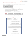
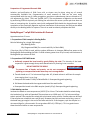
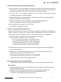
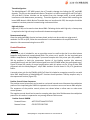
MethylMagnet™ mCpG DNA Isolation Kit User Manual Version B 06‐11‐09 Catalog # MM101‐K Table of Contents Kit Components and Storage ………………………………………………………….. 2 MethylMagnet™ Work Flow …………………………………………………………… 3 About MethylMagnet™ Proteins……………………………………………………… 4 MethylMagnet™ DNA Isolation Kit Overview…………………………………… 5 Advantages of MethylMagnet………………………………………………………….. 6 Preparation of Genomic DNA…………………………………………………………... 7 MethylMagnet™ Protocol ……………………………………………………..………… 7 Control Reactions …………………………………………………………………………… 9 Troubleshooting Guide……………………………………………………….……...... 11 Kit Components and Storage Component (Three 1.5 ml tubes, green cap) (One 1.5 ml tube, yellow cap) (Two bottles) (One bottle) (Five 1.5 ml tubes) (One 1.5 ml tube, blue cap) Volume 3.3 ml 1.32 ml 26.4 ml 13.2 ml 6.6 ml 165 µl 55 µl Wash Buffer 1 Binding Buffer Wash Buffer 2 TE Buffer Glutathione Elution Buffer GST‐MBD magnetic beads Control HeLa DNA 110 ng, MseI cut, 2ng/µl (One 0.6 ml tube, green dot) Positive Control Primers, 10 µM each (One 0.6 ml tube, red dot) 16.5 µl Negative Control Primers, 10 µM each (One 0.6 ml tube, blue dot) 16.5 µl User Manual 1 Kits are shipped on ‘blue ice’. All components should be stored at ‐20˚C. Do not expose beads to temperatures below ‐20˚C. Store beads in an insulated cooler if the freezer has a defrost cycle. www.ribomed.com MethylMagnet™ mCpG DNA Isolation Kit User Manual V2 Storage ‐20˚C ‐20˚C ‐20˚C ‐20˚C ‐20˚C ‐20˚C ‐20˚C ‐20˚C ‐20˚C IMPORTANT! 2 Please read entire manual before starting. Reagents are included for 30 samples. This can be 30 samples of the user’s DNA or 25 samples of the user’s DNA and 5 controls with HeLa DNA. User‐supplied Materials/Equipment • • • • • • Microcentrifuge tubes (1.7 ml polypropylene) and microcentrifuge. Filter pipette tips. Sample mixing equipment (e.g. Eppendorf Thermomixer, shaker or tube rotator). Thermocycler and PCR components for control reactions DNAse‐free ultrapure water Rare‐earth Magnet or magnetic rack (e.g. RiboMed Cat #MR22‐01) Caution: Rare earth magnets can be extremely powerful. Care should be taken when handling them. Keep magnetized parts away from instruments that may be damaged by high magnetic fields. MethylMagnet™ Work Flow www.ribomed.com MethylMagnet™ mCpG DNA Isolation Kit User Manual V2 3 About MethylMagnet™ Proteins The MethylMagnet™ mCpG DNA isolation Figure 1 kit utilizes RiboMed’s GST‐MBD protein (MethylMagnet™). MethylMagnet™ proteins are versatile tools for the study of CpG methylation in DNA. These proteins contain the methyl binding domain (MBD) of the mouse MBD2 protein fused to the glutathione‐S‐ transferase protein (GST) from S japonicum. The two domains are separated by a linker containing a thrombin cleavage site (Figure 1A). The MBD from the MBD2b protein was chosen because MBD2b has the highest affinity among the known methyl CpG binding proteins for Me‐CpG sites and the lowest cross reactivity with unmethylated CpGs. Additionally, there are no sequence context effects on MBD2 CpG recognition, as there are for MeCP2, which requires a run of A‐Ts near a CpG site, therefore a greater number of mCpG sites will be recognized by MethylMagnet™. In addition to its high specificity for methylated DNA, MethylMagnet™ proteins have several unique features that make them useful tools in the study of DNA methylation. The GST allows the fusion protein or its complexes with methylated DNA to be isolated on glutathione agarose, magnetic beads or plates and eluted intact with glutathione. The GST group further allows the eluted protein‐DNA complexes to be immobilized to beads or microtiter plates with GST antibodies. Alternatively, the GST group can be used for visualization of the DNA‐protein complexes by using labeled GST antibodies or GST antibodies and labeled secondary antibodies, or other detection methods for GST. The GST group contains surface cysteine groups (Figure 1B) that can be chemically modified to add other reporters or affinity tags, such as biotin, fluorescein or other functional groups. MethylMagnet™ has high affinity for fully methylated CpG sites MethylMagnet™ proteins Figure 2 A. have high affinity and specificity for binding to DNA containing 5‐Methyl‐ CpG groups. They have much higher affinity for DNA methylated on both strands, versus hemi‐ methylated (not shown) or unmethylated DNA (Figure 2). Purified MethylMagnet™ protein (Cat# MM101) was incubated with a 550 bp p16 amplicon that was either fully methylated or unmethylated or with no DNA (Figure 2A). The biotinylated DNA target was immobilized to magnetic beads. The MethylMagnet™ protein was incubated with the immobilized DNA for 1 hour. After washing, bound MethylMagnet™ was detected with an anti‐GST antibody‐horseradish peroxidase conjugate (Figure 2B). The HRP reaction was performed for 5 min. The filled and unfilled bars represent duplicate measurements. www.ribomed.com MethylMagnet™ mCpG DNA Isolation Kit User Manual V2 4 MethylMagnet™ mCpG DNA Isolation Kit Overview The MethylMagnet™ mCpG DNA Isolation Kit is used to isolate methylated dsDNA from a genomic sample. The MethylMagnet™ kit utilizes the MethylMagnet™ GST‐MBD fusion protein and glutathione magnetic beads for capture of methylated dsDNA. It can be used to isolate mCpG DNA from 1 ng to 1 µg of genomic DNA per sample, or it can be scaled up to isolate much larger quantities. The MethylMagnet™ kit can capture DNA containing 6 or more methylated CpG sites. DNA eluted with glutathione is ready for downstream applications without further processing. How it Works Step 1: DNA Isolation and Fragmentation DNA isolation from cells or tissues may be done using a kit of choice. Unlike some other methods, MethylMagnet™ binds methylated double‐stranded DNA, so it is not necessary to denature the DNA once it is isolated. It is imperative that the DNA be fragmented so that the region of interest is physically separated from other regions of DNA that may be methylated. Failure to adequately separate CpG islands could lead to co‐purification of unmethylated islands with neighboring methylated regions. Fragmentation by restriction enzyme digestion is recommended. An incomplete restriction digest can lead to isolation of unmethylated islands by association with adjacent methylated regions. The island detection assays that utilize abscription® for detection using the MethylMeter™ kits have been designed to work with DNA that has been cut with the restriction endonuclease Mse I. Mse I cuts at the sequence TTAA and therefore is not affected by CpG methylation. However, MethylMagnet™ can be used with DNA that has been fragmented with other restriction enzymes. Step 2: Capture of Methylated DNA The fragmented DNA is incubated with the MethylMagnet™ GST‐MBD protein which has already been attached to glutathione magnetic beads. The DNA population will generally contain a mixture of methylated and unmethylated CpG sites. Methylated CpG islands will bind to the beads via interaction of the mCpG sites and the MBD domain, while unmethylated islands will remain in the supernatant fraction. www.ribomed.com MethylMagnet™ mCpG DNA Isolation Kit User Manual V2 5 Step 3: Elution of Methylated DNA Methylated DNA can be eluted from the glutathione beads several ways, although the use of glutathione is recommended because the DNA is ready for downstream applications without further protease treatment or precipitation. However, several options are available. A. Elution of the GST‐MBD‐mCpG DNA complex with Glutathione (recommended) B. Elution of free DNA and denatured GST‐MBD with heat C. Elution of free DNA and digested GST‐MBD with Proteinase K D. Elution of the MBD‐mCpG DNA complex with Thrombin E. Elution of mCpG DNA with salt Following elution with glutathione or heat, the DNA is ready for downstream applications. If PCR, qPCR, or abscription® ‐ based assays are used for downstream analysis, then no further clean up of the DNA is required. If other methods are going to be used, please refer to the manufacturer’s recommendations. If eluting with high salt, Proteinase K, or Thrombin, a DNA clean‐up step is required before use. Advantages of MethylMagnet™: High sensitivity without loss of specificity The MethylMagnet™ mCpG DNA Isolation Kits can be used with 1 ng of input DNA up to 1 µg of DNA (not shown). Shown below (Figure 3), HeLa genomic DNA or artificially methylated HeLa DNA were purified with the MethylMagnet™ mCpG DNA Isolation Kit and eluted in 20 µl of glutathione Figure 3 buffer. 2 µl samples (1/10th of input) were tested for p16 DNA by PCR (40 cycles) and analyzed by gel electrophoresis and ethidium bromide staining. 2 ng input corresponds to approximately 300 cells, so methylated P16 from 200 pg of genomic DNA, or 30 cells is detected here. P16 was only detected with artificially methylated DNA. Shown in Figure 4, 1 ng of HeLa DNA or artificially methylated HeLa DNA was purified with the MethylMagnet™ mCpG DNA Isolation Kit. DNA was eluted in 50 µl and 2 µl samples were used for amplification of P16. This corresponds to detection from 6 cells, or 40 pg of DNA and 12 copies of P16. The gel was stained with SYBR® Green and imaged with a Typhoon fluorescence reader. MethylMagnet™ mCpG DNA Isolation Kits are very specific for methylated DNA. When unmethylated DNA is processed, nothing is captured and detected in the eluted (bound) fraction, even with 40 cycles of PCR. When methylated DNA is captured, nothing is detected in the supernatant (unbound) fraction. Figure 4 www.ribomed.com MethylMagnet™ mCpG DNA Isolation Kit User Manual V2 6 Preparation of Fragmented Genomic DNA Isolation and purification of DNA from cells or tissues may be done using any of several commercially available kits. Fragmentation of small amounts of DNA can be most easily accomplished with the use of 4‐base‐recognition restriction endonucleases that are biased to A‐T rich sequences (e.g. MseI, TTAA, and Tsp509I, AATT). The completeness of digestion can be tested by performing PCR with a primer pair flanking the restriction site verses a primer pair that does not have an intervening site. A positive control with undigested DNA should also be performed. Upon completion of the restriction digestion and heat inactivation of the enzyme, the DNA can be added directly to the binding buffer for capture. There is no need to ethanol precipitate the DNA. MethylMagnet™ mCpG DNA Isolation Kit Protocol Approximate time: 2 hours 1. Preparation of DNA samples in Binding Buffer Mix the following for a single DNA sample: 40 μl Binding Buffer 10 μl fragmented DNA (For controls add 10 μl of HeLa DNA) If less than 10 μl of DNA is used, add the volume difference in ultrapure DNAse free water to the Binding Buffer before adding the DNA. If a DNA volume greater than 10 μl is used, add 4 volumes of Binding Buffer per volume of DNA. 2. Preparation of GST‐MBD beads a. Uniformly suspend the bead stock by gently flicking the tube. The viscosity of the bead suspension is great enough that protein denaturation by foaming is not a concern. DO NOT VORTEX THE BEADS. IMPORTANT! To prevent loss of beads and protein, we do not recommend pipetting to resuspend the beads when they are in storage buffer. b. Transfer beads to a 1.7 ml microcentrifuge tube. A 5 μl bead volume is sufficient for sample sizes ranging from 1 ng to 1 μg. c. Resuspend the beads in 100 μl of Wash Buffer 1. Resuspend by gentle pipetting. d. Pull‐down the beads with the magnet and discard the wash buffer. e. Resuspend the beads in the DNA samples (usually 50 μl). Resuspend by gentle pipetting. 3. DNA binding reaction Incubate the beads at room temperature (18‐23°C) for 1 hour. The beads should be mixed during the incubation (e.g. with an Eppendorf Thermomixer set at 1,000 rpm). A 1.7 ml microcentrifuge tube is optimal if the samples are mixed with a horizontal rotary motion. Smaller tubes can be used if the samples are mixed end‐over‐end (8 rpm). Be aware that with end‐over‐end mixing, some of the beads may get caught in the seal of the tube and the lid. If this happens, spin the samples in a microcentrifuge for a few seconds. Do not go above 500 rcf (≈2700 rpm). This is a good time to thaw the elution buffer at room temperature. www.ribomed.com MethylMagnet™ mCpG DNA Isolation Kit User Manual V2 7 4. Removal of unbound (unmethylated) DNA fragments. a. Spin the samples in a microcentrifuge for a few seconds if droplets have collected on the walls of the tubes. Do not go above 500 rcf. The beads will settle to the bottom of the tubes but will rapidly form pellets on the sides of the tubes in response to the magnet. b. Place the tubes in the magnetic separation rack and remove the supernatant fraction containing the unbound, unmethylated DNA. c. Wash the beads with 0.4 ml of Wash Buffer 2. Rinse the tube walls while initially adding the buffer and completely suspend the beads. d. Incubate the beads for 5 min with mixing at room temperature and 1000 rpm. Perform one additional wash and incubation with Wash Buffer 2. e. Wash the beads one time with 0.4 ml of TE buffer. No incubation is needed for this step. 5. Elution of methylated DNA with glutathione Note: Elution buffer can lose potency with extended storage due to oxidation of the glutathione. Buffer is provided in 5 tubes. Once a tube is opened, the glutathione should be used within 2 months. Invert the elution buffer several times prior to use to be sure it is homogeneous. a. Suspend the beads in elution buffer and incubate 10 min with mixing (e.g. Thermomixer, 1000 rpm, room temperature). For 1 ng to < 20 ng input DNA, use 10 µl elution buffer. For 20 ng to < 50 ng input DNA, use 50 µl elution buffer. For 50 ng to 1 µg input DNA, use 100 µl elution buffer. b. Pull down the beads with the magnet and remove the supernatant fraction containing the eluted, methylated DNA. c. Although most of the DNA will be recovered in the first elution, a second elution can be performed to maximize methylated DNA recovery. For DNA input above 500 ng, two elutions are recommended DNA can be used in PCR without further purification. Alternative methods to recover methylated DNA Although we recommend elution with glutathione, other elution methods may be used. Heat treatment a. Suspend the beads in 50 μl 1X TE, pH 8 (100 μl when using > 50 ng input DNA). Incubate 10 minutes at 80˚C with mixing. b. Pull down the beads and save the supernatant fraction containing the eluted DNA. c. A second elution can be performed to maximize methylated DNA recovery. DNA can be used in PCR without further purification. www.ribomed.com MethylMagnet™ mCpG DNA Isolation Kit User Manual V2 8 Thrombin digestion The MethylMagnet™ GST‐MBD protein has a Thrombin cleavage site linking the GST and MBD domains. This site can be cleaved with Bovine Thrombin in a buffer containing 10 mM TrisCl pH 8, 150 mM NaCl. Calcium chloride can be omitted from the cleavage buffer to avoid possible interference with downstream processing. Thrombin digestion can release DNA containing the bound MBD domain. While Bovine Thrombin does not interfere with PCR, the samples should be treated to inactivate potential contaminating nuclease activities. High salt elution High salt (1‐2M) can be used to elute bound DNA. Following elution with high salt, a cleanup step is required as the high salt may interfere with downstream applications. Downstream Analysis Once the methylated DNA fraction has been eluted, analysis can be carried out using several methods. For PCR or qPCR, follow the manufacturer’s recommendations for amplification and detection. We recommend using hot start PCR. Control Reactions Overview A PCR primer pair is included for use as a positive control to confirm that the kit can discriminate between methylated and unmethylated DNAs. The primer set is specific for the imprinted gene SNRPN. Amplification of MethylMagnet™ processed samples with this primer pair will produce a 230 bp amplicon in both the supernatant fraction of the binding reaction (the unbound, unmethylated copy) and the DNA fraction eluted from the GST‐MBD beads (the methylated copy). Figure 5A shows the results of control amplifications with this primer set for 20 ng of HeLa DNA processed with the MethylMagnet™ mCpG DNA Isolation Kit and eluted in 50 µl of glutathione buffer. A second primer pair for the CDKN2A (p16) CpG island is included as a negative control for use with HeLa DNA. Amplification of MethylMagnet™ fractions should produce a 381 bp amplicon only in the supernatant fraction (Figure 5B). Positive Control Primer Sequences The positive control primer pair can be used as a universal internal control because the methylation pattern of the imprinted gene SNRPN is expected to be consistent over most human DNA samples. The sequences of the positive control primers are shown below to allow users to order extra control primers. The negative control should not be used on samples other than HeLa DNA because the methylation pattern for this marker is expected to vary among different DNA sources. Positive Control: SNRPN Forward Primer: ACCTCCGCCTAAAATCCCTATG Reverse Primer: GTATCCTGTCCGCTCGC www.ribomed.com MethylMagnet™ mCpG DNA Isolation Kit User Manual V2 9 Protocol A total of 110 ng (2 ng/µl) of fragmented HeLa DNA is included in the kit, together with primers for 5 control samples plus 1 no‐DNA control for each sample. Control PCR reactions should be done after processing 20 ng of fragmented HeLa DNA with the MethylMagnet™ kit, as described above, and eluting in 50 µl of glutathione elution buffer. PCR reactions should be done on both the supernatant fraction and the first elution fraction. A protocol using Finnzyme’s Phire Hot Start DNA polymerase is shown in Table 1. Table 1. Phire DNA hot start PCR (volumes for a single reaction) Component Volume per 20 µl Final Concentration 5x Phire PCR buffer 4.0 µl 1x Ultrapure water 10.0 µl ‐ dNTPs (10 mM, 2.5 mM each) 1.6 µl 800 µM (200 µM each) Control Primer mix (10 µM each) 1.0 µl 0.5 µM each primer DMSO 1.0 µl 5% v/v 1 MethylMagnet™ DNA fraction 2.0 µl Phire DNA polymerase 0.4 µl ‐ 1 Unbound supernatant fraction (unmethylated) or bound/eluted fraction (methylated). Replace with 2 μl of ultrapure water for the no‐DNA control Phire Cycling Conditions 1. 98°C, 15 sec 2. 98°C, 10 sec 3. 65.7°C 10 sec 4. 72°C 10 sec 5. Repeat steps 2‐4 35 times 6. 72°C 1 min 7. HOLD 4° A S E1 E2 E3 B S E1 S: Unbound Supernatant E1-E3: Elutions 1-3 Figure 5. Amplification of fractionated fragmented HeLa DNA with the positive control primer pair. MethylMagnet™ fractions (2 µl) from an input of 20 ng of HeLa DNA were subjected to PCR with positive control primers that target the imprinted gene SNRPN (A), and the negative control primers that target the unmethylated p16 CpG island (B). S is the fraction containing the unmethylated DNA. E1‐E3 represent 3 serial elutions of the methylated DNA target from the beads. Virtually all of the methylated DNA is recovered in the first elution. www.ribomed.com MethylMagnet™ mCpG DNA Isolation Kit User Manual V2 10 Interpretation of unexpected results for control reactions There are two situations in which unexpected results can arise when control primers are used with DNA from sources other than HeLa. A single DNA sample might have an unexpected methylation pattern for the imprinted marker if that sample has altered imprinting functions. For example, the unmethylated copy might have become methylated to produce amplicon only in the eluted fraction. Alternatively, the positive control might show signal only in the supernatant. If these results are associated with only a single sample then they likely reflect an abnormal methylation status of SNRPN in that sample. An unexpected result that includes all of the samples in an experiment indicates a failure of the kit. A systematic failure of the kit will most likely manifest itself in the positive control when the signal is all found in the supernatant fraction. This could be due to a failure of the beads to bind methylated DNA or a failure of the bound DNA to be released in the presence of elution buffer. Possible aberrant results and possible solutions are listed in the Troubleshooting table. Table 2. Troubleshooting Problem No PCR product in any of the fractions No PCR product from the eluted fraction Unmethylated DNA in the elution fraction Methylated DNA in the supernatant Signal appears only in the supernatant fractions of all samples High background in the eluted fraction of the negative control. Cause Primers flank a restriction site Remedy Be sure that the region you want to amplify does not contain any restriction sites for the enzyme you are using. DNA was not eluted because the Open a fresh tube of glutathione elution buffer glutathione in the elution buffer or try eluting with 1X TE buffer at 80˚C. has oxidized Beads not washed thoroughly Be sure to follow all wash instructions so that all unmethylated DNA is removed before eluting methylated DNA Binding capacity of the beads Be sure to use less than or equal to 1 μg of DNA has been exceeded per 5 μl of beads. If processing more DNA, increase the volume of beads used. Binding capacity of the beads is Use methylated DNA to confirm decreased binding capacity. Increase volume of beads. reduced because of improper storage or handling Binding capacity of the beads is Use only the reagents supplied with the reduced because of non‐optimal MethylMagnet™ kit. Reagents can be ordered at our website (www.ribomed.com). buffer conditions Failure of the beads to bind • Check kit expiration date. methylated DNA • Ensure that the beads are thoroughly mixed Failure to release methylated during the binding reaction DNA from the beads with • Incubate the beads at 80C for 10 min in TE glutathione buffer. buffer and amplify the eluted fraction. Incomplete washing • Ensure that the walls of the tubes are rinsed when applying Wash Buffer 2 • Briefly spin the binding reactions at 500 rcf before washing to collect droplets from the tube walls. www.ribomed.com MethylMagnet™ mCpG DNA Isolation Kit User Manual V2 11