1
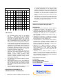
For Research Use Only Not for Diagnostic Use HEPATITIS B – anti HBc IgM Catalog #: WB2796 IgM ANTIBODIES TO HEPATITIS B VIRUS Core Antigen ELISA KIT Two-Step Incubation, Antibody Capture Principle are pre-coated with antibodies directed to human immunoglobulin M proteins (anti-µ chain). The patient’s serum/plasma sample is added and during the first incubation step, any IgM-class antibodies will be captured inside the wells. After washing out all other components of the sample and in particular IgG-class antibodies, the specific anti-HBc IgM captured on the solid phase is detected by the addition of purified HBcAg, labeled with anti-HBc monoclonal antibody conjugated to horseradish peroxidase (HRP-Conjugate). During the second incubation, the HRP-conjugated antigens will specifically react only with anti-HBc IgM antibodies, and after washing to remove the unbound HRP-conjugate, Chromogen solutions are added to the wells. This anti-HBc IgM kit is an enzyme-linked immunosorbent assay (ELISA) for qualitative determination of IgM class antibodies to hepatitis B virus core antigen in human serum or plasma. For Research Use Only. In presence of the (anti-µ chain)-(anti-HBc IgM)-(HBcAg-Ab (HRP)) immunocomplex, the colorless Chromogens are hydrolyzed by the bound HRP-conjugate to a blue-colored product. The blue color turns yellow after stopping the reaction with sulfuric acid. The amount of color intensity can be measured and is proportional to the amount of antibody captured in the wells, and to the sample respectively. Wells containing samples negative for antiHBc IgM remain colorless. SUMMARY COMPONENTS 96 Tests INSTRUCTIONS FOR USE Hepatitis B virus (HBV) is an enveloped, double-stranded DNA virus belonging to the Hepadnaviridae family and is recognized as the major cause of blood transmitted hepatitis together with hepatitis C virus (HCV). Infection with HBV induces a spectrum of clinical manifestations ranging from mild, unapparent disease to fulminant hepatitis, severe chronic liver diseases, which in some cases can lead to cirrhosis and carcinoma of the liver. Classification of a hepatitis B infection requires the identification of a number of serological markers expressed during three phases (incubation, acute and convalescent) of the infection. Hepatitis B “core” antigen (HBcAg) is a major component of the viral structure. HBcAg is composed of a single polypeptide of about 17 kD that is released upon disaggregation of the core particles; the antigen contains at least one immunological determinant. Antibodies to HBcAg (antiHBc total antibody and IgM) appear shortly after the appearance of HBsAg and persist for life both in persons who have recovered from a hepatitis B infection and in those who develop HBsAg-carrier status but in rare cases, an HBV infection can also run its course without the appearance of immunologically detectable anti-HBc (usually in immunosuppressed patients). In chronic hepatitis, however, spikes of anti-HBc IgM synthesis are present, confirming reactivation of HBV in hepatocites and giving origin to permanent IgM low titers. Presence of IgM and total anti-HBc indicates an ongoing or recent HBV infection. PRINCIPLE OF THE ASSAY This kit is a two-step incubation, solid phase antibody capture ELISA assay, in which polystyrene microwell strips MICROWELL PLATE 1 plate Blank microwell strips fixed on white strip holder. The plate is sealed in aluminum pouch with desiccant. 8×12/12×8well strips per plate. Each well contains anti-IgM antibodies (anti-µ chain). The microwell strips can be broken to be used separately. Place unused wells or strips in the plastic sealable storage bag together with the desiccant and return to 2~8ºC. NEGATIVE CONTROL 1 vial Yellowish liquid filled in a vial with green screw cap. 0.5ml per vial. Protein-stabilized buffer tested non reactive for anti-HBc IgM. Preservatives: 0.1% ProClin 300. Ready to use as supplied. Once open, stable for one month at 2-8ºC. POSITIVE CONTROL 1 vial Red-colored liquid filled in a vial with red screw cap. 0.5ml per vial. anti-HBc IgM antibodies diluted in proteinstabilized buffer. Preservatives: 0.1% ProClin 300. Ready to use as supplied. Once open, stable for one month at 2-8ºC. HRP-CONJUGATE REAGENT 1 vial Red-colored liquid filled in a white a vial with red screw cap. 12ml per vial. Horseradish peroxidase-conjugated purified HBcAg, labeled with monoclonal anti-HBc. Ready to use as supplied. Once open, stable for one month at 28ºC. STOCK WASH BUFFER 1 bottle Colorless liquid filled in a clear bottle with white screw cap. 50ml per bottle. pH 7.4 20 × PBS (Contains Tween-20 as a detergent). DILUTE BEFORE USE -The concentration must be diluted 1 to 20 with distilled/deionized water before use. Once diluted, stable for one week at room temperature or for two weeks at 2-8ºC. CHROMOGEN SOLUTION A 1 vial Colorless liquid filled in a white vial with green screw cap. 7ml per vial. Urea peroxide solution. Ready to use as supplied. Once open, stable for one month at 2-8ºC. CHROMOGEN SOLUTION B Colorless liquid filled in a black vial with black screw cap. 7ml per vial. TMB solution. (Tetramethylbenzidine dissolved in citric acid). Ready to use as supplied. Once open, stable for one month at 2-8ºC. STOP SOLUTION 1 vial Colorless liquid filled in a white vial with yellow screw cap. 7ml per vial. Diluted sulfuric acid solution (2.0M H2SO4). PLASTIC SEALABLE BAG 1 unit For enclosing the strips not in use. CARDBOARD PLATE COVER 2 sheets To cover the plates during incubation and prevent evaporation or contamination of the wells. PACKAGE INSERTS 1 copy ADDITIONAL MATERIALS AND INSTRUMENTS REQUIRED BUT NOT PROVIDED 1. 2. 3. Freshly distilled or deionized water. Disposable gloves and timer. Appropriate waste containers for potentially contaminated materials. 4. Disposable V-shaped troughs. 5. Dispensing system and/or pipette (single or multichannel), disposable pipette tips. 6. Absorbent tissue or clean towel. 7. Dry incubator or water bath, 37±0.5ºC. 8. Microshaker for dissolving and mixing conjugate with samples. 9. Microwell prate reader, single wavelength 450nm or dual wavelength 450nm and 630nm. 10. Microwell aspiration/wash system. 11. Normal saline solution for dilution of the samples. 1vial 3. 2. Sample Collection: Either fresh serum or plasma samples can be used for this assay. Blood collected by venipuncture should be allowed to clot naturally and completely – the serum/plasma must be separated from the clot as early as possible as to avoid hemolysis of the RBC. Care should be taken to ensure that the serum samples are clear and not contaminated by microorganisms. Any visible particulate matters in the sample should be removed by centrifugation at 3000 RPM for at least 20 minutes at room temperature, or by filtration on 0.22u filters. Plasma samples collected into EDTA, sodium citrate or heparin may be tested, but highly lipaemic, icteric, or hemolized samples should not be used as they could give erroneous results in the assay. Do not heat inactivate samples. This can cause sample deterioration. Transportation and Storage: Store samples at 28ºC. Samples not required for assaying within 3 Sample preparation: The samples must be diluted 1:1000 with normal saline. SPECIAL INSTRUCTIONS FOR WASHING 1. 2. 3. 4. 5. 6. 7. SPECIMEN COLLECTION, TRANSPORTATION AND STORAGE 1. days should be stored frozen (-20ºC or lower). Multiple freeze-thaw cycles should be avoided. For shipment, samples should be packaged and labeled in accordance with the existing local and international regulations for transport of clinical samples and ethological agents. A good washing procedure is essential to obtain correct and precise analytical data. It is therefore recommended to use a good quality ELISA microplate washer, maintained at the best level of washing performances. In general, no less than 5 automatic washing cycles with dispensing of 350-400µl/well, are sufficient to avoid false positive reactions and high background (all wells turn yellow). To avoid cross-contaminations of the plate with sample or HRP-conjugate, after incubation do not discard the content of the wells, but allow the plate washer to aspirate it automatically. Anyway, we recommend calibrating the washing system on the kit itself in order to match the declared analytical performances. Assure that the microplate washer’s liquid dispensing channels are not blocked or contaminated, and sufficient volume of Wash buffer is dispensed each time into the wells. In case of manual washing, we suggest to perform at least 5 cycles, dispensing 350-400µl/well and aspirating the liquid for 5 times. If poor results (high background) are observed, increase the washing cycles or soaking time per well. In any case, the liquid aspirated out the strips should be treated with a sodium hypochlorite solution (final concentration of 2.5%) for 24 hours, before liquids are disposed in an appropriate way. The concentrated Washing solution should be diluted 1 to 20 before use. For one plate, mix 50 ml of the concentrate with 950 ml of water for a final volume of 1000ml diluted Wash Buffer. If less than a whole plate is used, prepare the proportional volume of solution. STORAGE AND STABILITY The components of the kit will remain stable through the expiration date indicated on the label and package when stored between 2-8 ºC, do not freeze. To assure maximum performance of this anti-HCV ELISA kit, during storage protect the reagents from contamination with microorganism or chemicals. PRECAUTIONS AND SAFETY This kit is intended FOR RESEARCH USE ONLY The ELISA assay is a time and temperature sensitive method. To avoid incorrect result, strictly follow the test procedure steps and do not modify them. 1. Do not exchange reagents from different lots, or use 2. 3. 4. 5. 6. 7. 8. 9. 10. 11. 12. 13. 14. 15. 16. 17. reagents from other commercially available kits. The components of the kit are precisely matched as to achieve optimal performance during testing. Make sure that all reagents are within the validity indicated on the kit box and are of the same lot. Never use reagents beyond the expiry date stated on reagents labels or on the kit box. CAUTION - CRITICAL STEP: Allow the reagents and samples to stabilize at room temperature (1830ºC) before use. Shake reagent gently before, and return to 2-8ºC immediately after use. Use only sufficient volume of sample as indicated in the procedure steps. Failure to do so may cause in low sensitivity of the assay. Do not touch the bottom exterior of the wells; fingerprints or scratches may interfere with microwell reading. When reading the results, ensure that the plate bottom is dry and there are no air-bubbles inside the wells. Never allow the microplate wells to dry after the washing step. Immediately proceed to the next step. Avoid the formation of air bubbles when adding the reagents. Avoid assay steps long time interruptions. Assure same working conditions for all wells. Calibrate the pipette frequently to assure the accuracy of samples/reagents dispensing. Always use different disposal pipette tips for each specimen and reagents as to avoid cross-contaminations. Never pipette solutions by mouth. The use of automatic pipettes is recommended. Assure that the incubation temperature is 37ºC inside the incubator. When adding samples, avoid touching the well’s bottom with the pipette tip. When reading the results with a plate reader, it is recommended to determine the absorbance at 450nm or at 450nm with reference at 630nm. All specimens from human origin should be considered as potentially infectious. Materials from human origin may have been used in the kit. These materials have been tested with tests kits with accepted performance and found negative for antibodies to HIV ½, HCV, TP and HBsAg. However, there is no analytical method that can assure that infectious agents in the specimens or reagents are completely absent. Therefore, handle reagents and specimens with extreme caution as if capable of transmitting infectious diseases. Strict adherence to GLP (Good Laboratory Practice) regulations can ensure the personal safety. Never eat, drink, smoke, or apply cosmetics in the assay laboratory. Bovine derived sera may have been used in this kit. Bovine serum albumin (BSA) and fetal calf sera (FCS) are derived from animals from BSE/TSE freegeographical areas. The pipette tips, vials, strips and sample containers should be collected and autoclaved for 1hour at 18. 19. 20. 21. 121ºC or treated with 10% sodium hypochlorite for 30 minutes to decontaminate before any further steps for disposal. The Stop solution (2M H2SO4) is a corrosive, strong acid. Use with appropriate care. Wipe up spills immediately or wash with water if come into contact with the skin or eyes. ProClin 300 used as a preservative can cause sensation of the skin. The enzymatic activity of the HRP-conjugate might be affected from dust, reactive chemical, and substances like sodium hypochlorite, acids, alkalis etc. Do not perform the assay in the presence of such substances. Materials Safety Data Sheet (MSDS) available upon request. If using fully automated microplate processing system, during incubation, do not cover the plates with the plate cover. The tapping out of the remainders inside the plate after washing, can also be omitted. ASSAY PROCEDURE Step 1 Step 2 Step 3 Step 4 Step 5 Reagents Preparation: Allow the reagents and samples to reach room temperature (18-30ºC) for at least 15-30 minutes. Check the Wash buffer concentrate for the presence of salt crystals. If crystals have formed in the solution, resolubilize by warming at 37ºC until crystals dissolve. Dilute the Wash Buffer 1 to 20 with distilled or deionized water. Use only clean vessels to dilute the Wash buffer. Mark three wells as Negative control (e.g. B1, C1, D1), two wells as Positive control (e.g. E1, F1) and one Blank. (e.g. A1, neither samples nor HRPConjugate should be added into the Blank well). If the results will be determined by using dual wavelength plate reader, the requirement for use of Blank well could be omitted. Use only number of strips required for the test. Diluting Sample: Dilute each sample 1:1000 with normal saline (Do not dilute the controls, they are ready to use as supplied). Adding Sample: Add 100µl of samples and 100µl Positive and Negative controls and into their respective wells. Note: Use a separate disposal pipette tip for each specimen, Negative and Positive Controls as to avoid cross-contamination. Sample Incubation(1): Cover the plate with the plate cover and incubate for 30 minutes at 37ºC. It is recommended to use thermostatcontrolled water tank to assure the temperature stability and humidity during the incubation. If dry incubator is used, do not open the door frequently. Washing (2): At the end of the incubation, remove and discard the plate cover. Wash each well 5 times with diluted Washing buffer. Each time allow the microwells to soak for 30-60 seconds. After the final washing cycle, turn down the plate onto blotting paper or clean towel and tap it to remove any remaining liquids. Step 6 Adding HRP Conjugate: Add 100µl of HRPConjugate Reagent into each well except for the Blank. Step 7 HRP-Conjugate Incubation (2): Cover the plate with the plate cover and incubate for 30 minutes at 37ºC. Step 8 Washing (2): Remove and discard the plate cover. Aspirate the liquid and rinse each well 5 times with Wash buffer (as step 5). After the final washing cycle, turn the strip plate and tap out any remainders. Step 9 Coloring: Add 50µl of Chromogen A and 50µl Chromogen B solution into each well including the Blank. Incubate the plate at 37ºC for 15 minutes avoiding light. The enzymatic reaction between the Chromogen solutions and the HRPConjugate produces blue color in Positive control and anti-HBc IgM positive sample wells. Step 10 Stopping Reaction: Using a multichannel pipette or manually, add 50µl Stop solution into each well and mix gently. Intensive yellow color develops in Positive control and anti-HBc IgM positive sample wells. Step 11 Measuring the Absorbance: Calibrate the plate reader with the Blank well and read the absorbance at 450 nm. If a dual filter instrument is used, set the reference wavelength at 630 nm. Calculate the Cut-off value and evaluate the results (Note: read the absorbance within 5 minutes after stopping the reaction). INTERPRETATION OF RESULTS AND QUALITY CONTROL Each microplate should be considered separately when calculating and interpreting results of the assay, regardless of the number of plates concurrently processed. The results are calculated by relating each sample’s optical density (OD) value to the Cut-off value (C.O.) of the plate. If the Cut-off reading is based on single filter plate reader, the results should be calculated by subtracting the Blank well OD value from the print report values of samples and controls. In case the reading is based on dual filter plate reader, do not subtract the Blank well OD from the print report values of samples and controls. 1. Calculation of Cut-off value (C.O.) = *NC × 2.1 *NC = the mean absorbance value for three negative controls. Important: If the mean OD value of the negative control is lower than 0.05, take it as 0.05. N Example: 1. Calculation of NC: Well No B1 C1 D1 Negative controls OD value 0.02 0.012 0.016 NC=0.016 (NC is lower than 0.05 so take it as 0.05) 2. Calculation of Cut-off value (C.O.)= 0.05 x 2.1= 0.105 If one of the Negative control values does not meet the Quality control range specifications, it should be discarded, and the mean value is calculated again using the remaining two values. If more than one negative control OD value does not meet the Quality control range specifications, the test is invalid and must be repeated. 2. Quality control range: The test results are valid if the Quality Control criteria are verified. It is recommended that each laboratory must establish appropriate quality control system with quality control material similar to or identical with the patient sample being analyzed. 1. The OD value of the Blank well, which contains only Chromogens and Stop solution, is less than 0.080 at 450 nm. 2. The OD value of the Positive control must be equal to or greater than 0.800 at 450/630 nm or at 450 nm after blanking. 3. The OD value of the Negative control must be less than 0.100 at 450/630 nm or at 450 nm after blanking. 3. Interpretations of the results: (S = the individual absorbance (OD) of each specimen) Negative Results (S/C.O. <1) : samples giving absorbance less than the Cut-off value are negative for this assay, which indicates that no IgM-class antibodies to hepatitis B core antigen have been detected with this antiHBc IgM ELISA kit. Positive Results (S/C.O.≥1) : samples giving an absorbance greater than, or equal to the Cut-off value are initially reactive, which indicates that IgM-class antibodies to hepatitis B core antigen have probably been detected with this anti-HBc IgM ELISA kitBorderline (S/CO =0.91.1): Samples with absorbance to Cut-off ratio between 0.9 and 1.1 are considered borderline samples and retesting of these samples in duplicates is recommended. Repeatedly positive samples can be considered positive for anti-HBc IgM. TEST PERFORMANCE AND EXPECTED RESULTS Example of controls/samples dispensing scheme 1 2 A Blan k S 3 B Neg. … C Neg … D Neg. E Pos. F Pos. G S1 H S2 3 4 5 6 7 … … 12 LIMITATIONS 1. 2. 3. 4. 5. 6. Non- repeatable reactive results may be obtained with an ELISA test due to the general characteristics of this diagnostic method. A negative result with an antibody detection test does not preclude the possibility of infection. Antibodies could also be undetectable during the early stages of the disease and in some immunosuppressed individuals. If, after retesting of the initially reactive samples, the assay results are negative , these samples should be considered as non-repeatable (false positive) and interpreted as negative. As with many very sensitive ELISA assays, false positive results can occur due to the several reasons, most of which are related but not limited to inadequate washing step. Common sources for mistakes: kits beyond the expiry date, bad washing procedures and wrong washing buffer concentration, contaminated reagents, incorrect assay procedure steps, insufficient aspiration during washing, failure to add samples or reagents, equipment, timing, volumes, sample nature and quality. The prevalence of the marker will affect the assay’s predictive values. False negative results can occur from inhibition of specific IgM in the presence of high titers of specific IgG. The removal of IgG can be helpful to prevent false negative results and methods for this are given elsewhere. This is a qualitative assay and the results cannot be used to measure antibodies concentrations. INDICATIONS OF INSTABILITY OR DETERIORATION OF THE REAGENTS 1. Values of the Positive or Negative controls ,which are 2. out of the indicated Quality control range, are indicator of possible deterioration of the reagents and/or operator or equipment errors. In such case, the results should be considered as invalid and the samples must be retested. In case of constant erroneous results classified as due to deterioration or instability of the reagents, immediately substitute the reagents with new ones. If after mixing of the Chromogen A and B solutions into the wells, the, the color of the mixture turns blue within few minutes, do not continue carrying out the testing and replace the reagents with fresh ones. VALIDITY Please do not use this kit beyond the expiry date indicated on the kit box and reagent labels. REFERENCES: 1. Hansson, B.G. (1977). Persistence of Serum Antibody to Hepatitis B Core Antigen. J. Clin. Microbiol. 6, 209. 2. Hoofnagle, J.H., Gerety, R.J. and Barker, L.F. (1973). Antibody to Hepatitis B Virus Core in man. Lancet, 869. 3. Hoofnagle, J.H., Gerety, R.J., Ni, L.Y. and Barker, L.F. (1974). Antibody to Hepatitis B Core Antigen. N. Engl. J. Med., 290, 1336. 4. Mushahwar, I.K.,Dienstag,J .L.,Polesky,H.F et al (1981) Interpretation of Various Serological Profiles of Hepatitis B Virus Infection.Am J. Clin Pathol,76,773. 5. WHO/BCT/BTS/01.4, BLOOD SAFETY AND CLINICAL TECHNOLOGY. Hepatitis Serologic Markers and Nucleic Acid Testing NACB: Laboratory Guidelines for Screening, Diagnosis and Monitoring of Hepatic Injury Dufour, Lott, Nolte, Gretch, Koff, Seeff. 6. Kessler, H. H., K. Pierer, E. Dragon, H. Lackner, B. Santner, D. Stu¨nzner,E. Stelzl, B. Waitzl, and E. Marth. 1998. Evaluation of a new assay for HBA DNA quantitation in patients with chronic hepatitis B. Clin. Diagn.Virol.9:37–43. 7. Pawlotsky, J. M., A. Bastie, C. Hezode, I. Lonjon, F. Darthuy, J. Remire, and D. Dhumeaux. 2000. Routine detection and quantification of hepatitis B virus DNA in clinical laboratories: performance of three commercial assays.J. Virol. Methods. 85:11–21 Express Biotech International P.O. BOX 458 Thurmont, MD 21788 USA Tel: 301-228-2444 Fax: 301-560-6570 Toll Free: 888-562-8914 www.xpressbio.com [email protected]