1
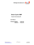
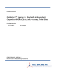
BioVision rev. 12/14 For research use only cAMP Direct Immunoassay Kit (Colorimetric) I. II. (Catalog #K371-100; 100 assays; Store at -20°C) Introduction: Adenosine 3’,5’-cyclic monophosphate (cyclic AMP, cAMP) is an important “second messenger” involved in many physiological processes. BioVision’s cAMP Assay Kit provides a direct competitive immunoassay for sensitive and quantitative determination of cAMP level in biological samples. The kit utilizes recombinant Protein G coated 96-well plate to efficiently anchor cAMP polyclonal antibody onto the plate. cAMP-HRP conjugate directly competes with cAMP from sample binding to the cAMP antibody on the plate. After incubation and washing, the amount of cAMP-HRP bound to the plate can easily be determined by reading HRP activity at OD450 nm. The intensity of OD450 nm is inversely proportional to the cAMP concentration in samples. The kit provides a new acetylation procedure to improve detection sensitivity significantly. The kit can detect ~0.1-10 pmol/well (or ~ 0.02 - 2 µM) cAMP samples. 10X cAMP Assay Buffer Standard cAMP (10 nmol) Neutralizing Buffer Acetylating Reagent A Acetylating Reagent B Rabbit Anti-cAMP pAb cAMP-HRP HRP Developer Protein G Coated Plate K371-100 Color Code Storage Part 100 assays Cap Color Temperature Number 25 ml 1 vial 7.5 ml 0.75 ml 1.5 ml 1 vial 1 vial 10 ml 1 each WM Yellow NM Violet Black Red Green Amber ------ +4°C -20°C +4°C +4°C +4°C -20°C -20°C +4°C -20°C K371-100-1 K371-100-2 K371-100-3 K371-100-4 K371-100-5 K371-100-6 K371-100-7 K371-100-8 K371-100-9 cAMP Assay Protocol: A. Reagent Preparations: Dilute the 10X cAMP Assay Buffer to 1X Assay Buffer with MilliQ water. Store at 4°C. Reconstitute the Standard cAMP (pellet may not be visible) in 1 ml of 0.1M HCl (not provided), vertex for 10 seconds to generate 10 pmol/µl cAMP standard stock solution. Dilute the rabbit anti-cAMP pAb and cAMP-HRP each with 1.1 ml of the 1X Assay Buffer as stock solutions, keep frozen. Unused Protein G coated strips can be kept at –20°C with descants, stable for up to 1 month after opening. The kit should be stored at –20°C. After opening and reconstitution, components can be stored as instructed in the kit contents above, stable for 1 to 2 months. B. General Consideration: Esterases in samples may degrade cAMP. Therefore, prepare samples in 0.1N HCl to inactivate esterase, and store at -80°C. Dilute your samples to ~0.1- 10 pmol/well (0.02-2 µM) cAMP range. Urine and tissue culture supernatant can be diluted in 10% 1M HCl and assayed directly. Plasma, serum, whole blood, and tissue homogenates often contain phosphodiesterases and large amount of immunoglobulins (IgGs) which may interfere with the assay. However, diluting these samples with 0.1M HCl can generally inactivate phosphodiesterases and lower the concentration of IgGs, making the samples suitable for the assay. Phosphodiesterases and IgGs can also be removed by 5% TCA precipitation or 10 kD molecular weight cut off micro centrifuge filters (BioVision Cat #1997-25). To determine whether interference is presence in your sample, you may make two different dilutions. If the two different dilutions of sample show good correlation in the final calculated cAMP concentrations, purification is not required; otherwise use TCA precipitation or 10 Kd molecular weight cut off microcentrifuge filters to remove any enzymes from samples. BioVision Incorporated 155 S. Milpitas Boulevard, Milpitas, CA 95035 USA Organic solvents in samples may interfere with the assay, which may need to be removed prior to the assay. C. Sample Preparation: Urine, Plasma and Culture Medium Samples: Urine and plasma may be tested directly with 1:20 to 1:100 dilutions in 0.1M HCl. Culture medium can also be tested with 1:10 to 1:200 dilutions in 0.1M HCl. (Note: RPMI medium may contain >350 fmol/µl cAMP). Cell Samples: Aspirate medium. Add 1 ml of 0.1M HCl for every 35 cm2 of surface area. Incubate at room temperature for 20 minutes. Scrape cells off the surface with a cell scraper. Dissociate sample by pipetting up and down until suspension is homogeneous. Transfer to a centrifuge tube and centrifuge at top speed for 10 min. The supernatant can be assayed directly. Protein concentration >1 mg/ml is recommended for reproducible results. Tissue Samples: Cyclic nucleotides may be metabolized quickly in tissue, so it is important to rapidly freeze tissues after collection (e.g., using liquid nitrogen). Weigh the frozen tissue and add 5-10 volume of 0.1M HCl. Homogenize the sample on ice using a Polytron-type homogenizer. Spin at top speed for 5 min and collect the supernatant. The supernatant may be assayed directly. Kit Contents: Component III. D. cAMP Assay: 1. 2. 3. 4. 5. 6. 7. 8. 9. The procedure described here includes an acetylation step which makes the cAMP assay much more sensitive and avoid the interferences of many components in samples. However, for routine assay of the well known samples, non-acetylation procedure may also be used, just skip the acetylation steps (Step 7 and 8). Prepare cAMP Standard Curve and Samples: Dilute 25 µl of the 10 pmol/l standard cAMP stock with 975 µl of 0.1M HCl to generate 0.25 pmol/l cAMP working solution. The diluted cAMP should be fresh, and used within 1 hour. Label 8 microcentrifuge tubes, 1.25, 0.625, 0.3125, 0.156, 0.078, 0.039, 0 , 0_B pmol/50 µl. (Note: these concentrations represent what will finally be in the wells after the dilutions mentioned below). Add 200 µl of the 0.25 pmol/µl cAMP standard into the tube labeled 1.25 pmol tube (enough for 20 assays). Add100 µl 0.1M HCl into the rest of tubes. Transfer 100 µl from the tube labeled 1.25 pmol tube to the labeled 0.625 pmol tube, mix, then transfer 100 µl into the labeled 0.3125 pmol tube. Continue the serial dilution by transferring 100 µl to 0.156, 0.078, 0.039 pmol tubes. Discard 100 µl from the 0.039 pmol tube. The diluted cAMP should be used within 1 hour. Label new tubes for test samples, add 100 µl each test sample per tube. We suggest using different dilutions for each sample (dilute with 0.1M HCl). Add 50 µl of Neutralizing Buffer to each tube (all standards cAMP and testing samples). Prepare Acetylating Reagent Mix: Mix 1 volume of Acetylating Reagent A (Violet cap) with two volumes of Acetylating Reagent B (Black cap) in a microtube. Prepare enough for the experiment (need 5 µl each sample and standard tubes). Use within 1 hour. Add 5 µl of the Acetylating Reagent Mix directly into each test solution (all standards and samples), IMMEDIATELY vortex 2-3 seconds following each addition without delay, one tube at a time, and incubate at room temperature for 10 min to acetylate cAMP. Add 845 µl 1X Assay Buffer into each tube to dilute the acetylation reagents, mix well. The acetylated standard and samples are ready for quantification. (If cAMP in your samples are very low, the acetylation reagents can be dried after step 8, without dilution step 9 to minimize the volume increase. Then reconstitute in a 50 -100 µl of Assay Buffer). Tel: 408-493-1800 | Fax: 408-493-1801 www.biovision.com | [email protected] Page 1 of 3 BioVision rev. 12/14 Quantification cAMP: 1. Add 50 µl of the acetylated Standard cAMP and test samples from Step 9 to the Protein G coated 96-well plate. 2. Add 10 µl of the reconstituted cAMP antibody per well to the standard cAMP and sample wells except the well with 0_B pmol cAMP. (Note: Do not add cAMP antibody into the well with 0_B pmol cAMP, instead add 10 µl of 1X Assay Buffer for background reading). Incubate for 1 hour at room temperature with gentle agitation. Note: It is recommended to use a repeating pipette to minimize pipette errors. 3. Add 10 µl of cAMP-HRP to each well, incubate for 1 hr at room temperature with agitation. 4. Wash 5 times with 200 µl 1X Assay Buffer each wash. Completely empty the wells by tapping the plate on a new paper towel after each wash step 5. Add 100 µl of HRP developer and develop for 1 hour at room temperature with agitation. 6. Stop the reaction by adding 100 µl of 1M HCl (not provided) to each well (sample color should change from blue to yellow). 7. Read the plate at OD 450 nm. Note: The OD450 nm reading may vary significantly among experiments depend on lot numbers, kit storage and experiment conditions. Therefore, samples and standard curve must be performed at the same time and using the same kit reagents. 8. Subtract OD450 nm background reading (the well with 0_B pmol cAMP) from all samples and standards. Plot standard curve to observe the linear portion, then replot only the linear portion and in Excel add a trendline, then use the trend line linear formula (y=mx+b) to calculate your sample concentrations. Calculate amount of cAMP in samples after correcting the for dilution factors. 9. Calculations: cAMP Concentration = Sa/Sv (pmol/µl or nmol/ml or µM) Where: Sa is cAMP amount (pmol) from standard curve. Sv is sample volume (µl) added into the assay wells after dilution factor correction. (b) (a) 0.3 cAMP (pm ole/µg protein) 1.2 1 O.D. 0.8 0.6 0.4 y = -0.55x + 1.0697 R² = 0.9617 0.2 0 0 0.5 1 cAMP (pmole) 1.5 0.25 0.2 0.15 0.1 0.05 0 Jurkat Cells HepG2 Cells Figure: (a) cAMP Standard Curve. (b) Measurement of cAMP in Jurkat Cells and HepG2 Cells. For suspension cultures, use between 2-4 x 10⁶ cells. Resuspend in ~ 500 µl 0.1 N HCl and prepare cell lysate as mentioned in the protocol. Use 100 µl of this cell lysate for acetylation reaction. Add 50 µl of the final acetylated reaction to the protein G coated wells and make up the volume to 50 µl using 1X Assay Buffer. Assay was performed following the kit protocol. For research use only RELATED PRODUCTS: Apoptosis Detection Kits & Reagents Annexin V Kits & Bulk Reagents Caspase Assay Kits & Reagents Mitochondrial Apoptosis Kits & Reagents Apoptotic DNA Ladder Detection Kits TUNEL-based DNA Fragmentation Assay Kits Apoptosis Inducers and siRNA Vectors Cell Fractionation System Mitochondria/Cytosol Fractionation Kit Nuclear/Cytosol Fractionation Kit Membrane Protein Extraction Kit Cytosol/Particulate Rapid Separation Kit Mammalian Cell Extraction Kit FractionPREP Fractionation System Cell Proliferation & Senescence Quick Cell Proliferation Assay Kit Senescence Detection Kit High Throughput Apoptosis/Cell Viability Assay Kits LDH-Cytotoxicity Assay Kit Bioluminescence Cytotoxicity Assay Kit Live/Dead Cell Staining Kit Cell Damage & Repair HDAC Fluorometric & Colorimetric Assays & Drug Discovery Kits HAT Colorimetric Assay Kit & Reagents DNA Damage Quantification Kit Glutathione & Nitric Oxide Fluorometric & Colorimetric Assay Kits Signal Transduction cAMP & cGMP Assay Kits Akt & JNK Activity Assay Kits Beta-Secretase Activity Assay Kit Obesity Research Recombinant Adiponectin, Survivin, & Leptin CETP & PLTP Activity Assay & Drug Discovery Kits Cholesterol and HDL/LDL Quantification Kits Fatty acid and Triglyceride Quantification Kits NAD(P)/NAD(P)H and ADP/ATP Assay Kits Antibodies & Recombinant Proteins (many) Adiponectin Proteins & Antibodies C5a Recombinant Protein Protein G & Protein A Bulk Quantity Growth Factors, Cytokines & Chemokines Antibodies to Apoptosis & Cell Signaling Molecules Metabolism Assay Kits Cholesterol Assay Kit HDL/LDL assay kit Glucose Assay Kit Lactate Assay Kit Pyruvate Assay Kit NAD(P)H/NAD(P) Assay Kit Glutathione Assay Kit Nitric Oxide Detection Kit FOR RESEARCH USE ONLY! Not to be used on humans. BioVision Incorporated 155 S. Milpitas Boulevard, Milpitas, CA 95035 USA Tel: 408-493-1800 | Fax: 408-493-1801 www.biovision.com | [email protected] Page 2 of 3 BioVision rev. 12/14 For research use only GENERAL TROUBLESHOOTING GUIDE: Problems Cause Solution Assay not working • Use of ice-cold assay buffer • Assay buffer must be at room temperature • Omission of a step in the protocol • Refer and follow the data sheet precisely • Plate read at incorrect wavelength • Check the wavelength in the data sheet and the filter settings of the instrument • Use of a different 96-well plate Samples with erratic readings • Use of an incompatible sample type • Samples prepared in a different buffer • Samples were not deproteinated • Cell/ tissue samples were not completely homogenized Lower/ Higher readings in Samples and Standards Unanticipated results • Use the assay buffer provided in the kit or refer data sheet for instructions • Use the 10 kDa spin cut-off filter or PCA precipitation as indicated or use 0.1 M HCl to inactivate phosphodiesterases • Use Dounce homogenizer (increase the number of strokes); observe for lysis under microscope • Samples used after multiple free-thaw cycles • Aliquot and freeze samples if needed to use multiple times • Presence of interfering substance in the sample • Troubleshoot if needed, deproteinize samples • Use of old or inappropriately stored samples • Use fresh samples or store at correct temperatures till use • Improperly thawed components • Thaw all components completely and mix gently before use • Use of expired kit or improperly stored reagents • Always check the expiry date and store the components appropriately • Allowing the reagents to sit for extended times on ice • Always thaw and prepare fresh reaction mix before use • Incorrect volumes used or protocol properly followed • Refer datasheet & verify correct incubation times and temperatures aceylated and treated one at a time through this treatment. • Use calibrated pipettes and aliquot correctly • Use of partially thawed components • Thaw and resuspend all components before preparing the reaction mix • Pipetting errors in the standard • Avoid pipetting small volumes • Pipetting errors in the reaction mix • Prepare a master reaction mix whenever possible • Air bubbles formed in well • Pipette gently against the wall of the tubes • Standard stock is at an incorrect concentration • Always refer the dilutions in the data sheet • Calculation errors • Recheck calculations after referring the data sheet; Use only the linear portion of curve. • Substituting reagents from older kits/ lots • Use fresh components from the same kit • Measured at incorrect wavelength • Check the equipment and the filter setting • Samples contain interfering substances • Troubleshoot if it interferes with the kit • Use of incompatible sample type • Refer data sheet to check if sample is compatible with the kit or optimization is needed • Sample readings above/below the linear range • Concentrate/ Dilute sample so as to be in the linear range • Incorrect incubation times or temperatures Readings do not follow a linear pattern for Standard curve • Fluorescence: Black plates (clear bottoms) ; Luminescence: White plates ; Colorimeters: Clear plates • Refer data sheet for details about incompatible samples (Samples should initially be at ≥ 1 mg/ml protein) ● Samples/standards Note: The most probable list of causes is under each problem section. Causes/ Solutions may overlap with other problems. BioVision Incorporated 155 S. Milpitas Boulevard, Milpitas, CA 95035 USA Tel: 408-493-1800 | Fax: 408-493-1801 www.biovision.com | [email protected] Page 3 of 3