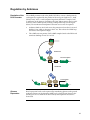
1
pBAD Directional TOPO® Expression Kit Five-minute, directional TOPO® cloning of blunt-end PCR products into vectors for soluble, regulated expression and purification in E. coli Catalog no. K4202-01 Revision date : 7 June 2010 Manual part no. 25-0526 MAN0000286 Corporate Headquarters Invitrogen Corporation 1600 Faraday Avenue Carlsbad, CA 92008 T: 1 760 603 7200 F: 1 760 602 6500 E: [email protected] For country-specific contact information visit our web site at www.invitrogen.com User Manual ii Contents Kit Contents and Storage ..................................................................................................................................... v Introduction ................................................................................................................... 1 Description of the System.....................................................................................................................................1 How Directional TOPO® Cloning Works ...........................................................................................................2 Arabinose Regulation and Thioredoxin .............................................................................................................3 Experiment Outline ...............................................................................................................................................4 Methods ......................................................................................................................... 5 Designing PCR Primers ........................................................................................................................................5 Producing Blunt-End PCR Products ...................................................................................................................9 Performing the TOPO® Cloning Reaction ........................................................................................................10 Transforming One Shot® TOP10 Competent Cells..........................................................................................12 Analyzing Transformants...................................................................................................................................15 Expressing the PCR Product ..............................................................................................................................17 Analyzing Samples..............................................................................................................................................19 Troubleshooting ...................................................................................................................................................22 Appendix...................................................................................................................... 24 Recipes...................................................................................................................................................................24 Purifying the PCR Products ...............................................................................................................................26 Performing the Control Reactions .....................................................................................................................28 Map and Features of pBAD202/D-TOPO® ......................................................................................................30 Map of pBAD202/D/lacZ ..................................................................................................................................32 Regulation by Arabinose ....................................................................................................................................33 Accessory Products..............................................................................................................................................34 Technical Support ................................................................................................................................................36 Purchaser Notification ........................................................................................................................................37 References .............................................................................................................................................................39 iii iv Kit Contents and Storage Shipping/Storage The pBAD Directional TOPO® Expression Kit is shipped on dry ice. Each kit contains a box with pBAD/D-TOPO® reagents (Box 1), a box with One Shot® TOP10 Chemically Competent E. coli (Box 2), and a stab of LMG194. Store Box 1 at –20°C and Box 2 at –80°C. Store the LMG194 stab at 4°C. pBAD/D-TOPO® Reagents pBAD/D-TOPO® reagents (Box 1) are listed below. Note that you must supply a thermostable, proofreading polymerase, and the appropriate PCR buffer. Store Box 1 at –20°C. Item Concentration ® Amount pBAD202/D-TOPO vector 15–20 ng/μL plasmid DNA in: 50% glycerol 50 mM Tris-HCl, pH 7.4 (at 25°C) 1 mM EDTA 1 mM DTT 0.1% Triton X-100 100 μg/mL BSA 30 M bromophenol blue 20 μL dNTP Mix 12.5 mM dATP; 12.5 mM dCTP 12.5 mM dGTP; 12.5 mM dTTP in water, pH 8 10 μL Salt Solution 1.2 M NaCl 0.06 M MgCl2 50 μL Sterile Water -- 1 mL 20% L-Arabinose 20% in sterile water 1 mL TrxFus Forward Sequencing Primer 0.1 μg/μL in TE Buffer, pH 8 20 μL pBAD Reverse Sequencing Primer 0.1 μg/μL in TE Buffer, pH 8 20 μL Control PCR Primers 0.1 μg/μL each in TE Buffer, pH 8 10 μL Control PCR Template 0.1 μg/μL in TE Buffer, pH 8 10 μL pBAD202/D/lacZ Expression Control Plasmid 0.01 μg/μL in TE buffer, pH 8 10 μL Sequences of the Primers The table below provides the sequences of the Trx Forward and pBAD Reverse sequencing primers. Two micrograms of each primer are supplied. Primer Sequence pMoles Supplied TrxFus Forward 5´-TTCCTCGACGCTAACCTG-3´ 371 pBAD Reverse 5´-GATTTAATCTGTATCAGG-3´ 363 Continued on next page v Kit Contents and Storage, Continued One Shot® TOP10 Reagents The table below lists the items included in the One Shot® TOP10 Chemically Competent E. coli kit (Box 2). Transformation efficiency is 1 109 cfu/μg DNA. Store Box 2 at –80°C. Item Amount 21 50 μL TOP10 cells -- S.O.C. Medium 2% Tryptone 0.5% Yeast Extract 10 mM NaCl 2.5 mM KCl 10 mM MgCl2 10 mM MgSO4 20 mM glucose 6 mL 10 pg/μL in: 5 mM Tris-HCl 0.5 mM EDTA, pH 8.0 50 μL (may be stored at room temperature or 4°C) pUC19 Control DNA Genotype of TOP10 Composition Use this strain for general cloning of blunt-end PCR products into the pBAD202/D-TOPO® vector. Genotype: F– mcrA (mrr-hsdRMS-mcrBC) 80lacZM15 lac74 recA1 araD139 (ara-leu)7697 galU galK rpsL (StrR) endA1 nupG Genotype of LMG194 F- lacX74 galE thi rpsL phoA (Pvu II) ara714 leu::Tn10. Note: This strain is deleted for araBADC. It is also streptomycin and tetracycline resistant. Preparing LMG194 Store the LMG194 E. coli stab supplied with the kit at 4°C. Upon receipt, we recommend that you prepare a set of LMG194 glycerol master stocks within two Glycerol Stocks weeks of receiving the kit. 1. Streak a small portion of the LMG194 cells from the stab on an LB plate containing the appropriate antibiotics and incubate at 37°C overnight. 2. Isolate a single colony and inoculate into 5–10 mL of LB medium with the appropriate antibiotics. 3. Grow the culture to stationary phase (OD600 = 1–2). 4. Mix 0.8 mL of culture with 0.2 mL of sterile glycerol and transfer to a cryovial. Store at –80°C. Use one master stock to create working stocks for regular use. vi Introduction Description of the System Product Features The pBAD Directional TOPO® Expression Kit utilizes a highly efficient, 5-minute cloning strategy ("TOPO® Cloning") to directionally clone a blunt-end PCR product into a vector for soluble, regulated expression and simplified protein purification in E. coli. Blunt-end PCR products clone directionally at greater than 90% efficiency with no ligase, post-PCR procedures, or restriction enzymes required. In addition, pBAD202/D-TOPO® vector contains the His-Patch (HP) thioredoxin leader for increased translation efficiency and solubility of recombinant fusion proteins. Expression in E. coli is driven by the araBAD promoter (PBAD). The AraC gene product encoded on the pBAD202/D-TOPO® vector positively regulates this promoter. pBAD202/ D-TOPO® Vector pBAD202/D-TOPO® is designed to facilitate rapid, directional TOPO® Cloning of blunt-end PCR products for regulated expression in E. coli. Features of the vector include: araBAD promoter (PBAD) for tight, dose-dependent regulation of heterologous gene expression N-terminal His-Patch thioredoxin for increased translation efficiency and solubility of heterologous proteins Directional TOPO® Cloning site for rapid and efficient directional cloning of a blunt-end PCR product (see next page for more information) C-terminal fusion tag for detection and purification of recombinant fusion proteins Kanamycin resistance gene for selection in E. coli araC gene encoding a regulatory protein for tight regulation of the PBAD promoter pUC origin for maintenance in E. coli. Note: Although the pBAD202/D-TOPO® vector contains a pUC origin, they act as lowcopy number plasmids, resulting in lower yields of the vectors. 1 How Directional TOPO® Cloning Works How Topoisomerase I Works Topoisomerase I from Vaccinia virus binds to duplex DNA at specific sites and cleaves the phosphodiester backbone after 5-CCCTT in one strand (Shuman, 1991). The energy from the broken phosphodiester backbone is conserved by formation of a covalent bond between the 3 phosphate of the cleaved strand and a tyrosyl residue (Tyr-274) of topoisomerase I. The phospho-tyrosyl bond between the DNA and enzyme can subsequently be attacked by the 5 hydroxyl of the original cleaved strand, reversing the reaction and releasing topoisomerase (Shuman, 1994). TOPO® Cloning exploits this reaction to efficiently clone PCR products. Directional TOPO® Cloning Directional joining of double-strand DNA using TOPO®-charged oligonucleotides occurs by adding a 3 single-stranded end (overhang) to the incoming DNA (Cheng and Shuman, 2000). This single-stranded overhang is identical to the 5 end of the TOPO®-charged DNA fragment. At Invitrogen, this idea has been modified by adding a 4 nucleotide overhang sequence to the TOPO®-charged DNA and adapting it to a ‘whole vector’ format. In this system, PCR products are directionally cloned by adding four bases to the forward primer (CACC). The overhang in the cloning vector (GTGG) invades the 5 end of the PCR product, anneals to the added bases, and stabilizes the PCR product in the correct orientation. Inserts can be cloned in the correct orientation with efficiencies equal to or greater than 90%. Topoisomerase Tyr-274 P O ----CCCTT ----GGGAAGTGG Overhang CACC ATG NNN --- --- --- NNN GTGG TAC NNN --- --- --- NNN PCR product Overhang invades double-stranded DNA, displacing the bottom strand. Tyr-274 AAG GG---TTC CC---- O P Topoisomerase GT GG ----CCCTTCACC ATG NNN --- --- --- NNN AAG GG- ------GGGAAGTGG TAC NNN --- --- --- NNN TTC CC- --- 2 Arabinose Regulation and Thioredoxin Regulation of Expression by Arabinose In the presence of arabinose, expression from PBAD is induced while only very low levels of transcription are observed from PBAD in the absence of arabinose (Lee, 1980; Lee et al., 1987). Uninduced levels are repressed even further by growth in the presence of glucose (0.1% to 0.2%). Glucose reduces the levels of 3´, 5´-cyclic AMP, lowering expression from the catabolite-repressed PBAD promoter (Miyada et al., 1984). By varying the concentration of arabinose, protein expression levels can be optimized to ensure maximum expression of protein. In addition, the tight regulation of PBAD by AraC is useful for expression of potentially toxic or essential genes (Carson et al., 1991; Dalbey and Wickner, 1985; Guzman et al., 1992; Kuhn and Wickner, 1985; Russell et al., 1989; San Millan et al., 1989). For more information on the mechanism of expression and repression of the ara regulon, see page 33 or refer to Schleif, 1992. Thioredoxin The 11.7 kDa thioredoxin protein is found in yeast, plants, and mammals, as well as in bacteria. It was originally isolated from E. coli as a hydrogen donor for ribonuclease reductase (see Holmgren, 1985 for a review). The gene has been completely sequenced (Wallace and Kushner, 1984). The protein has been crystallized and its three-dimensional structure determined (Katti et al., 1990). When overexpressed in E. coli, thioredoxin is able to accumulate to approximately 40% of the total cellular protein and still remains soluble. When used as a fusion partner, thioredoxin can increase translation efficiency and, in some cases, solubility of eukaryotic proteins expressed in E. coli. Examples of eukaryotic proteins that have been produced as soluble C-terminal fusions to the thioredoxin protein in E. coli (LaVallie et al., 1993) include: His-Patch Thioredoxin Murine interleukin-2 Human interleukin-3 Murine interleukin-4 Murine interleukin-5 Human macrophage colony stimulating factor Murine steel factor Murine leukemia inhibitory factor Human bone morphogenetic protein-2 The thioredoxin protein has been mutated to contain a metal binding domain, and is termed “His-Patch thioredoxin”. To create a metal binding domain in the thioredoxin protein, the glutamate residue at position 32 and the glutamine residue at position 64 were mutated to histidine residues. When His-Patch thioredoxin folds, the histidines at positions 32 and 64 interact with a native histidine at position 8 to form a “patch”. This histidine patch has been shown to have high affinity for divalent cations (Lu et al., 1996). His-Patch thioredoxin (HP-thioredoxin) proteins can therefore be purified on metal chelating resins (e.g. ProBond™). 3 Experiment Outline Experiment Outline The table below describes the general steps needed to clone and express your gene of interest. For more details, refer to the pages indicated. Step Page 1 Design PCR primers to clone your gene of interest in frame with the N-terminal His-Patch thioredoxin and C-terminal V5 epitope and polyhistidine tag, if desired. Consult the diagram on page 8 to help you design your PCR primers. 2 Produce your blunt-end PCR product. 3 4 4 Action ® 5–8 9 ® TOPO Clone your PCR product into pBAD202/D-TOPO and transform into One Shot® TOP10 E. coli. Select transformants on LB plates containing the appropriate antibiotic. Analyze transformants by restriction digestion or PCR. 10–14 15 5 Select a transformant with the correct restriction pattern and sequence it to confirm that your gene is cloned in frame with the N-terminal His-Patch thioredoxin and C-terminal V5 epitope and polyhistidine tag, if desired. 6 Select positive transformant and induce expression with arabinose. 17–18 7 Assay for expression of your protein of interest. 19–21 15 Methods Designing PCR Primers Designing Your PCR Primers Specific Features for Expression Guidelines to Design the Forward PCR Primer Example of Forward Primer Design The design of the PCR primers to amplify your gene of interest is critical for expression. Consider the following when designing your PCR primers: Sequences required to facilitate directional cloning (see below). Whether or not you wish to clone your PCR product in frame with the C-terminal V5 epitope and polyhistidine region. The pBAD202/D-TOPO® vector contains the following features to facilitate expression: Initiation ATG that is properly spaced from the optimized ribosome binding site to ensure optimal translation. HP-thioredoxin, which acts as a translation leader to facilitate high-level expression and in some cases, solubility. HP-thioredoxin can be removed after protein purification using enterokinase (e.g. EKMax™, see page 34). When designing your forward PCR primer, consider the following points below. Refer to page 8 for a diagram of the TOPO® Cloning site for the pBAD202/D-TOPO® vector. To enable directional cloning, the forward PCR primer must contain the sequence CACC at the 5 end of the primer (see Example below). The 4 nucleotides, CACC, base pair with the overhang sequence, GTGG, in the pBAD202/D-TOPO® vector. If you wish to include the N-terminal thioredoxin, design the forward PCR primer to ensure that your protein is in frame with the N-terminal leader peptide. Below is the DNA sequence of the N-terminus of a theoretical protein and the proposed sequence for your forward PCR primer: DNA sequence: 5-GTA GGA TCT GAT AAA Proposed Forward PCR primer: 5-C ACC GTA GGA TCT GAT AAA The first three base pairs of the PCR product following the 5 CACC overhang will constitute a functional codon. Continued on next page 5 Designing PCR Primers, Continued Guidelines to Design the Reverse Primer Example #1 of Reverse Primer Design When designing your reverse PCR primer, consider the points below. Refer to page 8 for a diagram of the TOPO® Cloning site on the pBAD202/D-TOPO® vector. To ensure that your PCR product clones directionally with high efficiency, the reverse PCR primer MUST NOT be complementary to the overhang sequence GTGG at the 5 end. A one base pair mismatch can reduce the directional cloning efficiency from 90% to 50%, increasing the likelihood of your ORF cloning in the opposite orientation (see Example #1 below). We have not observed evidence of PCR products cloning in the opposite orientation from a two base pair mismatch. To fuse your PCR product in frame with the C-terminal V5 epitope tag and polyhistidine region, design the reverse PCR primer to remove the native stop codon in the gene of interest (see Example #2 on the next page). If you do NOT wish to fuse your PCR product in frame with the C-terminal V5 epitope tag and polyhistidine region, include the native sequence containing the stop codon in the reverse primer or make sure the stop codon is upstream from the reverse PCR primer binding site (see Example #2 on the next page). Below is the sequence of the C-terminus of a theoretical protein. You want to fuse the protein in frame with a C-terminal tag. The stop codon is underlined. DNA sequence: AAG TCG GAG CAC TCG ACG ACG GTG TAG-3 One solution is to design the reverse PCR primer to start with the codon just upstream of the stop codon, but the last two codons contain GTGG (underlined below), which is identical to the 4 bp overhang sequence. As a result, the reverse primer will be complementary to the 4 bp overhang sequence, increasing the probability that the PCR product will clone in the opposite orientation. You want to avoid this situation. DNA sequence: AAG TCG GAG CAC TCG ACG ACG GTG TAG-3 Proposed Reverse PCR primer sequence: TG AGC TGC TGC CAC AAA-5 Another solution is to design the reverse primer so that it hybridizes just downstream of the stop codon, but still includes the C-terminus of the ORF. Note that you need to replace the stop codon with a codon for an innocuous amino acid such as glycine, alanine, or lysine. Continued on next page 6 Designing PCR Primers, Continued Example #2 of Reverse Primer Design Below is the sequence of the C-terminus of a theoretical protein. The stop codon is underlined. …GCG GTT AAG TCG GAG CAC TCG ACG ACT GCA TAG-3 To fuse the ORF in frame with a C-terminal tag, remove the stop codon by starting with nucleotides homologous to the last codon (TGC) and continue upstream. The reverse primer will be: 5-TGC AGT CGT CGA GTG CTC CGA CTT-3 This will amplify the C-terminus without the stop codon and allow you to join the ORF in frame with a C-terminal tag. If you do not want to join the ORF in frame with a C-terminal tag, simply design the reverse primer to include the stop codon. 5-CTA TGC AGT CGT CGA GTG CTC CGA CTT-3 Remember that the pBAD202/D-TOPO® vector accepts blunt-end PCR products. Refer to the diagram of the TOPO® Cloning site on page 8 to help you design your primers. When synthesizing PCR primers, do not add 5’ phosphates to the primers, because 5’ phosphates prevent the synthesized PCR product from ligating into the pBAD202/D-TOPO® vector. We recommend that you gel-purify your oligonucleotides, especially if they are long (> 30 nucleotides). Important Continued on next page 7 Designing PCR Primers, Continued TOPO® Cloning Site Use the diagram below to design suitable PCR primers to clone and express your PCR product in pBAD202/D-TOPO®. Restriction sites are labeled to indicate the actual cleavage site. The vector sequence of pBAD202/D-TOPO® is available for downloading at www.invitrogen.com or by contacting Technical Support (page 36). O2 Region 1 AAGAAACCAA TTGTCCATAT TGCATCAGAC ATTGCCGTCA CTGCGTCTTT TACTGGCTCT TCTCGCTAAC CAAACCGGTA 81 ACCCCGCTTA TTAAAAGCAT TCTGTAACAA AGCGGGACCA AAGCCATGAC AAAAACGCGT AACAAAAGTG TCTATAATCA CAP binding site O1 Region 161 CGGCAGAAAA GTCCACATTG ATTATTTGCA CGGCGTCACA CTTTGCTATG CCATAGCATT TTTATCCATA AGATTAGCGG -35 I2 and I1 Region -10 241 ATCCTACCTG ACGCTTTTTA TCGCAACTCT CTACTGTTTC TCCATACCCG TTTTTTTGGG CTAGAAATAA TTTTGTTTAA RBS His-patch (HP) thioredoxin site Nco I 321 CTTTAAGAAG GAGATATACA TACCC ATG GGA TCT GAT AAA ATT ATT CAT CTG ACT GAT GAT TCT TTT GAT Met Gly Ser Asp Lys Ile Ile His Leu Thr Asp Asp Ser Phe Asp 391 ACT GAT GTA CTT AAG GCA GAT GGT GCA ATC CTG GTT GAT TTC TGG GCA CAC TGG TGC GGT CCG TGC Thr Asp Val Leu Lys Ala Asp Gly Ala Ile Leu Val Asp Phe Trp Ala His Trp Cys Gly Pro Cys 457 AAA ATG ATC GCT CCG ATT CTG GAT GAA ATC GCT GAC GAA TAT CAG GGC AAA CTG ACC GTT GCA AAA Lys Met Ile Ala Pro Ile Leu Asp Glu Ile Ala Asp Glu Tyr Gln Gly Lys Leu Thr Val Ala Lys 523 CTG AAC ATC GAT CAC AAC CCG GGC ACT GCG CCG AAA TAT GGC ATC CGT GGT ATC CCG ACT CTG CTG Leu Asn Ile Asp His Asn Pro Gly Thr Ala Pro Lys Tyr Gly Ile Arg Gly Ile Pro Thr Leu Leu 589 CTG TTC AAA AAC GGT GAA GTG GCG GCA ACC AAA GTG GGT GCA CTG TCT AAA GGT CAG TTG AAA GAG Leu Phe Lys Asn Gly Glu Val Ala Ala Thr Lys Val Gly Ala Leu Ser Lys Gly Gln Leu Lys Glu TrxFus Forward priming site NgoM I Nae I EK recognition site EK cleavage site 655 TTC CTC GAC GCT AAC CTG GCC GGC TCT GGA TCC GGT GAT GAC GAT GAC AAG CTG GGA ATT GAT CCC Phe Leu Asp Ala Asn Leu Ala Gly Ser Gly Ser Gly Asp Asp Asp Asp Lys Leu Gly Ile Asp Pro V5 epitope Sac I Hind III ... ... AAG GGC GAG CTC AAG CTT GAA GGT AAG CCT ATC CCT AAC CCT CTC CTC GGT CTC ... ... TTC CCG Lys Gly Glu Leu Lys Leu Glu Gly Lys Pro Ile Pro Asn Pro Leu Leu Gly Leu G TG 721 TTC ACC AAG TGG Phe Thr G ... ... Polyhistidine (6xHis) region Pme I 781 GAT TCT ACG CGT ACC GGT CAT CAT CAC CAT CAC CAT TGA GTTTAAACGG TCTCCAGCTT GGCTGTTTTG Asp Ser Thr Arg Thr Gly His His His His His His *** pBAD Reverse priming site 850 GCGGATGAGA GAAGATTTTC AGCCTGATAC AGATTAAATC AGAACGCAGA AGCGGTCTGA TAAAACAGAA TTTGCCTGGC rrnB T1 and T2 transcriptional terminator 930 GGCAGTAGCG CGGTGGTCCC ACCTGACCCC ATGCCGAACT CAGAAGTGAA ACGCCGTAGC GCCGATGGTA GTGTGGGGTC 1010 TCCCCATGCG AGAGTAGGGA ACTGCCAGGC ATCAAATAAA ACGAAAGGCT CAGTCGAAAG ACTGGGCCTT TCGTTTTATC 8 Producing Blunt-End PCR Products Introduction After you have decided on a PCR strategy and have synthesized the primers, produce your blunt-end PCR product using any thermostable, proofreading polymerase. We recommend Platinum® Pfx DNA Polymerase, AccuPrime™ Pfx DNA Polymerase, or Pfx50™ DNA Polymerase, available separately from Invitrogen (see page 34 for ordering information). Follow the guidelines below to produce your blunt-end PCR product. Materials Supplied by the User Thermocycler and thermostable, proofreading polymerase 10X PCR buffer appropriate for your polymerase DNA template and primers for PCR product Note: dNTPs (adjusted to pH 8) are provided in the kit. Set up a 25 μL or 50 μL PCR reaction using the guidelines below. Producing BluntEnd PCR Products Follow the instructions and recommendations provided by the manufacturer of your thermostable, proofreading polymerase to produce blunt-end PCR products. Checking the PCR Product Use the cycling parameters suitable for your primers and template. Make sure to optimize PCR conditions to produce a single, discrete PCR product. Use a 7 to 30 minute final extension to ensure that all PCR products are completely extended. After cycling, place the tube on ice or store at –20ºC for up to 2 weeks. Proceed to Checking the PCR Product, below. After you have produced your blunt-end PCR product, use agarose gel electrophoresis to verify the quality and quantity of your PCR product. Check for the following outcomes below. Be sure you have a single, discrete band of the correct size. If you do not have a single, discrete band, follow the manufacturer’s recommendations to optimize your PCR with the polymerase of your choice. Alternatively, gelpurify the desired product (see pages 26–27). Estimate the concentration of your PCR product. Use this information when setting up your TOPO® Cloning reaction (see Amount of PCR Product to Use in the TOPO® Cloning Reaction, next page for details). 9 Performing the TOPO® Cloning Reaction Introduction Once you have produced the desired PCR product, you are ready to TOPO® Clone it into the pBAD202/D-TOPO® vector and transform the recombinant vector into One Shot® TOP10 E. coli. It is important to have everything you need set up and ready to use to ensure best results. We suggest that you read this section and the section entitled Transforming One Shot® TOP10 Competent Cells (pages 12–14) before beginning. If this is the first time you have TOPO® Cloned, perform the control reactions on pages 28–29 in parallel with your samples. Amount of PCR Product to Use in the TOPO® Cloning Reaction When performing directional TOPO® Cloning, we have found that the molar ratio of PCR product:TOPO® vector used in the reaction is critical to its success. To obtain the highest TOPO® Cloning efficiency, use a 0.5:1 to 2:1 molar ratio of PCR product:TOPO® vector (see figure below). Note that the TOPO® Cloning efficiency decreases significantly if the ratio of PCR product: TOPO® vector is <0.1:1 or >5:1. These results are generally obtained if too little PCR product is used (i.e. PCR product is too dilute) or if too much PCR product is used in the TOPO® Cloning reaction. If you have quantitated the yield of your PCR product, you may need to adjust the concentration of your PCR product before proceeding to TOPO® Cloning. Tip: For the pBAD202/D-TOPO® vector, using 1–5 ng of a 1 kb PCR product or 5–10 ng of a 2 kb PCR product in a TOPO® Cloning reaction generally results in a suitable number of colonies. Relative Activity (colonies/reaction) 100% 50% 0% 0.1 1 10 PCR Product:Ve ctor (Molar Ratio) Continued on next page 10 Performing the TOPO® Cloning Reaction, Continued Perform TOPO® Cloning in a reaction buffer containing salt (i.e. using the stock salt solution provided in the kit). Note that the amount of salt added to the TOPO® Cloning reaction varies depending on whether you plan to transform chemically competent cells (provided) or electrocompetent cells (see page 34 for ordering information). Using Salt Solution in the TOPO® Cloning Reaction If you are transforming chemically competent E. coli, use the stock Salt Solution as supplied and set up the TOPO® Cloning reaction as directed below. If you are transforming electrocompetent E. coli, reduce the amount of salt in the TOPO® Cloning reaction to 50 mM NaCl, 2.5 mM MgCl2 to prevent arcing during electroporation. Dilute the stock Salt Solution 4-fold with water to prepare a 300 mM NaCl, 15 mM MgCl2 Dilute Salt Solution. Use the Dilute Salt Solution to set up the TOPO® Cloning reaction as directed below. Use the procedure below to perform the TOPO® Cloning reaction. Set up the TOPO® Cloning reaction depending on whether you plan to transform chemically competent E. coli or electrocompetent E. coli. For optimal results, use a 0.5:1 to 2:1 molar ratio of PCR product:TOPO® vector in your TOPO® Cloning reaction. Performing the TOPO® Cloning Reaction Note: The blue color of the TOPO® vector solution is normal and is used to visualize the solution. Reagents* Chemically Competent E. coli Electrocompetent E. coli Fresh PCR product 0.5 to 4 μL 0.5 to 4 μL Salt Solution 1 μL -- Dilute Salt Solution (1:4) -- 1 μL Sterile Water add to a final volume of 5 μL add to a final volume of 5 μL ® TOPO vector 1 μL 1 μL Final volume 6 μL 6 μL *Store all reagents at –20°C when finished. Store salt solutions and water at room temperature or 4°C. 1. Mix reaction gently and incubate for 5 minutes at room temperature (22–23°C). Note: For most applications, 5 minutes yields plenty of colonies for analysis. Depending on your needs, you can vary the length of the TOPO® Cloning reaction from 30 seconds to 30 minutes. For routine subcloning of PCR products, 30 seconds may be sufficient. For large PCR products (> 1 kb) or if you are TOPO® Cloning a pool of PCR products, increasing the reaction time may yield more colonies. 2. Place the reaction on ice and proceed to Transforming One Shot® TOP10 Competent Cells, next page. Note: You may store the TOPO® Cloning reaction at –20°C overnight. 11 Transforming One Shot® TOP10 Competent Cells Introduction Once you have performed the TOPO® Cloning reaction, you will transform your pBAD202/D-TOPO® construct into competent E. coli. One Shot® TOP10 Chemically Competent E. coli are included with the kit to facilitate transformation, however, you may also transform electrocompetent cells. This section provides protocols to transform chemically competent or electrocompetent E. coli. Materials Supplied by the User Preparing for Transformation 42°C water bath (or electroporator with cuvettes, optional) LB plates containing 50 μg/mL kanamycin (two for each transformation) 37°C shaking and non-shaking incubator For each transformation, you will need one vial of competent cells and two selective plates. Equilibrate a water bath to 42°C (for chemical transformation) or set up your electroporator if you are using electrocompetent E. coli. Warm the vial of S.O.C. medium from Box 2 to room temperature. Warm LB plates containing 50 μg/mL kanamycin at 37°C for 30 minutes. Thaw on ice 1 vial of One Shot® TOP10 cells from Box 2 for each transformation. Continued on next page 12 Transforming One Shot® TOP10 Competent Cells, Continued Important The number of colonies obtained after transforming the pBAD202/D-TOPO® vector into One Shot® TOP10 cells is generally much lower when compared to the number of colonies obtained after transforming other TOPO® vectors (e.g. pCR®T7 TOPO®). Directional TOPO® Cloning generally yields 2 to 5-fold fewer colonies than traditional bidirectional TOPO TA Cloning®. Transforming low-copy number TOPO® plasmids generally yields 2 to 5-fold fewer colonies than transforming high-copy number TOPO® plasmids. To compensate for the lower transformation efficiency using the pBAD202/D-TOPO®: Increase the amount of TOPO® Cloning reaction that you transform into TOP10 cells (use 3 μL). Increase the amount of transformed cells that you plate (use 100–200 μL for chemically competent cells and 50–100 μL for electrocompetent cells). Example: When directionally TOPO® Cloning a 750 bp test insert into the pBAD202/D-TOPO® vector, we generally obtain 500–1,500 total colonies. Although fewer total colonies are obtained, greater than 90% of the colonies contain plasmid with your PCR insert in the correct orientation. One Shot® TOP10 Chemical Transformation Protocol 1. Add 3 μL of the TOPO® Cloning reaction from Performing the TOPO® Cloning Reaction, Step 2, page 11 into a vial of One Shot® TOP10 Chemically Competent E. coli and mix gently. Do not mix by pipetting up and down. 2. Incubate on ice for 5 to 30 minutes. Note: Longer incubations on ice seem to have a minimal effect on transformation efficiency. The length of the incubation is at the user’s discretion. 3. Heat-shock the cells for 30 seconds at 42°C without shaking. 4. Immediately transfer the tubes to ice. 5. Add 250 μL of room temperature S.O.C. medium to the tubes. 6. Cap the tube tightly and shake the tube horizontally (200 rpm) at 37°C for 1 hour. 7. Spread 100–200 μL from each transformation on a prewarmed selective plate and incubate overnight at 37°C. We recommend that you plate two different volumes to ensure that at least one plate will have well-spaced colonies. 8. An efficient TOPO® Cloning reaction may produce several hundred colonies. Pick ~5 colonies for analysis (see Analyzing Positive Clones, page 15). Continued on next page 13 Transforming One Shot® TOP10 Competent Cells, Continued Transformation by Electroporation Use ONLY electrocompetent cells for electroporation to avoid arcing. Do not use the One Shot® TOP10 chemically competent cells for electroporation. 1. Add 3 μL of the TOPO® Cloning reaction from Performing the TOPO® Cloning Reaction, Step 2, page 11 into a 0.1 cm cuvette containing 50 μL of electrocompetent E. coli and mix gently. Do not mix by pipetting up and down. Avoid formation of bubbles. 2. Electroporate your samples using your own protocol and your electroporator. MEND ION AT RECOM Note: If you have problems with arcing, see below. 3. Immediately add 250 μL of room temperature S.O.C. medium to the cuvette. 4. Transfer the solution to a 15 mL snap-cap tube (e.g. Falcon) and shake for at least 1 hour at 37°C to allow expression of the antibiotic resistance gene. 5. Spread 50–100 μL from each transformation on a prewarmed selective plate and incubate overnight at 37°C. We recommend that you plate two different volumes to ensure that at least one plate will have well-spaced colonies. 6. An efficient TOPO® Cloning reaction may produce several hundred colonies. Pick ~5 colonies for analysis (see Analyzing Positive Clones, page 15). To prevent arcing of your samples during electroporation, the volume of cells should be between 50 μL and 80 μL (0.1 cm cuvettes) or 100 μL to 200 μL (0.2 cm cuvettes). If you experience arcing during transformation, try one of the following suggestions: 14 Reduce the voltage normally used to charge your electroporator by 10% Reduce the pulse length by reducing the load resistance to 100 ohms Ethanol precipitate the TOPO® Cloning reaction and resuspend in water prior to electroporation Analyzing Transformants Analyzing Positive 1. Pick 5 colonies and culture them overnight in LB or SOB medium containing 50 μg/mL kanamycin. Clones 2. Isolate plasmid DNA using your method of choice. We recommend using the PureLink™ HQ Mini Plasmid Purification or PureLink™ HiPure Plasmid Miniprep kits (see page 34 for ordering information). Refer to www.invitrogen.com or contact Technical Support for more information on a large selection of plasmid purification columns. Note: Because the pBAD202/D-TOPO® vector acts as a low-copy number plasmid, you may need to increase the amount of bacterial culture to obtain enough plasmid DNA for sequencing or analysis purposes. Use extra care during purification to obtain plasmid DNA of sufficiently pure quality for sequencing (see below). 3. Sequencing Important Analyze the plasmids by restriction analysis to confirm the presence and correct orientation of the insert. Use a restriction enzyme or a combination of enzymes that cut once in the vector and once in the insert. You may sequence your construct to confirm that your gene is in frame with the N-terminal His-Patch thioredoxin peptide and the C-terminal V5 epitope and polyhistidine (6His) tag. The TrxFus Forward and pBAD Reverse primers are included in the kit to help you sequence your insert. Refer to the diagram on page 8 for the location of the primer binding sites. If you download the sequence from www.invitrogen.com, note that the overhang sequence (GTGG) is shown already hybridized to CACC. No DNA sequence analysis program allows us to show the overhang without the complementary sequence. Continued on next page 15 Analyzing Transformants, Continued Analyzing Transformants by PCR You may analyze positive transformants using PCR. For PCR primers, use a combination of the TrxFus Forward primer or the pBAD Reverse primer and a primer that hybridizes within your insert. Determine the amplification conditions based on the size of your insert and the sequence of your insert-specific primer. If you are using this technique for the first time, we recommend performing restriction analysis in parallel. Artifacts may be obtained because of mispriming or contaminating template. The protocol below is provided for your convenience. Other protocols are also suitable. Materials Needed PCR SuperMix High Fidelity (see page 34) Appropriate forward and reverse PCR primers (20 μM each) Procedure Important Long-Term Storage 16 1. For each sample, aliquot 48 μL of PCR SuperMix High Fidelity into a 0.5 mL microcentrifuge tube. Add 1 μL each of the forward and reverse PCR primer. 2. Pick 5 colonies and resuspend them individually in 50 μL of the PCR cocktail from Step 1, above. 3. Incubate reaction for 10 minutes at 94°C to lyse cells and inactivate nucleases. 4. Amplify for 20 to 30 cycles. 5. For the final extension, incubate at 72°C for 10 minutes. Store at 4°C. 6. Analyze by agarose gel electrophoresis. If you have problems obtaining transformants or the correct insert, perform the control reactions described on pages 28–29. These reactions will help you troubleshoot your experiment. After you have identified the correct clone, purify the colony and make a glycerol stock for long term storage. We recommend that you also store a stock of plasmid DNA at –20°C. 1. Streak the original colony out for single colony on LB plates containing 50 μL/mL kanamycin. 2. Isolate a single colony and inoculate into 1–2 mL of LB containing 50 μL/mL kanamycin. 3. Grow until culture reaches stationary phase. 4. Mix 0.85 mL of culture with 0.15 mL of sterile glycerol and transfer to a cryovial. 5. Store at –80°C. Expressing the PCR Product Introduction Because each recombinant protein has different characteristics that may affect optimal expression, it is helpful to vary the arabinose concentration and/or run a time course of expression to determine the best conditions for optimal expression of your particular protein. Using LMG194 The E. coli strain LMG194 (Guzman et al., 1995) is included in the kit to allow additional repression for low basal level expression of toxic genes. This strain is capable of growth on minimal medium (RM medium) which allows repression of PBAD by glucose. After you have determined that you have the correct construct, transform it into LMG194 prior to performing expression experiments. Follow the guidelines below for using LMG194: Plasmid Preparation Induce the pBAD promoter when cells are growing in LB or RM-Glucose. If you are growing your construct under maximal repression, i.e. with D-glucose in RM media, then you must spin down the culture and resuspend it in RM containing 0.2% glycerol and Arabinose (i.e. substitute glycerol for the glucose in the media recipe on page 25). You may prepare plasmid DNA using any method. We recommend using the PureLink™ HQ Mini Plasmid Purification Kit or the PureLink™ HiPure Plasmid Miniprep Kit (see page 34). Refer to www.invitrogen.com or contact Technical Support for more information on a large selection of plasmid purification columns. Note that, because you are purifying a vector that acts as a low-copy number plasmid, you may need to increase the amount of bacterial culture that you use to prepare your plasmid construct. Positive Control pBAD202/D/lacZ is included in the kit as an expression control. This control plasmid contains directionally TOPO® Cloned gene encoding -galactosidase (see page 32 for a map). Transform 10 ng of the control plasmid into One Shot® TOP10 cells using the procedure on page 13 or page 14. Basic Strategy Once you have some clones that you wish to characterize, we recommend the following strategy to determine the optimal expression level. 1. Pilot Expression. Vary the amount of arabinose over a 10,000-fold range (0.00002% to 0.2%) to determine the approximate amount of arabinose needed for maximum expression of your protein. See next page for protocol. 2. To optimize expression of your protein, try arabinose concentrations spanning the amount determined in Step 1, or perform a time course. Note: If your protein is insoluble, analyze the supernatant and the pellet of lysed cells for expression of soluble protein (see page 19). Continued on next page 17 Expressing the PCR Product, Continued Materials Required Pilot Expression SOB or LB containing 50 μg/mL kanamycin 37°C shaking incubator 20% L-arabinose (provided). Additional L-arabinose is available from Sigma (Cat. no. A3256). In addition to testing your transformants, we recommend that you include pBAD202/D/lacZ as a positive control and cells without vector as a negative control. 1. For each transformant or control, inoculate 2 mL of SOB or LB containing 50 μg/mL kanamycin with a single recombinant E. coli colony. Note: If you are using LMG194 as a host, use RM medium containing glucose and 100 μg/mL ampicillin for overnight growth (see page 25 for a recipe), and then substitute glycerol for glucose in medium at Step 3 below (see Using LMG194, previous page). 2. Grow overnight at 37°C with shaking (225-250 rpm) to OD600 = 1–2. 3. The next day, label five tubes 1 through 5 and add 10 mL of SOB or LB containing 50 μg/mL kanamycin. 4. Inoculate each tube with 0.1 mL of the overnight culture. 5. Grow the cultures at 37°C with vigorous shaking to an OD600 = ~0.5 (the cells should be in mid-log phase). 6. While the cells are growing, prepare four 10-fold serial dilutions of 20% arabinose with sterile water using aseptic technique (e.g. 2%, 0.2%, 0.02%, and 0.002%). 7. Remove a 1 mL aliquot of cells from each tube, centrifuge at maximum speed in a microcentrifuge for 30 seconds, and aspirate the supernatant. 8. Freeze the cell pellet at –20°C. This is the zero time point sample. 9. Use the stock solutions prepared in Step 6 and add arabinose to the five 9 mL cultures as follows. Note: For the positive and negative controls, it is not necessary to test all concentrations of arabinose. Use only the highest concentration of arabinose. Tube Stock Solution Volume (mL) Final Concentration 1 0.002% 0.09 0.00002% 2 0.02% 0.09 0.0002% 3 0.2% 0.09 0.002% 4 2% 0.09 0.02% 5 20% 0.09 0.2% 10. Grow at 37°C with shaking for 4 hours. 11. Take 1 mL samples at 4 hours and treat as in Step 7 and 8. You will have a total of ten samples for each transformant and two samples for each control. Proceed to Analyzing Samples, next page. 18 Analyzing Samples Preparing Samples Before starting, prepare SDS-PAGE gels or use one of the pre-cast polyacrylamide gels available from Invitrogen (see below) to analyze all the samples you have collected. Note: To analyze your samples for soluble protein, see the next section. Preparing Samples for Soluble/Insoluble Protein 1. When all the samples have been collected from Steps 8 and 11 on page 18, resuspend each cell pellet in 80 μL of 1X SDS-PAGE sample buffer. 2. Boil 5 minutes and centrifuge briefly. 3. Load 5–10 μL of each sample on an SDS-PAGE gel and electrophorese. Save your samples by storing them at –20°C. 1. Thaw and resuspend each pellet in 500 μL of Lysis Buffer (see page 25 for recipe). 2. Freeze sample in dry ice or liquid nitrogen, and then thaw it at 42°C. Repeat 2 to 3 times. Note: To facilitate lysis, you may add lysozyme to the sample or sonicate the cells. Polyacrylamide Gel Electrophoresis 3. Centrifuge samples at maximum speed in a microcentrifuge for 1 minute at 4°C to pellet insoluble proteins. Transfer supernatant to a fresh tube and store on ice. 4. Mix together equivalent amounts of supernatant and 2X SDS-PAGE sample buffer and boil for 5 minutes. 5. Add 500 μL of 1X SDS-PAGE sample buffer to the pellets from Step 3 and boil 5 minutes. 6. Load 10 μL of the supernatant sample and 5 μL of the pellet sample onto an SDS-PAGE gel and electrophorese. To facilitate separation and visualization of your recombinant fusion protein by polyacrylamide gel electrophoresis, a wide range of pre-cast NuPAGE® and Novex® Tris-Glycine polyacrylamide gels and electrophoresis apparatus are available from Invitrogen. The NuPAGE® Gel System avoids the protein modifications associated with LaemmLi-type SDS-PAGE, ensuring optimal separation for protein analysis. In addition, Invitrogen also carries a large selection of molecular weight protein standards and staining kits. For more information about the appropriate gels, standards, and stains to use to visualize your recombinant protein, refer to www.invitrogen.com or contact Technical Support (page 36). Continued on next page 19 Analyzing Samples, Continued Analyzing Samples Detecting Recombinant Fusion Proteins To determine the success of your expression experiment, perform the following types of analyses: 1. Stain the polyacrylamide gel with Coomassie blue and look for a band of increasing intensity in the expected size range for the recombinant protein. Use the uninduced culture as a negative control. 2. Perform a western blot to confirm that the overexpressed band is your desired protein (see below); this is the preferred method for analyzing your samples. 3. Use the expression control plasmid to confirm that growth and induction were performed properly. The size of the -galactosidase fusion protein expressed from the positive control plasmid when induced with 0.02% arabinose is approximately 133 kDa. 4. Determine the approximate arabinose concentration for maximum expression. To detect expression of your recombinant fusion protein by western blot analysis, you may use antibodies against the appropriate epitope (see page 35 for ordering information) or an antibody to your protein of interest. In addition, the Positope™ Control Protein is available from Invitrogen for use as a positive control for detection of fusion proteins containing a thioredoxin, V5, or C-terminal 6His epitope. The ready-to-use WesternBreeze® Chromogenic Kits and WesternBreeze® Chemiluminescent Kits are available from Invitrogen to facilitate detection of antibodies by colorimetric or chemiluminescent methods. For more information, refer to www.invitrogen.com or contact Technical Support (page 36). Expressing your protein with the N-terminal thioredoxin and/or the C-terminal peptide tags increases the size of your recombinant protein. The table below lists the expected increase in the molecular weight of your recombinant fusion protein. Be sure to account for any additional amino acids between the tag and your fusion protein. Peptide Tag Expected Size Increase (kDa) N-terminal Thioredoxin 13 kDa C-terminal V5, 6His 3 kDa Continued on next page 20 Analyzing Samples, Continued Optimizing Expression Expressing Toxic Proteins After you have detected expression of your protein of interest, perform some experiments to further optimize expression. Use the Pilot Expression protocol on page 18, but vary the arabinose concentration over a smaller range. For example, if you obtained the best expression at 0.002% arabinose in the medium, try 0.0004%, 0.0008%, 0.001%, 0.004%, and 0.008%. You may also perform a time course of induction to determine if varying the time increases expression. Take time points every hour, over a 5 to 6 hour period. If your protein is insoluble, analyze the supernatant and pellet of lysed cells when you vary the arabinose concentration (see Preparing Samples for Soluble/Insoluble Protein, page 19). Store your cell lysates at –20°C. To ensure low levels of expression, you may find it useful to utilize glucose or glycerol to further repress the araBAD promoter. Follow the steps below to express your protein. 1. Transform your construct into LMG194. LMG194 can be grown in RM medium that enables repression of araBAD promoter by glucose or glycerol (see Using LMG194, page 17). 2. Follow the Pilot Expression protocol (page 18) using RM medium containing 0.2% glucose or glycerol (see page 25 for recipe) to grow the cells. 3. Monitor the OD600, because the cells grow more slowly in RM medium. 4. Induce with various concentrations of arabinose as described in the Pilot Expression protocol. 5. Monitor OD600 over time to make sure that the cells are growing. Purifying Recombinant Fusion Proteins You may use the ProBond™ Purification System, the Ni-NTA Purification System, or a similar product to purify your 6His-tagged protein (see page 35 for ordering information). Both purification systems contain a metal-chelating resin specifically designed to purify 6His-tagged proteins. Before starting, consult the ProBond™ or Ni-NTA Purification System manual to familiarize yourself with the buffers and the binding and elution conditions. If you are using another resin, follow the manufacturer’s instructions. Removing the N-terminal Leader by Enterokinase The enterokinase (EK) recognition site can be used to remove the N-terminal leader from your recombinant fusion protein after purification. Note that after digestion with enterokinase, there will be seven vector-encoded amino acids remaining at the N-terminus of the protein (see page 8). A recombinant preparation of the catalytic subunit of bovine enterokinase (EKMax™) is available from Invitrogen. Instructions for digestion are included with the product. To remove EKMax™ from the digest, you may use EK-Away™ Resin, also available from Invitrogen (see page 34 for ordering information). 21 Troubleshooting TOPO® Cloning Reaction and Transformation Problem Few or no colonies obtained from sample reaction and the transformation control gave colonies The table below lists some potential problems and possible solutions that may help you troubleshoot the TOPO® Cloning and transformation reactions. To help evaluate your results, we recommend that you perform the control reactions in parallel with your samples (see pages 28–29). Reason Solution Suboptimal ratio of PCR product:TOPO® vector used in the TOPO® Cloning reaction Use a 0.5:1 to 2:1 molar ratio of PCR product:TOPO® vector. Too much PCR product used in the TOPO® Cloning reaction Dilute the PCR product. Use a 0.5:1 to 2:1 molar ratio of PCR product:TOPO® vector. PCR product too dilute Concentrate the PCR product. Use a 0.5:1 to 2:1 molar ratio of PCR product:TOPO® vector. PCR primers contain 5 phosphates Do not add 5 phosphates to your PCR primers. Incorrect PCR primer design Make sure that the forward PCR primer contains the sequence CACC at the 5 end. Make sure that the reverse PCR primer does not contain the sequence CACC at the 5 end. Used Taq polymerase or a Taq/proofreading polymerase mixture for PCR Use a proofreading polymerase for PCR. Long PCR product PCR reaction contains artifacts (i.e. does not run as a single, discrete band on an agarose gel) Cloning large pool of PCR products or a toxic gene Increase the incubation time of the TOPO® reaction from 5 minutes to 30 minutes. Gel-purify the PCR product to remove primer-dimers and other artifacts. Optimize your PCR using the proofreading polymerase of choice. Gel-purify your PCR product to remove primer-dimers and smaller PCR products. Increase the incubation time of the TOPO® reaction from 5 minutes to 30 minutes. Use a0.5:1 to 2:1 molar ratio of PCR product:TOPO® vector. Continued on next page 22 Troubleshooting, Continued TOPO® Cloning Reaction and Transformation, continued Problem Large percentage of inserts cloned in the incorrect orientation Reason Incorrect PCR primer design Make sure that the forward PCR primer contains the sequence CACC at the 5 end. Reverse PCR primer is complementary to the GTGG overhang at the 5 end Make sure that the reverse PCR primer does not contain the sequence CACC at the 5 end. Large number of PCR reaction contains artifacts incorrect inserts cloned (i.e. does not run as a single, discrete band on an agarose gel) Incorrect PCR primer design Few or no colonies obtained from sample reaction and the transformation control gave no colonies Solution One Shot® competent E. coli stored incorrectly Optimize your PCR using the proofreading polymerase of choice. Gel-purify your PCR product to remove primer-dimers and smaller PCR products. Make sure that the forward PCR primer contains the sequence CACC at the 5 end. Make sure that the reverse PCR primer does not contain the sequence CACC at the 5 end. Store One Shot® competent E. coli at –80C. If you are using another E. coli strain, follow the manufacturer’s instructions. One Shot® transformation protocol not followed correctly Follow the One Shot® transformation protocol provided on page 13 or page 14. Insufficient amount of E. coli plated Increase the amount of E. coli plated. Transformants plated on selective plates containing the wrong antibiotic Use the appropriate antibiotic for selection. 23 Appendix Recipes LB (Luria-Bertani) Medium and Plates Composition: 1.0% Tryptone 0.5% Yeast Extract 1.0% NaCl pH 7.0 1. For 1 liter, dissolve 10 g tryptone, 5 g yeast extract, and 10 g NaCl in 950 mL deionized water. 2. Adjust the pH of the solution to 7.0 with NaOH and bring the volume up to 1 liter. 3. Autoclave on liquid cycle for 20 minutes. Allow solution to cool to ~55°C and add antibiotic if needed. 4. Store at room temperature or at 4°C. LB agar plates SOB Medium 1. Prepare LB medium as above, but add 15 g/L agar before autoclaving. 2. Autoclave on liquid cycle for 20 minutes. 3. After autoclaving, cool to ~55°C, add antibiotic if needed, and pour into 10 cm plates. 4. Let the plates harden, then invert them, and store at 4°C, in the dark. 2% Tryptone 0.5% Yeast Extract 0.05% NaCl 2.5 mM KCl 10 mM MgCl2 1. Dissolve 20 g tryptone, 5 g yeast extract, and 0.5 g NaCl in 950 mL deionized water. 2. Make a 250 mM KCl solution by dissolving 1.86 g of KCl in 100 mL of deionized water. Add 10 mL of this stock KCl solution to the solution in Step 1. 3. Adjust pH to 7.5 with 5 M NaOH and add deionized water to 1 liter. 4. Autoclave this solution, cool to ~55°C, and add 10 mL of sterile 1 M MgCl2. You may also add antibiotic, if needed. 5. Store the medium at 4°C. Medium is stable for only 1–2 weeks. Continued on next page 24 Recipes, Continued RM Medium + Glucose 1X M9 Salts (see below for recipe for 10X M9 Salts) 2% Casamino Acids 0.2% glucose 1 mM MgCl2 antibiotic to the appropriate concentration 1. For 1 liter of RM medium, mix 20 g Casamino Acids and 890 mL deionized water. 2. Autoclave 20 minutes on liquid cycle. 3. After the autoclaved solution has cooled, add the following sterile solutions aseptically: 10X M9 Salts 1 M MgCl2 20% glucose antibiotic 4. 10X M9 Salts Lysis Buffer 100 mL 1 mL 10 mL Mix well and store medium containing antibiotic at 4°C. Medium is good for 1 month at 4°C. For 1 liter: Na2HPO4 KH2PO4 NaCl NH4Cl Water 60 g 30 g 5g 10 g 900 mL 1. Dissolve reagents in the water and adjust the pH to 7.4 with 10 M NaOH. 2. Add water to 1 liter and autoclave for 20 minutes on liquid cycle. 3. Store the solution at room temperature. 50 mM potassium phosphate, pH 7.8 400 mM NaCl 100 mM KCl 10% glycerol 0.5% Triton X-100 10 mM imidazole 1. Prepare 1 M stock solutions of KH2PO4 and K2HPO4. 2. For 100 mL, dissolve the following reagents in 90 mL of deionized water: 0.3 mL KH2PO4 4.7 mL K2HPO4 2.3 g NaCl 0.75 g KCl 10 mL glycerol 0.5 mL Triton X-100 68 mg imidazole 3. Mix thoroughly and adjust pH to 7.8 with HCl. Bring the volume to 100 mL. 4. Store the buffer at 4°C. 25 Purifying the PCR Products Introduction Smearing, multiple banding, primer-dimer artifacts, or large PCR products (>3 kb) may necessitate gel purification. If you intend to purify your PCR product, be extremely careful to remove all sources of nuclease contamination. There are many protocols to isolate DNA fragments or remove oligonucleotides. Refer to Current Protocols in Molecular Biology, Unit 2.6 (Ausubel et al., 1994) for the most common protocols. Two simple protocols are described below. Using the PureLink™ Quick Gel Extraction Kit The PureLink™ Quick Gel Extraction Kit allows you to rapidly purify PCR products from regular agarose gels (see page 34 for ordering information). 1. Equilibrate a water bath or heat block to 50°C. 2. Cut the area of the gel containing the desired DNA fragment using a clean, sharp blade. Minimize the amount of surrounding agarose excised with the fragment. Weigh the gel slice. 3. Add Gel Solubilization Buffer (GS1) supplied in the kit as follows: For <2% agarose gels, place up to 400 mg gel into a sterile, 1.5-mL polypropylene tube. Divide gel slices exceeding 400 mg among additional tubes. Add 30 μL Gel Solubilization Buffer (GS1) for every 10 mg of gel. For >2% agarose gels, use sterile 5-mL polypropylene tubes and add 60 μL Gel Solubilization Buffer (GS1) for every 10 mg of gel. 4. Incubate the tube at 50°C for 15 minutes. Mix every 3 minutes to ensure gel dissolution. After gel slice appears dissolved, incubate for an additional 5 minutes. 5. Preheat an aliquot of TE Buffer (TE) to 65–70°C 6. Place a Quick Gel Extraction Column into a Wash Tube. Pipette the mixture from Step 4, above onto the column. Use 1 column per 400 mg agarose. 7. Centrifuge at >12,000 g for 1 minute. Discard the flow-through. Place the column back into the Wash Tube. 8. Optional: Add 500 μL Gel Solubilization Buffer (GS1) to the column. Incubate at room temperature for 1 minute. Centrifuge at >12,000 g for 1 minute. Discard the flow-through. Place the column back into the Wash Tube. 9. Add 700 μL Wash Buffer (W9) with ethanol (add 96–100% ethanol to the Wash Buffer according to instructions on the label of the bottle) to the column and incubate at room temperature for 5 minutes. Centrifuge at >12,000 g for 1 minute. Discard flow-through. 10. Centrifuge the column at >12,000 g for 1 minute to remove any residual buffer. Place the column into a 1.5 mL Recovery Tube. 11. Add 50 μL warm (65–70C) TE Buffer (TE) to the center of the cartridge. Incubate at room temperature for 1 minute. 12. Centrifuge at >12,000 g for 2 minutes. The Recovery Tube contains the purified DNA. Store DNA at –20C. Discard the column. 13. Use 4 μL of the purified DNA for the TOPO® Cloning reaction. Continued on next page 26 Purifying the PCR Products, Continued Low-Melt Agarose Method Note that gel purification will dilute your PCR product. Use only chemically competent cells for transformation. 1. Electrophorese as much as possible of your PCR reaction on a low-melt agarose gel (0.8 to 1.2%) in TAE buffer. 2. Visualize the band of interest and excise the band. 3. Place the gel slice in a microcentrifuge tube and incubate the tube at 65°C until the gel slice melts. 4. Place the tube at 37°C to keep the agarose melted. 5. Use 4 μL of the melted agarose containing your PCR product in the TOPO® Cloning reaction (page 10). 6. Incubate the TOPO® Cloning reaction at 37°C for 5 to 10 minutes to keep the agarose melted. 7. Transform 2 to 4 μL directly into TOP10 One Shot® cells using the method on page 13. Cloning efficiency may decrease with purification of the PCR product. To produce a single band, optimize your PCR conditions. 27 Performing the Control Reactions Introduction We recommend performing the following control TOPO® Cloning reactions the first time you use the kit to help you evaluate your results. Performing the control reactions involves producing a control PCR product using the reagents included in the kit and using this product directly in TOPO® Cloning reaction. Before Starting For each transformation, prepare two LB plates containing 50 μg/mL kanamycin (see page 24 for recipe). Producing the Control PCR Product Use your thermostable, proofreading polymerase and the appropriate buffer to amplify the control PCR product. Follow the manufacturer’s recommendations for the polymerase you are using. 1. To produce the 750 bp control PCR product, set up the following 50 μL PCR: Control DNA Template (10 ng) 1 μL 10X PCR Buffer (appropriate for enzyme) 5 μL dNTP Mix 0.5 μL Control PCR Primers (0.1 μg/μL) Sterile Water 41.5 μL Thermostable polymerase (1–2.5 unit/μL) Total Volume 2. 1 μL 1 μL 50 μL Amplify using the following cycling parameters: Step Time Temperature Cycles Initial Denaturation 2 minutes 94°C 1X Denaturation 1 minute 94°C Annealing 1 minute 55°C Extension 1 minute 72°C Final Extension 7 minutes 72°C 25X 1X 3. Remove 10 μL from the reaction and analyze by agarose gel electrophoresis. A discrete 750 bp band should be visible. 4. Estimate the concentration of the PCR product, and adjust the concentration as necessary such that the amount of PCR product used in the control TOPO® Cloning reaction results in an optimal molar ratio of PCR product:TOPO® vector (i.e. 0.5:1 to 2:1). Proceed to the Control TOPO® Cloning Reactions, next page. Continued on next page 28 Performing the Control Reactions, Continued Control TOPO® Cloning Reactions Using the control PCR product produced on the previous page and the pBAD/D-TOPO® vector, set up two 6 μL TOPO® Cloning reactions as described below. 1. Set up control TOPO® Cloning reactions: Reagent "Vector Only" "Vector + PCR Insert" Sterile Water 4 μL 3 μL Salt Solution 1 μL 1 μL -- 1 μL pBAD/D-TOPO vector 1 μL 1 μL Final volume 6 μL 6 μL Control PCR Product ® 2. Incubate at room temperature for 5 minutes and place on ice. 3. Transform 3 μL of each reaction into separate vials of One Shot® TOP10 competent cells (page 13 or 14). 4. Spread 100–200 μL of each transformation mix onto LB plates containing 50 μg/mL kanamycin. Plate two different volumes to ensure that at least one plate has well-spaced colonies. Incubate overnight at 37°C. 5. Analyzing Results The vector + PCR insert reaction should yield hundreds of colonies. To analyze the transformations, isolate plasmid DNA, and digest with Hind III or another appropriate restriction enzyme. The table below lists the Hind III digestion pattern that you should see for inserts that are cloned in the correct orientation or in the reverse orientation. Vector Restriction Enzyme Expected Digestion Pattern (bp) ® pBAD202/D-TOPO Hind III Correct orientation: 560, 4642 Reverse orientation: 226, 4976 Empty vector: 4448 Transformation Control pUC19 plasmid is included as a control to check the transformation efficiency of One Shot® TOP10 competent cells. Transform one vial of One Shot® TOP10 cells with 10 pg of pUC19 using the protocol on page 13 or 14. Plate 10 μL of the transformation mixture plus 20 μL of S.O.C. on LB plates containing 100 μg/mL ampicillin. Transformation efficiency should be ~1 109 cfu/μg DNA. 29 Map and Features of pBAD202/D-TOPO® Nco I TOPO HP thioredoxin CCC TT GGG AAG TGG EK Site AAG GGC TTC CCG V5 epitope 6x His Stop Pme I The map below shows the features of pBAD202/D-TOPO®. The complete sequence of the vector is available for downloading at www.invitrogen.com or from Technical Support (page 36). Sac I Hind III Map of pBAD202/ D-TOPO® P TOPO D BA amycin Kan ® pBAD202/D-TOPO a ra C pUC or i pBAD202/D-TOPO® 4448 nucleotides Arabinose O2 operator region: Arabinose O1 operator region: CAP binding site: Arabinose I1 and I2 region: Arabinose minimal promoter: Ribosome binding site: His-Patch Thioredoxin ORF: TrxFus Forward priming site: Enterokinase recognition site: TOPO® recognition site 1: Overhang: TOPO® recognition site 2: V5 epitope: Polyhistidine (6xHis) region: pBAD Reverse priming site: rrnB transcriptional termination region: Kanamycin resistance gene (c): pUC origin: araC ORF (c): (c) = complementary strand 4-19 161-182 203-216 213-251 248-276 329-332 346-674 655-672 691-705 718-722 723-726 727-731 748-789 799-816 872-889 922-1079 1448-2242 2340-3013 3544-4422 Continued on next page 30 Map and Features of pBAD202/D-TOPO®, Continued The pBAD202/D-TOPO® vector contains the following elements. All features have been functionally tested. Features of pBAD202/ D-TOPO® Feature Benefit araBAD promoter (PBAD) Provides tight, dose-dependent regulation of heterologous gene expression (Guzman et al., 1995). O2 region Binding site of AraC that represses transcription from PBAD. O1 region Binding site of AraC that represses transcription of the araC promoter (PC) (transcribed on the opposite strand). CAP binding site Site where CAP (cAMP binding protein) binds to activate transcription from PBAD and PC. I2 and I1 regions Binding sites of AraC that activate transcription from PBAD. –10 and –35 regions Binding sites of RNA polymerase for transcription from PBAD. Ribosome binding site Increases efficiency of recombinant fusion protein expression. HP-thioredoxin Provides a highly efficient fusion partner for translation of the fusion protein. TrxFus forward priming site Allows sequencing of the insert in the sense orientation. Enterokinase recognition site (Asp-Asp-Asp-Asp-Lys) Allows removal of the N-terminal tag from the recombinant fusion protein using an enterokinase such as EKMax™. TOPO® Cloning site (directional) Allows rapid cloning of your PCR product for expression in E. coli. C-terminal V5 epitope tag Allows detection of the recombinant fusion protein by the Anti-V5 Antibodies (Southern et al., 1991). (Gly-Lys-Pro-Ile-Pro-Asn-ProLeu-Leu-Gly-Leu-Asp-Ser-Thr) C-terminal 6His tag Allows purification of the recombinant fusion protein on metal-chelating resins (e.g. ProBond™). Allows detection of the recombinant fusion protein with the Anti-His(C-term) Antibodies (Lindner et al., 1997). rrnB transcription termination region Strong transcription termination region. Kanamycin resistance gene Allows selection of the plasmid in E. coli. pUC origin Allows low-copy replication and growth in E. coli. araC gene Encodes the regulatory protein for tight regulation of the PBAD promoter (Lee, 1980; Schleif, 1992). 31 Map of pBAD202/D/lacZ Map of pBAD202/D/lacZ The figure below shows the elements of pBAD202/D/lacZ. The complete sequence of the vector is available at www.invitrogen.com or by contacting Technical Support (page 36). EK Site P HP thioredoxin lacZ V5 epitope 6x His Stop D BA amycin Kan pBAD202/D/lacZ a ra C o pUC ri pBAD202/D/lacZ 7,520 nucleotides Arabinose O2 operator region: Arabinose O1 operator region: CAP binding site: Arabinose I1 and I2 region: Arabinose minimal promoter: Ribosome binding site: His-Patch Thioredoxin ORF: TrxFus Forward priming site: Enterokinase recognition site: lacZ ORF: V5 epitope: Polyhistidine (6xHis) region: pBAD Reverse priming site: rrnB transcriptional termination region: Kanamycin resistance gene (c): pUC origin: araC ORF (c): (c) = complementary strand 32 4-19 161-182 203-216 213-251 248-276 329-332 346-674 655-672 691-705 730-3801 3820-3861 3871-3888 3944-3961 3994-4151 4520-5314 5412-6085 6616-7494 Pme I pBAD202/D/lacZ (7,520 bp) is a control vector that contains the gene for -galactosidase. The lacZ gene was amplified and directionally TOPO® Cloned into pBAD202/D-TOPO® in frame with HP-thioredoxin and the C-terminal peptide containing the V5 epitope and a polyhistidine (6His) tag. The size of the -galactosidase fusion protein is approximately 133 kDa. Nco I Description Regulation by Arabinose Regulation of the PBAD Promoter The araBAD promoter used in the pBAD/D-TOPO® vectors is both positively and negatively regulated by the product of the araC gene (Ogden et al., 1980; Schleif, 1992). AraC is a transcriptional regulator that forms a complex with L-arabinose. In the absence of arabinose the AraC dimer contacts the O2 and I1 half sites of the araBAD operon, forming a 210 bp DNA loop (see the figure below). For maximum transcriptional activation two events are required. Arabinose binds to AraC and causes the protein to release the O2 site and bind the I2 site which is adjacent to the I1 site. This releases the DNA loop and allows transcription to begin. The cAMP activator protein (CAP)-cAMP complex binds to the DNA and stimulates binding of AraC to I1 and I2. O2 C AraC dimer N N Pc C No transcription I1 I2 pBAD + arabinose N N Transcription Pc Glucose Repression CAP C I1 C I2 pBAD Basal expression levels can be repressed by introducing glucose to the growth medium. Glucose acts by lowering cAMP levels, which in turn decreases the binding of CAP. As cAMP levels are lowered, transcriptional activation is decreased. 33 Accessory Products Additional Products Many of the reagents supplied with the pBAD Directional TOPO® Expression Kit and other reagents suitable for use with the kit are available separately from Invitrogen. Ordering information for these reagents is provided below. For details, visit www.invitrogen.com. Product ™ 100 reactions K1220-01 PCR SuperMix High Fidelity 100 reactions 10790-020 10 reactions C4040-10 20 reactions C4040-03 PureLink HQ Mini Plasmid Purification Kit 100 preps K2100-01 PureLink™ HiPure Plasmid Miniprep Kit 25 preps K2100-02 100 preps K2100-03 EKMax™ Enterokinase 250 units E180-01 EK-Away™ Resin 7.5 mL R180-01 Ampicillin Sodium Salt, irradiated 200 mg 11593-027 -Gal Antiserum 50 μL R901-25 -Gal Assay Kit 100 reactions K1455-01 -Gal Staining Kit 1 kit K1465-01 X-gal 100 mg 15520-034 One Shot TOP10 Chemically Competent Cells ™ Invitrogen offers a variety of proofreading, thermostable DNA polymerases for generating blunt-end PCR products. Ordering information is provided below. Item Amount Cat. no. 100 units 11708-013 AccuPrime™ Pfx DNA Polymerase 200 reactions 12344-024 Pfx50™ DNA Polymerase 100 reactions 12355-012 ® Platinum Pfx DNA Polymerase Electrocompetent Cells Cat. no. PCR Optimizer Kit ® Proofreading DNA Polymerases Amount TOP10 cells are also available as electrocompetent cells. See the table below for ordering information. Kit One Shot® TOP10 Electrocomp™ E. coli ™ TOP10 Electrocomp Kits Reactions Cat. no. 10 C4040-50 20 C4040-52 20 C664-55 40 C664-11 120 C664-24 Continued on next page 34 Accessory Products, Continued Detecting Recombinant Protein Expression of your recombinant fusion protein can be detected using Anti-Thio, Anti-V5, or Anti-His(C-term) antibodies available from Invitrogen. Horseradish peroxidase (HRP) or alkaline phosphatase (AP)-conjugated antibodies allow onestep detection using chemiluminescent or colorimetric detection methods. The amount of antibody supplied is sufficient for 25 Western blots. Product ™ Anti-Thio Antibody Epitope Detects His-Patch thioredoxin fusion proteins. Cat. no. R920-25 Note: The exact epitope detected by this antibody has not been mapped. Anti-V5 Antibody Anti-V5-HRP Antibody Anti-V5-AP Antibody Detects 14 amino acid epitope derived from the P and V proteins of the paramyxovirus, SV5 (Southern et al., 1991). R960-25 R961-25 R962-25 GKPIPNPLLGLDST Anti-His(C-term) Antibody Anti-His(C-term)-HRP Antibody Anti-His(C-term)-AP Antibody Purifying Recombinant Protein Detects the C-terminal polyhistidine (6His) tag, requires the free carboxyl group for detection (Lindner et al., 1997). HHHHHH-COOH R930-25 R931-25 R932-25 If your gene of interest in is frame with the C-terminal peptide containing the polyhistidine (6His) tag, you may use Invitrogen’s ProBond™ or Ni-NTA Purification System to purify your recombinant fusion protein. See the table below for ordering information. Amount Cat. no. ™ Product 6 purifications K850-01 ™ 50 mL R801-01 150 mL R801-15 Ni-NTA Purification System 6 purifications K950-01 Ni-NTA Agarose 10 mL R901-01 25 mL R901-15 50 columns R640-50 ProBond Purification System ProBond Nickel-Chelating Resin Purification Columns (10 mL polypropylene columns) 35 Technical Support Web Resources Visit the Invitrogen website at www.invitrogen.com for: Technical resources, including manuals, vector maps and sequences, application notes, MSDSs, FAQs, formulations, citations, handbooks, etc. Complete technical support contact information Access to the Invitrogen Online Catalog Additional product information and special offers Contact Us For more information or technical assistance, call, write, fax, or email. Additional international offices are listed on our website (www.invitrogen.com). Corporate Headquarters: 5791 Van Allen Way Carlsbad, CA 92008 USA Tel: 1 760 603 7200 Tel (Toll Free): 1 800 955 6288 Fax: 1 760 602 6500 E-mail: [email protected] Japanese Headquarters: LOOP-X Bldg. 6F 3-9-15, Kaigan Minato-ku, Tokyo 108-0022 Tel: 81 3 5730 6509 Fax: 81 3 5730 6519 E-mail: [email protected] European Headquarters: Inchinnan Business Park 3 Fountain Drive Paisley PA4 9RF, UK Tel: 44 (0) 141 814 6100 Tech Fax: 44 (0) 141 814 6117 E-mail: [email protected] MSDS MSDSs (Material Safety Data Sheets) are available at www.invitrogen.com/msds. Certificate of Analysis The Certificate of Analysis (CofA) provides detailed quality control information for each product and is searchable by product lot number, which is printed on each box. CofAs are .available on our website at www.invitrogen.com/support. Limited Warranty Invitrogen (a part of Life Technologies Corporation) is committed to providing our customers with high-quality goods and services. Our goal is to ensure that every customer is 100% satisfied with our products and our service. If you should have any questions or concerns about an Invitrogen product or service, contact our Technical Support Representatives. All Invitrogen products are warranted to perform according to specifications stated on the certificate of analysis. The Company will replace, free of charge, any product that does not meet those specifications. This warranty limits the Company’s liability to only the price of the product. No warranty is granted for products beyond their listed expiration date. No warranty is applicable unless all product components are stored in accordance with instructions. The Company reserves the right to select the method(s) used to analyze a product unless the Company agrees to a specified method in writing prior to acceptance of the order. Invitrogen makes every effort to ensure the accuracy of its publications, but realizes that the occasional typographical or other error is inevitable. Therefore the Company makes no warranty of any kind regarding the contents of any publications or documentation. If you discover an error in any of our publications, please report it to our Technical Support Representatives. Life Technologies Corporation shall have no responsibility or liability for any special, incidental, indirect or consequential loss or damage whatsoever. The above limited warranty is sole and exclusive. No other warranty is made, whether expressed or implied, including any warranty of merchantability or fitness for a particular purpose. 36 Purchaser Notification Introduction Use of the pBAD Directional TOPO® Expression Kit is covered under the licenses detailed below. Limited Use Label License No. 5: Invitrogen Technology The purchase of this product conveys to the buyer the non-transferable right to use the purchased amount of the product and components of the product in research conducted by the buyer (whether the buyer is an academic or for-profit entity). The buyer cannot sell or otherwise transfer (a) this product (b) its components or (c) materials made using this product or its components to a third party or otherwise use this product or its components or materials made using this product or its components for Commercial Purposes. The buyer may transfer information or materials made through the use of this product to a scientific collaborator, provided that such transfer is not for any Commercial Purpose, and that such collaborator agrees in writing (a) not to transfer such materials to any third party, and (b) to use such transferred materials and/or information solely for research and not for Commercial Purposes. Commercial Purposes means any activity by a party for consideration and may include, but is not limited to: (1) use of the product or its components in manufacturing; (2) use of the product or its components to provide a service, information, or data; (3) use of the product or its components for therapeutic, diagnostic or prophylactic purposes; or (4) resale of the product or its components, whether or not such product or its components are resold for use in research. For products that are subject to multiple limited use label licenses, the terms of the most restrictive limited use label license shall control. Life Technologies Corporation will not assert a claim against the buyer of infringement of patents owned or controlled by Life Technologies Corporation which cover this product based upon the manufacture, use or sale of a therapeutic, clinical diagnostic, vaccine or prophylactic product developed in research by the buyer in which this product or its components was employed, provided that neither this product nor any of its components was used in the manufacture of such product. If the purchaser is not willing to accept the limitations of this limited use statement, Life Technologies is willing to accept return of the product with a full refund. For information about purchasing a license to use this product or the technology embedded in it for any use other than for research use please contact Out Licensing, Life Technologies, 5791 Van Allen Way, Carlsbad, California 92008 or [email protected] Limited Use Label License No. 22: Vectors and Clones Encoding Histidine Hexamer This product is licensed under U.S. Patent Nos. 5,284,933 and 5,310,663 and foreign equivalents from Hoffmann-LaRoche, Inc., Nutley, NJ and/or HoffmannLaRoche Ltd., Basel, Switzerland and is provided only for use in research. Information about licenses for commercial use is available from QIAGEN GmbH, Max-Volmer-Str. 4, D-40724 Hilden, Germany. Continued on next page 37 Purchaser Notification, Continued Limited Use Label License No. 29: ThioFusion™ The ThioFusion™ Expression System is licensed under U.S. Patent No. 5,270,181 from Genetics Institute, Inc. for research use only. Licenses for commercial manufacture or use may be obtained directly from Genetics Institute, Inc., 87 Cambridgepark Drive, Cambridge, MA 02140. Information for European Customers The LMG194 cell line is genetically modified. As a condition of sale, this product must be in accordance with all applicable local legislation and guidelines including EC Directive 90/219/EEC on the contained use of genetically modified organisms. 38 References Ausubel, F. M., Brent, R., Kingston, R. E., Moore, D. D., Seidman, J. G., Smith, J. A., and Struhl, K. (1994). Current Protocols in Molecular Biology (New York: Greene Publishing Associates and WileyInterscience). Brownstein, M. J., Carpten, J. D., and Smith, J. R. (1996). Modulation of Non-Templated Nucleotide Addition by Taq DNA Polymerase: Primer Modifications that Facilitate Genotyping. BioTechniques 20, 1004-1010. Carson, M. J., Barondess, J. J., and Beckwith, J. (1991). The FtsQ Protein of Escherichia coli: Membrane Topology, Abundance, and Cell Division Phenotypes Due to Overproduction and Insertion Mutations. J. Bacteriol. 173, 2187-2195. Dalbey, R. E., and Wickner, W. (1985). Leader Peptidase Catalyzes the Release of Exported Proteins from the Outer Surface of the Escherichia coli Plasma Membrane. J. Biol. Chem. 260, 15925-15931. Deutscher, M. P. (1990) Guide to Protein Purification. In Methods in Enzymology, Vol. 182. (J. N. Abelson and M. I. Simon, eds.) Academic Press, San Diego, CA. Guzman, L.-M., Barondess, J. J., and Beckwith, J. (1992). FtsL, an Essential Cytoplasmic Membrane Protein Involved in Cell Division in Escherichia coli. J. Bacteriol. 174, 7716-7728. Guzman, L.-M., Belin, D., Carson, M. J., and Beckwith, J. (1995). Tight Regulation, Modulation, and HighLevel Expression by Vectors Containing the Arabinose PBAD Promoter. J. Bacteriol. 177, 4121-4130. Holmgren, A. (1985). Thioredoxin. Ann. Rev. Biochem. 54, 237-271. Innis, M. A., Gelfand, D. H., Sninsky, J. J., and White, T. S. (1990) PCR Protocols: A Guide to Methods and Applications. Academic Press, San Diego, CA. Katti, S. K., LeMaster, D. M., and Eklund, H. (1990). Crystal Structure of Thioredoxin from E. coli at 1.68 Angstroms Resolution. J. Mol. Biol. 212, 167-184. Kuhn, A., and Wickner, W. (1985). Isolation of Mutants in M13 Coat Protein That Affect its Synthesis, Processing and Assembly into Phage. J. Biol. Chem. 260, 15907-15913. LaVallie, E. R., DiBlasio, E. A., Kovacic, S., Grant, K. L., Schendel, P. F., and McCoy, J. M. (1993). A Thioredoxin Gene Fusion Expression System That Circumvents Inclusion Body Formation in the E. coli Cytoplasm. Bio/Technology 11, 187-193. Lee, N. (1980) Molecular Aspects of ara Regulation. In The Operon, J. H. Miller and W. S. Reznikoff, eds. (Cold Spring Harbor, N.Y.: Cold Spring Harbor Laboratory), pp. 389-410. Lee, N., Francklyn, C., and Hamilton, E. P. (1987). Arabinose-Induced Binding of AraC Protein to araI2 Activates the araBAD Operon Promoter. Proc. Natl. Acad. Sci. USA 84, 8814-8818. Lindner, P., Bauer, K., Krebber, A., Nieba, L., Kremmer, E., Krebber, C., Honegger, A., Klinger, B., Mocikat, R., and Pluckthun, A. (1997). Specific Detection of His-tagged Proteins With Recombinant Anti-His Tag scFv-Phosphatase or scFv-Phage Fusions. BioTechniques 22, 140-149. Continued on next page 39 References, Continued Lu, Z., DiBlasio-Smith, E. A., Grant, K. L., Warne, N. W., LaVallie, E. R., Collins-Racie, L. A., Follettie, M. T., Williamson, M. J., and McCoy, J. M. (1996). Histidine Patch Thioredoxins. J. Biol. Chem. 271, 50595065. Miyada, C. G., Stoltzfus, L., and Wilcox, G. (1984). Regulation of the araC Gene of Escherichia coli: Catabolite Repression, Autoregulation, and Effect on araBAD Expression. Proc. Natl. Acad. Sci. USA 81, 4120-4124. Ogden, S., Haggerty, D., Stoner, C. M., Kolodrubetz, D., and Schleif, R. (1980). The Escherichia coli LArabinose Operon: Binding Sites of the Regulatory Proteins and a Mechanism of Positive and Negative Regulation. Proc. Natl. Acad. Sci. USA 77, 3346-3350. Russell, C. B., Stewart, R. C., and Dahlquist, F. W. (1989). Control of Transducer Methylation Levels in Escherichia coli: Investigation of Components Essential for Modulation of Methylation and Demethylation Reactions. J. Bacteriol. 171, 3609-3618. Sambrook, J., Fritsch, E. F., and Maniatis, T. (1989). Molecular Cloning: A Laboratory Manual, Second Edition (Plainview, New York: Cold Spring Harbor Laboratory Press). San Millan, J. L., Boyd, D., Dalbey, R., Wickner, W., and Beckwith, J. (1989). Use of phoA Fusions to Study the Topology of the Escherichia coli Inner Membrane Protein Leader Peptidase. J. Bacteriol. 171, 55365541. Schleif, R. S. (1992). DNA Looping. Ann. Rev. Biochem. 61, 199-223. Shuman, S. (1994). Novel Approach to Molecular Cloning and Polynucleotide Synthesis Using Vaccinia DNA Topoisomerase. J. Biol. Chem. 269, 32678-32684. Southern, J. A., Young, D. F., Heaney, F., Baumgartner, W., and Randall, R. E. (1991). Identification of an Epitope on the P and V Proteins of Simian Virus 5 That Distinguishes Between Two Isolates with Different Biological Characteristics. J. Gen. Virol. 72, 1551-1557. Wallace, B. J., and Kushner, S. R. (1984). Genetic and Physical Analysis of the Thioredoxin (trxA) Gene of Escherichia coli K-12. Gene 32, 399-408. ©2009 Life Technologies Corporation. All rights reserved. For research use only. Not intended for any animal or human therapeutic or diagnostic use. 40 Corporate Headquarters Invitrogen Corporation 5791 Van Allen Way Carlsbad, CA 92008 T: 1 760 603 7200 F: 1 760 602 6500 E: [email protected] For country-specific contact information, visit our web site at www.invitrogen.com User Manual