1
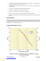
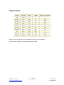
B-Bridge International, Inc.
Cortisol
Enzyme Immunoassay Kit
User Manual
Catalog # K3003-1
K3003-5
K3003-W1
K3003-W5
1 Strip Plate Kit
5 Strip Plate Kit
1 Whole Plate Kit
5 Whole Plate Kit
1
B-Bridge International, Inc.
[email protected]
www.b-bridge.com
Cortisol 150626
+1-408-252-6200
Table of Contents
Intended Use...……………………………………………………………………..3
Background…………………………………………………………………………3
Assay Principle…..……………………………………….….…………………….3
Kit Components………………………………..…………………………………..4
Materials Required But Not Supplied…………………………………………....4
Precautions…………………………….…………………………………………..4
Reagent Preparation………………………………..……………………………..5
Sample Preparation…………………………..……………………………………5
Assay Protocol…………..………………………………………………………....6
Calculations…………….……………………………………………………….....7
Typical Standard Curve…………….…………………………………………......7
Notice to Purchaser
This product is to be used for Research Purposes Only. It is not to be used for Drug or
Diagnostic Purposes, nor is it intended for Human Use. B-Bridge products may not be
resold, modified for resale, or used to manufacture commercial products without the
express written consent of B-Bridge International, Inc.
EXCEPT AS OTHERWISE EXPRESSLY SET FORTH IN THIS USER MANUAL, B-BRIDGE
DOES NOT MAKE ANY REPRESENTATION OR WARRANTIES OR CONDITIONS OF ANY
KIND, EITHER EXPRESSED OR IMPLIED, WITH RESPECT TO THE PRODUCTS, OR
INFORMATION DISCLOSED HEREUNDER, INCLUDING, BUT NOT LIMITED TO, THE IMPLIED
WARRANTIES OF MERCHANTABILITY, FIT FOR A PARTICULAR PURPOSE, OR
NONINFRINGEMENT OF THE INTELLECTUAL PROPERTY RIGHTS OF THIRD PARTIES.
B-Bridge International, Inc. All Rights Reserved.
2
B-Bridge International, Inc.
[email protected]
www.b-bridge.com
Cortisol 150626
+1-408-252-6200
Intended Use
The B-Bridge Cortisol Enzyme Immunoassay Kit quantitatively measures cortisol present in dried fecal
extracts, saliva, urine, serum, EDTA and heparin plasma and tissue culture media (TCM). Please note that
this kit measures total cortisol in extracted samples, serum, and plasma but measures free cortisol in saliva
and urine samples. Moderate to severely hemolyzed samples should not be used in this kit. This assay is
species independent.
Background
Cortisol, C21H30O5, (hydrocortisone, compound F) is the primary glucocorticoid produced and secreted by the
adrenal cortex. It is often referred to as the “stress hormone” because of its role in the stress response.
Cortisol is known to affect blood pressure, blood sugar levels, and other physiological aspets of stress
adaptation. Immunologically, cortisol performs anti-inflammatory functions and plays a role in hypersensitivity,
immunosuppression, and disease resistance. Metabolically, cortisol promotes gluconeogenesis, liver
glycogen deposition, and the reduction of glucose utilization. Production of cortisol follows an
adrenocorticotropic horomone (ATCH)-dependent circadian rhythm, with peak levels in the morning and
decreasing levels throughout the day. About 96% of serum cortisol is bound to proteins, including
corticosteroid binding globulin (also known as Transcortin or CBG) and serum albumin. The remaining
unbound cortisol is available to receptors; through these receptors cortisol modulates physiological
processes. Abnormal cortisol levels are being investiaged in realtion to a variety of different conditions, such
as prostate cancer, depression, and schizophrenia. It is already known that abnormal levels of cortisol are
involved in Cushing’s Syndrome and Addision’s disease.
Assay Principle
The Cortisol Enzyme Immunoassay Kit is designed to quantitatively measure cortisol present in dried fecal
extracts, saliva, urine, serum, plasma and tissue culture media samples.
1. Sample or standard added to well in clear 96-well plate coated with goat anti-mouse IgG
2. A cortisol-peroxidase conjugate is added to the standards and samples in the wells.
3. The binding reaction is initiated by the addition of an anti-cortisol monoclonal antibody to each well.
3. Incubate for 1 hour and read optical density with plate reader at 450 nm wavelength.
4. Calculate cortisol concentration from standard curve.
The immunological reaction occurs between the limiting amount of added anti-cortisol monoclonal antibody,
the cortisol antigen in the sample or standard, and the limiting amount of added cortisol-peroxidase conjugate.
The cortisol in the sample is competing with the cortisol-peroxidase to bind to the anti-cortisol monoclonal
antibody. The monoclonal antibody binds to the goat anti-mouse IgG on the plate. As the concentration of
cortisol in the sample increases, the amount of cortisol-peroxidase conjugate bound decreases causing a
decrease in signal, and vice versa. The signal is generated from the cortisol-peroxidase bound to the
anti-cortisol antibody which itself is bound to the goat anti-mouse IgG coated plates. Excess
cortisol-peroxidase does not bind to the plates and is washed out of the well prior to the addition of substrate.
After an hour incubation the plate is washed and substrate added. The substrate reacts with the bound
cortisol-peroxidase. The intensity of color generated is measured by a plate reader at a 450 nm wavelength.
3
B-Bridge International, Inc.
[email protected]
www.b-bridge.com
Cortisol 150626
+1-408-252-6200
Kit Components
Component:
Cat #
K3003-1/W1
K3003-5/-W5
Clear 96-well plate coated with goat anti-mouse IgG
1 plate
5 plates
Cortisol Standard (32,000 pg/mL in stabilizing solution)
- Calibrated to NIST SRM 921
125 µl
625 µl
Cortisol Detection Mouse Monoclonal Antibody
3 ml
3 ml
Cortisol-Peroxidase Detection Conjugate
3 ml
3 ml
1X Assay Buffer
28 ml
55 ml
Dissociation Reagent
1 ml
5 ml
Allow to reach room temperature before use; for use only with serum and plasma samples
20X Wash Buffer Concentrate
30 ml
125 ml
TMB Substrate
11 ml
55 ml
Stop Solution (IM HCl) Caution: Caustic
5 ml
25 ml
Plate Sealer
1 each
5 each
Store all components at 4°C
Materials Required But Not Supplied
Deionized or distilled water
Repeater pipet with disposable tips capable of dispensing 25 μL, 50 μL and 100 μL.
Colorimetric 96-well microplate reader capable of reading optical density at 450 nm, preferably with
correction between 570 and 590 nm
Software for converting raw relative optical density readings from the plate reader and carrying out four
parameter logistic curve (4PLC) fitting. Contact your plate reader manufacturer for details
Precautions
As with all such products, this kit should only be used by qualified personnel who have had laboratory safety
instruction. The complete User Manual should be read and understood before attempting to use the product.
Antibody coated plate needs to be stored desiccated. The silica pack wrapped with the plate will keep the
plate dry.
This kit utilizes a peroxidase-based readout system. Buffers, including other manufacturers Wash Buffers,
containing sodium azide will inhibit color production from the enzyme. Make sure all buffers used for
samples are azide free. Ensure that any plate washing system is rinsed well with deionized water prior to
using the supplied Wash Buffer.
The Stop Solution is 1M hydrochloric acid. The solution should not come in contact with skin or eyes. Take
appropriate precautions when handling this reagent and please consult your institution’s safety procedures
for working with hazardous chemicals.
4
B-Bridge International, Inc.
[email protected]
www.b-bridge.com
Cortisol 150626
+1-408-252-6200
Reagent Preparation
Allow the kit reagents to come to room temperature for 30 minutes.
We recommend that all standards and samples be run in duplicate to accurately determine cortisol
concentrations. Ensure that all samples have reached room temperature and have been diluted as
appropriate prior to assaying.
Wash Buffer
Dilute 20x Wash Buffer 1:20 by adding one part of the concentrate to nineteen parts of deionized
water. Once diluted this is stable at room temperature for 3 months at room temperature.
Standard Preparation
Standards are prepared by labeling seven test tubes as #1 through #6. Pipet 450 μL of Assay Buffer into tube
#1 and 250 μL into tubes #2 to #6. The cortisol stock solution contains an organic solvent. Prerinse the pipet
tip several times to ensure accurate delivery. Carefully add 50 μL of the cortisol stock solution to tube #1 and
vortex completely. Take 250 μL of the cortisol solution in tube #1 and add it to tube #2 and vortex completely.
Repeat the serial dilutions for tubes #3 through #6. The concentration of cortisol in tubes 1 through 6 will be
3,200, 1,600, 800, 400, 200, and 100 pg/mL.
Use all standards within 2 hours of preparation.
Std 1
Std 2
Std 3
Std 4
Std 5
Std 6
Assay Buffer
450 µl
250 µl
250 µl
250 µl
250 µl
250 µl
Cortisol Standard
50 µl
-
-
-
-
-
Standard 1
-
250 µl
-
-
-
-
Standard 2
-
-
250 µl
-
-
-
Standard 3
-
-
-
250 µl
-
-
Standard 4
-
-
-
-
250 µl
-
Standard 5
-
-
-
-
-
250 µl
3200 pg/mL
1600 pg/mL
800 pg/mL
400 pg/mL
300 pg/mL
100 pg/mL
Concentration
Sample Preparation
Serum and plasma samples need to be treated with the supplied Dissociation Reagent. Addition of this
reagent will cause results of this assay to yield the total cortisol concentration in serum or plasma samples.
Dissociation reagent is to be used only with Serum and Plasma samples. Free cortisol can be measured in
saliva and urine samples as directed below.
Dried Fecal Samples
Please see detailed Extraction Protocol below. The ethanol concentration in the final Assay Buffer dilution
added to the well should be <5%.
Extraction Protocol:
To extract steroids from non-liquid matrices, such as dried solids or other organic matter, we
recommend an organic phase extraction using ethanol or ethyl acetate. We recommend centrifugal
vacuum devices to remove the solvent completely and recommend using ethanol to completely
solubilize the dried steroid because certain steroids have limited aqueous solubility. The protocol
uses ethanol or ethyl acetate to extract the organic soluble steroid. The organic layer is separated
and stored.
Materials Needed:
- Steroid standard to allow extraction efficiency determination
- ACS Grade Ethanol (or Ethyl Acetate)
- Glass test tubes
Procedure:
Note: Ensure that the sample is completely dry and powder the sample to improve extraction
5
B-Bridge International, Inc.
[email protected]
www.b-bridge.com
Cortisol 150626
+1-408-252-6200
efficiency. Remove any large particles, such as grass, if possible. We suggest checking the
efficiency of extraction by preparing a steroid solution of known concentration in the kit
Assay Buffer (AB). Spike one aliquot of your sample with a volume of the steroid solution in
AB (Control Spike) and one aliquot of sample with the same volume of AB (Control Sample).
Extract samples and both Controls with Ethanol or Ethyl Acetate as described below.
1. Weigh out ≥ 0.2 gm of dried fecal solid into a tube.
2. Add 1 mL of Ethanol (or Ethyl Acetate) for every 0.1 gm of solid.
3. Shake vigorously for at least 30 minutes.
4. Centrifuge samples at 5,000 rpm for 15 minutes. Transfer measured volume of
supernatant to a clean tube for evaporation.
5. Evaporate supernatant solution to dryness in a SpeedVac or under nitrogen. Keep dried
extracted samples frozen <-20°C in a desiccator.
6. Dissolve extracted sample with 100μL ethanol, followed by at least 400μL AB. Vortex well
and allow to sit 5 minutes at room temperature. Vortex and sit for 5 minutes twice more to
ensure complete steroid solubility.
7. Run reconstituted samples in assay immediately according to insert directions.
8. Determine the extraction efficiency by comparing the concentration of the steroid
measured in the extracted Control (Control Spike - Control Sample) with the concentration of
steroid before extraction.
Note: In step 5 if only a portion of the organic solvent is being evaporated, ensure final
amounts of measured steroid per gm solid accounts for volume of solution evaporated.
Serum and Plasma Samples
The normal reference range for human serum cortisol is 2-25 µg/dL (20-250 ng/mL). Allow the
Dissociation Reagent (DR) to warm completely to room temperature before use. We suggest
pipeting 5 µL of DR into 1 mL Eppendorf tubes. Add 5 µL of serum or plasma to the DR in the tube,
vortex gently and incubate at room temperature for 5 minutes or longer. Dilute by adding 490
µL of supplied Assay Buffer. This 1:100 dilution can be diluted further with Assay Buffer for higher
cortisol sample concentrations. Final serum and plasma dilutions must be ≥ 1:100.
NOTE: Dissociation Reagent is to be used only with Serum and Plasma samples.
Saliva Samples
Saliva samples should be diluted ≥ 1:4 or greater with the supplied Assay Buffer prior running in the assay.
Urine Samples
Urine samples should be diluted ≥ 1:8 with the supplied Assay Buffer prior running in the assay. Urinary
cortisol normally ranges from 0.7-119 μg/gram of cortisol or approximately 100,000 to 1,000,000 pg/m in 24
hour urine samples. Samples may need to be diluted substantially to read within the standard curve range.
Tissue Culture Media
For measuring cortisol in tissue culture media (TCM), samples should be read off a standard curve generated
in TCM. Samples may need to be diluted further in TCM.
Use all samples within 2 hours of preparation.
Assay Protocol
1. Pipet 50 µl of samples or standards in duplicate into wells in the clear microplate.
2. Pipet 75 µl of Assay Buffer into the non-specific binding (NSB) wells.
3. Pipet 50 µl of Assay Buffer into wells to act as maximum binding wells (Bo or 0 pg/mL).
4. Add 25 µl of Cortisol-Peroxidase Detection Conjugate to each well using a repeater pipet.
5. Add 25 µl of Cortisol Detection Mouse Monoclonal Antibody to each well except the NSB wells
using a repeater pipet.
6
B-Bridge International, Inc.
[email protected]
www.b-bridge.com
Cortisol 150626
+1-408-252-6200
6. Gently tap the sides of the plate to ensure adequate mixing of the reagents. Cover the plate with the
plate sealer and shake at room temperature for 1 hour.
7. Aspirate the plate and wash each well 4 times with 300 µl of Wash Buffer. Tap the plate dry on clean
absorbent towels.
8. Add 100 µl of the TMB Substrate to each well using a repeater pipet.
9. Incubate at room temperature for 30 minutes without shaking.
10. Add 50 µl of the Stop Solution to each well using a repeater pipet.
11. Read the optical density (OD) of each well in a plate reader at 450 nm wavelength.
12. Use the plate reader’s built-in 4PLC software capabilities to calculate cortisol concentration for each
sample.
Calculations
Average the duplicate optical density (OD) readings for each standard and sample. Create a standard curve
by reducing the data using computer software capable of generating a four-parameter logistic curve (4PLC)
fit, after subtracting the mean OD’s for the blank. The sample concentrations obtained should be multiplied by
the dilution factor to obtain neat sample values.
Typical Standard Curve
7
B-Bridge International, Inc.
[email protected]
www.b-bridge.com
Cortisol 150626
+1-408-252-6200
Typical Data
Always run your own standard curve for calculation of results. Do not use this data.
Conversion factor 100 pg/ml of cortisol is equivalent to 275.9 pM.
8
B-Bridge International, Inc.
[email protected]
www.b-bridge.com
Cortisol 150626
+1-408-252-6200


![TEMPLATE No1 [CPMP positive opinion full application]](http://vs1.manualzilla.com/store/data/005681628_1-1a3acb54fcca990dd8c826546eb4788f-150x150.png)