1
CHAPTER 6
Electrophoresis and Immunoblotting
INTRODUCTION
T
he development of powerful new technologies is a major driving force of scientific
progress. A good example of this is the role that electrophoretic techniques have played
in the evolution of modern cell biology. Electrophoresis and related applications have
contributed greatly to the understanding of the molecular bases of cell structure and function.
The combination of high resolution, ease of use, speed, low cost, and versatility of electrophoretic techniques is unmatched by any other method used to separate proteins. It is for this
reason that electrophoresis is an indispensable tool in any cell biology laboratory and that
papers describing basic techniques of protein electrophoresis (e.g., Laemmli, 1970, and
O’Farrell, 1975, to name just a couple) are among the most cited articles in this field.
Laemmli’s technique of discontinuous gel electrophoresis in the presence of SDS, for
example, continues to be widely used and referenced almost 30 years after publication. Thus,
no book of techniques in cell biology would be complete without a detailed description of
electrophoretic techniques.
Chapter 6 begins with UNIT 6.1, which is a collection of state-of-the-art protocols for analyzing
proteins by one-dimensional electrophoresis under denaturing conditions on polyacrylamide
gels. Sodium dodecyl sulfate (SDS), in combination with a reducing agent and heat, is most
often used as a denaturant. This type of electrophoresis is thus referred to as SDS-polyacylamide gel electrophoresis (SDS-PAGE). Denaturation of the proteins prior to electrophoresis
allows for enhanced resolution and discrimination of proteins on the basis of molecular size
rather than charge or shape. Utilization of a discontinuous system (i.e., the apposition of
“stacking” and “separating” gels) results in concentration of dilute samples and enhanced
band sharpness. UNIT 6.1 presents an overview of electricity and electrophoresis, followed by
detailed protocols for SDS-PAGE using either Laemmli’s buffers and gel system or modifications of this system (i.e., use of Tris-tricine buffers, higher concentrations of buffers,
gradient gels, single-concentration gels, and minigels). The unit also explains how to calculate
the apparent molecular weights of proteins from SDS-PAGE data.
The next unit in the chapter, UNIT 6.2, describes protocols for immunoblotting (also referred
to as western blotting). In this technique, proteins separated by any of the electrophoretic
techniques described in UNIT 6.1 are electrophoretically transferred (“electroblotted”) onto a
membrane. The membrane, which thus becomes a replica of the polyacrylamide gel, is
subsequently probed with antibodies to specific proteins. The primary antibodies can be
revealed by an additional incubation with 125I-labeled secondary antibodies or protein A,
followed by autoradiography (UNIT 6.3). In recent years, however, the use of radioiodinated
antibodies has been progressively replaced by nonradioactive detection with antibodies
coupled to enzymes such as alkaline phosphatase or horseradish peroxidase (see UNIT 16.5).
The enzymes act on substrates which are converted to colored, luminescent, or fluorescent
products. Nonradioactive methods are just as sensitive as radioactive methods, with the added
advantage that they do not require the special precautions associated with the use of
radioactivity. Nonradioactive detection is nowadays the method of choice for visualizing
immunoblotted proteins. A disadvantage of nonradioactive methods is that they have a
narrower linear range of detection, which can be a problem in experiments that require
accurate quantitation of protein levels.
Electrophoresis
and
Immunoblotting
Contributed by Juan S. Bonifacino
Current Protocols in Cell Biology (2002) 6.0.1-6.0.3
Copyright © 2002 by John Wiley & Sons, Inc.
6.0.1
Supplement 15
Radiolabeled proteins separated by electrophoresis or proteins detected by immunoblotting
with radioiodinated antibodies or protein A can be visualized by autoradiography, as described
in UNIT 6.3. In this technique, ionizing radiation emanating from the radionuclides impresses
a photographic film. The technique can be made more sensitive by the use of intensifying
screens or scintillating compounds, which emit light upon radiation absorption, which then
impresses the film. The unit contains several protocols for autoradiographic detection of
various radionuclides, including methods for enhancing the signal with intensifying screens
or by fluorography. Also included in UNIT 6.3 are discussions of the quantification of film
images by densitometry and the direct detection and quantification of radioactive samples in
gels by phosphor imaging.
The resolution of electrophoretic techniques can be enormously enhanced by combining two
different electrophoretic procedures performed successively in perpendicular directions (i.e.,
two-dimensional gel electrophoresis). The most common type of two-dimensional gel
electrophoresis is based on separation of proteins by isoelectric focusing on a tube gel (first
dimension) followed by SDS-PAGE on a slab gel (second dimension). The two processes
separate proteins on the basis of charge and size, respectively, allowing resolution of up to
several thousand proteins on a single two-dimensional gel. UNIT 6.4 describes several methods
for separating proteins by two-dimensional isoelectric focusing/SDS-PAGE. In addition, this
unit presents a protocol for two-dimensional nonreducing/reducing electrophoresis in which
proteins are separated by SDS-PAGE under nonreducing conditions in the first dimension
and under reducing conditions in the second dimension. This type of two-dimensional gel
electrophoresis allows analysis of intersubunit disulfide bonds in multiprotein complexes and,
in some cases, of intrasubunit disulfide bonds. Both types of two-dimensional gel electrophoresis can be used for either analytical or preparative purposes.
Another useful method for electrophoretic separation of cellular proteins is one-dimensional
electrophoresis under nondenaturing conditions. Two protocols describing variations of this
method are included in UNIT 6.5. What distinguishes this method from those described in UNIT
6.1 and UNIT 6.4 is that protein samples are not exposed to denaturing agents (i.e., SDS or urea)
either prior to or during electrophoresis. Thus, proteins migrate according to their native
properties, such as size, shape, and charge. This allows analysis of the oligomeric state of
proteins, conformational changes, charge heterogeneity, and post-translational modifications
that affect conformation or charge while having minimal effects on the molecular weights of
the proteins. In many cases, this method preserves the intrinsic function of the proteins, which
allows their detection with specific activity or binding assays. The first protocol describes
continuous electrophoresis on nondenaturing polyacrylamide gels. This system involves
electrophoresis on a single separating gel and uses the same buffer in the chambers and the
gel. The second protocol, discontinuous electrophoresis on nondenaturing polyacrylamide
gels, is a variation of SDS-PAGE in which SDS and reducing agents are omitted from all the
solutions. Determination of the migration of proteins on parallel gels made up of different
concentrations of acrylamide and bisacrylamide allows calculation of their molecular weights
using Ferguson plots.
Proteins separated by electrophoresis can be visualized by direct staining of the gels. Four
procedures for staining proteins in gels based on different principles are presented in UNIT 6.6.
These procedures involve staining with Coomassie blue, silver, SYPRO ruby or zinc ions. The
unit describes the basic protocols and provides guidelines for the selection of a specific protocol.
Introduction
The separation of proteins by polyacrylamide gel electrophoresis is limited to proteins
with molecular weights less than ∼300,000. The electrophoretic separation of larger
proteins or multiprotein complexes requires the use of other matrix materials. A suitable
material is agarose, which is most commonly used for the separation of DNA. UNIT 6.7
presents protocols for the electrophoretic separation of proteins on agarose gels. In
6.0.2
Supplement 15
Current Protocols in Cell Biology
addition to permitting the separation of very large proteins, these protocols allow the
analysis of multimerization or aggregation. Although these processes can also be analyzed
by sedimentation on sucrose gradients (UNIT 6.3) or gel filtration (UNIT 6.4), agarose gel
electrophoresis is more convenient for analysis of large numbers of samples.
LITERATURE CITED
Laemmli, U.K. 1970. Cleavage of structural proteins during the assembly of the head of bacteriophage T4.
Nature 227:680-685.
O’Farrell, P.H. 1975. High resolution two-dimensional polyacrylamide gel electrophoresis of proteins. J. Biol.
Chem. 250:4007-4021.
Juan S. Bonifacino
Electrophoresis
and
Immunoblotting
6.0.3
Current Protocols in Cell Biology
Supplement 15
One-Dimensional SDS Gel Electrophoresis
of Proteins
UNIT 6.1
Sean R. Gallagher1
1
UVP, Inc., Upland, California
ABSTRACT
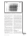
Electrophoresis is used to separate complex mixtures of proteins (e.g., from cells, subcellular fractions, column fractions, or immunoprecipitates), to investigate subunit compositions, and to verify homogeneity of protein samples. It can also serve to purify proteins
for use in further applications. In polyacrylamide gel electrophoresis, proteins migrate
in response to an electrical field through pores in a polyacrylamide gel matrix; pore size
decreases with increasing acrylamide concentration. The combination of pore size and
protein charge, size, and shape determines the migration rate of the protein. In this unit,
the standard Laemmli method is described for discontinuous gel electrophoresis under
denaturing conditions, that is, in the presence of sodium dodecyl sulfate (SDS). Both
full-size and minigel formats are detailed. Several modifications are provided for specific
applications. For separation of peptides and small proteins, the standard buffers are replaced with either a Tris-tricine buffer system or a modified Tris buffer in the absence of
urea. Continuous SDS-PAGE is a simplified method in which the same buffer is used for
both the gel and the electrode solutions and the stacking gel is omitted. Other protocols
cover the preparation and use of ultrathin gels and gradient gels, and the simultaneous
C 2007 by John
preparation of multiple gels. Curr. Protoc. Cell Biol. 37:6.1.1-6.1.38. Wiley & Sons, Inc.
Keywords: protein r electrophoresis r separation r polyacrylamide r SDS-PAGE
INTRODUCTION
Electrophoresis is used to separate complex mixtures of proteins (e.g., from cells, subcellular fractions, column fractions, or immunoprecipitates), to investigate subunit compositions, and to verify homogeneity of protein samples. It can also serve to purify
proteins for use in further applications. In polyacrylamide gel electrophoresis (PAGE),
proteins migrate in response to an electrical field through pores in the gel matrix; pore
size decreases with higher acrylamide concentrations. The combination of gel pore size
and protein charge, size, and shape determines the migration rate of the protein.
The standard Laemmli method (see Basic Protocol 1) is used for discontinuous gel
electrophoresis under denaturing conditions, that is, in the presence of sodium dodecyl
sulfate (SDS). The standard method for full-size gels (e.g., 14 × 14 cm) can be adapted
for the minigel format (e.g., 7.3 × 8.3 cm; see Basic Protocol 2). Minigels provide rapid
separation but give lower resolution.
Several alternate protocols are provided for specific applications. The first two alternate
protocols cover electrophoresis of peptides and small proteins, separations that require
modification of standard buffers: either a Tris-tricine buffer system (see Alternate Protocol 1) or a modified Tris buffer in the absence of urea (see Alternate Protocol 2).
Continuous SDS-PAGE is a simplified method in which the same buffer is used for
both gel and electrode solutions and the stacking gel is omitted (see Alternate Protocol 3). Other protocols cover the preparation and electrophoresis of various types of gels:
Electrophoresis
and
Immunoblotting
Current Protocols in Cell Biology 6.1.1-6.1.38, December 2007
Published online December 2007 in Wiley Interscience (www.interscience.wiley.com).
DOI: 10.1002/0471143030.cb0601s37
C 2007 John Wiley & Sons, Inc.
Copyright 6.1.1
Supplement 37
ultrathin gels (see Alternate Protocol 4), multiple single-concentration gels (see Support
Protocol 1), gradient gels (see Alternate Protocol 5), multiple gradient gels (see Support
Protocol 2), and multiple gradient minigels (see Support Protocol 3). Proteins separated
on gels can be subsequently analyzed by immunoblotting (UNIT 6.2), autoradiography or
phosphor imaging (UNIT 6.3), or staining with protein dyes (UNIT 6.6). Protein size is determined by comparing the mobility of the protein band to the mobility of the dye front
(see Support Protocol 4).
CAUTION: Before any protocols are used, it is extremely important to read the following
section about electricity and electrophoresis.
ELECTRICITY AND ELECTROPHORESIS
Many researchers are poorly informed concerning the electrical parameters of running
a gel. It is important to note that the voltages and currents used during electrophoresis
are dangerous and potentially lethal. Thus, safety should be an overriding concern.
A working knowledge of electricity is an asset in determining what conditions to use and
in troubleshooting the electrophoretic separation, if necessary. For example, an unusually
high or low voltage for a given current (milliampere) might indicate an improperly made
buffer or an electrical leak in the chamber.
Safety Considerations
1. Never remove or insert high-voltage leads unless the power supply voltage is turned
down to zero and the power supply is turned off. Always grasp high-voltage leads
one at a time with one hand only. Never insert or remove high-voltage leads with both
hands. This can shunt potentially lethal electricity through the chest and heart should
electrical contact be made between a hand and a bare wire. On older or homemade
instruments, the banana plugs may not be shielded and can still be connected to the
power supply at the same time they make contact with a hand. With commercial
modern power supplies, this is less of an issue. However, with age and use, wires
may become exposed through cracks in the insulation or poor connections. Carefully
inspect all cables and connections and replace frayed or exposed wires immediately.
2. Always start with the power supply turned off. Have the power supply controls turned
all the way down to zero. Then hook up the gel apparatus: generally, connect the
red high-voltage lead to the red outlet and the black high-voltage lead to the black
outlet. Turn the power supply on with the controls set at zero and the high-voltage
leads connected. Then turn up the voltage, current, or power to the desired level.
Reverse the process when the power supply is turned off: i.e., to disconnect the gel,
turn the power supply down to zero, wait for the meters to read zero, turn off the
power supply, and then disconnect the gel apparatus one lead at a time.
CAUTION: If the gel is first disconnected and then the power supply turned off, a considerable amount of electrical charge is stored internally. The charge will stay in the power supply
over a long time. This will discharge through the outlets even though the power supply is
turned off and can deliver an electrical shock.
One-Dimensional
SDS-PAGE
Ohm’s Law and Electrophoresis
Understanding how a gel apparatus is connected to the power supply requires a basic
understanding of Ohm’s law: voltage = current × resistance, or V = IR. A gel can be
viewed as a resistor and the power supply as the voltage and current source. Most power
supplies deliver constant current or constant voltage. Some will also deliver constant
power: power = voltage × current, or VI = I2 R. The discussion below focuses on constant
current because this is the most common mode in vertical SDS-PAGE.
6.1.2
Supplement 37
Current Protocols in Cell Biology
Most modern commercial equipment is color-coded so that the red or positive terminal
of the power supply can simply be connected to the red lead of the gel apparatus, which
goes to the lower buffer chamber. The black lead is connected to the black or negative
terminal and goes to the upper buffer chamber. This configuration is designed to work
with vertical slab gel electrophoreses in which negatively charged proteins or nucleic
acids move to the positive electrode in the lower buffer chamber (an anionic system).
When a single gel is attached to a power supply, the negative charges flow from the
negative cathode (black) terminal into the upper buffer chamber, through the gel, and
into the lower buffer chamber. The lower buffer chamber is connected to the positive
anode (red) terminal to complete the circuit. Thus, negatively charged molecules, such
as SDS-coated proteins and nucleic acids, move from the negative cathode attached
to the upper buffer chamber toward the positive anode attached to the lower chamber.
SDS-PAGE is an anionic system because of the negatively charged SDS.
Occasionally, proteins are separated in cationic systems. In these gels, the proteins are
positively charged because of the very low pH of the gel buffers (e.g., acetic acid/urea
gels for histone separations) or the presence of a cationic detergent (e.g., cetyltrimethylammonium bromide, CTAB). Proteins move toward the negative electrode (cathode) in
cationic gel systems, and the polarity is reversed compared to SDS-PAGE: the red lead
from the lower buffer chamber is attached to the black outlet of the power supply, and
the black lead from the upper buffer chamber is attached to the red outlet of the power
supply.
Most SDS-PAGE separations are performed under constant current (consult instructions
from the manufacturer to set the power supply for constant current operation). The
resistance of the gel will increase during SDS-PAGE in the standard Laemmli system.
If the current is constant, then the voltage will increase during the run as the resistance
goes up.
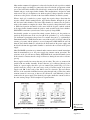
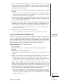
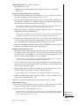
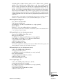
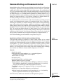
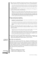
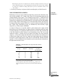
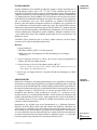
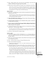
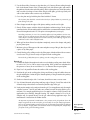
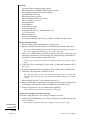
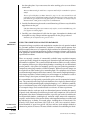
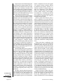
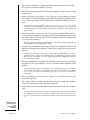
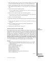
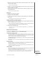
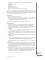
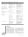
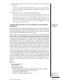
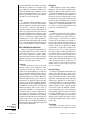
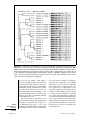
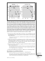
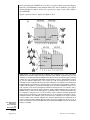
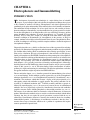
Power supplies usually have more than one pair of outlets. The pairs are connected in
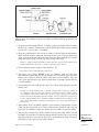
parallel with one another internally. If more than one gel is connected directly to the
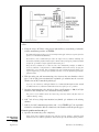
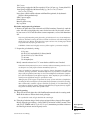
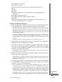
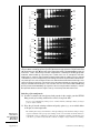
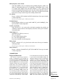
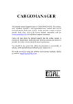
outlets of a power supply, then these gels are connected in parallel (Fig. 6.1.1). In a
parallel circuit, the voltage is the same across each gel. In other words, if the power
supply reads 100 V, then each gel has 100 V across its electrodes. The total current,
however, is the sum of the individual currents going through each gel. Therefore, under
constant current it is necessary to increase the current for each additional gel that is
connected to the power supply. Two identical gels require double the current to achieve
the same starting voltages and electrophoresis separation times.
Figure 6.1.1
Series and parallel connections of gel tanks to power supply.
Electrophoresis
and
Immunoblotting
6.1.3
Current Protocols in Cell Biology
Supplement 37
Multiple gel apparatuses can also be connected to one pair of outlets on a power supply.
This is useful with older power supplies that have a limited number of outlets. When
connecting several gel units to one outlet, make certain the connections between the
units are shielded and protected from moisture. The gels can be connected in parallel or
in series (Fig. 6.1.1). When gels are connected through the same outlet in parallel, the
principle of additive currents is the same as for gels connected through different outlets
in parallel. In the case of two or more gels running off the same outlet in series, the
current is the same for every gel. If 10 mA is displayed by the power supply meter, for
example, each gel in series will experience 10 mA. The voltage, however, is additive for
each gel. If one gel at a constant 10 mA produces 100 V, then two identical gels in series
will produce 200 V (100 V each) and so on. Thus, the voltage can limit the number of
units connected in series on low-voltage power supplies.
Gel thickness affects the above relationships. A 1.5-mm gel can be thought of as two
0.75-mm-thick gels run in parallel. Because currents are additive in parallel circuits, a
0.75-mm gel will require half the current of the 1.5-mm gel to achieve the same starting
voltage and separation time. If gel thickness is doubled, then the current must also be
doubled. There are limits to the amount of current that can be applied. Thicker gels
require more current, generating more heat that must be dissipated. Unless temperature
control is available in the gel unit, a thick gel should be run more slowly than a thin gel.
NOTE: Milli-Q-purified water or equivalent should be used throughout the protocols.
BASIC
PROTOCOL 1
DENATURING (SDS) DISCONTINUOUS GEL ELECTROPHORESIS:
LAEMMLI GEL METHOD
One-dimensional gel electrophoresis under denaturing conditions (i.e., in the presence of
0.1% SDS) separates proteins based on molecular size as they move through a polyacrylamide gel matrix toward the anode. The polyacrylamide gel is cast as a separating gel
(sometimes called resolving or running gel) topped by a stacking gel and secured in an
electrophoresis apparatus. After sample proteins are solubilized by boiling in the presence
of SDS, an aliquot of the protein solution is applied to a gel lane, and the individual
proteins are separated electrophoretically. The stacking gel, through a combination of
low porosity and a lower buffer concentration and pH, concentrates proteins into a thin
stack before they enter the resolving gel. 2-Mercaptoethanol (2-ME) or dithiothreitol
(DTT) is added during solubilization to reduce disulfide bonds.
This protocol is designed for a vertical slab gel with a maximum size of 0.75 mm × 14 cm
× 14 cm. For thicker gels or minigels (see Basic Protocol 2 and Support Protocol 3), the
volumes of stacking and separating gels and the operating current must be adjusted.
Additional protocols describe the preparation of ultrathin gels (see Alternate Protocol 4)
and gradient gels (see Alternate Protocol 5), as well as the use of gel casters to make
multiple gels, both single-concentration gels (see Support Protocol 1) and gradient gels
(see Support Protocol 2).
Materials
One-Dimensional
SDS-PAGE
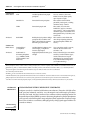
Separating and stacking gel solutions (Table 6.1.1)
H2 O-saturated isobutyl alcohol
1× Tris·Cl/SDS, pH 8.8 (dilute 4× Tris·Cl/SDS, pH 8.8; Table 6.1.1)
Protein sample, on ice
2× and 1× SDS sample buffer (see recipe)
Protein molecular weight standards (Tables 6.1.2 and 6.1.3)
6× SDS sample buffer (see recipe; optional)
1× SDS electrophoresis buffer (see recipe)
6.1.4
Supplement 37
Current Protocols in Cell Biology
Electrophoresis apparatus: e.g., Protean II 16-cm cell (Bio-Rad) or SE 600/400
16-cm unit (Hoefer) with clamps, glass plates, casting stand, and buffer
chambers
0.75-mm spacers
0.45-µm filters (used in stock solution preparation)
25-ml Erlenmeyer side-arm flasks
Vacuum pump with cold trap
0.75-mm Teflon comb with 1, 3, 5, 10, 15, or 20 teeth
Screw-top microcentrifuge tubes (recommended)
25- or 100-µl syringe with flat-tipped needle
Constant-current power supply (see Electricity and Electrophoresis above)
Pour the separating gel
1. Assemble the glass-plate sandwich of the electrophoresis apparatus according to
manufacturer’s instructions using two clean glass plates and two 0.75-mm spacers.
If needed, clean the glass plates in liquid Alconox or RBS-35 (Pierce). These aqueousbased solutions are compatible with silver and Coomassie blue staining procedures.
2. Lock the sandwich to the casting stand.
3. Prepare the separating gel solution as directed in Table 6.1.1, degassing using a
rubber-stoppered 25-ml Erlenmeyer side-arm flask connected with vacuum tubing
to a vacuum pump with a cold trap. After adding the specified amount of 10%
ammonium persulfate and TEMED to the degassed solution, stir gently to mix.
The desired percentage of acrylamide in the separating gel depends on the molecular
size of the protein being separated. Generally, use 5% gels for SDS-denatured proteins of
60 to 200 kDa, 10% gels for SDS-denatured proteins of 16 to 70 kDa, and 15% gels for
SDS-denatured proteins of 12 to 45 kDa (Table 6.1.1).
The stacking gel is the same regardless of the separating gel used.
4. Using a Pasteur pipet, apply the separating gel solution to the sandwich along an
edge of one of the spacers until the height of the solution between the glass plates
is ∼11 cm.
Use the solution immediately; otherwise it will polymerize in the flask.
Sample volumes <10 µl do not require a stacking gel. In this case, cast the resolving
gel as usual, but extend the resolving gel into the comb (step 10) to form the wells. The
proteins are then separated under the same conditions as used when a stacking gel is
present. Although this protocol works well with single-concentration gels, a gradient gel
is recommended for maximum resolution (see Alternate Protocol 5).
5. Using another Pasteur pipet, slowly cover the top of the gel with a layer (∼1 cm
thick) of H2 O-saturated isobutyl alcohol, by gently layering the isobutyl alcohol
against the edge of one and then the other of the spacers.
Be careful not to disturb the gel surface. The overlay provides a barrier to oxygen, which
inhibits polymerization, and allows a flat interface to form during gel formation.
The H2 O-saturated isobutyl alcohol is prepared by shaking isobutyl alcohol and H2 O in
a separatory funnel. The aqueous (lower) phase is removed. This procedure is repeated
several times. The final upper phase is H2 O-saturated isobutyl alcohol.
6. Allow the gel to polymerize 30 to 60 min at room temperature.
A sharp optical discontinuity at the overlay/gel interface will be visible on polymerization. Failure to form a firm gel usually indicates a problem with the ammonium persulfate,
TEMED, or both. Ammonium persulfate solution should be made fresh before use. Ammonium persulfate should “crackle” when added to the water. If not, fresh ammonium
persulfate should be purchased. Purchase TEMED in small bottles so, if necessary, a new
previously unopened source can be tried.
Current Protocols in Cell Biology
Electrophoresis
and
Immunoblotting
6.1.5
Supplement 37
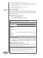
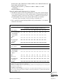
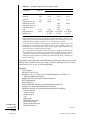
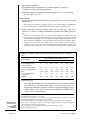
Table 6.1.1 Recipes for Polyacrylamide Separating and Stacking Gelsa
SEPARATING GEL
Final acrylamide concentration in separating gel (%)c
Stock solutionb
5
6
7
7.5
8
9
10
12
30% (w/v) acrylamide/ 2.50
0.8% (w/v)
bisacrylamide
3.00
3.50
3.75
4.00
4.50
5.00
6.00
6.50 7.50
4× Tris·Cl/SDS,
pH 8.8
3.75
3.75
3.75
3.75
3.75
3.75
3.75
3.75
3.75 3.75
H2 O
8.75
8.25
7.75
7.50
7.25
6.75
6.25
5.25
4.75 3.75
10% (w/v) ammonium 0.05
persulfated
0.05
0.05
0.05
0.05
0.05
0.05
0.05
0.05 0.05
TEMEDe
0.01
0.01
0.01
0.01
0.01
0.01
0.01
0.01 0.01
0.01
13
15
Preparation of separating gel
In a 25-ml side-arm flask, mix 30% acrylamide/0.8% bisacrylamide solution, 4×
Tris·Cl/SDS, pH 8.8 (see reagents, below), and H2 O. Degas under vacuum ∼5 min.
Add 10% ammonium persulfate and TEMED. Swirl gently to mix. Use immediately.
STACKING GEL (3.9% w/v acrylamide)
In a 25-ml side-arm flask, mix 0.65 ml of 30% acrylamide/0.8% bisacrylamide, 1.25 ml
of 4× Tris·Cl/SDS, pH 6.8 (see reagents, below), and 3.05 ml H2 O. Degas under vacuum
10 to 15 min. Add 25 µl of 10% ammonium persulfate and 5 µl TEMED. Swirl gently to
mix. Use immediately.
REAGENTS USED IN GELS
30% (w/v) acrylamide/0.8% (w/v) bisacrylamide
Mix 30.0 g acrylamide and 0.8 g N,N -methylenebisacrylamide with H2 O in a total volume
of 100 ml. Filter the solution through a 0.45-µm filter and store at 4◦ C in the dark. The
2× crystallized grades of acrylamide and bisacrylamide are recommended. Discard after
30 days, as acrylamide gradually hydrolyzes to acrylic acid and ammonia.
CAUTION: Acrylamide monomer is neurotoxic. A mask should be worn when weighing
acrylamide powder. Gloves should be worn while handling the solution, and the solution
should not be pipetted by mouth.
4× Tris·Cl/SDS, pH 6.8 (0.5 M Tris·Cl containing 0.4% w/v SDS)
Dissolve 6.05 g Tris base in 40 ml H2 O. Adjust to pH 6.8 with 1 N HCl. Add H2 O to
100 ml total volume. Filter the solution through a 0.45-µm filter, add 0.4 g SDS, and
store at 4◦ C up to 1 month.
4× Tris·Cl/SDS, pH 8.8 (1.5 M Tris·Cl containing 0.4% w/v SDS)
Dissolve 91 g Tris base in 300 ml H2 O. Adjust to pH 8.8 with 1 N HCl. Add H2 O to
500 ml total volume. Filter the solution through a 0.45-µm filter, add 2 g SDS, and
store at 4◦ C up to 1 month.
a The recipes produce 15 ml of separating gel and 5 ml of stacking gel, which are adequate for a gel of dimensions
One-Dimensional
SDS-PAGE
0.75 mm × 14 cm × 14 cm. The recipes are based on the SDS (denaturing) discontinuous buffer system of Laemmli
(1970).
b All reagents and solutions used in the protocol must be prepared with Milli-Q-purified water or equivalent.
c Volumes are in milliliters. The desired percentage of acrylamide in the separating gel depends on the molecular size
of the protein being separated. See annotation to step 3, Basic Protocol 1.
d Best to prepare fresh. Failure to form a firm gel usually indicates a problem with the ammonium persulfate, TEMED,
or both.
e TEMED, N,N,N,N-tetramethylethylenediamine.
6.1.6
Supplement 37
Current Protocols in Cell Biology
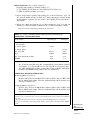
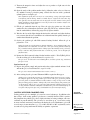
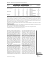
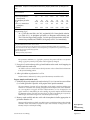
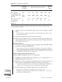
Table 6.1.2 Molecular Weights of Protein Standards for Polyacrylamide Gel Electrophoresisa
Protein
Molecular weight (Da)
Cytochrome c
11,700
α-Lactalbumin
14,200
Lysozyme (hen egg white)
14,300
Myoglobin (sperm whale)
16,800
β-Lactoglobulin
18,400
Trypsin inhibitor (soybean)
20,100
Trypsinogen, PMSF treated
24,000
Carbonic anhydrase (bovine erythrocytes)
29,000
Glyceraldehyde-3-phosphate dehydrogenase (rabbit muscle)
36,000
Lactate dehydrogenase (porcine heart)
36,000
Aldolase
40,000
Ovalbumin
45,000
Catalase
57,000
Bovine serum albumin
66,000
Phosphorylase b (rabbit muscle)
97,400
β-Galactosidase
116,000
RNA polymerase, E. coli
160,000
Myosin, heavy chain (rabbit muscle)
205,000
a Protein standards are commercially available as prepared mixtures (see Table 6.1.3).
Pour the stacking gel
7. Pour off the layer of H2 O-saturated isobutyl alcohol and rinse with 1× Tris·Cl/SDS,
pH 8.8.
Residual isobutyl alcohol can reduce resolution of the protein bands; therefore, it must
be completely removed. The isobutyl alcohol overlay should not be left on the gel longer
than 2 hr.
8. Prepare the stacking gel solution as directed in Table 6.1.1.
Use the solution immediately to keep it from polymerizing in the flask.
9. Using a Pasteur pipet, allow the stacking gel solution to trickle slowly into the center
of the sandwich along an edge of one of the spacers until the height of the solution
in the sandwich is ∼1 cm from the top of the plates.
Be careful not to introduce air bubbles into the stacking gel.
10. Insert a 0.75-mm Teflon comb into the layer of stacking gel solution. If necessary,
add additional stacking gel to fill the spaces in the comb completely.
Again, be careful not to trap air bubbles in the tooth edges of the comb; they will cause
small circular depressions in the well after polymerization that will lead to distortion in
the protein bands during separation.
11. Allow the stacking gel solution to polymerize 30 to 45 min at room temperature.
A sharp optical discontinuity will be visible around the wells on polymerization. Again,
failure to form a firm gel usually indicates a problem with the ammonium persulfate,
TEMED, or both.
Electrophoresis
and
Immunoblotting
6.1.7
Current Protocols in Cell Biology
Supplement 37
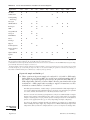
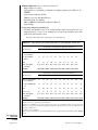
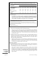
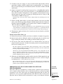
Table 6.1.3 Protein Standard Mixtures Available from Selected Suppliers
Applicationsa
1-D
2-Db
Bio-Rad
X
X
CalBiochem
X
Cell Signaling
Technology
X
X
Favorgen
X
X
GE Healthcare
X
X
X
Invitrogen
X
X
X
NEB
X
Norgen Biotek
X
X
Novagen
X
X
PerkinElmer
X
Pierce
X
Promega
X
Qiagen
X
R & D Systems
X
Roche Applied
Science
X
Sigma-Aldrich
X
Upstate
X
X
USB
X
X
Im
Prec
Fluor
Gly
Phos
X
Bio
Tag
IEF
X
X
X
X
X
X
X
Nat
X
X
X
X
X
X
X
X
X
X
X
X
X
X
X
X
X
X
X
X
X
X
X
X
X
X
X
X
X
a Abbreviations: 1-D, one-dimensional gels; 2-D, two-dimensional gels; Im, immunoblotting; Pre, prestained; Fluor, fluorescent; Gly, glycoprotein;
Phos, phosphoprotein; Bio, biotinylated; Tag, tagged; IEF, isoelectic focusing; Nat, native.
b 2-D standards are useful as independently characterized internal controls or reference standards for 2-D SDS-PAGE. Many investigators simply
use an internally characterized test sample as a reference set.
c Prestained standards, while not as sharply delineated as unstained standards, can be used to monitor progress of the separation since the bands
are visible through the gel cassette during electrophoresis. They are also useful for marking the position of a band after electroblotting to a
nitrocellulose or PVDF membrane prior to immunoassay or analysis by mass spectrometry.
Prepare the sample and load the gel
12. Dilute a portion of the protein sample to be analyzed 1:1 (v/v) with 2× SDS sample
buffer and heat 3 to 5 min at 100◦ C in a sealed screw-cap microcentrifuge tube. If
the sample is a precipitated protein pellet, dissolve the protein in 50 to 100 µl of 1×
SDS sample buffer and boil 3 to 5 min at 100◦ C. Dissolve protein molecular weight
standards in 1× SDS sample buffer according to supplier’s instructions; use these
standards as a control (Tables 6.1.2 and 6.1.3).
For dilute protein solutions, consider using 5:1 protein solution/6× SDS sample buffer to
increase the amount of protein loaded. Proteins can also be concentrated by precipitation
in acetone, ethanol, or trichloroacetic acid (TCA), but losses will occur.
For a 0.8-cm-wide well, 25 to 50 µg total protein in <20 µl is recommended for a complex
mixture when staining with Coomassie blue, and 1 to 10 µg total protein is needed for
samples containing one or a few proteins. If silver staining is used, 10- to 100-fold less
protein can be applied (0.01 to 5 µg in <20 µl depending on sample complexity).
One-Dimensional
SDS-PAGE
To achieve the highest resolution possible, the following precautions are recommended.
Prior to adding the sample buffer, keep samples at 0◦ C. Add the SDS sample buffer (room
temperature) directly to the 0◦ C sample (still on ice) in a screw-top microcentrifuge tube.
6.1.8
Supplement 37
Current Protocols in Cell Biology
Cap the tube to prevent evaporation, vortex, and transfer directly to a 100◦ C water bath
for 3 to 5 min. Let immunoprecipitates dissolve for 1 hr at 56◦ C in 1× SDS sample buffer
prior to boiling. DO NOT leave the sample in SDS sample buffer at room temperature
without first heating to 100◦ C to inactivate proteases (see Critical Parameters and
Troubleshooting). Endogenous proteases are very active in SDS sample buffer and will
cause severe degradation of the sample proteins after even a few minutes at room temperature. To test for possible proteases, mix the sample with SDS sample buffer without
heating and leave at room temperature for 1 to 3 hr. A loss of high-molecular-weight
bands and a general smearing of the banding pattern indicate a protease problem.
Once heated, the samples can sit at room temperature for the time it takes to load
samples.
13. Carefully remove the Teflon comb without tearing the edges of the polyacrylamide
wells. After the comb is removed, rinse wells with 1× SDS electrophoresis buffer.
The rinse removes unpolymerized monomer; otherwise, the monomer will continue to
polymerize after the comb is removed, creating uneven wells that will interfere with
sample loading and subsequent separation.
14. Using a Pasteur pipet, fill the wells with 1× SDS electrophoresis buffer.
If well walls are not upright, they can be manipulated with a flat-tipped needle attached
to a syringe.
15. Attach gel sandwich to upper buffer chamber following manufacturer’s instructions.
16. Fill lower buffer chamber with the recommended amount of 1× SDS electrophoresis
buffer.
17. Place sandwich attached to upper buffer chamber into lower buffer chamber.
18. Partially fill the upper buffer chamber with 1× SDS electrophoresis buffer so that
the sample wells of the stacking gel are filled with buffer.
Monitor the upper buffer chamber for leaks and, if necessary, reassemble the unit. A
slow leak in the upper buffer chamber may cause arcing around the upper electrode and
damage the upper buffer chamber.
19. Using a 25- or 100-µl syringe with a flat-tipped needle, load the protein sample(s)
into one or more wells by carefully applying the sample as a thin layer at the bottom of
the wells. Load control wells with molecular weight standards. Add an equal volume
of 1× SDS sample buffer to any empty wells to prevent spreading of adjoining
lanes.
Disposable loading tips can be used with automatic pipettors to simplify loading.
Preparing the samples at approximately the same concentration and loading an equal
volume to each well will ensure that all lanes are the same width and that the proteins run
evenly. If unequal volumes of sample buffer are added to wells, the lane with the larger
volume will spread during electrophoresis and constrict the adjacent lanes, causing
distortions.
The samples will layer on the bottom of the wells because the glycerol added to the
sample buffer gives the solution a greater density than the electrophoresis buffer. To keep
bands tight, hold the tip of the needle near the bottom of the well and load the samples
slowly. The bromphenol blue in the sample buffer makes sample application easy to follow
visually.
20. Fill the remainder of the upper buffer chamber with additional 1× SDS electrophoresis buffer so that the upper platinum electrode is completely covered. Do this slowly
so that samples are not swept into adjacent wells.
Electrophoresis
and
Immunoblotting
6.1.9
Current Protocols in Cell Biology
Supplement 37
Run the gel
21. Connect the power supply to the cell and run at 10 mA of constant current for a slab
gel 0.75 mm thick, until the bromphenol blue tracking dye enters the separating gel.
Then increase the current to 15 mA.
For a standard 16-cm gel sandwich, 4 mA per 0.75-mm-thick gel will run ∼15 hr (i.e.,
overnight); 15 mA per 0.75-mm gel will take 4 to 5 hr. To run two gels or a 1.5-mm-thick
gel, simply double the current. When running a 1.5-mm gel at 30 mA, the temperature
must be controlled (10◦ to 20◦ C) with a circulating constant-temperature water bath to
prevent “smiling” (curvature in the migratory band). Temperatures <5◦ C should not be
used because SDS in the running buffer will precipitate.
If the level of buffer in the upper chamber decreases, a leak has occurred.
22. After the bromphenol blue tracking dye has reached the bottom of the separating
gel, disconnect the power supply.
Refer to Safety Considerations under Electricity and Electrophoresis.
Disassemble the gel
23. Discard electrophoresis buffer and remove the upper buffer chamber with the attached
gel sandwich.
24. Orient the gel so that the order of the sample wells is known, remove the sandwich
from the upper buffer chamber, and lay the sandwich on a sheet of absorbent paper
or paper towels.
25. Carefully slide one of the spacers halfway from the edge of the sandwich along its
entire length. Use the exposed spacer as a lever to pry open the glass plate, exposing
the gel.
26. Carefully remove the gel from the lower plate. Cut a small triangle off one corner
of the gel so the lane orientation is not lost during staining and drying. Proceed with
protein detection.
Gradient gels are most easily picked up without tearing from the high-concentration end
of the gel using gloved fingers. Single-concentration gels <10% can be picked up and
placed in fixative, but are more easily removed if first immersed in fixative while left on
the plate, allowing the gel to float off.
The gel can be stained with Coomassie blue or silver (UNIT 6.6), or proteins can be
electroeluted, electroblotted onto a polyvinylidene difluoride (PVDF) membrane for subsequent staining or sequence analysis, or transferred to a membrane for immunoblotting
(UNIT 6.2). If the proteins are radiolabeled, they can be detected by autoradiography
(UNIT 6.3).
ALTERNATE
PROTOCOL 1
ELECTROPHORESIS IN TRIS-TRICINE BUFFER SYSTEMS
Separation of peptides and proteins under 10 to 15 kDa is not possible in the traditional
Laemmli discontinuous gel system (see Basic Protocol 1). This is due to the comigration
of SDS and smaller proteins, obscuring the resolution. Two approaches to obtain the
separation of small proteins and peptides in the range of 5 to 20 kDa are presented: the
following Tris-tricine method and a system using increased buffer concentrations (see
Alternate Protocol 2). The Tris-tricine system uses a modified buffer to separate the SDS
and peptides, thus improving resolution. Several precast gels are available for use with
the tricine formulations (Table 6.1.4).
One-Dimensional
SDS-PAGE
6.1.10
Supplement 37
Current Protocols in Cell Biology
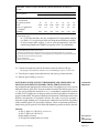
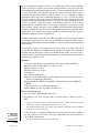
Table 6.1.4 Precast Gels Available from Selected Suppliers
Format
Bio-Rad
Large
Mini
2-Da
X
X
X
X
X
X
X
X
Cambrex
Jule
Application
X
Invitrogen
Instrument Compatibility
Native Peptide
SDS
Bio-Rad
Cambrex
Hoefer
Invitrogen
X
X
X
X
X
X
X
X
X
X
X
X
X
X
X
X
X
X
X
X
X
X
X
X
X
X
a Two-dimensional analysis.
Table 6.1.5 Recipes for Tricine Peptide Separating and Stacking Gelsa
SEPARATING AND STACKING GELS
Stock solutionb
Separating gel
Stacking gel
30% (w/v) acrylamide/0.8% (w/v)
bisacrylamide
9.80 ml
1.62 ml
Tris·Cl/SDS, pH 8.45
10.00 ml
3.10 ml
H2 O
7.03 ml
7.78 ml
4.00 g (3.17 ml)
—
10% (w/v) ammonium persulfate
50 µl
25 µl
TEMED
10 µl
5 µl
Glycerol
c
Prepare separating and stacking gel solutions separately.
In a 50-ml side-arm flask, mix 30% acrylamide/0.8% bisacrylamide solution
(Table 6.1.1), Tris·Cl/SDS, pH 8.45 (see reagents, below), and H2 O. Add glycerol
to separating gel only. Degas under vacuum 10 to 15 min. Add 10% ammonium
persulfate and TEMED. Swirl gently to mix; use immediately.
ADDITIONAL REAGENTS USED IN GELS
Tris·Cl/SDS, pH 8.45 (3.0 M Tris·Cl containing 0.3% w/v SDS)
Dissolve 182 g Tris base in 300 ml H2 O. Adjust to pH 8.45 with 1 N HCl. Add
H2 O to 500 ml total volume. Filter the solution through a 0.45-µm filter, add 1.5
g SDS, and store at 4◦ C up to 1 month.
a The recipes produce 30 ml of separating gel and 12.5 ml of stacking gel, which are adequate for two gels of dimensions
0.75 mm × 14 cm × 14 cm. The recipes are based on the Tris-tricine buffer system of Schagger and von Jagow (1987).
b All reagents and solutions used in the protocol must be prepared with Milli-Q-purified water or equivalent.
c Best to prepare fresh. Failure to form a firm gel usually indicates a problem with the persulfate, TEMED, or both.
Additional Materials (also see Basic Protocol 1)
Separating and stacking gel solutions (Table 6.1.5)
2× tricine sample buffer (see recipe)
Peptide molecular weight standards (Table 6.1.6)
Cathode buffer (see recipe)
Anode buffer (see recipe)
Coomassie blue G-250 staining solution (see recipe)
10% (v/v) acetic acid
50-ml Erlenmeyer side-arm flasks
Electrophoresis
and
Immunoblotting
6.1.11
Current Protocols in Cell Biology
Supplement 37
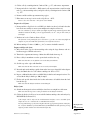
Table 6.1.6 Molecular Weights of Peptide Standards for Polyacrylamide Gel Electrophoresisa
Peptide
Molecular weight (Da)
Myoglobin (polypeptide backbone)
16,950
Myoglobin 1-131
14,440
Myoglobin 56-153
10,600
Myoglobin 56-131
8,160
Myoglobin 1-55
6,210
Glucagon
3,480
Myoglobin 132-153
2,510
a Peptide standards are commercially available from Sigma-Aldrich. See Sigma-
Aldrich Technical Bulletin MWSDS70-L for molecular weight markers for proteins.
1. Prepare and pour the separating and stacking gels (see Basic Protocol 1, steps 1 to
11) using the recipes in Table 6.1.5.
2. Prepare the sample (see Basic Protocol 1, step 12), but substitute 2× tricine sample
buffer for the 2× SDS sample buffer, and heat the sample at 40◦ C for 30 to 60 min
to improve solubilization prior to loading. Use peptide molecular weight standards
(Table 6.1.6).
If proteolytic activity is a problem (see Basic Protocol 1, step 12), heating samples to
100◦ C for 3 to 5 min may be required.
3. Set up the electrophoresis apparatus and load the gel (see Basic Protocol 1, steps 13
to 20), but use cathode buffer or water to rinse the wells, use cathode buffer in the
upper buffer chamber, and use anode buffer in the lower buffer chamber.
The cathode buffer contains the tricine.
4. Connect the power supply to the cell and run 1 hr at 30 V (constant voltage)
followed by 4 to 5 hr at 150 V (constant voltage). Use a heat exchanger to keep
the electrophoresis chamber at room temperature.
5. After the tracking dye has reached the bottom of the separating gel, disconnect the
power supply.
Refer to Safety Considerations under Electricity and Electrophoresis.
Coomassie blue G-250 is used as a tracking dye instead of bromphenol blue because it
moves ahead of the smallest peptides.
6. Disassemble the gel (see Basic Protocol 1, steps 23 to 26). Stain proteins in the gel
for 1 to 2 hr in Coomassie blue G-250 staining solution. Follow by destaining with
10% acetic acid, changing the solution every 30 min until background is clear (3 to
5 changes). For higher sensitivity, use silver staining as a recommended alternative.
Prolonged staining and destaining will result in the loss of resolution of the smaller
proteins (<10 kDa). Proteins diffuse within the gel and out of the gel, resulting in a loss
of staining intensity and resolution.
ALTERNATE
PROTOCOL 2
One-Dimensional
SDS-PAGE
NONUREA PEPTIDE SEPARATIONS WITH TRIS BUFFERS
A simple modification of the traditional Laemmli buffer system presented in Basic
Protocol 1, in which the increased concentration of buffers provides better separation
between the stacked peptides and the SDS micelles, permits reasonable separation of
peptides as small as 5 kDa.
6.1.12
Supplement 37
Current Protocols in Cell Biology
Additional Materials (also see Basic Protocol 1)
Separating and stacking gel solutions (Table 6.1.7)
2× Tris·Cl/SDS, pH 8.8 (dilute 4× Tris·Cl/SDS, pH 8.8; Table 6.1.1)
2× SDS electrophoresis buffer (see recipe)
1. Prepare and pour the separating and stacking gels (see Basic Protocol 1, steps 1 to
11), using the modified recipes in Table 6.1.7. After removing the isobutyl alcohol
overlay from the separating gel, rinse with 2× Tris·Cl/SDS, pH 8.8, rather than 1×
Tris·Cl/SDS.
2. Prepare the sample and load the gel (see Basic Protocol 1, steps 12 to 20), but
substitute 2× SDS electrophoresis buffer for the 1× SDS electrophoresis buffer.
Table 6.1.6 lists the standards for small protein separations.
Table 6.1.7 Recipes for Modified Laemmli Peptide Separating and Stacking Gelsa
SEPARATING AND STACKING GELS
Stock solutionb
Separating gel
Stacking gel
30% (w/v) acrylamide/0.8% (w/v)
bisacrylamide
10.00 ml
0.65 ml
8× Tris·Cl, pH 8.8
3.75 ml
—
4× Tris·Cl, pH 6.8
—
1.25 ml
0.15 ml
50 µl
1.00 ml
3.00 ml
10% (w/v) ammonium persulfate
50 µl
25 µl
TEMED
10 µl
5 µl
c
10% (w/v) SDS
H2 O
c
Prepare separating and stacking gel solutions separately.
In a 25-ml side-arm flask, mix 30% acrylamide/0.8% bisacrylamide solution
(see Table 6.1.1), 8× Tris·Cl, pH 8.8, or 4× Tris·Cl, pH 6.8 (see reagents below),
10% SDS, and H2 O. Degas under vacuum 10 to 15 min. Add 10% ammonium
persulfate and TEMED. Swirl gently to mix. Use immediately.
ADDITIONAL REAGENTS USED IN GELS
4× Tris·Cl, pH 6.8 (0.5 M Tris·Cl)
Dissolve 6.05 g Tris base in 40 ml H2 O. Adjust to pH 6.8 with 1 N HCl. Add
H2 O to 100 ml total volume. Filter the solution through a 0.45-µm filter and
store up to 1 month at 4◦ C.
8× Tris·Cl, pH 8.8 (3.0 M Tris·Cl)
Dissolve 182 g Tris base in 300 ml H2 O. Adjust to pH 8.8 with 1 N HCl. Add
H2 O to 500 ml total volume. Filter the solution through a 0.45-µm filter and
store up to 1 month at 4◦ C.
a The recipes produce 15 ml of separating gel and 5 ml of stacking gel, which are adequate for one gel of dimensions
0.75 mm × 14 cm × 14 cm. The recipes are based on the modified Laemmli peptide separation system of Okajima
et al. (1993).
b All reagents and solutions used in the protocol must be prepared with Milli-Q-purified water or equivalent.
c Best to prepare fresh. Failure to form a firm gel usually indicates a problem with the ammonium persulfate, TEMED,
or both.
Electrophoresis
and
Immunoblotting
6.1.13
Current Protocols in Cell Biology
Supplement 37
3. Run the gel (see Basic Protocol 1, steps 21 and 22).
Note that the separations will take ∼25% longer than those using Basic Protocol 1. The
increased buffer concentrations lead to faster transit through the stacking gel but lower
mobility in the resolving gel.
4. Disassemble the gel (see Basic Protocol 1, steps 23 to 26).
Proteins in the gel may now be stained.
ALTERNATE
PROTOCOL 3
CONTINUOUS SDS-PAGE
With continuous SDS-PAGE, the same buffer is used for both the gel and electrode
solutions. Although continuous gels lack the resolution of the discontinuous systems,
they are extremely versatile, less prone to mobility artifacts, and much easier to prepare.
The stacking gel is omitted.
Additional Materials (also see Basic Protocol 1)
Separating gel solution (Table 6.1.8)
2× and 1× phosphate/SDS sample buffer (see recipe)
1× phosphate/SDS electrophoresis buffer (see recipe)
Table 6.1.8 Recipes for Separating Gels for Continuous SDS-PAGEa
SEPARATING GEL
Final acrylamide concentration in the separating gel (%)c
Stock solutionb
5
30% (w/v)
acrylamide/0.8% (w/v)
bisacrylamide
2.5
4× phosphate/SDS,
pH 7.2
3.75 3.75 3.75 3.75 3.75 3.75 3.75 3.75 3.75 3.75 3.75
H2 O
8.75 8.25 7.75 7.25 6.75 6.25 5.75 5.25 4.75 4.25 3.75
10% (w/v)
ammonium
persulfated
0.05 0.05 0.05 0.05 0.05 0.05 0.05 0.05 0.05 0.05 0.05
TEMED
0.01 0.01 0.01 0.01 0.01 0.01 0.01 0.01 0.01 0.01 0.01
6
7
8
9
10
11
12
13
14
15
3.00 3.50 4.00 4.50 5.00 5.50 6.00 6.50 7.00 7.50
Preparation of separating gel
In a 25-ml side-arm flask, mix 30% acrylamide/0.8% bisacrylamide solution
(see Table 6.1.1), 4× phosphate/SDS, pH 7.2, and H2 O. Degas under vacuum
about 5 min. Add 10% ammonium persulfate and TEMED. Swirl gently to mix.
Use immediately.
ADDITIONAL REAGENTS USED IN GELS
4× phosphate/SDS, pH 7.2 (0.4 M sodium phosphate/0.4% w/v SDS)
Mix 46.8 g NaH2 PO4 ·H2 O, 231.6 g Na2 HPO4 ·7H2 O, and 12 g SDS in 3 liters
H2 O. Store at 4◦ C for up to 3 months.
a The recipes produce 15 ml of separating gel, which is adequate for one gel of dimensions 0.75 mm × 14 cm × 14
cm. The recipes are based on the original continuous phosphate buffer system of Weber et al. (1972).
b All reagents and solutions used in the protocol must be prepared with Milli-Q-purified water or equivalent.
c Volumes are in milliliters. The desired percentage of acrylamide in the separating gel depends on the molecular size
One-Dimensional
SDS-PAGE
of the protein being separated. See Basic Protocol 1, annotation to step 3.
d Best to prepare fresh. Failure to form a firm gel usually indicates a problem with the ammonium persulfate, TEMED,
or both.
6.1.14
Supplement 37
Current Protocols in Cell Biology
1. Prepare and pour a single separating gel (see Basic Protocol 1, steps 1 to 4), except
use the recipe in Table 6.1.8 and fill the gel sandwich to the top. Omit the stacking
gel. Insert the comb (see Basic Protocol 1, step 10) and allow the gel to polymerize
30 to 60 min at room temperature.
2. Mix the protein sample 1:1 with 2× phosphate/SDS sample buffer and heat to 100◦ C
for 2 min.
For large sample volumes or samples suspended in high-ionic-strength buffers (>50 mM),
dialyze against 1× sample buffer prior to electrophoresis. Note that the precautions about
proteases (see Basic Protocol 1, step 12) apply here.
3. Assemble the electrophoresis apparatus and load the sample (see Basic Protocol 1,
steps 13 to 20) using the phosphate/SDS electrophoresis buffer. Load empty wells
with 1× phosphate/SDS sample buffer.
4. Connect the power supply and start the run with 15 mA per 0.75-mm-thick gel until
the tracking dye has entered the gel. Continue electrophoresis at 30 mA for 3 hr (5%
gel), 5 hr (10% gel), 8 hr (15% gel), or until the dye reaches the bottom of the gel.
Use temperature control if available to maintain the gel at 15◦ to 20◦ C. SDS will precipitate
below 15◦ C in this system.
5. Disassemble the gel (see Basic Protocol 1, steps 23 to 26).
See Safety Considerations in introduction. Proteins in the gel may now be stained.
CASTING AND RUNNING ULTRATHIN GELS
Ultrathin gels provide superb resolution but are difficult to handle. In this application,
gels are cast on GelBond, a Mylar support material. Silver staining is recommended for
the best resolution. Combs and spacers for gels <0.5 mm thick are not readily available
for most protein electrophoresis units. However, by adapting combs and spacers used for
DNA sequencing, casting gels from 0.2 to 0.5 mm thick is straightforward.
ALTERNATE
PROTOCOL 4
Additional Materials (also see Basic Protocol 1)
95% (v/v) ethanol
GelBond (Lonza) cut to a size slightly smaller than the gel plate dimensions
Glue stick
Ink roller (available from art supply stores)
Combs and spacers (0.19 to 0.5 mm; sequencing gel spacers and combs can be cut
to fit)
1. Wash gel plates with water-based laboratory detergent followed by successive rinses
with hot tap water, deionized water, and finally 95% ethanol. Allow to air dry.
Gel plates must be extremely clean for casting thin gels. Gloves are needed throughout
these procedures to prevent contamination by proteins on the surface of skin.
2. Apply a streak of adhesive from a glue stick to the bottom edge of the glass plate.
Quickly position the GelBond with the hydrophobic side down (a drop of water will
bead up on the hydrophobic surface). Apply pressure with Kimwipe tissue to attach
the GelBond firmly to the plate. Finally, pull the top portion of the GelBond back,
place a few drops of water underneath, and roll flat with an ink roller.
Make sure the GelBond does not extend beyond the edges of the upper and lower sealing
surface of the plate. This will cause it to buckle on sealing. Reposition the GelBond if
needed to prevent it from extending beyond the glass plate. Material may also be trimmed
to fit flush with the plate edge.
Electrophoresis
and
Immunoblotting
6.1.15
Current Protocols in Cell Biology
Supplement 37
3. Assemble the gel cassette according to the manufacturer’s instructions (also see
Basic Protocol 1, steps 1 and 2). Just prior to assembly, blow air over the surface of
both the GelBond and the opposing glass surface to remove any particulate material
(e.g., dust).
Sequencing gel spacers can be easily adapted. First, cut the spacers slightly longer than
the length of the gel plate. Position a spacer along each edge of the glass plate and
assemble the gel sandwich, clamping in place. With a razor blade, trim the excess spacer
at top and bottom to get a reusable spacer exactly the size of the plate.
4. Prepare and pour the separating and stacking gels (see Basic Protocol 1, steps 3 to
9). In place of the Teflon comb, insert a square well sequencing comb cut to fit
within the gel sandwich. Allow the stacking gel to polymerize 30 to 45 min at room
temperature.
Less solution is needed for ultrathin gels. For example, a 0.5-mm-thick gel requires 33%
less gel solution than a 0.75-mm gel.
5. Prepare the sample and load the gel (see Basic Protocol 1, steps 12 to 20).
When preparing protein samples for ultrathin gels, 3 to 4 µl at 5 µg protein/µl is required
for Coomassie blue R-250 staining, whereas 10-fold less is needed for silver staining.
6. Run the gel (see Basic Protocol 1, steps 21 and 22), except conduct the electrophoresis
at 7 mA/gel (0.25-mm-thick gels) or 14 mA/gel (0.5-mm-thick gels) for 4 to 5 hr.
7. When the separation is complete, disassemble the unit and remove the gel (see Basic
Protocol 1, steps 23 to 26) with the GelBond still attached. With a gloved hand, wash
away the adhesive material from the back of the GelBond under a stream of water
before proceeding to protein detection.
Either Coomassie blue or silver staining may be used, but silver staining produces
particularly fine resolution with thin GelBond-backed gels. Compared to staining thicker
(>0.75 mm) gels, thin (<0.75 mm) gels stain and destain more quickly. Although the
optimum staining times must be empirically determined, all steps in Coomassie blue and
silver staining procedures are generally reduced by half.
SUPPORT
PROTOCOL 1
CASTING MULTIPLE SINGLE-CONCENTRATION GELS
Casting multiple gels at one time has several advantages. All the gels are identical, so
sample separation is not affected by gel-to-gel variation. Furthermore, casting ten gels
is only slightly more difficult than casting two gels. Once cast, gels can be stored for
several days in a refrigerator.
Additional Materials (also see Basic Protocol 1)
Separating and stacking gels for single-concentration gels (Table 6.1.9)
Multiple gel caster (Bio-Rad, Hoefer)
100-ml disposable syringe and flat-tipped needle
Extra plates and spacers
14 × 14–cm acrylic blocks or polycarbonate sheets
250- and 500-ml side-arm flasks (used in gel preparation)
Long razor blade or plastic wedge (Wonder Wedge, Hoefer)
Resealable plastic bags
Pour the separating gel
1. Assemble the multiple gel caster according to the manufacturer’s instructions.
One-Dimensional
SDS-PAGE
With the Hoefer unit, make sure to insert the large triangular space filler plug in the base
of the caster. The plug is removed when casting gradient gels (see Support Protocol 2).
6.1.16
Supplement 37
Current Protocols in Cell Biology
Table 6.1.9 Recipes for Multiple Single-Concentration Polyacrylamide Gelsa
SEPARATING GEL
Final acrylamide concentration in the separating gel (%)c
Stock solutionb
5
6
7
8
9
10
11
12
13
14
15
30% (w/v) acrylamide/
0.8% (w/v)
bisacrylamide
52
62
72
83
93
103
114
124
134
145
155
4× Tris·Cl/SDS,
pH 8.8
78
78
78
78
78
78
78
78
78
78
78
H2 O
181
171
160
150
140
129
119
109
98
88
78
10% (w/v) ammonium
persulfated
1.0
1.0
1.0
1.0
1.0
1.0
1.0
1.0
1.0
1.0
1.0
TEMED
0.21
0.21
0.21
0.21
0.21
0.21
0.21
0.21
0.21
0.21
0.21
Preparation of separating gel
In a 500-ml side-arm flask, mix 30% acrylamide/0.8% bisacrylamide solution (see Table 6.1.1), 4×
Tris·Cl/SDS, pH 8.8 (Table 6.1.1), and H2 O. Degas under vacuum ∼5 min. Add 10% ammonium
persulfate and TEMED. Swirl gently to mix. Use immediately.
STACKING GEL
In a 250-ml side-arm flask, mix 13.0 ml 30% acrylamide/0.8% bisacrylamide solution, 25 ml 4×
Tris·Cl/SDS, pH 6.8 (Table 6.1.1), and 61 ml H2 O. Degas under vacuum ∼5 min. Add 0.25 ml
10% ammonium persulfate and 50 µl TEMED. Swirl gently to mix. Use immediately.
a The recipes produce about 300 ml of separating gel and 100 ml of stacking gel, which are adequate for ten gels of dimensions 1.5 mm ×
14 cm × 14 cm, with extra solution should there be a leak or spill while casting the gels. For thinner spacers or fewer gels, calculate volumes
using the equation in the annotation to step 4. The recipes are based on the SDS (denaturing) discontinuous buffer system of Laemmli (1970).
b All reagents and solutions used in the protocol must be prepared with Milli-Q-purified water or equivalent.
c Volumes in table body are in milliliters. The desired percentage of acrylamide in separating gel depends on the molecular size of the protein
being separated. See Basic Protocol 1, annotation to step 3.
d Best to prepare fresh. Failure to form a firm gel usually indicates a problem with the persulfate, TEMED, or both.
2. Assemble glass sandwiches and stack them in the casting chamber. Stack up to ten
1.5-mm gels and fill in extra space with acrylic blocks or polycarbonate sheets to
hold the sandwiches tightly in place. Make sure the spacers are straight along the
top, right, and left edges of the glass plates and that all edges of the stack are flush.
The presence of loosely fitting sandwiches in the caster will lead to unevenly cast gels,
creating distortions during electrophoresis. Polycarbonate inhibits gel polymerization.
Therefore, if polycarbonate sheets are placed in the caster before and after the set of
glass sandwiches, the entire set will slide out as one block after polymerization. Placing
polycarbonate sheets between each gel sandwich makes them easier to separate from one
another after polymerization.
3. Place the front sealing plate on the casting chamber, making sure the stack fits snugly.
Secure the plate with four spring clamps and tighten the bottom thumb screws.
4. Prepare the separating (resolving) gel solution (Table 6.1.9).
A 12-cm separating gel with a 4-cm stacking gel is recommended.
If fewer than ten gels are prepared (Table 6.1.9), use the following formula to estimate
the amount of separating gel volume needed:
volume = gel no. × height (cm) × width (cm) × thickness (cm) + 4 × gel no. + 10 ml.
Electrophoresis
and
Immunoblotting
6.1.17
Current Protocols in Cell Biology
Supplement 37
5. Using a 100-ml disposable syringe with flat-tipped needle, inject the resolving gel
solution down the side of one spacer into the multiple caster. A channel in the silicone
plug distributes the solution throughout the whole caster. Avoid introducing bubbles
by giving the caster a quick tap on the benchtop once the caster is filled.
6. Overlay the center of each gel with 100 µl H2 O-saturated isobutyl alcohol and let
polymerize for 1 to 2 hr.
7. Drain off the overlay and rinse the surface with 1× Tris·Cl/SDS, pH 8.8. If the gels
will not be used immediately, skip to step 12.
Pour the stacking gel
8. Prepare the stacking gel solution either singly (see Basic Protocol 1, step 8) or for
all the gels at once (Table 6.1.9).
The stacking gel solution should be prepared just before it is poured.
9. Fill each sandwich in the caster with stacking gel solution.
10. Insert a comb into each sandwich and let the gel polymerize for 2 hr.
Insert the combs at a 45◦ angle to avoid trapping air underneath the comb teeth. Air
bubbles will inhibit polymerization and cause dents in the wells and a distorted pattern
of protein bands.
11. Remove the combs and rinse wells with 1× SDS electrophoresis buffer.
Remove the gels from the caster
12. Remove the gels from the caster and separate by carefully inserting a long razor
blade or knife between the gel sandwiches.
A plastic wedge (Hoefer’s Wonder Wedge) also works well.
13. Clean the outside of each gel plate with running water to remove the residual polymerized and unpolymerized acrylamide.
14. Overlay gels to be stored with 1× Tris·Cl/SDS, pH 8.8, place in a resealable plastic
bag, and store at 4◦ C until needed (up to 1 week).
ALTERNATE
PROTOCOL 5
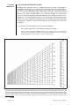
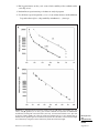
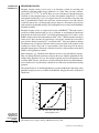
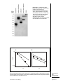
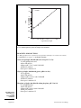
SEPARATION OF PROTEINS ON GRADIENT GELS
Gels that consist of a gradient of increasing polyacrylamide concentration resolve a
much wider size range of proteins than standard single-concentration gels (see Critical
Parameters and Troubleshooting). The protein bands are also much sharper, particularly in
the low-molecular-weight range. Unlike single-concentration gels, gradient gels separate
proteins in a way that can be represented easily to give a linear plot from 10 to 200 kDa.
This facilitates molecular weight estimations.
The quantities given below provide separating gel solution sufficient for two 0.75-mm
gels (∼7 ml of each concentration) or one 1.5-mm gel (∼14 ml of each concentration).
Volumes can be adjusted to accommodate gel sandwiches of different dimensions.
Additional Materials (also see Basic Protocol 1)
Light and heavy acrylamide gel solutions (Table 6.1.10)
Bromphenol blue (optional; for checking practice gradient)
10% ammonium persulfate (prepare fresh)
TEMED
One-Dimensional
SDS-PAGE
6.1.18
Supplement 37
Current Protocols in Cell Biology
Gradient maker (30 to 50 ml, Hoefer SG30 or SG50; or 30 to 100 ml, Bio-Rad 385)
Tygon tubing with micropipet tip
Peristaltic pump (optional; e.g., Markson A-13002, A-34040, or A-34105
minipump)
Whatman 3MM filter paper
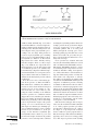
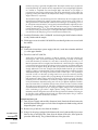
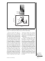
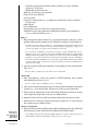
Set up the gradient maker and prepare the gel solutions
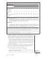
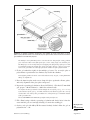
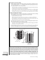
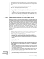
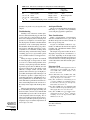
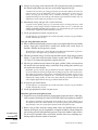
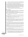
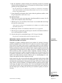
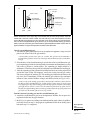
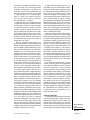
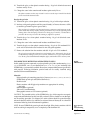
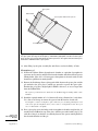
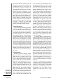
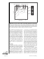
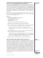
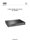
1. Assemble the magnetic stirrer and gradient maker on a ring stand as shown in Figure
6.1.2. Connect the outlet valve of the gradient maker to Tygon tubing attached to
a micropipet tip that is placed over the vertical gel sandwich. If desired, place a
peristaltic pump in line between the gradient maker and the gel sandwich.
A peristaltic pump will simplify casting by providing a smooth flow rate.
2. Place a small stir-bar into the mixing chamber of the gradient maker (i.e., the chamber
connected to the outlet).
Table 6.1.10 Light and Heavy Acrylamide Gel Solutions for Gradient Gels
Acrylamide concentration in light gel solution (%)a,b
Stock solution
5
30% acrylamide/0.8% 2.5
bisacrylamidec
6
7
8
9
10
11
12
13
14
3.0
3.5
4.0
4.5
5.0
5.5
6.0
6.5
7.0
4× Tris·Cl/SDS,
pH 8.8c
3.75 3.75 3.75 3.75 3.75 3.75 3.75 3.75 3.75 3.75
H2 O
8.75 8.25 7.75 7.25 6.75 6.25 5.75 5.25 4.75 4.25
10% ammonium
persulfated
0.05 0.05 0.05 0.05 0.05 0.05 0.05 0.05 0.05 0.05
TEMEDd
0.005 0.005 0.005 0.005 0.005 0.005 0.005 0.005 0.005 0.005
Acrylamide concentration in heavy gel solution (%)a,b
Stock solution
10
11
12
13
14
15
16
17
18
19
20
30% acrylamide/0.8% 5.0
bisacrylamidec
5.5
6.0
6.5
7.0
7.5
8.0
8.5
9.0
9.5
10.0
4× Tris·Cl/SDS,
pH 8.8c
3.75 3.75 3.75 3.75 3.75 3.75 3.75 3.75 3.75 3.75
3.75
H2 O
5.0
Sucrose (g)
2.25 2.25 2.25 2.25 2.25 2.25 2.25 2.25 2.25 2.25
2.25
10% ammonium
persulfated
0.05 0.05 0.05 0.05 0.05 0.05 0.05 0.05 0.05 0.05
0.05
TEMEDd
0.005 0.005 0.005 0.005 0.005 0.005 0.005 0.005 0.005 0.005 0.005
4.5
4.0
3.5
3.0
2.5
2.0
1.5
1.0
0.5
0
a To survey proteins ≥10 kDa, 5% to 20% gradient gels are recommended. To expand the range between 10 and
200 kDa, a 10% to 20% gradient gel is recommended.
b Volumes are in milliliters (sucrose is in grams). Keep light gel solution at room temperature prior to use (no longer than
1 hr). Keep heavy solution on ice.
c See Table 6.1.1 for preparation.
d Ammonium persulfate and TEMED are added directly to the gradient chambers immediately before the gel is poured.
It is best to prepare ammonium persulfate fresh. Failure to form a firm gel usually indicates a problem with the ammonium
persulfate, TEMED, or both.
Electrophoresis
and
Immunoblotting
6.1.19
Current Protocols in Cell Biology
Supplement 37
Figure 6.1.2
control.
Gradient gel setup. A peristaltic pump, though not required, will provide better
3. Using the recipes in Table 6.1.10, prepare light and heavy acrylamide gel solutions
without ammonium persulfate or TEMED.
Recommended gradient ranges are 5% to 20% for most applications (to separate proteins
of 10 to several hundred kilodaltons).
Deaeration is not recommended for either the light or heavy solution. Omitting the
deaeration will allow polymerization to proceed more slowly, letting the gradient establish
itself in the gel sandwich before polymerization takes place.
Keep the heavy solution on ice until use. Once the ammonium persulfate is added to
the heavy solution, it will polymerize without TEMED, albeit more slowly; keeping the
solution on ice prevents this. The gel solution will come to room temperature during casting. The higher the percentage of acrylamide, the more severe the problem of premature
polymerization.
4. With the outlet port and interconnecting valve between the two chambers closed,
pipet 7 ml of light (low-concentration) acrylamide gel solution into the reservoir
chamber for one 0.75-mm-thick gradient gel.
A practice run with heavy and light solutions is recommended. Bromphenol blue should
be added to the heavy solution to demonstrate linearity of the practice gradient.
5. Open the interconnecting valve briefly to allow a small amount (∼200 µl) of light
solution to flow through the valve and into the mixing chamber.
The presence of air bubbles in the interconnecting valve may obstruct the flow between
chambers during casting.
6. Add 7 ml of heavy (high-concentration) acrylamide gel solution to the mixing
chamber.
7. Add 23 µl of 10% ammonium persulfate and ∼2.3 µl TEMED per 7 ml acrylamide
solution to each chamber. Mix the solutions in each chamber with a disposable pipet.
Form the gradient and cast the gel
8. Open the interconnecting valve completely.
One-Dimensional
SDS-PAGE
Some of the heavy solution will flow back into the reservoir chamber containing light
solution as the two chambers equilibrate. This will not affect the formation of the gradient.
6.1.20
Supplement 37
Current Protocols in Cell Biology
9. Turn on the magnetic stirrer and adjust the rate to produce a slight vortex in the
mixing chamber.
10. Open the outlet of the gradient maker slowly. Adjust the outlet valve to a flow rate
of 2 ml/min. If using a peristaltic pump, calibrate the flow rate with a graduated
cylinder prior to casting the gel.
Some adjustment of the flow rate may be necessary during casting. If the light solution
is not flowing into the mixing chamber, a bubble may be caught in the interconnecting
valve. Quickly close the outlet and cover the top of the reservoir chamber with a gloved
thumb. Push down with the thumb to increase the pressure in the chamber and force the
air bubble out of the center valve.
11. Fill the gel sandwich from the top. Place the pipet tip against one side of the
sandwich so the solution flows down one plate only. The heavy solution will flow
into the sandwich first, followed by progressively lighter solution.
12. Watch as the last of the light solution drains into the outlet tube and adjust the flow
rate to ensure that the last few milliliters of solution do not flow quickly into the gel
sandwich and disturb the gradient.
13. Overlay the gradient gel with H2 O-saturated isobutyl alcohol. Allow the gel to
polymerize ∼1 hr.
In this gel system, the gel will polymerize from the bottom (i.e., heavy solution) up. Because
polymerization is an exothermic reaction, heat can be felt evolving from the bottom of
the gel sandwich during polymerization. A sharp optical discontinuity at the gel-overlay
interface indicates that polymerization has occurred. In general, 1 hr is adequate for
polymerization.
14. Remove the H2 O-saturated isobutyl alcohol and rinse with 1× Tris·Cl/SDS, pH 8.8.
Cast the stacking gel (see Basic Protocol 1, steps 8 to 11).
The gel can be covered with 1× Tris·Cl/SDS, pH 8.8, sealed in a plastic bag, and stored
for up to 1 week.
Load and run the gel
15. Prepare the protein sample and protein molecular-weight-standards mixture. Load
and run the gel (see Basic Protocol 1, steps 13 to 26).
The gel can be stained with Coomassie blue or silver (UNIT 6.6).
16. After staining, dry the gels onto Whatman 3MM or equivalent filter paper.
Gradient gels >0.75 mm thick require special handling during drying to prevent cracking.
The simplest approach to drying gradient gels is to use thin gels; ≤0.75-mm gradient
gels with ≤20% acrylamide solutions will dry without cracking as long as the vacuum
pump is working properly and the cold trap is dry at the onset of drying. For gradient gels
>0.75 mm thick, add 3% (w/v) glycerol to the final destaining solution to help prevent
cracking. Another method is to dehydrate and shrink the gel in 30% methanol for up to
3 hr prior to drying. Then place the gel in distilled water for 5 min before drying.
CASTING MULTIPLE GRADIENT GELS
Casting gradient gels in a multiple gel caster has several advantages. In addition to the
time savings, batch casting produces gels that are essentially identical. This is particularly important for gradient gels, where slight variations in casting technique can cause
variations in protein mobility. The gels may be stored for up to 1 week after casting
to ensure internal consistency from run to run during the week. Furthermore, gels with
several ranges of concentrations (e.g., 5% to 20% and 10% to 20% acrylamide) can be
cast and stored, giving much more flexibility to optimize separations.
SUPPORT
PROTOCOL 2
Electrophoresis
and
Immunoblotting
6.1.21
Current Protocols in Cell Biology
Supplement 37
Additional Materials (also see Alternate Protocol 5)
Plug solution (see recipe)
Light and heavy acrylamide gel solutions for multiple gradient gels (Table 6.1.11)
TEMED
H2 O-saturated isobutyl alcohol
Multiple gel caster (Bio-Rad, Hoefer)
Peristaltic pump (25 ml/min)
500- or 1000-ml gradient maker (Bio-Rad, Hoefer)
Tygon tubing
Set up system and pour separating gel
1. Assemble the multiple caster as in casting multiple single-concentration gels (see
Support Protocol 1, steps 1 to 3), making sure to remove the triangular space filler
plugs in the bottom of the caster.
The plug is used only when casting single-concentration gels.
Table 6.1.11 Light and Heavy Acrylamide Gel Solutions for Casting Multiple Gradient Gels
Acrylamide concentration in light gel solution (%)a,b
Stock solution
5
6
7
8
9
10
11
12
13
14
30% acrylamide/0.8%
bisacrylamidec
28
33
39
44
50
55
61
66
72
77
4× Tris·Cl/SDS,
pH 8.8c
41
41
41
41
41
41
41
41
41
41
H2 O
96
91
85
80
74
69
63
58
52
47
10% ammonium
persulfated
0.55 0.55 0.55 0.55 0.55 0.55 0.55 0.55 0.55 0.55
TEMED
0.054 0.054 0.054 0.054 0.054 0.054 0.054 0.054 0.054 0.054
Acrylamide concentration in heavy gel solution (%)a,b
Stock solution
10
11
12
13
14
15
16
17
18
19
20
30% acrylamide/0.8%
bisacrylamidec
55
61
66
72
77
83
88
94
99
105
110
4× Tris·Cl/SDS, pH
8.8c
41
41
41
41
41
41
41
41
41
41
41
H2 O
55
50
44
39
33
28
22
17
11
5.5
0
Sucrose (g)
25
25
25
25
25
25
25
25
25
25
25
10% ammonium
persulfated
0.55 0.55 0.55 0.55 0.55 0.55 0.55 0.55 0.55 0.55
0.55
TEMED
0.054 0.054 0.054 0.054 0.054 0.054 0.054 0.054 0.054 0.054 0.054
a To survey proteins ≥10 kDa, 5% to 20% gradient gels are recommended. To expand the range between 10 and
200 kDa, a 10% to 20% gradient gel is recommended.
b Volumes are in milliliters (sucrose is in grams). Recipes produce ten 1.5-mm-thick gradient gels with 10 ml extra
solution to account for losses in tubing. Keep light gel solution at room temperature prior to use (no longer than 1 hr).
Keep heavy solution on ice.
c See Table 6.1.1 for preparation.
d Best to prepare fresh. Failure to form a firm gel usually indicates a problem with the ammonium persulfate, TEMED,
or both.
One-Dimensional
SDS-PAGE
6.1.22
Supplement 37
Current Protocols in Cell Biology
Figure 6.1.3 Setup for casting multiple gradient gels. Casting multiple gradient gels requires a
peristaltic pump and a multiple gel caster. Gel solution is introduced through the bottom of the
multiple caster.
2. Set up the peristaltic pump (Fig. 6.1.3). Using a graduated cylinder and water, adjust
the flow rate so that the volume of the gradient solution plus volume of plug solution
is poured in ∼15 to 18 min (∼25 ml/min).
3. Set up the gradient maker. Close all valves and place a stir-bar in the mixing chamber,
which is the one with the outlet port. Attach one end of a piece of Tygon tubing to the
outlet of the gradient maker. Run the other end of the tubing through the peristaltic
pump and attach it to the red inlet port at the bottom of the caster.
Choose a gradient maker that holds no more than four times the total volume of the
gradient solution to be poured (i.e., a 1000- or 500-ml gradient maker).
4. Prepare solutions for the gradient maker (Table 6.1.11).
Deaeration is not recommended for gradient gels.
5. Immediately after adding TEMED to the gel solutions, pour the light (lowconcentration) solution into the mixing chamber (the one with the port). Open the
mixing valve slightly to allow the tunnel to fill and to avoid air bubbles. Close the
valve again and pour the heavy (high-concentration) acrylamide solution into the
reservoir chamber.
6. Start the magnetic stirrer and open the outlet valve; then start the pump and open the
mixing valve.
In units for casting multiple gels, acrylamide solution flows in from the bottom. To
use a multiple casting unit, the light solution is placed in the mixing chamber and the
heavy solution in the reservoir. This is the reverse of casting a single gel (see Alternate
Protocol 5). Thus, light solution enters the multiple caster first, followed by progressively
heavier solution. Finally, the acrylamide solution is stabilized in the multiple caster
with a heavy plug solution and allowed to polymerize (see step 8 and manufacturer’s
instructions).
7. When almost all the acrylamide solution is gone from the gradient maker, stop the
pump and close the mixing valve. Tilt the gradient maker toward the outlet side and
remove the last milliliters of the mix. Do not allow air bubbles to enter the tubing.
8. Add the plug solution to the mixing chamber and start the pump. Make sure that
no bubbles are introduced. Continue pumping until the bottom of the caster is filled
with plug solution to just below the glass plates; then turn off the pump. Clamp the
tubing close to the red port of the casting chamber.
Electrophoresis
and
Immunoblotting
6.1.23
Current Protocols in Cell Biology
Supplement 37
9. Quickly overlay each separate gel sandwich with 100 µl H2 O-saturated isobutyl
alcohol. Use the same amount on each sandwich. Allow the gels to polymerize for
∼1 hr.
Failure to use the same amount of overlay solution will cause the gel sandwiches to
polymerize at different heights.
10. Drain off the overlay and rinse the surface of the gels with 1× Tris·Cl/SDS, pH 8.8.
Pour stacking gel and remove gels from caster
11. Prepare and cast the stacking gel as in casting multiple single-concentration gels (see
Support Protocol 1, steps 8 to 11).
12. Remove gels from the caster and clean the gel sandwiches (see Support Protocol 1,
steps 12 and 13). Store gels, if necessary, according to the instructions for multiple
single-concentration gels (see Support Protocol 1, step 14).
BASIC
PROTOCOL 2
ELECTROPHORESIS IN SINGLE-CONCENTRATION MINIGELS
Separation of proteins in a small-gel format is becoming increasingly popular for applications that range from isolating material for peptide sequencing to performing routine protein separations. The unique combination of speed and high resolution is the
foremost advantage of small gels. Additionally, small gels are easily adapted to singleconcentration, gradient, and two-dimensional SDS-PAGE procedures. The minigel procedures described are adaptations of larger gel systems.
This protocol describes the use of a multiple gel caster. The caster is simple to use,
and up to five gels can be prepared at one time with this procedure. Single gels can be
prepared using adaptations in the manufacturer’s instructions. A multiple gel caster is
the only practical way to produce small linear polyacrylamide gradient gels (see Support
Protocol 3).
Materials
Minigel vertical gel unit (Hoefer Mighty Small SE 250/280 or Bio-Rad
Mini-Protean II) with glass plates, clamps, and buffer chambers
0.75-mm spacers
Multiple gel caster (Hoefer SE-275/295 or Bio-Rad Mini-Protean II multicasting
chamber)
Acrylic plate (Hoefer SE-217 or Bio-Rad 165-1957) or polycarbonate separation
sheet (Hoefer SE-213 or Bio-Rad 165-1958)
10- and 50-ml syringes
Combs (Teflon, Hoefer SE-211A series or Bio-Rad Mini-Protean II)
Long razor blade
Micropipet
Additional reagents and equipment for standard denaturing SDS-PAGE (see Basic
Protocol 1)
Pour the separating gel
1. Assemble each gel sandwich by stacking, in order, the notched (Hoefer) or small
rectangular (Bio-Rad) plate, 0.75-mm spacers, and the larger rectangular plate. Be
sure to align the spacers properly, with the ends flush with the top and bottom
edge of the two plates, when positioning the sandwiches in the multiple gel caster
(Fig. 6.1.4).
One-Dimensional
SDS-PAGE
The protocol described is basically for the Hoefer system. For other systems, make
adjustments according to the manufacturer’s instructions. Alternatively, precast minigels
can be purchased from a number of suppliers (see Table 6.1.4).
6.1.24
Supplement 37
Current Protocols in Cell Biology
Figure 6.1.4 Minigel sandwiches positioned in the multiple gel caster. Extra glass or acrylic
plates or polycarbonate sheets are used to fill any free space in the caster and to ensure that the
gel sandwiches are held firmly in place.
The multiple casters from Hoefer have a notch in the base designed for casting gradient
gels. A silicone rubber insert fills up this space when casting single-concentration gels.
The Hoefer spacers are T-shaped to prevent slipping. The flanged edge of the spacer must
be positioned against the outside edge of the glass plate. Placing a sheet of wax paper
between the gel sandwiches will help separate the sandwiches after polymerization.
2. Fit the gel sandwiches tightly in the multiple gel caster. Use an acrylic plate or
polycarbonate separation sheet to eliminate any slack in the chamber.
Loosely fitting sandwiches in the caster will lead to unevenly cast gels, creating distortions
during electrophoresis.
3. Place the front faceplate on the caster, clamp it in place against the silicone gasket,
and verify alignment of the glass plates and spacers.
4. Prepare the separating gel solution as directed in Table 6.1.1. For five 0.75-mm-thick
gels, prepare ∼30 ml solution (i.e., double the volumes listed).
To compute the total gel volume needed, multiply the area of the gel (e.g., 7.3 × 8.3 cm)
by the thickness of the gel (e.g., 0.75 mm) and then by the number of gels in the caster. If
needed, add ∼4 to 5 ml of extra gel solution to account for the space around the outside
of the gel sandwiches.
Do not add TEMED and ammonium persulfate until just before use.
5. Fill a 50-ml syringe with the separating gel solution and slowly inject it into the
caster until the gels are 6 cm high, allowing 1.5 cm for the stacking gel.
6. Overlay each gel with 100 µl H2 O-saturated isobutyl alcohol. Allow the gels to
polymerize for ∼1 hr.
Electrophoresis
and
Immunoblotting
6.1.25
Current Protocols in Cell Biology
Supplement 37
Pour the stacking gel
7. Remove the isobutyl alcohol and rinse with 1× Tris·Cl/SDS, pH 8.8.
Stacking gels can be cast one at a time with the gel mounted on the electrophoresis unit,
or all at once in the multiple caster.
8. Practice placing a comb in the gel sandwiches before preparing the stacking gel
solution. Press the comb against the rectangular or taller plate so that all teeth of the
comb are aligned with the opening in the gel sandwich, then insert into the sandwich.
Remove combs after practicing.
9. Prepare the stacking gel solution (2 ml per gel) as directed in Table 6.1.1. Fill a 10-ml
syringe with stacking gel solution and inject the solution into each gel sandwich.
10. Insert combs, taking care not to trap bubbles. Allow the gels to polymerize 1 hr.
11. Remove the front faceplate. Carefully pull the gels out of the caster, using a long
razor blade to separate the sandwiches.
If the gels are left to polymerize for prolonged periods, they will be difficult to remove
from the caster.
The gels can be stored tightly wrapped in plastic wrap with the combs left in place inside
a sealable bag to prevent drying for ∼1 week. Without the stacking gel, the separating
gel can be stored for 2 to 3 weeks. Keep gels moist with 1× Tris·Cl/SDS, pH 8.8, at 4◦ C.
Do not store gels in the multiple caster.
Prepare the sample, load the gel, and conduct electrophoresis
12. Remove the combs and rinse the sample wells with 1× SDS electrophoresis buffer.
Place a line indicating the bottom of each well on the front glass plate with a marker.
13. Fill the upper and lower buffer chambers with 1× SDS electrophoresis buffer. The
upper chamber should be filled to 1 to 2 cm over the notched plate.
14. Prepare the protein sample and protein standards mixture (see Basic Protocol 1,
step 12).
15. Load the sample using a micropipet. Insert the pipet tip through the upper buffer and
into the well. The mark on the glass plate will act as a guide. Dispense the sample
into the well.
For a complex mixture, 20 to 25 µg protein in 10 µl SDS sample buffer will give a strongly
stained Coomassie blue pattern. Much smaller amounts (1 to 5 µg) are required for highly
purified proteins, and a 10- to 100-fold smaller amount of protein in the same volume
(e.g., 10 µl) is required for silver staining.
16. Electrophorese samples at 10 to 15 mA per 0.75-mm gel until the dye front reaches
the bottom of the gel (∼1 to 1.5 hr).
17. Disassemble the gel (see Basic Protocol 1, steps 23 to 26). Proceed with detection of
proteins.
SUPPORT
PROTOCOL 3
One-Dimensional
SDS-PAGE
PREPARING MULTIPLE GRADIENT MINIGELS
Polyacrylamide gradients not only enhance the resolution of larger-format gels but also
greatly improve protein separation in the small format. Casting gradient minigels one
at a time is not generally feasible because of the small volumes used, but multiple gel
casters make it easy to cast several small gradient gels at one time. The gels are cast from
the bottom in multiple casters, with the light acrylamide solution entering first. This is
the opposite of casting one gel at a time, in which the heavy solution enters from the top
of the gel sandwich and flows down to the bottom.
6.1.26
Supplement 37
Current Protocols in Cell Biology
Additional Materials (also see Basic Protocol 2)
Plug solution (see recipe)
Additional reagents and equipment for preparing gradient gels (see Alternate
Protocol 5)
Set up the system and prepare the gel solutions
1. Assemble minigel sandwiches in the multiple gel caster as described for singleconcentration minigels (see Basic Protocol 2, steps 1 to 3).
2. Set up the 30-ml gradient maker, magnetic stirrer, peristaltic pump (optional), and
Tygon tubing as in Figure 6.1.3. Connect the outlet of the 30-ml gradient maker to
the inlet at the base of the front faceplate of the caster.
The monomer solution will be introduced through the inlet at the bottom of the front
faceplate of the caster first, followed by progressively heavier solution.
3. Prepare light and heavy acrylamide gel solutions (Table 6.1.10). Use ∼12 ml of each
solution for five 0.75-mm-thick minigels.
Adjust volumes if a different thickness or number of gels is needed. Do not add ammonium
persulfate until just before use. Deaeration is not recommended for gradient gels.
4. With the outlet and interconnecting valve closed, add the heavy solution to the
reservoir chamber. Briefly open the interconnecting valve to let a small amount of
heavy solution through to the mixing chamber, clearing the valve of air.
5. Fill the mixing chamber with light solution. Add 4 µl TEMED per 12 ml acrylamide
solution to each chamber and mix with a disposable pipet.
Form the gradient and cast the gels
6. Turn on the magnetic stirrer. Open the interconnecting valve and allow the chambers
to equilibrate. Then slowly open the outlet port to allow the solution to flow from
the gradient maker to the multiple caster by gravity (a peristaltic pump may be used
for better control). Adjust the flow rate to 3 to 4 ml/min.
Faster flow rates are possible and will also produce good gradients. However, a fast flow
increases the potential for introduction of bubbles into the caster.
7. Close the outlet port as the last of the gradient solution leaves the mixing chamber,
just before air enters the outlet tube. Fill the two chambers with plug solution and
slowly open the outlet once again.
8. Allow the plug solution to push the acrylamide in the caster up into the plates. Close
the outlet when the plug solution reaches the bottom of the plates.
A discontinuity between the bottom of the gels and the plug solution will be obvious.
9. Quickly add 100 µl H2 O-saturated isobutyl alcohol to each gel sandwich. Let the
gels polymerize undisturbed for ∼1 hr.
10. Prepare and pour the stacking gel (see Basic Protocol 2, steps 9 and 10).
Disassemble the system
11. Disconnect the gradient maker, place the caster in a sink, and remove the front
faceplate. The plug solution will drain out from the bottom of the caster.
12. Remove the gels (see Basic Protocol 2, step 11).
Gradient minigels can be stored as described for single-concentration minigels (see Basic
Protocol 2, step 11 annotation). For instructions on preparing, loading, and running the
gels, see Basic Protocol 2, steps 12 to 17.
Electrophoresis
and
Immunoblotting
6.1.27
Current Protocols in Cell Biology
Supplement 37
SUPPORT
PROTOCOL 4
CALCULATING MOLECULAR MASS
Determining the molecular mass of an unknown protein or nucleic acid fragment is
straightforward given the use of calibration size standards in the same gel. Typically, a
tracking dye such as bromphenol blue is added to the sample prior to loading on the gel
(e.g., see recipe for SDS sample buffer). The tracking dye moves ahead of the proteins
and serves as a relative mobility marker. A set of protein standards is separated in the
same gel as the protein sample containing the unknown. The standards are used to create a
standard curve of relative mobility versus size or molecular mass. Although digital image
analysis has greatly simplified calculating the mass of an unknown protein separated by
electrophoresis, manual assessments of molecular mass are useful and use the same basic
calculations.
1. Calculate the relative mobility (Rf ) using following formula:
Rf = distance migrated by protein/distance migrated by marker.
Placing a molecular mass/Rf mobility acetate overlay calculator (Fig. 6.1.5) on the gel is
a quick way to determine Rf . Simply align the top and bottom of the overlay with the top
of the gel and the dye front, respectively, to get a read out of Rf .
Figure 6.1.5 Example of an Rf calculator. This sheet is copied to transparency film using a paper copier and used as an
overlay on the gel. When the transparency is placed on top of the gel, so that the top of the gel aligns with the top of the
calculator and the dye front aligns with the bottom of the calculator, the Rf can be read directly off the overlay. Note that the
calculator accommodates a range of gel lengths. The overlay should be copied at a 1:1 ratio so that the centimeter scale
remains accurate. However, as long as the overlay can fit the top and bottom of the gel, the Rf numbers will be accurate.
6.1.28
Supplement 37
Current Protocols in Cell Biology
2. Plot log protein mass on the y axis versus relative mobility of the standards on the
x axis (Fig. 6.1.6).
3. Perform linear regression using a calculator or analysis program.
4. Use the linear regression equation (y = mx + b) to estimate the mass of the unknown:
Log molecular weight = (slope)(mobility of unknown) + y intercept.
Figure 6.1.6 Standard protein molecular weight curves for (A) single concentration (5% and
12.5%) and (B) gradient (5% to 20%) gels. Protein standards are separated via SDS-PAGE,
visualized by staining with Coomassie blue (UNIT 6.6), and measured relative to the dye front
to give the relative mobility (Rf ). Note the single-concentration gel has a more limited range of
linearity than the gradient gel. The standard curve permits the calculation of the molecular weight
of an unknown by using the Rf of the unknown to predict the molecular weight.
Electrophoresis
and
Immunoblotting
6.1.29
Current Protocols in Cell Biology
Supplement 37
REAGENTS AND SOLUTIONS
Use Milli-Q water in all recipes and protocol steps. For common stock solutions, see APPENDIX 2A;
for suppliers, see SUPPLIERS APPENDIX.
Anode buffer
121.1 g Tris base (0.2 M final)
500 ml H2 O
Adjust to pH 8.9 with concentrated HCl
Dilute to 5 liters with H2 O
Store at 4◦ C up to 1 month
Final concentration is 0.2 M Tris·Cl, pH 8.9.
Cathode buffer
12.11 g Tris base (0.1 M final)
17.92 g tricine (0.1 M final)
1 g SDS [0.1% w/v final; recrystallization (see recipe) optional]
Dilute to 1 liter with H2 O
Do not adjust pH
Store at 4◦ C up to 1 month
Coomassie blue G-250 staining solution
200 ml acetic acid (20% v/v final)
1800 ml H2 O
0.5 g Coomassie blue G-250 (0.025% w/v final)
Mix 1 hr and filter (Whatman no. 1 paper)
Store at room temperature indefinitely
Phosphate/SDS electrophoresis buffer
Dilute 500 ml of 4× phosphate/SDS, pH 7.2 (Table 6.1.8) with H2 O to 2 liters.
Store at 4◦ C up to 1 month.
Final concentrations are 0.1 M sodium phosphate (pH 7.2)/0.1% (w/v) SDS.
Phosphate/SDS sample buffer, 2× ( for continuous systems)
0.5 ml 4× phosphate/SDS, pH 7.2 (Table 6.1.8; 20 mM sodium phosphate final)
0.2 g SDS [2% w/v final; recrystallization (see recipe) optional]
0.1 mg bromphenol blue (0.001% w/v final)
0.31 g DTT (0.2 M final)
2.0 ml glycerol (20% v/v final)
Add H2 O to 10 ml and mix
Plug solution
0.125 M Tris·Cl, pH 8.8 (APPENDIX 2A)
50% (w/v) sucrose
0.001% (w/v) bromphenol blue
Store at 4◦ C up to 1 month
Recrystallized SDS (optional)
High-purity SDS is available from several suppliers, but for some sensitive applications (e.g., protein sequencing) recrystallization is useful. Commercially available
electrophoresis-grade SDS is usually of sufficient purity for most applications.
One-Dimensional
SDS-PAGE
continued
6.1.30
Supplement 37
Current Protocols in Cell Biology
Add 100 g SDS to 450 ml ethanol and heat to 55◦ C. While stirring, gradually
add 50 to 75 ml hot H2 O until all SDS dissolves. Add 10 g activated charcoal
(Norit 1, Sigma) to solution. After 10 min, filter solution through Whatman no.
5 paper on a Buchner funnel to remove charcoal. Chill filtrate 24 hr at 4◦ C and
24 hr at −20◦ C. Collect crystalline SDS on a coarse-frit (porosity A) sintered-glass
funnel and wash with 800 ml −20◦ C ethanol (reagent grade). Repeat crystallization
without adding activated charcoal. Dry recrystallized SDS under vacuum overnight
at room temperature. Store in a desiccator over phosphorous pentoxide (P2 O5 ) in a
dark bottle.
If proteins will be electroeluted or electroblotted for protein sequence analysis, it may be
desirable to crystallize the SDS twice from ethanol/H2 O (Hunkapiller et al., 1983).
SDS electrophoresis buffer, 5×
15.1 g Tris base (0.125 M final)
72.0 g glycine (0.96 M final)
5.0 g SDS [0.5% w/v final; recrystallization (see recipe) optional]
H2 O to 1000 ml
Dilute to 1× or 2× for working solution, as appropriate
Do not adjust the pH of the stock solution, as the solution is pH 8.3 when diluted. Store at
0◦ to 4◦ C until use (up to 1 month).
SDS sample buffer, 2× ( for discontinuous systems)
25 ml 4× Tris·Cl/SDS, pH 6.8 (Table 6.1.1)
20 ml glycerol (20% final)
4 g SDS [4% w/v final; recrystallization (see recipe) optional]
2 ml 2-ME or 3.1 g DTT (0.2% v/v 2-ME or 0.2 M DTT final)
1 mg bromphenol blue (0.001% w/v final)
Add H2 O to 100 ml and mix
Store in 1-ml aliquots at −70◦ C
To avoid reducing proteins to subunits (if desired), omit 2-ME or DTT (reducing agent) and
add 10 mM iodoacetamide to prevent disulfide interchange.
SDS sample buffer, 6× ( for discontinuous systems)
7 ml 4× Tris·Cl/SDS, pH 6.8 (Table 6.1.1)
3.0 ml glycerol (30% v/v final)
1 g SDS [10% w/v final; recrystallization (see recipe) optional]
0.93 g DTT (0.6 M final)
1.2 mg bromphenol blue (0.012% w/v final)
Add H2 O to 10 ml (if needed)
Store in 0.5-ml aliquots at −70◦ C
Tricine sample buffer, 2×
2 ml 4× Tris·Cl/SDS, pH 6.8 (Table 6.1.1; 0.1 M)
2.4 ml (3.0 g) glycerol (24% v/v final)
0.8 g SDS [8% w/v final; recrystallization (see recipe) optional]
0.31 g DTT (0.2 M final)
2 mg Coomassie blue G-250 (0.02% w/v final)
Add H2 O to 10 ml and mix
Electrophoresis
and
Immunoblotting
6.1.31
Current Protocols in Cell Biology
Supplement 37
COMMENTARY
Background Information
One-Dimensional
SDS-PAGE
Although electrophoresis has been studied
for over two centuries, Arne Tiselius (University of Uppsala, Sweden) put the technique on
the map with his Ph.D. thesis in 1930, which
demonstrated moving zones of serum proteins
in a special square-shaped, free-solution electrophoresis cell. His original work showed
for the first time that protein components in
serum could be separated, giving the now familiar designations for α, β, and γ globulin.
Arne Tiselius was awarded the Nobel Prize
in Chemistry in 1948 (http://nobelprize.org/
chemistry/laureates/1948/index.html) “. . . for
his research on electrophoresis and adsorption analysis, especially for his discoveries
concerning the complex nature of the serum
proteins.”
Electrophoresis is the movement of a
charged particle, including large molecules
such as DNA and proteins, in a liquid medium
under the influence of an electric field. The
driving force (QE) on the charged molecule is
a product of the charge (Q) and the electric
field (E) across the separation gel. Larger proteins move more slowly, as do proteins with
a lower net charge. Furthermore, if a protein is in native (compact) form it will migrate more quickly than the same protein fully
denatured and extended, where it experiences
more frictional resistance with the surrounding
medium. Resistance in electrophoresis is defined as f = 6π rvη, where f is the resistance of
the medium to electrophoretic movement, r the
radius of the protein (assumed to be a sphere),
v the electrophoretic velocity, and η the viscosity of the fluid. The elements contributing
to driving force and resistance together indicate that protein charge, size, shape, solution
viscosity, and applied voltage are key factors
influencing electrophoretic separation.
Electrophoresis is used to separate complex
mixtures of proteins (e.g., from cells, subcellular fractions, column fractions, or immunoprecipitates), investigate subunit compositions,
and verify homogeneity of protein samples
(Table 6.1.12). It can also serve to purify proteins for use in further applications. In polyacrylamide gel electrophoresis (PAGE), proteins migrate in response to an electrical field
through pores in a gel matrix consisting of
polymers of cross-linked acrylamide. The pore
size is determined by acrylamide concentration. The combination of gel pore size and
protein charge, size, and shape determines the
migration rate of the protein.
Polyacrylamide gels form after polymerization of monomeric acrylamide into polymeric
polyacrylamide chains and cross-linking of the
chains by N,N -methylenebisacrylamide (Fig.
6.1.7). The polymerization reaction is initiated by the addition of ammonium persulfate,
and the reaction is accelerated by TEMED,
which catalyzes the formation of free radicals
from ammonium persulfate. Because oxygen
inhibits the polymerization process, deaerating the gel solution before the polymerization
catalysts are added will speed up polymerization; deaeration is not recommended for the
gradient gel protocols because slower polymerization facilitates casting of gradient gels.
Precast gels for commonly used vertical
minigel and standard-sized SDS-PAGE apparatuses are available from several manufacturers (Table 6.1.4). Flatbed (horizontal) isoelectric focusing (IEF) and SDS-PAGE gels are not
listed. Hoefer supplies a range of horizontal
gels for a variety of applications and should be
consulted for further information. When using
precast gels, pay strict attention to shelf life. In
general, manufacturers overrate the shelf life,
and the sooner the gels are used, the better.
When reasonably fresh, precast gels provide
excellent resolution that is as good or better
than a typical gel cast in the laboratory.
The most widely used method for discontinuous gel electrophoresis is the system described by Laemmli (1970). This is the denaturing (SDS) discontinuous method used in
Basic Protocol 1. A discontinuous buffer system uses buffers of different pH and composition to generate a discontinuous pH and
voltage gradient in the gel. Because the discontinuous gel system concentrates the proteins in each sample into narrow bands, the
applied sample may be more dilute than that
used for continuous electrophoresis.
In the discontinuous system the sample
first passes through a stacking gel, which has
large pores. The stacking gel buffer contains
chloride ions (called the leading ions) whose
electrophoretic mobility is greater than the
mobility of the proteins in the sample. The
electrophoresis buffer contains glycine ions
(called the trailing ions) whose electrophoretic
mobility is less than the mobility of the proteins in the sample. The net result is that the
faster migrating ions leave a zone of lower
conductivity between themselves and the migrating protein. The higher voltage gradient in
this zone allows the proteins to move faster
and to “stack” in the zone between the leading
6.1.32
Supplement 37
Current Protocols in Cell Biology
Table 6.1.12 Key Applications of Protein Electrophoresisa
For more
information
Technique
Separation principle
Application
Native PAGE
Native charge, size,
and shape
Purification, determination of
native protein size, and
identification of protein
complexes
UNIT 6.5
1-D SDS-PAGE
Size dependent: SDS
imparts a negative
charge to proteins,
giving a constant
charge to mass ratio
Size estimation, purity check,
purification, subunit
composition, protein
expression
UNIT 6.1
IEF
Intrinsic charge with
both native and
denatured proteins
Purification, purity check,
isoenzyme analysis
Ploegh (1995)
2-D SDS-PAGE
Isoelectric point in the Protein expression,
first dimension, size in purification, posttranslational
the second
analysis, proteomics
UNIT 6.4
a Abbreviations: 1-D, one-dimensional; 2-D, two-dimensional; IEF, isoelectric focusing.
Figure 6.1.7 Structures of acrylamide and bisacrylamide and the associated reaction producing
the polyacrylamide matrix used for protein separation.
and trailing ions. After leaving the stacking
gel, the protein enters the separating gel. The
separating gel has a smaller pore size, a higher
salt concentration, and higher pH compared
to the stacking gel. In the separating gel, the
glycine ions migrate past the proteins, and
the proteins are separated according to either
molecular size in a denaturing gel (containing
SDS) or molecular shape, size, and charge in a
nondenaturing gel.
Proteins are denatured by heating in the
presence of a low-molecular-weight thiol
Electrophoresis
and
Immunoblotting
6.1.33
Current Protocols in Cell Biology
Supplement 37
Figure 6.1.8 Structures of sodium dodecyl sulfate, dithiothreitol, and 2-mercaptoethanol: used
to break disulfide bonds in proteins so they are fully denatured.
One-Dimensional
SDS-PAGE
(2-ME or DTT) and SDS (Fig. 6.1.8). Most
proteins bind SDS in a constant-weight ratio,
leading to identical charge densities for the denatured proteins. Thus, the SDS-protein complexes migrate in the polyacrylamide gel according to size, not charge. Most proteins are
resolved on polyacrylamide gels containing
from 5% to 15% acrylamide and 0.2% to 0.5%
bisacrylamide (see Table 6.1.1). The relationship between the relative mobility and log
molecular weight is linear over these ranges
(Fig. 6.1.6). With the use of plots like those
shown in Figure 6.1.6, the molecular weight
of an unknown protein (or its subunits) may
be determined by comparison with known protein standards (Table 6.1.2). In general, all of
the procedures in this unit are suitable for radiolabeled and biotinylated proteins without
modification.
Basic Protocol 1 relies on denaturing proteins in the presence of SDS and 2-ME or
DTT. Under these conditions, the subunits of
proteins are dissociated and their biological
activities are lost. A true estimate of a protein’s molecular size can be made by comparing the relative mobility of the unknown
protein to proteins in a calibration mixture
(Support Protocol 4). Gradient gels (Alternate
Protocol 5) simplify molecular-weight determinations by producing a linear relationship
between log molecular weight of the protein
and log % T over a much wider size range
than single-concentration gels. Although percent acrylamide monomer is a more common
measure of gel concentration, % T, the percentage of total monomer (acrylamide plus
bisacrylamide) in the solution or gel, is used
for molecular weight calculations in gradient
gels. The % T of a stained protein is estimated
assuming the acrylamide gradient is linear. For
example, proteins in the gel shown in Figure
6.1.9 were separated in a 5.1% to 20.5% T
acrylamide gradient. The % T of the point
halfway through the resolving gel is 12.5%
T. Simply plotting log molecular mass versus
distance moved into the gel (or Rf ) also produces a relatively linear standard curve over a
fairly wide size range.
If two proteins have identical molecular
sizes, they more than likely will not be resolved
with one-dimensional SDS-PAGE, and twodimensional SDS-PAGE should be considered. Unusual protein compositions can cause
anomalous mobilities during electrophoresis
(see Critical Parameters and Troubleshooting), but similar-sized proteins of widely different amino acid composition or structure
may still be resolved from one another using
one-dimensional SDS-PAGE. Purified protein
complexes or multimeric proteins consisting
of subunits of different molecular size will be
resolved into constituent polypeptides. Comparison of the protein bands obtained under
nonreducing and reducing conditions provides
information about the molecular size of disulfide cross-linked component subunits. The individual polypeptides can be isolated by electroelution or electroblotting, and the amino
acid sequences can be determined.
Both the tricine (Schagger and von Jagow,
1987) and the modified Laemmli (Okajima
et al., 1993) peptide separation procedures
presented here (Alternate Protocols 1 and 2)
are simple to set up and provide resolution
down to 5 kDa. To separate peptides below 5
kDa, the tricine procedure must be modified by
preparing a 16.5% T, 2.7% C resolving gel that
uses a 10% T spacer gel between the stacking
6.1.34
Supplement 37
Current Protocols in Cell Biology
Figure 6.1.9 Separation of membrane proteins by 5.1% to 20.5% T polyacrylamide gradient
SDS-PAGE. Approximately 30 µl of 1× SDS sample buffer containing 30 µg of Alaskan pea
(Pisum sativum) membrane proteins was loaded in wells of a 14 × 14–cm, 0.75-mm-thick gel.
Standard proteins were included in the outside lanes. The gel was run at 4 mA for ≈15 hr.
and resolving gel (Schagger and von Jagow,
1987). The % C is the percentage of crosslinker (bisacrylamide) in the total monomer
(acrylamide plus bisacrylamide).
Continuous electrophoresis, where the
same buffer is used throughout the tank and
gel, is popular because of its versatility and
simplicity. The phosphate system described in
Alternate Protocol 3 is based on that of Weber et al. (1972). Although unable to produce the high-resolution separations of the discontinuous SDS-PAGE procedures, continuous SDS-PAGE uses fewer solutions with one
basic buffer and no stacking gel. Artifacts are
also less likely to occur in continuous systems.
Pepsin, for example, migrates anomalously on
Laemmli-based discontinuous SDS-PAGE but
has the expected mobility after electrophoresis
in the phosphate-based continuous system described here. This is also true of cross-linked
proteins.
Multiple gel casting (Support Protocols 1
to 3) is appropriate when gel-to-gel consistency is paramount or when the number of
gels processed exceeds five a week. The variety of multiple gel casters, gradient makers,
and inexpensive pumps available from major
suppliers simplifies the process of casting gels
in the laboratory. Alternatively, precast gradient gels are available for most major brands of
gel apparatuses (Table 6.1.4).
Minigels (Basic Protocol 2) are generally
considered to be in the 8 × 10–cm size range,
although there is considerable variation in exact size. Every technique that is used on larger
systems can be translated with little difficulty
into the minigel format. This includes standard and gradient SDS-PAGE and separations
for immunoblotting and peptide sequencing.
Two-dimensional SDS-PAGE electrophoresis
also adapts well, but here the limitation of
separation area becomes apparent; for highresolution separations, large-format gels are
required. Gradient minigels (Support Protocol 3) are popular due to the combination of
separation range and resolution (Matsudaira
and Burgess, 1978). They are particularly useful for separation of proteins prior to peptide
sequencing.
Mylar support (GelBond) provides a practical way of casting, running, and, staining extremely thin gels. When gels <0.75 mm thick
are used, reagents have much better access
both into and out of the gel, reducing staining
time in both Coomassie blue and silver staining. Double and broadened images caused by
differential migration of the protein across the
thickness of the gel are minimized, improving
resolution.
Critical Parameters and
Troubleshooting
If an electrophoretically separated protein
will be electroeluted or electroblotted for
sequence analysis, the highest-purity reagents
available should be used. If necessary, SDS
Electrophoresis
and
Immunoblotting
6.1.35
Current Protocols in Cell Biology
Supplement 37
One-Dimensional
SDS-PAGE
can be purified by recrystallization following
the procedure given in Reagents and Solutions.
If the gels polymerize too fast, the amount
of ammonium persulfate should be reduced by
one-third to one-half. If the gels polymerize
too slowly or fail to polymerize all the way
to the top, use fresh ammonium persulfate or
increase the amount of ammonium persulfate
by one-third to one-half. The overlay should be
added slowly down the spacer edge to prevent
the overlay solution from crashing down and
disturbing the gel interface.
After a separating gel is poured, it may be
stored with an overlay of the same buffer used
in the gel. Immediately prior to use, the stacking gel should be poured; otherwise, there will
be a gradual diffusion-driven mixing of buffers
between the two gels, which will cause a loss
of resolution.
The protein of interest should be present in
0.2- to 1-µg amounts in a complex mixture of
proteins if the gel will be stained by Coomassie
blue (UNIT 6.6). Typically, 30 to 50 µg of a
complex protein mixture in a total volume of
<20 µl is loaded on a 0.75-mm-thick slab gel
(16 cm, 10 wells).
When casting multiple gradient gels, eliminate all bubbles in the outlet tubing of the
gradient maker. If air bubbles get into the outlet tube, they may flow into the caster and then
up through the gradient being poured, causing
an area of distortion in the polymerized gel.
Air bubbles are not so great a problem when
casting single gradient gels from the top. As
the gels are cast, the stirrer must be slowed so
that the vortex in the mixing chamber does not
allow air to enter the outlet.
Uneven heating of the gel causes differential migration of proteins, with the outer
lanes moving more slowly than the center lanes
(called smiling). Increased heat transfer eliminates smiling and can be achieved by filling
the lower buffer chamber with buffer all the
way to the level of the sample wells, by maintaining a constant temperature between 10◦ to
20◦ C, and by stirring the lower buffer with
a magnetic stirrer. Alternatively, decrease the
heat load by running at a lower current.
If the tracking dye band is diffuse, prepare fresh buffer and acrylamide monomer
stocks. If the protein bands are diffuse, increase the current by 25% to 50% to complete
the run more quickly and minimize band diffusion, use a higher percentage of acrylamide, or
try a gradient gel. Lengthy separations using
gradient gels generally produce good results
(Fig. 6.1.9). Check for possible proteolytic
degradation that may cause loss of highmolecular-weight bands and create a smeared
banding pattern.
If there is vertical streaking of protein
bands, decrease the amount of sample loaded
on the gel, further purify the protein of interest
to reduce the amount of contaminating protein applied to the gel, or reduce the current
by 25%. Another cause of vertical streaking
of protein bands is precipitation, which can
sometimes be eliminated by centrifuging the
sample or by reducing the percentage of acrylamide in the gel.
Proteins can migrate faster or slower than
their actual molecular weight would indicate.
Abnormal migration is usually associated with
a high proportion of basic or charged amino
acids (Takano et al., 1988). Other problems
can occur during isolation and preparation of
the protein sample for electrophoresis. Proteolysis of proteins during cell fractionation
by endogenous proteases can cause subtle
band splitting and smearing in the resulting
electrophoretogram (electrophoresis pattern).
Many endogenous proteases are very active in
SDS sample buffers and will rapidly degrade
the sample; thus, first heating the samples to
70◦ to 100◦ C for 3 min is recommended.
In some cases, heating to 100◦ C in sample
buffer will cause selective aggregation of proteins, creating a smeared layer of Coomassie
blue–stained material at the top of the gel
(Gallagher and Leonard, 1987). To avoid heating artifacts and also prevent proteolysis,
the use of specific protease inhibitors during
protein isolation and/or lower heating temperatures (70◦ to 80◦ C) have been effective
(Dhugga et al., 1988).
Although continuous gels suffer from poor
band sharpness, they are less prone to artifacts caused by aggregation and protein crosslinking. If streaking or aggregation appear to
be a problem with the Laemmli system, then
the same sample should be subjected to continuous SDS-PAGE to see if the problem is
intrinsic to the Laemmli gel or the sample.
If the protein bands spread laterally from
gel lanes, the time between applying the sample and running the gel should be reduced in
order to decrease the diffusion of sample out
of the wells. Alternatively, the acrylamide percentage should be increased in the stacking
gels from 4% to 4.5% or 5% acrylamide, or the
operating current should be increased by 25%
to decrease diffusion in the stacking gel. Use
caution when adding 1× SDS electrophoresis
buffer to the upper buffer chamber. Samples
6.1.36
Supplement 37
Current Protocols in Cell Biology
can get swept into adjacent wells and onto the
top of the well arm.
If the protein bands are uneven, the stacking gel may not have been adequately polymerized. This can be corrected by deaerating the stacking gel solution thoroughly or
by increasing the ammonium persulfate and
TEMED concentrations by one-third to onehalf. Another cause of distorted bands is salt in
the protein sample, which can be removed by
dialysis, gel filtration, or precipitation. Skewed
protein bands can be caused by an uneven interface between the stacking and separating
gels, which can be corrected by starting over
and being careful not to disturb the separating
gel while overlaying with isobutyl alcohol.
If a run takes too long, the buffers may be
too concentrated or the operating current too
low. If the run is too short, the buffers may be
too dilute or the operating current too high.
If double bands are observed, the protein
may be partially oxidized or partially degraded. Oxidation can be minimized by increasing the 2-ME concentration in the sample
buffer or by preparing a fresh protein sample.
If fewer bands than expected are observed and
there is a heavy protein band at the dye front,
increase the acrylamide percentage in the gel.
Anticipated Results
Polyacrylamide gel electrophoresis done
under denaturing and reducing conditions
should resolve any two proteins, except two
of identical size. Resolution of proteins in the
presence of SDS is a function of gel concentration and the size of the proteins being
separated. Under nondenaturing conditions,
the biological activity of a protein will be
maintained.
Comparison of reducing and nonreducing
denaturing gels can also provide valuable information about the number of disulfide crosslinked subunits in a protein complex. If the
subunits are held together by disulfide linkages, the protein will separate in denaturing
gels as a complex or as smaller-sized subunits under nonreducing or reducing conditions, respectively. However, proteins separated on nonreducing denaturing gels appear
more diffuse and exhibit less overall resolution
than those separated on reducing gels.
Gradient gels provide superior proteinband sharpness and resolve a larger size range
of proteins, making them ideal for most types
of experiments in spite of being more difficult to prepare. Molecular-weight calculations are simplified because of the extended
linear relationship between size and protein
position in the gel. Increased band sharpness
of both high- and low-molecular-weight proteins on the same gel greatly simplifies survey
experiments, such as gene expression studies where the characteristics of the responsive
protein are not known. Furthermore, the increased resolution dramatically improves autoradiographic analysis. Preparation of gradient gels is straightforward, although practice
with gradient solutions containing dye is recommended. The gradient gels can be stored
for several days at 0◦ to 4◦ C before casting the
stacking gel.
Time Considerations
Preparation of separating and stacking gels
requires 2 to 3 hr. Gradient gels generally
take 5 min to cast singly. Casting multiple
single-concentration gels requires an additional 10 min for assembly. Casting multiple
gradient gels takes 15 to 20 min plus assembly
time. It takes 4 to 5 hr to run a 14 × 14–cm,
0.75-mm gel at 15 mA (70 to 150 V), and 3 to
4 hr to run a 0.75-mm gel at 20 mA (80 to 200
V). Overnight separations of ∼12 hr require
4 mA per 0.75-mm gel. It takes 4 to 5 hr to
run a 1.5-mm gel at 30 mA. Electrophoresis
is normally performed at 15◦ to 20◦ C, with
the temperature held constant using a circulating water bath. For air-cooled electrophoresis
units, lower currents and thus longer run times
are recommended.
It takes ∼1 hr to run a 0.75-mm minigel at
20 mA (100 to 120 V). Separation times are not
significantly different for gradient minigels.
Literature Cited
Dhugga, K.S., Waines, J.G., and Leonard, R.T.
1988. Correlated induction of nitrate uptake
and membrane polypeptides in corn roots. Plant
Physiol. 87:120-125.
Gallagher, S.R. and Leonard, R.T. 1987. Electrophoretic characterization of a detergenttreated plasma membrane fraction from corn
roots. Plant Physiol. 83:265-271.
Hunkapiller, M.W., Lujan, E., Ostrander, F., and
Hood, L.E. 1983. Isolation of microgram quantities of proteins from polyacrylamide gels for
amino acid sequence analysis. Methods Enzymol. 91:227-236.
Laemmli, U.K. 1970. Cleavage of structural proteins during the assembly of the head of bacteriophage T4. Nature 227:680-685.
Matsudaira, P.T. and Burgess, D.R. 1978. SDS microslab linear gradient polyacrylamide gel electrophoresis. Anal. Biochem. 87:386-396.
Okajima, T., Tanabe, T., and Yasuda, T. 1993.
Nonurea
sodium
dodecyl
sulfatepolyacrylamide gel electrophoresis with
Electrophoresis
and
Immunoblotting
6.1.37
Current Protocols in Cell Biology
Supplement 37
high-molarity buffers for the separation of
proteins and peptides. Anal. Biochem. 211:293300.
Ploegh, H.L. 1995. One-Dimensional Isoelectric
Focusing of Proteins in Slab Gels. In Current
Protocols in Protein Science (J.E. Coligan, B.M.
Dunn, D.W. Speicher, and P.T. Wingfield, eds.)
pp. 10.2.1-10.2.8. John Wiley & Sons, Hoboken,
N.J.
Schagger, H. and von Jagow, G. 1987. Tricinesodium dodecyl sulfate-polyacrylamide gel
electrophoresis for the separation of proteins in
the range from 1 to 100 kDa. Anal. Biochem.
166:368-379.
Takano, E., Maki, M., Mori, H., Hatanaka, N.,
Marti, T., Titani, K., Kannagi, R., Ooi, T., and
Murachi, T. 1988. Pig heart calpastatin: Identification of repetitive domain structures and
anomalous behavior in polyacrylamide gel electrophoresis. Biochemistry 27:1964-1972.
Weber, K., Pringle, J.R., and Osborn, M. 1972.
Measurement of molecular weights by electrophoresis on SDS-acrylamide gel. Methods
Enzymol. 26:3-27.
Key Reference
Hames, B.D. and Rickwood, D. (eds.) 1990.
Gel Electrophoresis of Proteins: A Practical
Approach, 2nd ed. Oxford University Press,
New York.
An excellent book describing gel electrophoresis of
proteins.
One-Dimensional
SDS-PAGE
6.1.38
Supplement 37
Current Protocols in Cell Biology
,PPXQREORWWLQJDQG,PPXQRGHWHFWLRQ
81,7
,PPXQREORWWLQJRIWHQUHIHUUHGWRDVZHVWHUQEORWWLQJLVXVHGWRLGHQWLI\VSHFLILFDQWLJHQV
UHFRJQL]HG E\ SRO\FORQDO RU PRQRFORQDO DQWLERGLHV 3URWHLQ VDPSOHV DUH VROXELOL]HG
XVXDOO\ZLWKVRGLXPGRGHF\OVXOIDWH6'6DQGUHGXFLQJDJHQWVVXFKDVGLWKLRWKUHLWRO
'77RUPHUFDSWRHWKDQRO0()ROORZLQJVROXELOL]DWLRQWKHPDWHULDOLVVHSDUDWHG
E\6'63$*(81,77KHDQWLJHQVDUHWKHQHOHFWURSKRUHWLFDOO\WUDQVIHUUHGLQDWDQN
VHH%DVLF3URWRFRORUDVHPLGU\WUDQVIHUDSSDUDWXVVHH$OWHUQDWH3URWRFROWRD
QLWURFHOOXORVH39')RUQ\ORQPHPEUDQHDSURFHVVWKDWFDQEHPRQLWRUHGE\UHYHUVLEOH
VWDLQLQJ VHH 6XSSRUW 3URWRFRO RU E\ 3RQFHDX 6 VWDLQLQJ VHH 6XSSRUW 3URWRFRO 3UHYLRXVO\VWDLQHGJHOVPD\DOVREHEORWWHGVHH$OWHUQDWH3URWRFRO
7KHWUDQVIHUUHGSURWHLQVDUHERXQGWRWKHVXUIDFHRIWKHPHPEUDQHSURYLGLQJDFFHVVIRU
UHDFWLRQ ZLWK LPPXQRGHWHFWLRQ UHDJHQWV $OO UHPDLQLQJ ELQGLQJ VLWHV DUH EORFNHG E\
LPPHUVLQJWKHPHPEUDQHLQDVROXWLRQFRQWDLQLQJHLWKHUDSURWHLQRUGHWHUJHQWEORFNLQJ
DJHQW $IWHU SURELQJ ZLWK WKH SULPDU\ DQWLERG\ WKH PHPEUDQH LV ZDVKHG DQG WKH
DQWLERG\DQWLJHQFRPSOH[HVDUHLGHQWLILHGZLWKKRUVHUDGLVKSHUR[LGDVH+532RUDONDOLQH
SKRVSKDWDVHHQ]\PHVFRXSOHGWRWKHVHFRQGDU\DQWL,J*DQWLERG\HJJRDWDQWL¤UDEELW
,J*7KHHQ]\PHVDUHDWWDFKHGGLUHFWO\VHH%DVLF3URWRFRORUYLDDQDYLGLQELRWLQ
EULGJHVHH$OWHUQDWH3URWRFROWRWKHVHFRQGDU\DQWLERG\&KURPRJHQLFRUOXPLQHVFHQW
VXEVWUDWHVVHH%DVLF3URWRFRODQG$OWHUQDWH3URWRFRODUHWKHQXVHGWRYLVXDOL]HWKH
DFWLYLW\)LQDOO\PHPEUDQHVPD\EHVWULSSHGDQGUHSUREHGVHH6XSSRUW3URWRFRO
127(:KHQKDQGOLQJJHOVDQGPHPEUDQHVZHDUSRZGHUIUHHJORYHV
3527(,1%/277,1*:,7+7$1.75$16)(56<67(06
,Q WKLV SURFHGXUH EORWWLQJ LV SHUIRUPHG LQ D WDQN RI EXIIHU ZLWK WKH JHO LQ D YHUWLFDO
RULHQWDWLRQFRPSOHWHO\VXEPHUJHGEHWZHHQWZRODUJHHOHFWURGHSDQHOV,QVRPHV\VWHPV
XSWRIRXUJHOVFDQEHWUDQVIHUUHGDWRQHWLPH)RUGLIILFXOWWRWUDQVIHUSURWHLQV!N'D
RUK\GURSKRELFHJP\RVLQWDQNEORWWLQJLVSUHIHUDEOHWRVHPLGU\V\VWHPVVHH%DVLF
3URWRFROEHFDXVHSURORQJHGWUDQVIHUVDUHSRVVLEOHZLWKRXWEXIIHUGHSOHWLRQ+RZHYHU
WUDQVIHUV!KUDWKLJKSRZHUUHTXLUHFRROLQJXVLQJDKHDWH[FKDQJHUDQGDFLUFXODWLQJ
ZDWHUEDWKWKDWFDQPDLQWDLQDFRQVWDQWWUDQVIHUWHPSHUDWXUHRI°WR°&
%$6,&
35272&2/
0DWHULDOV
6DPSOHVIRUDQDO\VLV
3URWHLQPROHFXODUZHLJKWVWDQGDUGV81,7SUHVWDLQHG6LJPDRU
%LR5DGRUELRWLQ\ODWHG9HFWRU/DEVRU6LJPD
7UDQVIHUEXIIHUVHHUHFLSH
3RZGHUIUHHJORYHV
6FRWFK%ULWHSDGV0RUHTXLYDOHQWVSRQJH
:KDWPDQ00ILOWHUSDSHURUHTXLYDOHQW
7UDQVIHUPHPEUDQHµPQLWURFHOOXORVH0LOOLSRUHRU6FKOHLFKHU6FKXHOO
39')0LOOLSRUH,PPRELORQ3QHXWUDOQ\ORQ3DOO%LRG\QH$RUSRVLWLYHO\
FKDUJHGQ\ORQ3DOO%LRG\QH%%LR5DG=HWDELQGPHPEUDQH
(OHFWUREORWWLQJDSSDUDWXV(&$SSDUDWXV%LR5DGRU$PHUVKDP3KDUPDFLD
%LRWHFK
,QGHOLEOHSHQHJ3DSHU0DWHEDOOSRLQWRUVRIWOHDGSHQFLO
$GGLWLRQDOUHDJHQWVDQGHTXLSPHQWIRUJHOHOHFWURSKRUHVLV81,7DQGVWDLQLQJ
SURWHLQVLQJHOVDQGRQPHPEUDQHVVHH6XSSRUW3URWRFRO
127('HLRQL]HGGLVWLOOHGZDWHUVKRXOGEHXVHGWKURXJKRXWWKLVSURWRFRO
(OHFWURSKRUHVLV
DQG
,PPXQREORWWLQJ
&RQWULEXWHGE\6HDQ*DOODJKHU6FRWW(:LQVWRQ6WHYHQ$)XOOHUDQG-RKQ*5+XUUHOO
&XUUHQW3URWRFROVLQ&HOO%LRORJ\
&RS\ULJKWE\-RKQ:LOH\6RQV,QF
(OHFWURSKRUHVHWKHSURWHLQVDPSOH
3UHSDUHDQWLJHQLFVDPSOHVDQGVHSDUDWHSURWHLQVXVLQJVPDOORUVWDQGDUGVL]HGRQH
RUWZRGLPHQVLRQDOJHOV81,7,QFOXGHSUHVWDLQHGRUELRWLQ\ODWHGSURWHLQPROHFX
ODUZHLJKWVWDQGDUGVLQRQHRUPRUHJHOODQHV
7KHSURWHLQPDUNHUVZLOOWUDQVIHUWRWKHPHPEUDQHDQGFRQYHQLHQWO\LQGLFDWHPHPEUDQH
RULHQWDWLRQDQGVL]HVRISURWHLQVDIWHULPPXQRVWDLQLQJ
$YDULHW\RIJHOVL]HVDQGSHUFHQWDJHVRIDFU\ODPLGHFDQEHXVHG81,70RVWURXWLQHO\
XVHGDUHHLWKHUFP×FPפPPJHOVRUFP×FPפPPPLQLJHOV
$FU\ODPLGHFRQFHQWUDWLRQVYDU\IURPWREXWDUHXVXDOO\LQWKHWRUDQJH
$VVHPEOHWKHLPPXQREORWVDQGZLFK
:KHQHOHFWURSKRUHVLVLVFRPSOHWHGLVDVVHPEOHJHOVDQGZLFKDQGUHPRYHVWDFNLQJ
JHO(TXLOLEUDWHJHOPLQDWURRPWHPSHUDWXUHLQWUDQVIHUEXIIHU
2LOIURPKDQGVEORFNVWKHWUDQVIHU
0DWFKWKHDSSURSULDWHWUDQVIHUEXIIHUWRWKHPHPEUDQHVHH5HDJHQWVDQG6ROXWLRQV
*HOHTXLOLEUDWLRQLVUHTXLUHGWRSUHYHQWDFKDQJHLQWKHVL]HRIWKHJHOGXULQJWUDQVIHU$Q\
VKLIWLQJHOGLPHQVLRQZLOOUHVXOWLQDEOXUUHGWUDQVIHUSDWWHUQ
$VVHPEOHWUDQVIHUVDQGZLFKLQDWUD\ODUJHHQRXJKWRKROGWKHSODVWLFWUDQVIHUFDVVHWWH
)LOOZLWKWUDQVIHUEXIIHUVRWKDWFDVVHWWHLVFRYHUHG
7KHWUDQVIHUFDVVHWWHVKRXOGEHDVVHPEOHGXQGHUEXIIHUWRPLQLPL]HWUDSSLQJRIDLUEXEEOHV
8VH)LJXUHDVDJXLGHWRDVVHPEO\
2QERWWRPKDOIRISODVWLFWUDQVIHUFDVVHWWHSODFH6FRWFK%ULWHSDGRUVSRQJHIROORZHG
E\DVKHHWRIILOWHUSDSHUFXWWRVDPHVL]HDVJHODQGSUHZHWZLWKWUDQVIHUEXIIHU
pad
cathode
anode
+
+
plastic
support
filter paper
gel
nitrocellulose
electroblotting
buffer
direction of
protein transfer
,PPXQREORWWLQJ
DQG
,PPXQRGHWHFWLRQ
)LJXUH ,PPXQREORWWLQJ ZLWK D WDQN EORWWLQJ XQLW 7KH SRO\DFU\ODPLGH JHO FRQWDLQLQJ WKH
SURWHLQLVODLGRQDVKHHWRIILOWHUSDSHU7KHXQFRYHUHGVLGHRIWKHJHOLVRYHUODLGZLWKDVKHHWRI
PHPEUDQHSUHFXWWRWKHVL]HRIWKHJHOSOXVWRPPRQHDFKHGJHWKHQWKLVPHPEUDQHLVRYHUODLG
ZLWKDQRWKHUVKHHWRIILOWHUSDSHU7KHILOWHUSDSHUFRQWDLQLQJWKHJHODQGPHPEUDQHLVVDQGZLFKHG
EHWZHHQ6FRWFK%ULWHSDGV7KLVVDQGZLFKLVSODFHGLQDSODVWLFVXSSRUWDQGWKHHQWLUHDVVHPEO\
LV SODFHG LQ D WDQN FRQWDLQLQJ WUDQVIHU EXIIHU )RU WUDQVIHU RI QHJDWLYHO\ FKDUJHG SURWHLQ WKH
PHPEUDQHLVSRVLWLRQHGRQWKHDQRGHVLGHRIWKHJHO)RUWUDQVIHURISRVLWLYHO\FKDUJHGSURWHLQWKH
PHPEUDQHLVSODFHGRQWKHFDWKRGHVLGHRIWKHJHO&KDUJHGSURWHLQVDUHWUDQVIHUUHGHOHFWURSKRUHWL
FDOO\IURPWKHJHORQWRWKHPHPEUDQH7UDQVIHULVDFKLHYHGE\DSSO\LQJDYROWDJHRI9IRUWR
KUZLWKFRROLQJRU9RYHUQLJKW
&XUUHQW3URWRFROVLQ&HOO%LRORJ\
3ODFHJHORQWRSRIILOWHUSDSHU7KHVLGHRIWKHJHOWRXFKLQJWKHSDSHUDUELWUDULO\
EHFRPHVWKHFDWKRGHVLGHRIWKHJHOLHXOWLPDWHO\WRZDUGWKHQHJDWLYHHOHFWURGH
ZKHQSRVLWLRQHGLQWKHWDQN5HPRYHDQ\DLUEXEEOHVEHWZHHQJHODQGILOWHUSDSHU
E\JHQWO\UROOLQJDWHVWWXEHRUJODVVURGRYHUVXUIDFHRIJHO
$Q\EXEEOHVEHWZHHQWKHILOWHUSDSHUJHODQGPHPEUDQHZLOOEORFNFXUUHQWIORZDQGSUHYHQW
SURWHLQWUDQVIHU7KLVSUREOHPLVLQGLFDWHGRQWKHPHPEUDQHE\VKDUSO\GHILQHGZKLWHDUHDV
GHYRLGRIWUDQVIHUUHGSURWHLQ
3URWHLQVDUHXVXDOO\QHJDWLYHO\FKDUJHGLQWUDQVIHUEXIIHUDQGPRYHWRZDUGWKHSRVLWLYH
DQRGH+RZHYHUVRPHSURWHLQVPD\EHSRVLWLYHO\FKDUJHG$QDGGLWLRQDOPHPEUDQHSODFHG
RQWKHFDWKRGHVLGHRIWKHJHOZLOOELQGWKHVHSURWHLQV
3UHSDUHWUDQVIHUPHPEUDQH&XWPHPEUDQHWRVDPHVL]HDVJHOSOXVWRPPRQ
HDFKHGJH3ODFHLQWRGLVWLOOHGZDWHUVORZO\ZLWKRQHHGJHDWD°DQJOH(TXLOLEUDWH
WRPLQLQWUDQVIHUEXIIHU
7KHZDWHUZLOOZLFNXSLQWRWKHPHPEUDQHZHWWLQJWKHHQWLUHVXUIDFH,ILWLVLQVHUWHGWRR
TXLFNO\LQWRWKHZDWHUDLUJHWVWUDSSHGDQGZLOODSSHDUDVZKLWHEORWFKHVLQWKHPHPEUDQH
3URWHLQZLOOQRWWUDQVIHURQWRWKHVHDUHDV
7KLVZHWWLQJSURFHGXUHZRUNVIRUQLWURFHOOXORVHDQGQ\ORQPHPEUDQHVRQO\39')PHP
EUDQHVDUHK\GURSKRELFDQGZLOOQRWZHWVLPSO\IURPEHLQJSODFHGLQWRGLVWLOOHGZDWHURU
WUDQVIHUEXIIHU)RUWKHVHPHPEUDQHVILUVWLPPHUVHWRVHFLQPHWKDQROWKHQ
HTXLOLEUDWHWRPLQZLWKWUDQVIHUEXIIHU'RQRWOHWPHPEUDQHGU\RXWDWDQ\WLPH,I
WKLVRFFXUVZHWRQFHDJDLQZLWKPHWKDQRODQGWUDQVIHUEXIIHUDVGHVFULEHGDERYH
0RLVWHQVXUIDFHRIJHOZLWKWUDQVIHUEXIIHU3ODFHSUHZHWWHGPHPEUDQHGLUHFWO\RQ
WRSVLGHRIJHOLHDQRGHVLGHDQGUHPRYHDOODLUEXEEOHVDVLQVWHS
3RRUFRQWDFWEHWZHHQWKHJHODQGPHPEUDQHZLOOFDXVHDVZLUOHGSDWWHUQRIWUDQVIHUUHG
SURWHLQVRQWKHPHPEUDQH6RPHSURWHLQVZLOOWUDQVIHUDVVRRQDVWKHJHOLVSODFHGRQWKH
PHPEUDQHUHSRVLWLRQLQJWKHJHORUPHPEUDQHFDQUHVXOWLQDVPHDUHGRUGRXEOHLPDJHRQ
WKHGHYHORSHGEORW
7KHXVHRIµPPHPEUDQHVPD\LPSURYHUHWHQWLRQRIVPDOOHUPROHFXODUZHLJKWSUR
WHLQV
:HWDQRWKHUSLHFHRI:KDWPDQ00ILOWHUSDSHUSODFHRQDQRGHVLGHRIPHPEUDQH
DQGUHPRYHDOODLUEXEEOHV3ODFHDQRWKHU6FRWFK%ULWHSDGRUVSRQJHRQWRSRIWKLV
ILOWHUSDSHU
&RPSOHWHDVVHPEO\E\ORFNLQJWRSKDOIRIWKHWUDQVIHUFDVVHWWHLQWRSODFH)LJ
,WLVLPSRUWDQWWRRULHQWWKHVDQGZLFKVRWKDWWKHPHPEUDQHIDFHVWKHDQRGHSRVLWLYHO\
FKDUJHGVLGHRIWKHWDQN
7UDQVIHUSURWHLQVIURPJHOWRPHPEUDQH
)LOOWDQNZLWKWUDQVIHUEXIIHUDQGSODFHWUDQVIHUFDVVHWWHFRQWDLQLQJVDQGZLFKLQWR
HOHFWUREORWWLQJDSSDUDWXVLQFRUUHFWRULHQWDWLRQ&RQQHFWOHDGVRISRZHUVXSSO\WR
FRUUHVSRQGLQJDQRGHDQGFDWKRGHVLGHVRIHOHFWUREORWWLQJDSSDUDWXV
7UDQVIHU EXIIHU VKRXOG FRYHU WKH HOHFWURGH SDQHOV EXW VKRXOG QRW WRXFK WKH EDVH RI WKH
EDQDQDSOXJ
(OHFWURSKRUHWLFDOO\WUDQVIHUSURWHLQVIURPJHOWRPHPEUDQHIRUPLQWRKUDW
9ZLWKFRROLQJRURYHUQLJKWDW9FRQVWDQWYROWDJHLQDFROGURRP
7UDQVIHUWLPHLVGHSHQGHQWRQWKHWKLFNQHVVDQGWKHSHUFHQWDFU\ODPLGHRIWKHJHODVZHOO
DVWKHVL]HRIWKHSURWHLQEHLQJWUDQVIHUUHG,QJHQHUDOSURWHLQVDUHWUDQVIHUUHGZLWKLQWR
KU EXW KLJKPROHFXODUZHLJKW PROHFXOHV PD\ WDNH ORQJHU 2YHUQLJKW WUDQVIHU DW ORZ
YROWDJHLVUHOLDEOHDQGFRQYHQLHQW&RROLQJDW°WR°&LVUHTXLUHGIRUWUDQVIHUV!KU
DWKLJKSRZHU+HDWH[FKDQJHUFRROLQJFRUHVXVLQJDFLUFXODWLQJZDWHUEDWKDUHSODFHGLQWR
WKHWUDQVIHUXQLWIRUFRROLQJ
(OHFWURSKRUHVLV
DQG
,PPXQREORWWLQJ
&XUUHQW3URWRFROVLQ&HOO%LRORJ\
7XUQRIIWKHSRZHUVXSSO\DQGGLVDVVHPEOHWKHDSSDUDWXV5HPRYHPHPEUDQHIURP
EORWWLQJDSSDUDWXVDQGQRWHRULHQWDWLRQE\FXWWLQJDFRUQHURUPDUNLQJZLWKDVRIWOHDG
SHQFLORU3DSHU0DWHEDOOSRLQWSHQ
0DQ\EDOOSRLQWLQNVFRPHRIIEXW3DSHU0DWHVWD\VRQWKHPHPEUDQH
0HPEUDQHVFDQEHGULHGDQGVWRUHGLQUHVHDODEOHSODVWLFEDJVDW°&IRU\HDURUORQJHU
DWWKLVSRLQW3ULRUWRIXUWKHUSURFHVVLQJGULHG39')PHPEUDQHVPXVWEHSODFHGLQWRD
VPDOODPRXQWRIPHWKDQROWRZHWWKHPHPEUDQHWKHQLQGLVWLOOHGZDWHUWRUHPRYH
WKHPHWKDQRO
6WDLQJHOIRUWRWDOSURWHLQZLWK&RRPDVVLHEOXHWRYHULI\WUDQVIHUHIILFLHQF\,IGHVLUHG
VWDLQPHPEUDQHUHYHUVLEO\WRYLVXDOL]HWUDQVIHUUHGSURWHLQVVHH6XSSRUW3URWRFRO
RULUUHYHUVLEO\ZLWK&RRPDVVLHEOXH,QGLDLQNQDSKWKROEOXHRUFROORLGDOJROG
7KHVHVWDLQLQJSURFHGXUHVDUHLQFRPSDWLEOHZLWKQ\ORQPHPEUDQHV
,I PHPEUDQH VKRZV VLJQLILFDQW VWDLQLQJ RQ WKH EDFNVLGH HLWKHU WKH JHO ZDV KHDYLO\
RYHUORDGHGRUWKHPHPEUDQHKDVSRRUSURWHLQELQGLQJFDSDFLW\VHH7URXEOHVKRRWLQJ,Q
HLWKHUFDVHSURWHLQELQGLQJVLWHVRQWKHVLGHIDFLQJWKHJHODUHVDWXUDWHGDOORZLQJSURWHLQ
WR PLJUDWH WR WKH RWKHU VLGH RI WKH PHPEUDQH 1LWURFHOOXORVH LQ SDUWLFXODU ZLOO VKRZ
GLPLQLVKHGELQGLQJFDSDFLW\ZLWKDJHRUSRRUVWRUDJHFRQGLWLRQVHJKLJKWHPSHUDWXUH
DQGKXPLGLW\,QDGGLWLRQVRPHSURWHLQVVLPSO\GRQRWELQGZHOOWRDSDUWLFXODUPDWUL[
%\XVLQJVHYHUDOPHPEUDQHVKHHWVLQSODFHRIRQHWKHSURWHLQFDQEHGHWHFWHGDVLWSDVVHV
WKURXJKHDFKFRQVHFXWLYHVKHHW7KLVZLOOJLYHDQLQGLFDWLRQRIKRZHIILFLHQWO\WKHPHPEUDQH
ELQGVWRDSDUWLFXODUSURWHLQ
3URFHHGZLWKLPPXQRSURELQJDQGYLVXDOGHWHFWLRQRISURWHLQVVHH%DVLF3URWRFROV
DQGDQG$OWHUQDWH3URWRFROVDQG
$/7(51$7(
35272&2/
3527(,1%/277,1*:,7+6(0,'5<6<67(06
(YHQ DQG HIILFLHQW WUDQVIHU RI PRVW SURWHLQV LV DOVR SRVVLEOH ZLWK VHPLGU\ EORWWLQJ D
FRQYHQLHQWDOWHUQDWLYHWRWDQNWUDQVIHUV\VWHPV,QVWHDGRIEHLQJSODFHGYHUWLFDOO\LQWRD
WDQN ILOOHG ZLWK WUDQVIHU EXIIHU WKH JHO LV KHOG KRUL]RQWDOO\ EHWZHHQ EXIIHUVDWXUDWHG
EORWWLQJ SDSHU WKDW LV LQ FRQWDFW ZLWK WKH HOHFWURGHV )LJ JUHDWO\ UHGXFLQJ WKH
DPRXQWRIEXIIHUUHTXLUHG7KHHOHFWURGHVDUHFORVHWRJHWKHUJLYLQJKLJKILHOGVWUHQJWKV
DQGUDSLGWUDQVIHUZLWKDVWDQGDUGHOHFWURSKRUHVLVSRZHUVXSSO\3URORQJHGWUDQVIHUV!
KUDUHQRWUHFRPPHQGHGWDQNEORWWLQJVHH%DVLF3URWRFROVKRXOGEHXVHGIRUSURWHLQV
WKDWUHTXLUHORQJEORWWLQJWLPHVIRUHIILFLHQWWUDQVIHU
$GGLWLRQDO0DWHULDOVDOVRVHH%DVLF3URWRFRO
6L[VKHHWVRI:KDWPDQ00ILOWHUSDSHURUHTXLYDOHQWFXWWRVL]HRIJHO
DQGVDWXUDWHGZLWKWUDQVIHUEXIIHU
6HPLGU\WUDQVIHUXQLW$PHUVKDP3KDUPDFLD%LRWHFK%LR5DGRU6DUWRULXV
3UHSDUHVDPSOHVDQGVHSDUDWHSURWHLQVXVLQJVPDOORUVWDQGDUGVL]HGRQHRUWZRGL
PHQVLRQDOJHOV81,7
%HFDXVHWUDQVIHUHIILFLHQF\GHSHQGVRQPDQ\IDFWRUVHJJHOFRQFHQWUDWLRQDQGWKLFNQHVV
SURWHLQVL]HVKDSHDQGQHWFKDUJHUHVXOWVPD\YDU\%HORZLVDJXLGHOLQHIRU¤PP¤
WKLFN6'63$*(JHOVWUDQVIHUUHGE\VHPLGU\EORWWLQJ
,PPXQREORWWLQJ
DQG
,PPXQRGHWHFWLRQ
3HUFHQWDFU\ODPLGH
UHVROYLQJJHO
6L]HUDQJHWUDQVIHUUHG
(∼HIILFLHQF\
¤
¤
¤
¤
¤N'D
¤N'D
N'D
N'D
&XUUHQW3URWRFROVLQ&HOO%LRORJ\
-
cathode
buffer-soaked filter paper
gel
membrane
buffer-soaked filter paper
transfer
stack
cellophane
gel
membrane
buffer-soaked filter paper
transfer
stack
buffer-soaked filter paper
Mylar mask
+ anode
)LJXUH ,PPXQREORWWLQJZLWKDVHPLGU\WUDQVIHUXQLW*HQHUDOO\WKHORZHUHOHFWURGHLVWKH
DQRGHDQGRQHJHOLVWUDQVIHUUHGDWDWLPH$0\ODUPDVNRSWLRQDOLQVRPHXQLWVLVSXWLQSODFH
RQWKHDQRGH7KLVLVIROORZHGE\WKUHHVKHHWVRIWUDQVIHUEXIIHU¤VRDNHGILOWHUSDSHUWKHPHPEUDQH
WKHJHODQGILQDOO\WKUHHPRUHVKHHWVRIEXIIHUVRDNHGILOWHUSDSHU7RWUDQVIHUPXOWLSOHJHOVFRQVWUXFW
WUDQVIHUVWDFNVDVLOOXVWUDWHGDQGVHSDUDWHHDFKZLWKDVKHHWRISRURXVFHOORSKDQH)RUWUDQVIHURI
QHJDWLYHO\FKDUJHGSURWHLQWKHPHPEUDQHLVSRVLWLRQHGRQWKHDQRGHVLGHRIWKHJHO)RUWUDQVIHU
RISRVLWLYHO\FKDUJHGSURWHLQWKHPHPEUDQHLVSODFHGRQWKHFDWKRGHVLGHRIWKHJHO7UDQVIHULV
DFKLHYHGE\DSSO\LQJDPD[LPXPFXUUHQWRIP$FPRIJHODUHD)RUDW\SLFDOPLQLJHO×
FPDQGVWDQGDUGVL]HGJHO×FPWKLVPHDQVDQGP$UHVSHFWLYHO\
3UHSDUHWUDQVIHUPHPEUDQHVHH%DVLF3URWRFROVWHS
'LVDVVHPEOHJHOVDQGZLFK5HPRYHDQGGLVFDUGVWDFNLQJJHO
(TXLOLEUDWLRQRIWKHVHSDUDWLQJJHOZLWKWUDQVIHUEXIIHULVQRWQRUPDOO\UHTXLUHGIRUVHPLGU\
EORWWLQJEXWLWPD\LPSURYHWUDQVIHULQVRPHFDVHV
3ODFHWKUHHVKHHWVRIILOWHUSDSHUVDWXUDWHGZLWKWUDQVIHUEXIIHURQWKHDQRGH)LJ
0RVW WUDQVIHU XQLWV DUH GHVLJQHG VR WKDW QHJDWLYHO\ FKDUJHG SURWHLQV PRYH GRZQZDUG
WRZDUGHLWKHUDSODWLQXPRUJUDSKLWHSRVLWLYHHOHFWURGHDQRGH
&$36WUDQVIHUEXIIHUS+VHHUHFLSHIRUWUDQVIHUEXIIHUFDQEHXVHGLQSODFHRIWKH
7ULVJO\FLQHPHWKDQROWUDQVIHUEXIIHURI%DVLF3URWRFRO&$36EXIIHUVKRXOGEHXVHGLI
WKHSURWHLQLVWREHVHTXHQFHGULJKWRQWKHPHPEUDQH0RRVDVJO\FLQHZLOOLQWHUIHUH
ZLWKWKLVSURFHGXUH
7KHILOWHUSDSHUVKRXOGEHFXWWRWKHH[DFWVL]HRIWKHJHO7KLVIRUFHVWKHFXUUHQWWRIORZ
RQO\WKURXJKWKHJHODQGQRWWKURXJKRYHUODSSLQJILOWHUSDSHU6RPHPDQXIDFWXUHUVHJ
$PHUVKDP3KDUPDFLD%LRWHFKUHFRPPHQGSODFLQJD0\ODUPDVNRQWKHORZHUSODWLQXP
DQRGH:LWKDQRSHQLQJWKDWLVVOLJKWO\OHVVWKDQWKHVL]HRIWKHJHOWKHPDVNIRUFHVWKH
FXUUHQWWRIORZWKURXJKWKHJHODQGQRWWKHVXUURXQGLQJHOHFWURGHDUHDGXULQJWUDQVIHU
3ODFHHTXLOLEUDWHGWUDQVIHUPHPEUDQHRQWRSRIILOWHUSDSHUVWDFN5HPRYHDOOEXEEOHV
EHWZHHQPHPEUDQHDQGILOWHUSDSHUE\UROOLQJDWHVWWXEHRYHUVXUIDFHRIPHPEUDQH
$Q\EXEEOHVLQWKHILOWHUSDSHUVWDFNRUEHWZHHQWKHILOWHUSDSHUPHPEUDQHDQGJHOZLOO
EORFNFXUUHQWIORZDQGSUHYHQWSURWHLQWUDQVIHU7KLVSUREOHPLVLQGLFDWHGRQWKHPHPEUDQH
E\VKDUSO\GHILQHGZKLWHDUHDVGHYRLGRIWUDQVIHUUHGSURWHLQ
(OHFWURSKRUHVLV
DQG
,PPXQREORWWLQJ
&XUUHQW3URWRFROVLQ&HOO%LRORJ\
3ODFHJHORQWRSRIPHPEUDQH*HQWO\UROODWHVWWXEHRYHUVXUIDFHRIJHOWRHQVXUH
LQWLPDWHFRQWDFWEHWZHHQJHODQGPHPEUDQHDQGWRUHPRYHDQ\LQWHUIHULQJEXEEOHV
3RRUFRQWDFWEHWZHHQWKHJHODQGPHPEUDQHZLOOFDXVHDVZLUOHGSDWWHUQRIWUDQVIHUUHG
SURWHLQVRQWKHPHPEUDQH6RPHSURWHLQVZLOOWUDQVIHUDVVRRQDVWKHJHOLVSODFHGRQWKH
PHPEUDQHUHSRVLWLRQLQJWKHJHORUPHPEUDQHFDQUHVXOWLQDVPHDUHGRUGRXEOHLPDJHRQ
WKHGHYHORSHGEORW
&RPSOHWHWKHWUDQVIHUVWDFNE\SXWWLQJWKHWKUHHUHPDLQLQJVKHHWVRIILOWHUSDSHURQ
WRSRIJHO5ROORXWEXEEOHVDVGHVFULEHGDERYH
0XOWLSOH JHOV FDQ EH WUDQVIHUUHG XVLQJ VHPLGU\ EORWWLQJ 6LPSO\ SXW D VKHHW RI SRURXV
FHOORSKDQH$PHUVKDP3KDUPDFLD%LRWHFKRUGLDO\VLVPHPEUDQH%LR5DGRU6DUWRULXV
HTXLOLEUDWHGZLWKWUDQVIHUEXIIHUEHWZHHQHDFKWUDQVIHUVWDFN)LJ7UDQVIHUHIIL
FLHQF\LVGHSHQGHQWRQWKHSRVLWLRQRIWKHWUDQVIHUVWDFNLQWKHEORWWLQJXQLWDQGIRUFULWLFDO
DSSOLFDWLRQVWUDQVIHUULQJRQHJHODWDWLPHLVUHFRPPHQGHG7KHJHOQH[WWRWKHDQRGHWHQGV
WREHPRUHHIILFLHQWO\WUDQVIHUUHGZKHQEORWWLQJPRUHWKDQRQHJHODWDWLPH
7UDQVIHUSURWHLQVIURPJHOWRPHPEUDQH
3ODFHWRSHOHFWURGHRQWRWUDQVIHUVWDFN
0RVW XQLWV KDYH VDIHW\LQWHUORFN IHDWXUHV DQG FDQ RQO\ EH DVVHPEOHG RQH ZD\ &RQVXOW
PDQXIDFWXUHU©VLQVWUXFWLRQVIRUGHWDLOV
2QFHDVVHPEOHGGRQRWPRYHWKHWRSHOHFWURGH7KLVFDQVKLIWWKHWUDQVIHUVWDFNDQGPRYH
WKHJHOUHODWLYHWRWKHPHPEUDQH6RPHWUDQVIHUZLOORFFXUDVVRRQDVWKHJHOFRQWDFWVWKH
PHPEUDQHDQGDQ\VKLIWLQJRIWKHWUDQVIHUVWDFNDIWHUDVVHPEO\ZLOOGLVWRUWWKHWUDQVIHU
SDWWHUQ
&DUHIXOO\ FRQQHFW KLJKYROWDJH OHDGV WR WKH SRZHU VXSSO\ VHH 81,7 IRU VDIHW\
SUHFDXWLRQV$SSO\FRQVWDQWFXUUHQWWRLQLWLDWHSURWHLQWUDQVIHU7UDQVIHUVRIKUDUH
JHQHUDOO\VXIILFLHQW
,QJHQHUDOGRQRWH[FHHGP$FPRIJHODUHD)RUDW\SLFDOPLQLJHO×FPDQG
VWDQGDUGVL]HGJHO×FPWKLVPHDQV∼DQGP$UHVSHFWLYHO\
0RQLWRUWKHWHPSHUDWXUHRIWKHWUDQVIHUXQLWGLUHFWO\DERYHWKHJHOE\WRXFK7KHXQLWVKRXOG
QRWH[FHHG°&,IWKHRXWVLGHRIWKHXQLWLVZDUPWRRPXFKFXUUHQWLVEHLQJDSSOLHG1RWH
WKDWXQLWVZLWKJUDSKLWHHOHFWURGHVDUHPRUHSURQHWRKHDWLQJEHFDXVHJUDSKLWHKDVPXFK
PRUHUHVLVWDQFHWRFXUUHQWIORZWKDQSODWLQXPRUVWHHOHOHFWURGHV
$IWHUWUDQVIHUWXUQRIISRZHUVXSSO\DQGGLVDVVHPEOHXQLW5HPRYHPHPEUDQHIURP
WUDQVIHUVWDFNPDUNLQJRULHQWDWLRQDVLQVWHSRI%DVLF3URWRFRO3URFHHGZLWK
VWDLQLQJDQGLPPXQRSURELQJVHH%DVLF3URWRFROVWHSVDQG
$/7(51$7(
35272&2/
,PPXQREORWWLQJ
DQG
,PPXQRGHWHFWLRQ
%/277,1*2)67$,1('*(/6
*HOV VWDLQHG ZLWK &RRPDVVLH EOXH 5 FDQ EH HIIHFWLYHO\ LPPXQREORWWHG E\ WKH
IROORZLQJSURFHGXUHEDVHGRQ3HULGHVHWDODQG'LRQLVLHWDO%ULHIO\WKH
VWDLQHGJHOLVVRDNHGLQDVHULHVRIVROXWLRQVGHVLJQHGWRLQFUHDVHWKHVROXELOLW\RIWKH
SURWHLQVDIWHUVWDLQLQJDQGIL[DWLRQ$IWHUWUDQVIHUWKHPHPEUDQHVDUHWUHDWHGZLWK
RU PHWKDQRO WR GHFUHDVH WKH &RRPDVVLH EOXH ERXQG WR WKH PHPEUDQH SULRU WR
SURFHVVLQJIRUFKURPRJHQLFGHYHORSPHQW)RUFKHPLOXPLQHVFHQWGHYHORSPHQWUHPRYDO
RIWKH&RRPDVVLHEOXHLVJHQHUDOO\QRWQHHGHG
0DWHULDOV
'HVWDLQHGJHOFRQWDLQLQJSURWHLQVRILQWHUHVW
P07ULVEDVHP0JO\FLQH6'6
P07ULVEDVHP0JO\FLQH6'6
&XUUHQW3URWRFROVLQ&HOO%LRORJ\
6RDNGHVWDLQHGJHOFRQWDLQLQJSURWHLQVRILQWHUHVWLQGLVWLOOHGZDWHUIRUPLQ
(TXLOLEUDWHJHOZLWKP07ULVEDVHP0JO\FLQH6'6IRUKUZLWKJHQWOH
DJLWDWLRQ
7UDQVIHUJHOWRP07ULVEDVHP0JO\FLQH6'6DQGHTXLOLEUDWHPLQ
ZLWKJHQWOHDJLWDWLRQ
7RLQFUHDVHWUDQVIHUHIILFLHQF\RIODUJHUSURWHLQVWKHJHOVKRXOGEHWUDQVIHUUHGWRWKHDERYH
VROXWLRQZLWK0XUHDIRUDQDGGLWLRQDOPLQ
3URFHHGZLWKWUDQVIHUVHH%DVLF3URWRFROVWHSVWR
)RUWKHPRVWHIILFLHQWWUDQVIHUDQGELQGLQJWRWKHPHPEUDQHWKHWUDQVIHUEXIIHUVKRXOG
FRQWDLQ6'6
$IWHUWUDQVIHUVRDNPHPEUDQHVIRUWRPLQLQPHWKDQROQLWURFHOOXORVHRU
PHWKDQROQ\ORQRU39')WRUHPRYHWKHERXQG&RRPDVVLHEOXH
7KLVVWHSLVQRWQHHGHGLIXVLQJFKHPLOXPLQHVFHQWUHDFWLRQVRUUDGLRODEHOHGSURWHLQ$IRU
LPPXQRGHYHORSPHQW'HVWDLQLQJRIWKHQLWURFHOOXORVHPHPEUDQHLVHQKDQFHGE\DGGLQJD
VPDOOEDOORIODERUDWRU\WLVVXHWRWKHPHWKDQROWRDEVRUEWKH&RRPDVVLHEOXH
3URFHHGZLWKLPPXQRSURELQJDQGYLVXDOGHWHFWLRQRISURWHLQVVHH%DVLF3URWRFROV
DQGDQG$OWHUQDWH3URWRFROVDQG
5(9(56,%/(67$,1,1*2)75$16)(55('3527(,16
7R YHULI\ WUDQVIHU HIILFLHQF\ QLWURFHOOXORVH DQG 39') PHPEUDQHV FDQ EH UHYHUVLEO\
VWDLQHG7KLVPHWKRGZLOOQRWZRUNRQQ\ORQPHPEUDQHV
6833257
35272&2/
$GGLWLRQDO0DWHULDOVDOVRVHH%DVLF3URWRFRO
3RQFHDX6VROXWLRQVHHUHFLSH
$GGLWLRQDOUHDJHQWVDQGHTXLSPHQWIRUSKRWRJUDSKLQJPHPEUDQHV
)ROORZLQJ SURWHLQ WUDQVIHU WR QLWURFHOOXORVH RU 39') VHH %DVLF 3URWRFRO RU
$OWHUQDWH 3URWRFRO SODFH PHPEUDQH LQ 3RQFHDX 6 VROXWLRQ PLQ DW URRP
WHPSHUDWXUH
'HVWDLQPLQLQZDWHU3KRWRJUDSKPHPEUDQHLIUHTXLUHGDQGPDUNWKHPROHFXODU
ZHLJKWVWDQGDUGEDQGORFDWLRQVZLWKLQGHOLEOHLQN
&RPSOHWHO\GHVWDLQPHPEUDQHE\VRDNLQJDQDGGLWLRQDOPLQLQZDWHU
48$17,7$7,212)3527(,1:,7+321&($86
,QDGGLWLRQWRTXDOLWDWLYHO\YLVXDOL]LQJSURWHLQVRQPHPEUDQHVDIWHUEORWWLQJ3RQFHDX6
SURYLGHVDFRQYHQLHQWPHWKRGIRUTXDQWLI\LQJWKHDPRXQWRISURWHLQLQDJLYHQODQH%\
HOXWLQJWKHG\HRIIWKHVWULSDQGUHDGLQJLQDVSHFWURSKRWRPHWHU$DQLQWHUQDOFRQWURO
YDOXHRISURWHLQRQDODQHLVREWDLQHG7KLVYDOXHLVXVHGWRFRUUHFWIRUDQ\GLIIHUHQFHVLQ
SURWHLQORDGLQJIURPODQHWRODQH&RPSDULVRQRIWKH3RQFHDX6YDOXHWRWKHFKHPLOXPL
QHVFHQWRUFKURPRJHQLFLPPXQRGHWHFWLRQYDOXHGHWHUPLQHGE\GHQVLWRPHWU\SURYLGHVD
VWUDLJKWIRUZDUGFRUUHFWLRQIRUODQHWRODQHYDULDWLRQ7KLVPHWKRGZRUNVEHVWIRUFRPSOH[
PL[WXUHVZKHUHWKHLPPXQRGHWHFWHGSURWHLQUHSUHVHQWVDVPDOOSURSRUWLRQRIWKHWRWDO
SURWHLQ.OHLQHWDO
$GGLWLRQDO0DWHULDOVDOVRVHH%DVLF3URWRFRO
6SHFWURSKRWRPHWHUDQGPOFXYHWWH
6833257
35272&2/
(OHFWURSKRUHVLV
DQG
,PPXQREORWWLQJ
&XUUHQW3URWRFROVLQ&HOO%LRORJ\
)ROORZLQJSURWHLQWUDQVIHUWRQLWURFHOOXORVH39')RUQ\ORQVHH%DVLF3URWRFRORU
$OWHUQDWH3URWRFROVWDLQPHPEUDQHSKRWRJUDSKDQGGHVWDLQVHH6XSSRUW3URWR
FRO
0HPEUDQHVVKRXOGEHGHVWDLQHGXQWLOWKHEDFNJURXQGEHFRPHVZKLWH
0DUNODQHVZLWKDVRIWSHQFLODQGFXWODQHVLQWRVWULSV
3ODFHHDFKVWULSLQWRPORIGLVWLOOHGZDWHUIRUPLQDQGUHPRYHWKHUHVXOWLQJVROXWLRQ
,IDQ\SDUWLFXODWHVDUHYLVLEOHFHQWULIXJHPLQDWUSPWRUHPRYHWKHP
5HDG$LQDPOFXYHWWH
$Q\ YDULDWLRQ LQ JHOWRJHO VDPSOH ORDGLQJ DQG EORWWLQJ HIILFLHQF\ ZLOO EH UHIOHFWHG LQ D
FKDQJHLQ$RIWKHVDPSOHODQHVZKHQFRPSDUHGWRWKHFRQWURO7KHFKDQJHLQ$FDQEH
FDOLEUDWHGWRDNQRZQDPRXQWRISURWHLQORDGHGRQWKHFRQWUROODQH7KLVZLOOEHDUHODWLYH
YDOXHKRZHYHUVLQFHWKHWUDQVIHURXWRIWKHJHODQGELQGLQJWRWKHPHPEUDQHLVUDUHO\
%$6,&
35272&2/
,00812352%,1*:,7+',5(&7/<&21-8*$7('6(&21'$5<
$17,%2'<
,PPRELOL]HGSURWHLQVDUHSUREHGZLWKVSHFLILFDQWLERGLHVWRLGHQWLI\DQGTXDQWLWDWHDQ\
DQWLJHQVSUHVHQW7KHPHPEUDQHLVLPPHUVHGLQEORFNLQJEXIIHUWRILOODOOSURWHLQELQGLQJ
VLWHVZLWKDQRQUHDFWLYHSURWHLQRUGHWHUJHQW1H[WLWLVSODFHGLQDVROXWLRQFRQWDLQLQJ
WKH DQWLERG\ GLUHFWHG DJDLQVW WKH DQWLJHQ SULPDU\ DQWLERG\ 7KH EORW LV ZDVKHG DQG
H[SRVHGWRDQHQ]\PHDQWLERG\FRQMXJDWHGLUHFWHGDJDLQVWWKHSULPDU\DQWLERG\VHFRQ
GDU\ DQWLERG\ HJ JRDW DQWLUDEELW ,J* $QWLJHQV DUH LGHQWLILHG E\ FKURPRJHQLF RU
OXPLQHVFHQWYLVXDOL]DWLRQVHH%DVLF3URWRFRODQG$OWHUQDWH3URWRFRORIWKHDQWL
JHQSULPDU\ DQWLERG\VHFRQGDU\ DQWLERG\HQ]\PH FRPSOH[ ERXQG WR WKH PHPEUDQH
7ZHHQLVDFRPPRQDOWHUQDWLYHWRSURWHLQEORFNLQJDJHQWVZKHQXVLQJQLWURFHOOXORVH
RU39')ILOWHUV
0DWHULDOV
0HPEUDQHZLWKWUDQVIHUUHGSURWHLQVVHH%DVLF3URWRFRORU$OWHUQDWH3URWRFRO
%ORFNLQJEXIIHUDSSURSULDWHIRUPHPEUDQHDQGGHWHFWLRQSURWRFROVHHUHFLSH
3ULPDU\DQWLERG\VSHFLILFIRUSURWHLQRILQWHUHVW
77%6QLWURFHOOXORVHRU39')RU7%6Q\ORQVHH$33(1',;$IRUUHFLSHV
6HFRQGDU\DQWLERG\FRQMXJDWHKRUVHUDGLVKSHUR[LGDVH+532¤RUDONDOLQH
SKRVSKDWDVH$3¤DQWL,JFRQMXJDWH&DSSHO9HFWRU/DEV.LUNHJDDUG3HUU\
RU6LJPDGLOXWHDVLQGLFDWHGE\PDQXIDFWXUHUDQGVWRUHIUR]HQLQµODOLTXRWV
XQWLOXVH
+HDWVHDODEOHSODVWLFEDJ
3RZGHUIUHHJORYHV
3ODVWLFER[
3ODFHPHPEUDQHLQKHDWVHDODEOHSODVWLFEDJZLWKPOEORFNLQJEXIIHUDQGVHDOEDJ
,QFXEDWHPLQWRKUDWURRPWHPSHUDWXUHZLWKDJLWDWLRQRQDQRUELWDOVKDNHURU
URFNLQJSODWIRUP
8VXDOO\POEXIIHULVVXIILFLHQWIRUWZRWRWKUHHPHPEUDQHVפFPVL]H,IPHPEUDQH
LVWREHVWULSSHGDQGUHSUREHGVHH6XSSRUW3URWRFROEORFNLQJEXIIHUPXVWFRQWDLQFDVHLQ
IRU$3V\VWHPVRUQRQIDWGU\PLON
3ODVWLFLQFXEDWLRQWUD\VDUHRIWHQXVHGLQSODFHRIKHDWVHDODEOHEDJVDQGFDQEHHVSHFLDOO\
XVHIXOZKHQSURFHVVLQJODUJHQXPEHUVRIVWULSVLQGLIIHUHQWSULPDU\DQWLERG\VROXWLRQV
,PPXQREORWWLQJ
DQG
,PPXQRGHWHFWLRQ
'LOXWHSULPDU\DQWLERG\LQEORFNLQJEXIIHU
3ULPDU\DQWLERG\GLOXWLRQLVGHWHUPLQHGHPSLULFDOO\EXWLVW\SLFDOO\WRIRUD
SRO\FORQDODQWLERG\)LJWRIRUK\EULGRPDVXSHUQDWDQWVDQG≥
&XUUHQW3URWRFROVLQ&HOO%LRORJ\
1/6400
1/3200
1/1600
1/800
1/400
1/200
1/100
1/50
Serum
dilution
Mr (kDa)
200
116
97
66
)LJXUH 6HULDOGLOXWLRQRISULPDU\
DQWLERG\GLUHFWHGDJDLQVWWKHN'D
FDWDO\WLFVXEXQLWRIWKHSODQWSODVPD
PHPEUDQH$73DVH%ORWZDVGHYHORSHGZLWK
+532FRXSOHGDYLGLQELRWLQUHDJHQWV
DFFRUGLQJWRWKHVHFRQGDOWHUQDWHSURWRFRO
DQGYLVXDOL]HGZLWKFKORURQDSKWKRO
&11RWHKRZEDFNJURXQGLPSURYHVZLWK
GLOXWLRQ
43
24
18
IRUPXULQHDVFLWHVIOXLGFRQWDLQLQJPRQRFORQDODQWLERGLHV7HQWRRQHKXQGUHGIROGKLJKHU
GLOXWLRQVFDQEHXVHGZLWKDONDOLQHSKRVSKDWDVH¤RUOXPLQHVFHQFH¤EDVHGGHWHFWLRQV\V
WHPV %RWK SULPDU\ DQG VHFRQGDU\ DQWLERG\ VROXWLRQV FDQ EH XVHG DW OHDVW WZLFH EXW
ORQJWHUPVWRUDJHLH!GD\VDW°&LVQRWUHFRPPHQGHG
2SHQEDJDQGSRXURXWEORFNLQJEXIIHU5HSODFHZLWKGLOXWHGSULPDU\DQWLERG\DQG
LQFXEDWHPLQWRKUDWURRPWHPSHUDWXUHZLWKFRQVWDQWDJLWDWLRQ
8VXDOO\POGLOXWHGSULPDU\DQWLERG\VROXWLRQLVVXIILFLHQWIRUWZRWRWKUHHPHPEUDQHV
פFPVL]H,QFXEDWLRQWLPHPD\YDU\GHSHQGLQJRQFRQMXJDWHXVHG
:KHQXVLQJSODVWLFWUD\VWKHSULPDU\DQGVHFRQGDU\DQWLERG\VROXWLRQYROXPHVKRXOGEH
LQFUHDVHGWRWRPO)RUPHPEUDQHVWULSVLQFXEDWLRQWUD\VZLWKLQGLYLGXDOVORWVDUH
UHFRPPHQGHG7\SLFDOO\WRPOVROXWLRQVORWLVQHHGHG
5HPRYHPHPEUDQHIURPSODVWLFEDJZLWKJORYHGKDQG3ODFHLQSODVWLFER[DQGZDVK
IRXUWLPHVE\DJLWDWLQJZLWKPO77%6QLWURFHOOXORVHRU39')RU7%6Q\ORQ
WRPLQHDFKWLPH
'LOXWHVHFRQGDU\DQWLERG\+532RU$3DQWL,JFRQMXJDWHLQEORFNLQJEXIIHU
&RPPHUFLDOO\DYDLODEOHHQ]\PH¤FRQMXJDWHGVHFRQGDU\DQWLERG\LVXVXDOO\GLOXWHG
WRSULRUWRXVH+DUORZDQG/DQH
3ODFHPHPEUDQHLQQHZKHDWVHDODEOHSODVWLFEDJDGGGLOXWHG+532RU$3DQWL,J
FRQMXJDWHDQGLQFXEDWHPLQWRKUDWURRPWHPSHUDWXUHZLWKFRQVWDQWDJLWDWLRQ
:KHQXVLQJSODVWLFLQFXEDWLRQWUD\VVHHVWHSDQQRWDWLRQIRUSURSHUDQWLERG\VROXWLRQ
YROXPHV
5HPRYHPHPEUDQHIURPEDJDQGZDVKDVLQVWHS'HYHORSDFFRUGLQJWRDSSURSULDWH
YLVXDOL]DWLRQSURWRFROVHH%DVLF3URWRFRORU$OWHUQDWH3URWRFRO
(OHFWURSKRUHVLV
DQG
,PPXQREORWWLQJ
&XUUHQW3URWRFROVLQ&HOO%LRORJ\
$/7(51$7(
35272&2/
,00812352%,1*:,7+$9,',1%,27,1&283/,1*72
6(&21'$5<$17,%2'<
7KH IROORZLQJ SURFHGXUH LV EDVHG RQ WKH 9HFWDVWDLQ $%& NLW IURP 9HFWRU /DEV VHH
6833/,(56 $33(1',;,WXVHVDQDYLGLQELRWLQFRPSOH[WRDWWDFKKRUVHUDGLVKSHUR[LGDVH
+532RUDONDOLQHSKRVSKDWDVH$3WRWKHELRWLQ\ODWHGVHFRQGDU\DQWLERG\$YLGLQELR
WLQV\VWHPVDUHFDSDEOHRIH[WUHPHO\KLJKVHQVLWLYLW\GXHWRWKHPXOWLSOHUHSRUWHUHQ]\PHV
ERXQG WR HDFK VHFRQGDU\ DQWLERG\ ,Q DGGLWLRQ WKH GHWHUJHQW 7ZHHQ LV D SRSXODU
DOWHUQDWLYHWRSURWHLQEORFNLQJDJHQWVZKHQXVLQJQLWURFHOOXORVHRU39')ILOWHUV
$GGLWLRQDO0DWHULDOVDOVRVHH%DVLF3URWRFRO
%ORFNLQJEXIIHUDSSURSULDWHIRUPHPEUDQHDQGGHWHFWLRQSURWRFROVHHUHFLSH
77%6QLWURFHOOXORVHRU39')RU7%6QHXWUDORUSRVLWLYHO\FKDUJHGQ\ORQVHH
$33(1',;$IRUUHFLSHV
9HFWDVWDLQ$%&+532RU$%&$3$3NLW9HFWRU/DEVFRQWDLQLQJWKH
IROORZLQJUHDJHQW$DYLGLQUHDJHQW%ELRWLQ\ODWHG+532RU$3DQG
ELRWLQ\ODWHGVHFRQGDU\DQWLERG\UHTXHVWPHPEUDQHLPPXQRGHWHFWLRQSURWRFROV
ZKHQRUGHULQJ
(TXLOLEUDWHPHPEUDQHLQDSSURSULDWHEORFNLQJEXIIHULQKHDWVHDOHGSODVWLFEDJZLWK
FRQVWDQWDJLWDWLRQXVLQJDQRUELWDOVKDNHURUURFNLQJSODWIRUP)RUQLWURFHOOXORVHDQG
39')LQFXEDWHWRPLQDWURRPWHPSHUDWXUH)RUQ\ORQLQFXEDWH≥KUDW°&
77%6 LV ZHOO VXLWHG IRU DYLGLQELRWLQ V\VWHPV )RU Q\ORQ SURWHLQELQGLQJ DJHQWV DUH
UHFRPPHQGHG%HFDXVHQRQIDWGU\PLONFRQWDLQVUHVLGXDOELRWLQZKLFKZLOOLQWHUIHUHZLWK
WKHLPPXQRDVVD\LWPXVWEHXVHGLQWKHEORFNLQJVWHSRQO\,IPHPEUDQHLVWREHVWULSSHG
DQG UHSUREHG VHH 6XSSRUW 3URWRFRO EORFNLQJ EXIIHU PXVW FRQWDLQ FDVHLQ IRU $3
V\VWHPVRUQRQIDWGU\PLON
3ODVWLFLQFXEDWLRQWUD\VDUHRIWHQXVHGLQSODFHRIKHDWVHDODEOHEDJVDQGFDQEHHVSHFLDOO\
XVHIXOZKHQSURFHVVLQJODUJHQXPEHUVRIVWULSVLQGLIIHUHQWSULPDU\DQWLERG\VROXWLRQV
3UHSDUHSULPDU\DQWLERG\VROXWLRQLQ77%6QLWURFHOOXORVHRU39')RU7%6Q\ORQ
'LOXWLRQVRIVHUDFRQWDLQLQJSULPDU\DQWLERG\JHQHUDOO\UDQJHIURPWR
7KLVGHSHQGVLQODUJHSDUWRQWKHVHQVLWLYLW\RIWKHGHWHFWLRQV\VWHP:LWKKLJKVHQVLWLYLW\
DYLGLQELRWLQV\VWHPVGLOXWLRQVIURPWRDUHFRPPRQ+LJKHUGLOXWLRQV
FDQEHXVHGZLWK$3RUOXPLQHVFHQFHEDVHGGHWHFWLRQV\VWHPV7RGHWHUPLQHWKHDSSUR
SULDWHFRQFHQWUDWLRQRIWKHSULPDU\DQWLERG\DGLOXWLRQVHULHVLVHDVLO\SHUIRUPHGZLWK
PHPEUDQHVWULSV6HSDUDWHDQWLJHQVRQDSUHSDUDWLYHJHOLHDVLQJOHODUJHVDPSOHZHOO
DQGLPPXQREORWWKHHQWLUHJHO&XWWRPPVWULSVE\KDQGRUZLWKDPHPEUDQHFXWWHU
6FKOHLFKHUDQG6FKXHOO,QRWHFKDQGLQFXEDWHLQGLYLGXDOVWULSVLQDVHWRIVHULDOGLOXWLRQV
RISULPDU\DQWLERG\7KHFRUUHFWGLOXWLRQVKRXOGJLYHORZEDFNJURXQGDQGKLJKVSHFLILFLW\
)LJ
2SHQEDJUHPRYHEORFNLQJEXIIHUDQGDGGHQRXJKSULPDU\DQWLERG\VROXWLRQWRFRYHU
PHPEUDQH,QFXEDWHPLQDWURRPWHPSHUDWXUHZLWKJHQWOHURFNLQJ
8VXDOO\POGLOXWHGSULPDU\DQWLERG\VROXWLRQLVVXIILFLHQWIRUWZRWRWKUHHPHPEUDQHV
פFPVL]H,QFXEDWLRQWLPHPD\YDU\GHSHQGLQJRQFRQMXJDWHXVHG
:KHQXVLQJSODVWLFWUD\VWKHSULPDU\DQGVHFRQGDU\DQWLERG\VROXWLRQYROXPHVKRXOGEH
LQFUHDVHGWRWRPO)RUPHPEUDQHVWULSVLQFXEDWLRQWUD\VZLWKLQGLYLGXDOVORWVDUH
UHFRPPHQGHG7\SLFDOO\WRPOVROXWLRQVORWLVQHHGHG
,PPXQREORWWLQJ
DQG
,PPXQRGHWHFWLRQ
5HPRYHPHPEUDQHIURPEDJDQGSODFHLQSODVWLFER[:DVKPHPEUDQHWKUHHWLPHV
RYHUDPLQVSDQLQ77%6QLWURFHOOXORVHRU39')RU7%6Q\ORQ$GGHQRXJK
77%6RU7%6WRIXOO\FRYHUWKHPHPEUDQHHJWRPOVWULSRUWRPOZKROH
PHPEUDQH
&XUUHQW3URWRFROVLQ&HOO%LRORJ\
3UHSDUHELRWLQ\ODWHGVHFRQGDU\DQWLERG\VROXWLRQE\GLOXWLQJWZRGURSVELRWLQ\ODWHG
DQWLERG\ZLWKWRPO77%6QLWURFHOOXORVHRU39')RU7%6Q\ORQ
7KLVGLOXWLRQJLYHVERWKKLJKVHQVLWLYLW\DQGHQRXJKYROXPHWRHDVLO\FRYHUDODUJH×
¤FPPHPEUDQH
7UDQVIHU PHPEUDQH WR IUHVK SODVWLF EDJ FRQWDLQLQJ VHFRQGDU\ DQWLERG\ VROXWLRQ
,QFXEDWHPLQDWURRPWHPSHUDWXUHZLWKVORZURFNLQJWKHQZDVKDVLQVWHS
:KHQXVLQJSODVWLFLQFXEDWLRQWUD\VVHHVWHSDQQRWDWLRQVIRUSURSHUDQWLERG\VROXWLRQ
YROXPHV
:KLOHPHPEUDQHLVEHLQJLQFXEDWHGZLWKVHFRQGDU\DQWLERG\SUHSDUHDYLGLQELRWLQ
+532RU$3FRPSOH[0L[WZRGURSV9HFWDVWDLQUHDJHQW$DQGWZRGURSVUHDJHQW
%LQWRPO77%6QLWURFHOOXORVHRU39')RU7%6Q\ORQ,QFXEDWHPLQDW
URRPWHPSHUDWXUHWKHQIXUWKHUGLOXWHWRPOZLWK77%6RU7%6
'LOXWLQJWKH$DQG%UHDJHQWVWRPOH[SDQGVWKHDPRXQWRIPHPEUDQHWKDWFDQEHSUREHG
ZLWKRXWJUHDWO\DIIHFWLQJVHQVLWLYLW\6RGLXPD]LGHLVDSHUR[LGDVHLQKLELWRUDQGVKRXOGQRW
EHXVHGDVDSUHVHUYDWLYH&DVHLQQRQIDWGU\PLONVHUXPDQGVRPHJUDGHVRI%6$PD\
LQWHUIHUHZLWKWKHIRUPDWLRQRIWKHDYLGLQELRWLQFRPSOH[DQGVKRXOGQRWEHXVHGLQWKH
SUHVHQFHRIDYLGLQRUELRWLQUHDJHQWV*LOOHVSLHDQG+XGVSHWK9HFWRU/DEV
7UDQVIHU PHPEUDQH WR DYLGLQELRWLQHQ]\PH VROXWLRQ ,QFXEDWH PLQ DW URRP
WHPSHUDWXUHZLWKVORZURFNLQJWKHQZDVKRYHUDPLQVSDQDVLQVWHS
'HYHORSPHPEUDQHDFFRUGLQJWRWKHDSSURSULDWHYLVXDOL]DWLRQSURWRFROVHH%DVLF
3URWRFRORU$OWHUQDWH3URWRFRO
9,68$/,=$7,21:,7+&+5202*(1,&68%675$7(6
%RXQG DQWLJHQV DUH W\SLFDOO\ YLVXDOL]HG ZLWK FKURPRJHQLF VXEVWUDWHV 7KH VXEVWUDWHV
&1'$%1L&ODQG70%DUHFRPPRQO\XVHGZLWKKRUVHUDGLVKSHUR[LGDVH+532¤
EDVHG LPPXQRGHWHFWLRQ SURFHGXUHV ZKLOH %&,31%7 LV UHFRPPHQGHG IRU DONDOLQH
SKRVSKDWDVH$3¤EDVHGSURFHGXUHVVHH7DEOH$IWHULQFXEDWLRQZLWKSULPDU\DQG
VHFRQGDU\ DQWLERGLHV WKH PHPEUDQH LV SODFHG LQ WKH DSSURSULDWH VXEVWUDWH VROXWLRQ
3URWHLQEDQGVXVXDOO\DSSHDUZLWKLQDIHZPLQXWHV
%$6,&
35272&2/
0DWHULDOV
0HPEUDQHZLWKWUDQVIHUUHGSURWHLQVDQGSUREHGZLWKDQWLERG\HQ]\PHFRPSOH[
VHH%DVLF3URWRFRORU$OWHUQDWH3URWRFRO
7%6$33(1',;$
&KURPRJHQLFYLVXDOL]DWLRQVROXWLRQ7DEOH
$GGLWLRQDOUHDJHQWVDQGHTXLSPHQWIRUJHOSKRWRJUDSK\
,IILQDOPHPEUDQHZDVKVHH%DVLF3URWRFROVWHSRUVHH$OWHUQDWH3URWRFRO
VWHSZDVSHUIRUPHGLQ77%6ZDVKPHPEUDQHPLQDWURRPWHPSHUDWXUHLQ
PO7%6
7KH7ZHHQLQ77%6LQWHUIHUHVZLWK&1GHYHORSPHQW%MHUUXPHWDO
3ODFHPHPEUDQHLQWRFKURPRJHQLFYLVXDOL]DWLRQVROXWLRQ%DQGVVKRXOGDSSHDULQ
WRPLQ
7HUPLQDWHUHDFWLRQE\ZDVKLQJPHPEUDQHLQGLVWLOOHGZDWHU$LUGU\DQGSKRWRJUDSK
IRUDSHUPDQHQWUHFRUG
(OHFWURSKRUHVLV
DQG
,PPXQREORWWLQJ
&XUUHQW3URWRFROVLQ&HOO%LRORJ\
7DEOH &KURPRJHQLFDQG/XPLQHVFHQW9LVXDOL]DWLRQ6\VWHPVD
6\VWHP
5HDJHQWE
5HDFWLRQ'HWHFWLRQ
&RPPHQWVF
&KURPRJHQLF
+532EDVHG
&1
2[LGL]HGSURGXFWVIRUPSXUSOH
SUHFLSLWDWH
1RWYHU\VHQVLWLYH7ZHHQ
LQKLELWVUHDFWLRQIDGHVUDSLGO\
XSRQH[SRVXUHWROLJKW
'$%1L&OG
)RUPVGDUNEURZQSUHFLSLWDWH
0RUHVHQVLWLYHWKDQ&1EXW
SRWHQWLDOO\FDUFLQRJHQLFUHVXOWLQJ
PHPEUDQHHDVLO\VFDQQHG
70%H
)RUPVGDUNSXUSOHVWDLQ
0RUHVWDEOHOHVVWR[LFWKDQ
'$%1L&OPD\EHVRPHZKDW
PRUHVHQVLWLYHHFDQEHXVHGZLWKDOO
PHPEUDQHW\SHVNLWVDYDLODEOHIURP
.LUNHJDDUG3HUU\76,0RVVDQG
9HFWRU/DEV
%&,31%7
%&,3K\GURO\VLVSURGXFHVLQGLJR
SUHFLSLWDWHDIWHUR[LGDWLRQZLWK
1%7UHGXFHG1%7SUHFLSLWDWHV
GDUNEOXHJUD\VWDLQUHVXOWV
0RUHVHQVLWLYHDQGUHOLDEOHWKDQ
RWKHU$3SUHFLSLWDWLQJVXEVWUDWHV
QRWHWKDWSKRVSKDWHLQKLELWV$3
DFWLYLW\
/XPLQRO+2
SLRGRSKHQRO
2[LGL]HGOXPLQROVXEVWUDWHJLYHV
RIIEOXHOLJKWSLRGRSKHQRO
LQFUHDVHVOLJKWRXWSXW
9HU\FRQYHQLHQWVHQVLWLYHV\VWHP
UHDFWLRQGHWHFWHGZLWKLQDIHZ
VHFRQGVWRKU
6XEVWLWXWHG
GLR[HWDQHSKRVSKDWHV
HJ$033'&63'
/XPLJHQ33'/XPL
3KRVI
'HSKRVSKRU\ODWHGVXEVWUDWHJLYHV
RIIOLJKW
3URWRFROGHVFULEHGJLYHVUHDVRQDEOH
VHQVLWLYLW\RQDOOPHPEUDQHW\SHV
FRQVXOWLQVWUXFWLRQVRIUHDJHQW
PDQXIDFWXUHUIRUPD[LPXPVHQVL
WLYLW\DQGPLQLPXPEDFNJURXQG
VHH7URXEOHVKRRWLQJ
$3EDVHG
/XPLQHVFHQW
+532EDVHG
$3EDVHG
D$EEUHYLDWLRQV$033'RU/XPLJHQ33'GLVRGLXPPHWKR[\VSLUR^GLR[HWDQH′WULF\FOR>@GHFDQ`\OSKHQ\OSKRVSKDWH$3
DONDOLQHSKRVSKDWDVH%&,3EURPRFKORURLQGRO\OSKRVSKDWH&1FKORURQDSWKRO&63'$033'ZLWKVXEVWLWXWHGFKORULQHPRLHW\RQ
DGDPDQWLQHULQJ'$%′GLDPLQREHQ]LGLQH+532KRUVHUDGLVKSHUR[LGDVH1%7QLWUREOXHWHWUD]ROLXP70%′′WHWUDPHWK\OEHQ]LGLQH
E5HFLSHVDQGVXSSOLHUVDUHOLVWHGLQ5HDJHQWVDQG6ROXWLRQVH[FHSWIRU703IRUZKLFKXVHRIDNLWLVUHFRPPHQGHG
F6HH&RPPHQWDU\IRUIXUWKHUGHWDLOV
G'$%1L&O FDQEHXVHGZLWKRXWWKHQLFNHOHQKDQFHPHQWEXWLWLVPXFKOHVVVHQVLWLYH
H0F.LPP%UHVFKNLQUHSRUWHGWKDWLIQLWURFHOOXORVHILOWHUVDUHILUVWWUHDWHGZLWKGH[WUDQVXOIDWHIRUPLQLQP0FLWUDWH('7$S+
70%SUHFLSLWDWHVRQWRWKHPHPEUDQHZLWKDVHQVLWLYLW\PXFKJUHDWHUWKDQ&1RU'$%DQGHTXDOWRRUEHWWHUWKDQWKDWRI%&,31%7
I/XPL3KRVFRQWDLQVGLR[HWDQHSKRVSKDWH0J&O &7$%FHW\OWULPHWK\ODPPRQLXPEURPLGHDQGIOXRUHVFHQWHQKDQFHULQDS+EXIIHU
$/7(51$7(
35272&2/
9,68$/,=$7,21:,7+/80,1(6&(1768%675$7(6
$QWLJHQVFDQDOVREHYLVXDOL]HGZLWKOXPLQHVFHQWVXEVWUDWHV'HWHFWLRQZLWKOLJKWRIIHUV
ERWK VSHHG DQG HQKDQFHG VHQVLWLYLW\ RYHU FKURPRJHQLF DQG UDGLRLVRWRSLF SURFHGXUHV
$IWHUWKHILQDOZDVKWKHEORWLVLPPHUVHGLQDVXEVWUDWHVROXWLRQFRQWDLQLQJOXPLQROIRU
KRUVHUDGLVKSHUR[LGDVH+532V\VWHPVRUGLR[HWDQHSKRVSKDWHIRUDONDOLQHSKRVSKDWDVH
$3V\VWHPVVHDOHGLQWKLQSODVWLFZUDSDQGSODFHGILUPO\DJDLQVWILOP([SRVXUHVUDQJH
IURPDIHZVHFRQGVWRVHYHUDOKRXUVDOWKRXJKW\SLFDOO\VWURQJVLJQDOVDSSHDUZLWKLQDIHZ
VHFRQGVRUPLQXWHV
,PPXQREORWWLQJ
DQG
,PPXQRGHWHFWLRQ
&XUUHQW3URWRFROVLQ&HOO%LRORJ\
$GGLWLRQDO0DWHULDOVDOVRVHH%DVLF3URWRFRO
/XPLQHVFHQWVXEVWUDWHEXIIHUP07ULV⋅&OS++532$33(1',;$RU
GLR[HWDQHSKRVSKDWHVXEVWUDWHEXIIHUDONDOLQHSKRVSKDWDVHVHHUHFLSH
1LWUR%ORFNVROXWLRQ$3UHDFWLRQVRQO\YY1LWUR%ORFN7URSL[LQ
GLR[HWDQHSKRVSKDWHVXEVWUDWHEXIIHUSUHSDUHGMXVWEHIRUHXVH
/XPLQHVFHQWYLVXDOL]DWLRQVROXWLRQ7DEOH
&OHDUSODVWLFZUDS
$GGLWLRQDOUHDJHQWVDQGHTXLSPHQWIRUDXWRUDGLRJUDSK\81,7
127( 6HH 7URXEOHVKRRWLQJ VHFWLRQ IRU VXJJHVWLRQV FRQFHUQLQJ RSWLPL]DWLRQ RI WKLV
SURWRFROSDUWLFXODUO\ZKHQHPSOR\LQJ$3EDVHGV\VWHPV
(TXLOLEUDWHPHPEUDQHLQWZRPLQZDVKHVZLWKPOVXEVWUDWHEXIIHU
)RUEORWVRIZKROHJHOVXVHPOVXEVWUDWHEXIIHUIRUVWULSVXVHWRPOVWULS
)RU $3 UHDFWLRQV XVLQJ QLWURFHOOXORVH RU 39') PHPEUDQHV ,QFXEDWH PLQ LQ
1LWUR%ORFNVROXWLRQIROORZHGE\PLQLQVXEVWUDWHEXIIHUYROXPHVDVLQVWHS
1LWUR%ORFNHQKDQFHVOLJKWRXWSXWIURPWKHGLR[HWDQHVXEVWUDWHLQUHDFWLRQVXVLQJ$033'
&63'RU/XPLJHQ33'FRQFHQWUDWH,WLVUHTXLUHGIRUQLWURFHOOXORVHDQGUHFRPPHQGHG
IRU 39') PHPEUDQHV ,W LV QRW QHHGHG IRU /XPL3KRV $3 UHDFWLRQV RQ Q\ORQ
PHPEUDQHVRU+532EDVHGUHDFWLRQVRQDQ\W\SHRIPHPEUDQH/XPL3KRVLVQRW
UHFRPPHQGHGIRUQLWURFHOOXORVHPHPEUDQHV
7UDQVIHUPHPEUDQHWRYLVXDOL]DWLRQVROXWLRQ6RDNVHF+532UHDFWLRQVWRPLQ
$3UHDFWLRQVYROXPHVDVLQVWHS
$OWHUQDWLYHO\OD\RXWDVTXDUHRISODVWLFZUDSDQGSLSHWWRPOYLVXDOL]DWLRQVROXWLRQ
LQWRWKHPLGGOH3ODFHPHPEUDQHRQWKHSODVWLFVRWKDWWKHYLVXDOL]DWLRQVROXWLRQVSUHDGV
RXWHYHQO\IURPHGJHWRHGJH)ROGZUDSEDFNRQWRPHPEUDQHVHDODQGSURFHHGWRVWHS
5HPRYHPHPEUDQHGUDLQDQGSODFHIDFHGRZQRQDVKHHWRIFOHDUSODVWLFZUDS)ROG
ZUDSEDFNRQWRPHPEUDQHWRIRUPDOLTXLGWLJKWHQFORVXUH
7RHQVXUHDQRSWLPDOLPDJHRQO\RQHOD\HURISODVWLFVKRXOGEHEHWZHHQWKHPHPEUDQH
DQGILOP6HDODEOHEDJVDUHDQHIIHFWLYHDOWHUQDWLYH0RLVWXUHPXVWQRWFRPHLQFRQWDFWZLWK
WKH;UD\ILOP
,QDGDUNURRPSODFHPHPEUDQHIDFHGRZQRQWRILOP
'RWKLVTXLFNO\DQGGRQRWUHSRVLWLRQDGRXEOHLPDJHZLOOEHIRUPHGLIWKHPHPEUDQHLV
PRYHGZKLOHLQFRQWDFWZLWKWKHILOP$EOXUUHGLPDJHLVXVXDOO\FDXVHGE\SRRUFRQWDFW
EHWZHHQPHPEUDQHDQGILOPXVHDILOPFDVVHWWHWKDWHQVXUHVDWLJKWILW
([SRVHILOPIRUDIHZVHFRQGVWRVHYHUDOKRXUV
7\SLFDOO\ LPPXQREORWV SURGXFH YHU\ VWURQJ VLJQDOV ZLWKLQ D IHZ VHFRQGV RU PLQXWHV
+RZHYHUZHDNVLJQDOVPD\UHTXLUHVHYHUDOKRXUVWRDQRYHUQLJKWH[SRVXUH,IQRLPDJHLV
GHWHFWHGH[SRVHILOPPLQWRKUDQGLIQHHGHGRYHUQLJKWVHH7URXEOHVKRRWLQJ
,IGHVLUHGZDVKPHPEUDQHLQWZRPLQZDVKHVRIPO7%6DQGSURFHVVIRU
FKURPRJHQLFGHYHORSPHQWVHH%DVLF3URWRFRO
&KHPLOXPLQHVFHQWDQGFKURPRJHQLFLPPXQREORWWLQJFDQEHHDVLO\FRPELQHGRQDVLQJOH
EORWWRSURYLGHDSHUPDQHQWYLVXDOPDUNHURIDNQRZQSURWHLQ)LUVWSUREHPHPEUDQHZLWK
WKHFKHPLOXPLQHVFHQWUHDFWLRQVWRUHFRUGRQILOP,IVWULSSLQJDQGUHSURELQJLVQHHGHGWKHQ
SURFHVVE\ZHWWLQJDQG1D2+WUHDWPHQWVHH6XSSRUW3URWRFRO)RUWKHODVWUHDFWLRQ
XVHFKURPRJHQLFGHYHORSPHQWWRSURGXFHDSHUPDQHQWYLVXDOUHFRUGRIWKHEORW$OWHUQD
WLYHO\RQFHWKHILOPUHFRUGRIWKHFKHPLOXPLQHVFHQWEORWLVUHFRUGHGWKHEORWFDQEHULQVHG
EULHIO\ ZLWK GLVWLOOHG ZDWHU DQG SODFHG LQ WKH DSSURSULDWH FKURPRJHQLF VROXWLRQ IRU
FKURPRJHQLFGHYHORSPHQWRIWKHEORW7KLVUHVXOWVLQDSHUPDQHQWUHIHUHQFHVWDLQRQWKH
EORWIRUFRPSDULVRQWRWKHPRUHHDVLO\VFDQQHGDQGTXDQWLWDWHGILOPUHFRUG
(OHFWURSKRUHVLV
DQG
,PPXQREORWWLQJ
&XUUHQW3URWRFROVLQ&HOO%LRORJ\
6833257
35272&2/
675,33,1*$1'5(86,1*0(0%5$1(6
7KLVVWULSSLQJSURFHGXUHZRUNVZLWKEORWWHGPHPEUDQHVIURPRQHDQGWZRGLPHQVLRQDO
JHOVDVZHOODVZLWKSURWHLQVEORWWHGIURPSUHYLRXVO\VWDLQHGJHOV6XFNDQG.UXSLQVND
5HSURELQJ39')PHPEUDQHVWKDWKDYHEHHQGHYHORSHGZLWKFKHPLOXPLQHVFHQW
UHDJHQWV LV VLPSOH DQG VWUDLJKWIRUZDUG $OO UHVLGXDO DQWLERGLHV DUH UHPRYHG IURP WKH
PHPEUDQHE\ILUVWUHZHWWLQJLWLQZDWHUDQGWKHQEULHIO\WUHDWLQJLWZLWK1D2+$OWKRXJK
UHSHDWHGUHSURELQJFDQOHDGWRORVVRIVLJQDOXSWRILYHUHSURELQJVJHQHUDOO\DUHIHDVLEOH
7KHEORWVKRXOGKDYHEHHQSUHYLRXVO\EORFNHGZLWKQRQIDWGU\PLONSULRUWRWUHDWPHQW
0DWHULDOV
01D2+
:DVKEORWPLQLQGLVWLOOHGZDWHU
,QRUGHUWRHIIHFWLYHO\UHSUREHWKHPHPEUDQHVFDVHLQIRU$3V\VWHPVRUQRQIDWGU\PLON
PXVWEHXVHGDVWKHEORFNLQJDJHQW&KURPRJHQLFGHYHORSPHQWOHDYHVDSHUPDQHQWVWDLQ
RQWKHPHPEUDQHWKDWLVGLIILFXOWWRUHPRYHDQGVKRXOGQRWEHXVHGZKHQUHSURELQJ7KH
VWDLQFDQLQWHUIHUHZLWKVXEVHTXHQWDQDO\VLVLIUHDFWLYHEDQGVIURPVHTXHQWLDOLPPXQRVWDLQ
LQJVDUHFORVHWRJHWKHU
7UDQVIHUWR01D2+DQGZDVKPLQ
:DVKEORWPLQLQGLVWLOOHGZDWHU
3URFHHGZLWKLPPXQRSURELQJSURFHGXUHVHH%DVLF3URWRFRODQG$OWHUQDWH3URWR
FRO
&DVHLQRUQRQIDWGU\PLONLVUHFRPPHQGHGDVEORFNLQJDJHQWZKHQUHSURELQJPHPEUDQHV
5($*(176$1'62/87,216
8VHGHLRQL]HGRUGLVWLOOHGZDWHULQDOOUHFLSHVDQGSURWRFROVWHSV)RUFRPPRQVWRFNVROXWLRQVVHH
$33(1',;$IRUVXSSOLHUVVHH6833/,(56$33(1',;)RUVHOHFWLRQRIDSSURSULDWHFKURPRJHQLFRU
OXPLQHVFHQWVROXWLRQVDQGIRUGHILQLWLRQRIDEEUHYLDWLRQVVHH7DEOH
$ONDOLQHSKRVSKDWHVXEVWUDWHEXIIHU
P07ULV⋅&OS+
P01D&O
P00J&O
%&,31%7YLVXDOL]DWLRQVROXWLRQ
0L[µO1%7VWRFNPJ1%7LQPODW'0)VWRUHG\HDUDW°&
DQGPODONDOLQHSKRVSKDWHVXEVWUDWHEXIIHUVHHUHFLSH$GGµO%&,3VWRFN
PJ%&,3LQPORI'0)VWRUHG\HDUDW°&DQGPL[6WDEOHKU
DWURRPWHPSHUDWXUH
5HFLSH LV IURP +DUORZ DQG /DQH $OWHUQDWLYHO\ %&,31%7 VXEVWUDWHV PD\ EH
SXUFKDVHGIURP6LJPD.LUNHJDDUG3HUU\0RVVDQG9HFWRU/DEV
%ORFNLQJEXIIHU
&RORULPHWULFGHWHFWLRQ
)RUQLWURFHOOXORVHDQG39')YY7ZHHQLQ7%677%6$33(1',;$
)RUQHXWUDODQGSRVLWLYHO\FKDUJHGQ\ORQ7ULVEXIIHUHGVDOLQH7%6 $33(1',;$
FRQWDLQLQJZYQRQIDWGU\PLON3UHSDUHMXVWEHIRUHXVH
77%6FDQEHVWRUHG≤ZHHNDW°&
,PPXQREORWWLQJ
DQG
,PPXQRGHWHFWLRQ
/XPLQHVFHQFHGHWHFWLRQ
)RUQLWURFHOOXORVH39')DQGQHXWUDOQ\ORQHJ3DOO%LRG\QH$FDVHLQHJ
+DPPDUVWHQJUDGHRU,%ORFN7URSL[LQ77%6$33(1',;$3UHSDUHMXVWEHIRUH
XVH
FRQWLQXHG
&XUUHQW3URWRFROVLQ&HOO%LRORJ\
)RU SRVLWLYHO\ FKDUJHG Q\ORQ ZY FDVHLQ YY SRO\YLQ\O S\UUROLGRQH
393LQ77%6$33(1',;$:LWKFRQVWDQWPL[LQJDGGFDVHLQDQG393WRZDUP
°&77%66WLUIRUPLQ&RROEHIRUHXVH3UHSDUHMXVWEHIRUHXVH
&1YLVXDOL]DWLRQVROXWLRQ
0L[POLFHFROGPHWKDQROZLWKPJ&16HSDUDWHO\PL[µORI+2
ZLWKPO7%6$33(1',;$DWURRPWHPSHUDWXUH5DSLGO\PL[WKHWZRVROXWLRQV
DQGXVHLPPHGLDWHO\
'$%1L&OYLVXDOL]DWLRQVROXWLRQ
POP07ULV⋅&OS+$33(1',;$
µO'$%VWRFNPJPOLQ+2VWRUHGLQµODOLTXRWVDW−°&
µO1L&OVWRFNPJPOLQ+2VWRUHGLQµODOLTXRWVDW−°&
µO+2
0L[MXVWEHIRUHXVH
&$87,21+DQGOH'$%FDUHIXOO\ZHDULQJJORYHVDQGPDVNLWLVDFDUFLQRJHQ
6XSSOLHUVRISHUR[LGDVHVXEVWUDWHVDUH6LJPD.LUNHJDDUG3HUU\0RVVDQG9HFWRU/DEV
'LR[HWDQHSKRVSKDWHVXEVWUDWHEXIIHU
P00J&O
0GLHWKDQRODPLQH
VRGLXPD]LGHRSWLRQDO
$GMXVWWRS+ZLWK+&ODQGXVHIUHVK
7UDGLWLRQDOO\WKH$033'VXEVWUDWHEXIIHUKDVEHHQDVROXWLRQFRQWDLQLQJP00J&ODQG
P0VRGLXPFDUERQDWHELFDUERQDWHS+*LOOHVSLHDQG+XGVSHWK7KHXVHRI
GLHWKDQRODPLQHUHVXOWVLQEHWWHUOLJKWRXWSXW7URSL[:HVWHUQ/LJKWLQVWUXFWLRQV
$OWHUQDWLYHO\P07ULV⋅&OS+P01D&OP00J&OFDQEHXVHG6DQGKXHW
DO
'LR[HWDQHSKRVSKDWHYLVXDOL]DWLRQVROXWLRQ
3UHSDUH PJPO $033' RU &63' 7URSL[ RU /XPLJHQ33' /XPLJHQ VHH
7DEOHVXEVWUDWHLQGLR[HWDQHSKRVSKDWHVXEVWUDWHEXIIHUVHHUHFLSH3UHSDUH
MXVWEHIRUHXVH/XPL3KRV%RHKULQJHU0DQQKHLPRU/XPLJHQLVDUHDG\WR
XVHVROXWLRQDQGFDQEHDSSOLHGGLUHFWO\WRWKHPHPEUDQH
7KLVFRQFHQWUDWLRQRI$033'VXEVWUDWHµ0LVWKHPLQLPXPUHFRPPHQGHGE\7URSL[
:HVWHUQ/LJKW7HQIROGORZHUFRQFHQWUDWLRQVFDQEHXVHGEXWUHTXLUHORQJHUH[SRVXUHV
/XPLQROYLVXDOL]DWLRQVROXWLRQ
PO×OXPLQROVWRFN>PJOXPLQRO6LJPDLQPO'062@
PO×SLRGRSKHQROVWRFN>RSWLRQDOPJ$OGULFKLQPO'062@
POP07ULV⋅&OS+$33(1',;$
µO+2
+2WRPO
3UHSDUHMXVWEHIRUHXVH
5HFLSHLVIURP6FKQHSSHQKHLPHWDO3UHPL[HGOXPLQROVXEVWUDWHPL[0DVW,PPX
QRV\VWHPV$PHUVKDP(&/'X3RQW1(15HQDLVVDQFH.LUNHJDDUG3HUU\/XPL*/2
PD\DOVREHXVHGSLRGRSKHQROLVDQRSWLRQDOHQKDQFLQJDJHQWWKDWLQFUHDVHVOLJKWRXWSXW
/XPLQRODQGSLRGRSKHQROVWRFNVFDQEHVWRUHGIRU≤PRQWKVDW−°&
3RQFHDX6VROXWLRQ
'LVVROYHJ3RQFHDX6LQPOJODFLDODFHWLFDFLG%ULQJWRPOZLWKZDWHU
3UHSDUHMXVWEHIRUHXVH
(OHFWURSKRUHVLV
DQG
,PPXQREORWWLQJ
&XUUHQW3URWRFROVLQ&HOO%LRORJ\
7UDQVIHUEXIIHU
$GGJ7ULVEDVHDQGJJO\FLQHWROLWHUVRIZDWHU$GGPOPHWKDQRO
DQGEULQJWROLWHUVZLWKZDWHU7KHS+RIWKHVROXWLRQLV∼WR)RUXVHZLWK
39') ILOWHUV GHFUHDVH PHWKDQRO FRQFHQWUDWLRQ WR IRU Q\ORQ ILOWHUV RPLW
PHWKDQRODOWRJHWKHU
&$36WUDQVIHUEXIIHUFDQDOVREHXVHG$GGJF\FORKH[\ODPLQRSURSDQHVXOIRQLFDFLG
&$36IUHHDFLGJ'77POPHWKDQRODQGZDWHUWROLWHU$GMXVWWRS+ZLWK
1D2+DQGFKLOOWR°&)RUSURWHLQV!N'DUHGXFHPHWKDQROFRQWHQWWR0RRV
&200(17$5<
%DFNJURXQG,QIRUPDWLRQ
,PPXQREORWWLQJ
DQG
,PPXQRGHWHFWLRQ
,PPXQRSUHFLSLWDWLRQKDVEHHQZLGHO\XVHG
WRYLVXDOL]HWKHDQWLJHQVUHFRJQL]HGE\YDULRXV
DQWLERGLHV ERWK SRO\FORQDO DQG PRQRFORQDO
81,7+RZHYHUWKHUHDUHVHYHUDOSUREOHPV
LQKHUHQW ZLWK WKLV PHWKRG LQFOXGLQJ WKH UH
TXLUHPHQWIRUUDGLRODEHOLQJRIDQWLJHQFRSUH
FLSLWDWLRQ RI WLJKWO\ DVVRFLDWHG PDFURPROH
FXOHV RFFDVLRQDO GLIILFXOW\ LQ REWDLQLQJ SUH
FLSLWDWLQJ DQWLERGLHV DQG LQVROXELOLW\ RI
YDULRXVDQWLJHQV7DOERWHWDO
7RFLUFXPYHQWWKHVHSUREOHPVHOHFWUREORW
WLQJ7RZELQHWDO¥VXEVHTXHQWO\SRSX
ODUL]HGDVZHVWHUQEORWWLQJRULPPXQREORWWLQJ
%XUQHWWH ¥ZDV FRQFHLYHG ,PPXQR
EORWWLQJLVDUDSLGDQGVHQVLWLYHDVVD\IRUWKH
GHWHFWLRQDQGFKDUDFWHUL]DWLRQRISURWHLQVWKDW
ZRUNVE\H[SORLWLQJWKHVSHFLILFLW\LQKHUHQWLQ
DQWLJHQDQWLERG\ UHFRJQLWLRQ ,W LQYROYHV WKH
VROXELOL]DWLRQDQGHOHFWURSKRUHWLFVHSDUDWLRQRI
SURWHLQVJO\FRSURWHLQVRUOLSRSRO\VDFFKDULGHV
E\ 6'63$*( 81,7 RU XUHD3$*( IRO
ORZHGE\TXDQWLWDWLYHWUDQVIHUDQGLUUHYHUVLEOH
ELQGLQJWRQLWURFHOOXORVH39')RUQ\ORQ7KLV
WHFKQLTXHKDVEHHQXVHIXOLQLGHQWLI\LQJVSH
FLILF DQWLJHQV UHFRJQL]HG E\ SRO\FORQDO RU
PRQRFORQDODQWLERGLHVDQGLVKLJKO\VHQVLWLYH
QJRIDQWLJHQFDQEHGHWHFWHG
(OHFWUREORWWLQJRISUHYLRXVO\VWDLQHGJHOVLV
DFRQYHQLHQWZD\WRYLVXDOL]HDQGGRFXPHQWWKH
JHOSULRUWRLPPXQREORWWLQJ7UDQVIHUHIILFLHQ
FLHVDWDOOPROHFXODUZHLJKWVZLOOEHORZHUZLWK
IL[HGDQGVWDLQHGJHOV7KLVLVSDUWLFXODUO\WUXH
RISURWHLQV!N'D3HULGHVHWDO7KH
DGGLWLRQDOLQFXEDWLRQLQ0XUHDZLOOVLJQLIL
FDQWO\ LQFUHDVH WUDQVIHU HIILFLHQF\ RI DOO SUR
WHLQVDQGLVUHTXLUHGIRUSURWHLQV!N'D
3RQFHDX6VWDLQLQJSURYLGHVDQHDV\PHWKRG
IRUFDOLEUDWLQJDQGTXDQWLWDWLQJWKHDPRXQWRI
PDWHULDORQDQLWURFHOOXORVHRU39')EORW$Q
DOWHUQDWLYHWRWKLVPHWKRGLVWRXVHDQLQWHUQDO
SURWHLQFRQWUROZLWKDVHSDUDWHDQWLERG\SUREH
EXWWKHVHWHQGWREHH[SHQVLYHDQGWLPHFRQ
VXPLQJWRXVH2WKHUDSSOLFDWLRQVIRU3RQFHDX
6FDOLEUDWLRQLQFOXGHPRQLWRULQJWUDQVIHUHIIL
FLHQF\XQGHUYDULHGFRQGLWLRQVIRURSWLPL]DWLRQ
RIWDQNDQGVHPLGU\EORWWLQJ
,PPXQREORWWHGSURWHLQVFDQEHGHWHFWHGE\
FKURPRJHQLFRUOXPLQHVFHQWDVVD\VVHH7DEOH
IRUDGHVFULSWLRQRIWKHUHDJHQWVDYDLODEOH
IRUHDFKV\VWHPWKHLUUHDFWLRQVDQGDFRPSDUL
VRQ RI WKHLU DGYDQWDJHV DQG GLVDGYDQWDJHV
/XPLQHVFHQW GHWHFWLRQ PHWKRGV RIIHU VHYHUDO
DGYDQWDJHVRYHUWUDGLWLRQDOFKURPRJHQLFSUR
FHGXUHV,QJHQHUDOOXPLQHVFHQWVXEVWUDWHVLQ
FUHDVHWKHVHQVLWLYLW\RIERWK+532DQGSKRV
SKDWDVHV\VWHPVZLWKRXWWKHQHHGIRUUDGLRLVR
WRSHV 6XEVWUDWHV IRU WKH ODWWHU KDYH RQO\
UHFHQWO\EHHQDSSOLHGWRSURWHLQEORWWLQJVHH
*LOOHVSLHDQG+XGVSHWK6DQGKXHWDO
%URQVWHLQHWDO/XPLQHVFHQWGH
WHFWLRQFDQEHFRPSOHWHGLQDVOLWWOHDVDIHZ
VHFRQGVH[SRVXUHVUDUHO\JRPRUHWKDQKU
'HSHQGLQJ RQ WKH V\VWHP WKH OXPLQHVFHQFH
FDQODVWIRUGD\VSHUPLWWLQJPXOWLSOHH[SR
VXUHVRIWKHVDPHEORW)XUWKHUPRUHWKHVLJQDO
LVGHWHFWHGE\ILOPDQGYDU\LQJWKHH[SRVXUH
FDQUHVXOWLQPRUHRUOHVVVHQVLWLYLW\/XPLQHV
FHQW EORWV FDQ EH HDVLO\ HUDVHG DQG UHSUREHG
EHFDXVHWKHUHDFWLRQSURGXFWVDUHVROXEOHDQG
GRQRWGHSRVLWRQWKHPHPEUDQHVHHEHORZ
&RPSDUHG WR FKURPRJHQLF GHYHORSPHQW WKH
OXPLQHVFHQWLPDJHUHFRUGHGRQILOPLVHDVLHUWR
SKRWRJUDSKDQGWRTXDQWLWDWHE\GHQVLWRPHWU\
$ONDOLQH SKRVSKDWDVH¤EDVHG OXPLQHVFHQW
SURWRFROV WKDW DFKLHYH PD[LPXP VHQVLWLYLW\
ZLWK PLQLPXP EDFNJURXQG FDQ EH FRPSOH[
DQGWKHPDQXIDFWXUHU©VLQVWUXFWLRQVVKRXOGEH
FRQVXOWHG VHH 5HDJHQWV DQG 6ROXWLRQV 7KH
SURFHGXUH GHVFULEHG LQ $OWHUQDWH 3URWRFRO JLYHVUHDVRQDEOHVHQVLWLYLW\RQQLWURFHOOXORVH
39')DQGQ\ORQPHPEUDQHVZLWKDPLQLPXP
RIVWHSV
&ULWLFDO3DUDPHWHUV
)LUVWDQGIRUHPRVWWKHDQWLERG\EHLQJXVHG
VKRXOGUHFRJQL]HGHQDWXUHGDQWLJHQ1RQVSH
FLILFELQGLQJRIDQWLERGLHVFDQRFFXUVRFRQWURO
DQWLJHQVDQGDQWLERGLHVVKRXOGDOZD\VEHUXQ
LQSDUDOOHO7LPHRIWUDQVIHUDQGSULPDU\DQWL
&XUUHQW3URWRFROVLQ&HOO%LRORJ\
ERG\DQGFRQMXJDWHGLOXWLRQVVKRXOGDOZD\VEH
RSWLPL]HG
$ YDULHW\ RI DJHQWV DUH FXUUHQWO\ XVHG WR
EORFNELQGLQJVLWHVRQWKHPHPEUDQHDIWHUEORW
WLQJ+DUORZDQG/DQH7KHVHLQFOXGH
7ZHHQ393QRQIDWGU\PLONFDVHLQ%6$
DQGVHUXP$YYVROXWLRQRI7ZHHQ
LQ 7%6 77%6 D FRQYHQLHQW DOWHUQDWLYH WR
SURWHLQEDVHG EORFNLQJ DJHQWV LV UHFRP
PHQGHG IRU FKURPRJHQLF GHYHORSPHQW RI QL
WURFHOOXORVH DQG 39') PHPEUDQHV%ODNHHW
DO,QFRQWUDVWWRGU\PLON7%6EORFNLQJ
VROXWLRQ%/277277%6LVVWDEOHDQGKDVD
ORQJVKHOIOLIHDW°&)XUWKHUPRUH77%6JHQ
HUDOO\SURGXFHVDFOHDQEDFNJURXQGDQGSHUPLWV
VXEVHTXHQWVWDLQLQJZLWK,QGLDLQN+RZHYHU
HYHQ ZLWK WKH DSSOLFDWLRQ RI VXFK VWDQGDUG
EORFNLQJSURFHGXUHVDVWRPLONSURWHLQ
RUWR7ZHHQEDFNJURXQGFDQ
VWLOOEHDVLJQLILFDQWSUREOHP,IWKLVKDSSHQV
XVLQJDEORFNLQJSURWHLQHJJRDWKRUVHRU
UDEELWQRUPDOVHUXPIURPWKHVDPHVSHFLHVDV
WKH SULPDU\ DQWLERG\ FDQ UHGXFH WKH EDFN
JURXQGSUHVXPDEO\E\UHGXFLQJFURVVUHDFWLY
LW\ EHWZHHQ WKH SULPDU\ DQWLERGLHV DQG WKH
EORFNLQJ DJHQW &RPELQDWLRQV RI EORFNLQJ
DJHQWVFDQDOVREHHIIHFWLYH7KXVKXPDQ
VHUXPDOEXPLQ+6$DQG7ZHHQLQ
7%6 LV UHFRPPHQGHG ZKHQ SURELQJ ,PPR
ELORQ3PHPEUDQHVZLWKKXPDQVHUXP&UDLJ
HW DO +RZHYHU WKLV FDQ DOVR OHDG WR
RYHUDOOORVVRIDQWLJHQVLJQDOUHTXLULQJDWHQ
IROGLQFUHDVHLQWKHSULPDU\DQWLERG\VHUXP
FRQFHQWUDWLRQ WR DFKLHYH DQ DGHTXDWH EDFN
JURXQGIUHHDQWLJHQVLJQDO
:KHQXVLQJFKHPLOXPLQHVFHQWGHWHFWLRQIRU
LPPXQREORWWLQJ KLJK EDFNJURXQG IUHTXHQWO\
RFFXUVSDUWLFXODUO\IRUVWURQJVLJQDOV3DPSRUL
HWDO6HYHUDOPHWKRGVDUHDYDLODEOHIRU
UHGXFLQJWKHEDFNJURXQGIURPFKHPLOXPLQHV
FHQWUHDFWLRQV7KHVHLQFOXGHFKDQJLQJWKHW\SH
DQG FRQFHQWUDWLRQ RI EORFNLQJ DJHQWV VHH
DERYH RSWLPL]LQJ DQWLERG\ FRQFHQWUDWLRQV
OHWWLQJWKHUHDFWLRQSURFHHGIRUVHYHUDOPLQXWHV
EHIRUHH[SRVLQJWRILOPRUVLPSO\OLPLWLQJWKH
H[SRVXUH WLPH RI WKH ILOP RQ WKH EORW 7KHVH
SURFHGXUHVDUHQRWDOZD\VVXFFHVVIXOKRZHYHU
DQGFDQOHDGWRLQFRQVLVWHQWUHVXOWV$QDOWHUQD
WLYHDSSURDFKLVWRUHGXFHWKHFRQFHQWUDWLRQRI
UHDJHQWVWHQIROG7KLVHIIHFWLYHO\UHPRYHVWKH
EDFNJURXQG DQG KDV D QXPEHU RI DGYDQWDJHV
ZKLFKLQFOXGHORZHUFRVWLQFUHDVHGVLJQDOWR
QRLVHUDWLRDQGUHGXFHGGHWHFWLRQRIFURVVUH
DFWLQJVSHFLHV
7ZRW\SHVRIQ\ORQPHPEUDQHDUHXVHGIRU
ZHVWHUQ WUDQVIHU¥QHXWUDO HJ 3DOO %LRG\QH
$DQGSRVLWLYHO\FKDUJHGHJ3DOO%LRG\QH
% $OWKRXJK WKH SRVLWLYHO\ FKDUJHG PHP
EUDQHVKDYHYHU\JRRGSURWHLQELQGLQJFKDUDF
WHULVWLFVWKH\WHQGWRJLYHDKLJKHUEDFNJURXQG
7KHVH PHPEUDQHV UHPDLQ SRVLWLYHO\ FKDUJHG
IURPS+WRS+1HXWUDOQ\ORQPHPEUDQHV
DUHDOVRFKDUJHGKDYLQJDPL[RIDPLQRDQG
FDUER[\OJURXSVWKDWJLYHDQLVRHOHFWULFSRLQW
RI%HFDXVHRIWKHLUKLJKELQGLQJFDSDFLW\
SRVLWLYHO\FKDUJHGPHPEUDQHVDUHSRSXODUIRU
SURWHLQDSSOLFDWLRQVXVLQJOXPLQHVFHQFH
1\ORQ PHPEUDQHV UHTXLUH PRUH VWULQJHQW
EORFNLQJVWHSV+HUHQRQIDWGU\PLONLQ
7%6LVUHFRPPHQGHGIRUFKURPRJHQLFGHYHO
RSPHQW 'XULQJ OXPLQHVFHQFH GHYHORSPHQW
KRZHYHU EDFNJURXQG LV D PRUH VLJQLILFDQW
SUREOHP&RPSDUHGWRGU\PLONSXULILHGFDVHLQ
KDVPLQLPDOHQGRJHQRXVDONDOLQHSKRVSKDWDVH
DFWLYLW\$3DFWLYLW\OHDGVWRKLJKEDFNJURXQG
DQG LV WKHUHIRUH UHFRPPHQGHG DV D EORFNLQJ
DJHQW IRU QLWURFHOOXORVH 39') DQG Q\ORQ
PHPEUDQHV3RVLWLYHO\FKDUJHGQ\ORQUHTXLUHV
PXFKPRUHVWULQJHQWEORFNLQJZLWKZY
FDVHLQ DQG YY SRO\YLQ\OS\UUROLGRQH
393%HFDXVHQRQIDWGU\PLONDQGFDVHLQPD\
FRQWDLQELRWLQWKDWZLOOLQWHUIHUHZLWKDYLGLQ
ELRWLQ UHDFWLRQV VXEVHTXHQW VWHSV DUH GRQH
ZLWKRXW SURWHLQEORFNLQJ DJHQWV ZKHQ XVLQJ
WKHVH V\VWHPV ,I EDFNJURXQG LV D SUREOHP
KLJKO\SXULILHGFDVHLQWRDGGHGWR
WKHDQWLERG\LQFXEDWLRQEXIIHUVPD\KHOS
,IUHSURELQJLVGHVLUHGEORWVFDQEHDLUGULHG
DQGVWRUHGDW°&IRUPRQWKVDIWHUFKHPLOX
PLQHVFHQFHGHWHFWLRQ$IWHUGU\LQJVWRUHLQD
VHDOHGIUHH]HUEDJXQWLOXVH5HSHDWHGSURELQJ
ZLOO OHDG WR D JUDGXDO ORVV RI VLJQDO DQG LQ
FUHDVHGEDFNJURXQG+RZHYHUWKLVZLOOGHSHQG
LQSDUWRQWKHSURSHUWLHVRIWKHVDPSOH
,IWKHSULPDU\SURFHGXUHLVSUREOHPDWLFGXH
WRORVVRIVHQVLWLYLW\RUDQLQFUHDVHLQWKHEDFN
JURXQG WKHQ WZR SRVVLEOH DOWHUQDWLYH SURFH
GXUHV IRU VWULSSLQJ PHPEUDQHV DUH UHFRP
PHQGHG7KHILUVWXVHVPHUFDSWRHWKDQRODQG
6'6.DXIPDQQHWDO7HVIDLJ]LHWDO
%ULHIO\WKHPHPEUDQHVDUHLQFXEDWHGLQ
6'6 P0 7ULV⋅&O S+ P0
PHUFDSWRHWKDQROIRUPLQDW°&HIIHF
WLYHO\ UHPRYLQJ SULPDU\ DQG VHFRQGDU\ DQWL
ERGLHV$VZLWKWKHSULPDU\SURFHGXUHUHFRP
PHQGHGDERYHWKHUHSHDWHGSURELQJVKRXOGEH
GRQHZLWKFDXWLRQGXHWRWKHSRWHQWLDOORVVRI
GHWHFWLRQ VLJQDO DQG QRQIDW GU\ PLON LV
UHTXLUHGDVDEORFNLQJDJHQW7KHPLONEORFNLQJ
DJHQWIDFLOLWDWHVDQWLERG\UHPRYDOIURPWKHEORW
.DXIPDQQ HW DO 7KH VHFRQG XVHV
JXDQLGLQH⋅+&O )RU Q\ORQ DQG 39') PHP
EUDQHVGRQRWXVHZLWKQLWURFHOOXORVHLQFX
EDWHWKHLPPXQREORWLQ0JXDQLGLQH⋅+&OIRU
(OHFWURSKRUHVLV
DQG
,PPXQREORWWLQJ
&XUUHQW3URWRFROVLQ&HOO%LRORJ\
PLQDWURRPWHPSHUDWXUH7KHVKRUWZDVK
WLPH LV FULWLFDO DV JXDQLGLQH⋅+&O LV D YHU\
VWURQJGHQDWXUDQWVRGRQRWOHDYHWKHILOWHULQ
WKLVVROXWLRQ!PLQ3RXURIIH[FHVVJXDQL
GLQH⋅+&ODQGWKHQULQVHWKHPHPEUDQHVHYHUDO
WLPHVLQ×77%65HEORFNWKHPHPEUDQHDQG
SURFHHGZLWKWKHVWDQGDUGLPPXQREORWWLQJSUR
FHGXUH0HPEUDQHVVWULSSHGXVLQJWKLVSURFH
GXUHFDQJHQHUDOO\EHUHXVHGWKUHHRUIRXUWLPHV
7URXEOHVKRRWLQJ
,PPXQREORWWLQJ
DQG
,PPXQRGHWHFWLRQ
7KHUHDUHVHYHUDOSUREOHPVDVVRFLDWHGZLWK
LPPXQREORWWLQJ7KHDQWLJHQLVVROXELOL]HGDQG
HOHFWURSKRUHVHGLQWKHSUHVHQFHRIGHQDWXULQJ
DJHQWVHJ6'6RUXUHDDQGVRPHDQWLERGLHV
PD\QRWUHFRJQL]HWKHGHQDWXUHGIRUPRIWKH
DQWLJHQWUDQVIHUUHGWRWKHPHPEUDQH7KHUH
VXOWV REVHUYHG PD\ EH HQWLUHO\ GHSHQGHQW RQ
WKHGHQDWXUDWLRQDQGWUDQVIHUV\VWHPXVHG)RU
H[DPSOH ]ZLWWHULRQLF GHWHUJHQWV KDYH EHHQ
VKRZQWRUHVWRUHWKHDQWLJHQLFLW\RIRXWHUPHP
EUDQH SURWHLQV LQ LPPXQREORWWLQJ 0DQGUHOO
DQG=ROOLQJHU*HOHOHFWURSKRUHVLVXQGHU
QRQGHQDWXULQJ FRQGLWLRQV FDQ DOVR EH SHU
IRUPHG
2WKHUSRWHQWLDOSUREOHPVLQFOXGHKLJKEDFN
JURXQGQRQVSHFLILFRUZHDNFURVVUHDFWLYLW\RI
DQWLERGLHVSRRUSURWHLQWUDQVIHURUPHPEUDQH
ELQGLQJHIILFLHQF\DQGLQVXIILFLHQWVHQVLWLYLW\
)RUDQH[WHQVLYHVXUYH\DQGGLVFXVVLRQRILP
PXQREORWWLQJSUREOHPVDQGDUWLIDFWVVHH%MHU
UXPHWDO
,IQRWUDQVIHURISURWHLQKDVRFFXUUHGFKHFN
WKHSRZHUVXSSO\DQGHOHFWUREORWDSSDUDWXVWR
PDNHVXUHWKDWWKHSURSHUHOHFWULFDOFRQQHFWLRQV
ZHUHPDGHDQGWKDWSRZHUZDVGHOLYHUHGGXULQJ
WUDQVIHU,QDGGLWLRQFKHFNWKDWWKHFRUUHFWRUL
HQWDWLRQRIILOWHUDQGJHOUHODWLYHWRWKHDQRGH
DQGFDWKRGHHOHFWURGHVZDVXVHG
,IWKHWUDQVIHUHIILFLHQF\XVLQJWKHWDQNV\V
WHPDSSHDUVWREHORZLQFUHDVHWKHWUDQVIHUWLPH
RU SRZHU &RROLQJ XVLQJ WKH XQLW©V EXLOWLQ
FRROLQJFRUHVLVJHQHUDOO\UHTXLUHGIRUWUDQV
IHUV!KU$WQRWLPHVKRXOGWKHEXIIHUWHP
SHUDWXUH JR DERYH °& 3URORQJHG WUDQVIHUV
!KUDUHQRWSRVVLEOHLQVHPLGU\WUDQVIHUXQLWV
GXHWRUDSLGEXIIHUGHSOHWLRQ
$OWHUQDWLYHO\ WKH WUDQVIHU EXIIHU FDQ EH
PRGLILHGWRLQFUHDVHHIILFLHQF\$GGLQJ6'6DW
DFRQFHQWUDWLRQRIWRWKHWUDQVIHUEXIIHU
LPSURYHVWKHWUDQVIHURIDOOSURWHLQVRXWRIWKH
JHOSDUWLFXODUO\WKRVHDERYHWRN'LQVL]H
/RZHULQJ WKH FRQFHQWUDWLRQ RI PHWKDQRO ZLOO
DOVRLPSURYHWKHUHFRYHU\RISURWHLQVIURPWKH
JHO7KHVHSURFHGXUHVDUHWUDGHRIIV0HWKDQRO
LPSURYHVWKHELQGLQJRISURWHLQVWR39')DQG
QLWURFHOOXORVH EXW DW WKH VDPH WLPH KLQGHUV
WUDQVIHU:LWK6'6SUHVHQWWUDQVIHUHIILFLHQF\
LV LPSURYHG EXW WKH 6'6 FDQ LQWHUIHUH ZLWK
SURWHLQ ELQGLQJ WRWKHPHPEUDQH1\ORQDQG
39')PHPEUDQHVDUHSDUWLFXODUO\VHQVLWLYHWR
6'6LQWHUIHUHQFH,IQHHGHGWR
6'6PD\EHXVHGLQ39')PHPEUDQHWUDQVIHU
EXIIHUV0LOOLSRUH6'6DQGPHWKDQRO
VKRXOG QRW EH XVHG LQ WKH WUDQVIHU EXIIHU IRU
Q\ORQ3HOXVRDQG5RVHQEHUJ
*HOFURVVOLQNLQJDQGWKLFNQHVVDOVRKDYHD
SURIRXQG HIIHFW RQ WKH WUDQVIHU HIILFLHQF\ ,Q
JHQHUDOWRPPWKLFNJHOVZLOOWUDQV
IHU PXFK PRUH HIILFLHQWO\ WKDQ WKLFNHU JHOV
HJPPWKLFN*HOVZLWKDKLJKHUDFU\O
DPLGH SHUFHQWDJH ZLOO DOVR WUDQVIHU OHVV HIIL
FLHQWO\3URWHLQVFDQEHSDUWLFXODUO\GLIILFXOWWR
WUDQVIHUIURPJUDGLHQWJHOVDQGDFRPELQDWLRQ
RI ORQJHU WUDQVIHU WLPHV WKLQ JHOV DQG WKH
DGGLWLRQRI6'6WRWKHWUDQVIHUEXIIHUPD\EH
QHHGHG
,IWKHSURWHLQEDQGVDUHGLIIXVHFKHFNWKH
WUDQVIHUFDVVHWWH7KHJHOPXVWEHKHOGILUPO\
DJDLQVW WKH PHPEUDQH GXULQJ WUDQVIHU ,I WKH
WUDQVIHUVDQGZLFKLVORRVHLQWKHFDVVHWWHDGG
DQRWKHU WKLQ VSRQJH RU PRUH EORWWHU SDSHU WR
ERWKVLGHV
2FFDVLRQDOO\DJULGSDWWHUQZLOOEHDSSDUHQW
RQ WKH PHPEUDQH DIWHU WDQN WUDQVIHU 7KLV LV
FDXVHGE\KDYLQJHLWKHUWKHJHORUWKHPHPEUDQH
WRRFORVHWRWKHVLGHVRIWKHFDVVHWWH&RUUHFW
WKLV E\ DGGLQJ PRUH OD\HUV RI ILOWHU SDSHU WR
GLIIXVHWKHFXUUHQWIORZLQJWKURXJKWKHJHODQG
PHPEUDQH 8VH D WKLQQHU VSRQJH DQG PRUH
ILOWHUSDSHULIQHFHVVDU\
,IDLUEXEEOHVDUHWUDSSHGEHWZHHQWKHILOWHU
DQGWKHJHOWKH\ZLOODSSHDUDVFOHDUZKLWHDUHDV
RQ WKH ILOWHU DIWHU EORWWLQJ DQG VWDLQLQJ 7DNH
H[WUD FDUH WR PDNH VXUH WKDW DOO EXEEOHV DUH
UHPRYHG
,QVXIILFLHQWEORFNLQJRUQRQVSHFLILFELQGLQJ
RIWKHSULPDU\RUVHFRQGDU\DQWLERG\ZLOOFDXVH
DKLJKEDFNJURXQGVWDLQ$FRQWUROXVLQJSUH
LPPXQHVHUXPRURQO\WKHVHFRQGDU\DQWLERG\
ZLOOGHWHUPLQHLIWKHVHSUREOHPVDUHGXHWRWKH
SULPDU\ DQWLERG\ 7U\ VZLWFKLQJ WR DQRWKHU
EORFNLQJ DJHQW SURWHLQ EORFNLQJ DJHQWV PD\
ZHDNO\ FURVVUHDFW /RZHULQJ WKH FRQFHQWUD
WLRQRISULPDU\DQWLERG\VKRXOGGHFUHDVHEDFN
JURXQGDQGLPSURYHVSHFLILFLW\)LJ
'XHWRWKHQDWXUHRIOLJKWDQGWKHPHWKRGRI
GHWHFWLRQ FHUWDLQ SUHFDXWLRQV DUH ZDUUDQWHG
ZKHQ XVLQJ OXPLQHVFHQW YLVXDOL]DWLRQ HJ
+DUSHUDQG0XUSK\9HU\VWURQJVLJQDOV
FDQRYHUVKDGRZQHDUE\ZHDNHUVLJQDOVRQWKH
PHPEUDQH%HFDXVHOLJKWZLOOSLSHWKURXJKWKH
PHPEUDQH DQG WKH VXUURXQGLQJ SODVWLF ZUDS
RYHUH[SRVXUHZLOOSURGXFHDEURDGGLIIXVHLP
&XUUHQW3URWRFROVLQ&HOO%LRORJ\
DJHRQWKHILOP7KHVLJQDOFDQDOVRVDWXUDWHWKH
ILOP H[SRVLQJ WKH ILOP WR D SRLQW ZKHUHE\
LQFUHDVHGH[SRVXUHZLOOQRWFDXVHDOLQHDULQ
FUHDVHLQWKHGHQVLW\RIWKHLPDJHRQWKHILOP
:LWK WKH DONDOLQH SKRVSKDWDVH VXEVWUDWH
$033' QLWURFHOOXORVH 39') DQG Q\ORQ
PHPEUDQHVUHTXLUHDQGWRKUUHVSHF
WLYHO\ WR UHDFK PD[LPXP OLJKW HPLVVLRQ ,Q
DGGLWLRQ39')LVUHSRUWHGWRJLYHDVWURQJHU
VLJQDO WKDQ QLWURFHOOXORVH 7URSL[ :HVWHUQ
/LJKW LQVWUXFWLRQV 3RVLWLYHO\ FKDUJHG Q\ORQ
UHTXLUHVVSHFLDOEORFNLQJSURFHGXUHVWRPLQL
PL]H EDFNJURXQG *LOOHVSLH DQG +XGVSHWK
7KHVHSURFHGXUHVLQFOXGHXVLQJDEORFN
LQJDQGSULPDU\DQWLERG\VROXWLRQFRQWDLQLQJ
FDVHLQSRO\YLQ\OS\UUROLGRQH393
P01D1P0('7$DQG3%6S+
3ULRUWRXVHWKHFDVHLQPXVWEHKHDWHGWR
°&WRUHGXFHDONDOLQHSKRVSKDWDVHDFWLYLW\LQ
WKHFDVHLQLWVHOI,QDGGLWLRQPD[LPXPVHQVL
WLYLW\ KDV EHHQ REVHUYHG ZKHQ IUHH ELRWLQ RU
ELRWLQ\ODWHGSURWHLQVDUHUHPRYHGE\SUHWUHDW
LQJWKHFDVHLQZLWKDYLGLQDJDURVH6LJPD
$QWLFLSDWHG5HVXOWV
,PPXQREORWWLQJVKRXOGUHVXOWLQWKHGHWHF
WLRQRIRQHRUPRUHEDQGV$OWKRXJKDQWLERGLHV
GLUHFWHGDJDLQVWDVLQJOHSURWHLQVKRXOGSURGXFH
DVLQJOHEDQGGHJUDGDWLRQRIWKHVDPSOHHJ
YLDHQGRJHQRXVSURWHRO\WLFDFWLYLW\PD\FDXVH
YLVXDOL]DWLRQRIPXOWLSOHEDQGVRIVOLJKWO\GLI
IHUHQWVL]H0XOWLPHUVZLOODOVRIRUPVSRQWDQH
RXVO\FDXVLQJKLJKHUPROHFXODUZHLJKWEDQGV
RQWKHEORW,IVLPXOWDQHRXVO\WHVWLQJPXOWLSOH
DQWLERGLHVGLUHFWHGDJDLQVWDFRPSOH[SURWHLQ
PL[WXUHHJXVLQJSDWLHQWVHUDDJDLQVW6'6
3$*(VHSDUDWHGYLUDOSURWHLQVLQ$,'6ZHVW
HUQEORWWHVWPXOWLSOHEDQGVZLOOEHYLVXDOL]HG
7\SLFDOO\SLFRJUDPWRQDQRJUDPVHQVLWLYLWLHV
DUHFRPPRQLQSURWHLQEORWWLQJDQGLPPXQRGH
WHFWLRQSURFHGXUHV
7LPH&RQVLGHUDWLRQV
7KH HQWLUH LPPXQREORWWLQJ SURFHGXUH FDQ
EH FRPSOHWHG LQ WR GD\V GHSHQGLQJ RQ
WUDQVIHUWLPHDQGW\SHRIJHO*HOHOHFWURSKRUH
VLVUHTXLUHVWRKURQDUHJXODUJHODQGKU
RQDPLQLJHO7UDQVIHUWLPHFDQEHKUKLJK
SRZHUWUDQVIHUWRRYHUQLJKW%ORFNLQJFRQMX
JDWHLQFXEDWLRQDQGZDVKLQJHDFKWDNHPLQ
WRKU)LQDOO\VXEVWUDWHLQFXEDWLRQUHTXLUHV
WRPLQFKURPRJHQDQGDIHZVHFRQGVWR
VHYHUDOKRXUVOXPLQHVFHQFH
/LWHUDWXUH&LWHG
%MHUUXP2-/DUVHQ.3DQG+HHJDDUG1++
1RQVSHFLILF ELQGLQJ DQG DUWLIDFWVVSHFL
ILFLW\SUREOHPVDQGWURXEOHVKRRWLQJZLWKDQDWODV
RILPPXQREORWWLQJDUWLIDFWV,Q&5&+DQGERRN
RI,PPXQREORWWLQJRI3URWHLQV9RO,7HFKQLFDO
'HVFULSWLRQV 2- %MHUUXP DQG 1++
+HHJDDUGHGVSS&5&3UHVV%RFD
5DWRQ)OD
%ODNH 06 -RKQVWRQ .+ 5XVVHOO-RQHV *-
DQG *RWVFKOLFK (& $ UDSLG VHQVLWLYH
PHWKRG IRU GHWHFWLRQ RI DONDOLQH SKRVSKDWDVH¤
FRQMXJDWHGDQWLDQWLERG\RQ:HVWHUQEORWV$QDO
%LRFKHP
%URQVWHLQ,9R\WD-&0XUSK\2-%UHVQLFN
/DQG.ULFND/-,PSURYHGFKHPLOXPL
QHVFHQW :HVWHUQ EORWWLQJ SURFHGXUH %LR7HFK
QLTXHV
%XUQHWWH :1 :HVWHUQ EORWWLQJ (OHFWUR
SKRUHWLF WUDQVIHU RI SURWHLQV IURP VRGLXP GR
GHF\OVXOIDWHSRO\DFU\ODPLGHJHOVWRXQPRGLILHG
QLWURFHOOXORVH DQG UDGLRJUDSKLF GHWHFWLRQ ZLWK
DQWLERG\ DQG UDGLRLRGLQDWHG SURWHLQ $ $QDO
%LRFKHP
&UDLJ:<3RXOLQ6(&ROOLQV0)/HGXH7%
DQG5LWFKLH5)%DFNJURXQGVWDLQLQJLQ
LPPXQREORWDVVD\V5HGXFWLRQRIVLJQDOFDXVHG
E\FURVVUHDFWLYLW\ZLWKEORFNLQJDJHQWV-,P
PXQRO0HWKRGV
'LRQLVL+0&KHFD6.DQG9LDOH$0
3URWHLQ LPPXQREORWWLQJ RI VWDLQHG JHOV
%LR7HFKQLTXHV
*LOOHVSLH3*DQG+XGVSHWK$-&KHPLOX
PLQHVFHQFH GHWHFWLRQ RI SURWHLQV IURP VLQJOH
FHOOV 3URF 1DWO $FDG 6FL 86$ +DUORZ(DQG/DQH',PPXQREORWWLQJ,Q
$QWLERGLHV$/DERUDWRU\0DQXDOSS
&6+/DERUDWRU\&ROG6SULQJ+DUERU1<
+DUSHU '5 DQG 0XUSK\ * 1RQXQLIRUP
YDULDWLRQLQEDQGSDWWHUQZLWKOXPLQROKRUVHUDG
LVKSHUR[LGDVH:HVWHUQEORWWLQJ$QDO%LRFKHP
.DXIPDQQ 6+ (ZLQJ &0 DQG 6KDSHU -+
7KHHUDVDEOH:HVWHUQEORW$QDO%LRFKHP
.OHLQ ' .HUQ 50 DQG 6RNRO 5= $
PHWKRGIRUTXDQWLILFDWLRQDQGFRUUHFWLRQRISUR
WHLQV DIWHU WUDQVIHU WR LPPRELOL]DWLRQ PHP
EUDQHV%LRFKHP0RO%LRO,QW
0DQGUHOO 5( DQG =ROOLQJHU :' 8VH RI
]ZLWWHULRQLFGHWHUJHQWIRUWKHUHVWRUDWLRQRIDQWL
ERG\ELQGLQJFDSDFLW\RIHOHFWUREORWWHGPHQLQ
JRFRFFDORXWHUPHPEUDQHSURWHLQV-,PPXQRO
0HWKRGV
0F.LPP%UHVFKNLQ -/ 7KH XVH RI WHWUD
PHWK\OEHQ]LGLQHIRUVROLGSKDVHLPPXQRDVVD\V
-,PPXQRORJLFDO0HWKRGV
0LOOLSRUH3URWHLQEORWWLQJSURWRFROVIRU,P
PRELORQ3WUDQVIHUPHPEUDQH%HGIRUG0DVV
(OHFWURSKRUHVLV
DQG
,PPXQREORWWLQJ
&XUUHQW3URWRFROVLQ&HOO%LRORJ\
0RRV0,VRODWLRQRISURWHLQVIRUPLFURVH
TXHQFHDQDO\VLV,Q&XUUHQW3URWRFROVLQ,PPX
QRORJ\ -( &ROLJDQ $0 .UXLVEHHN '+
0DUJXOLHV(06KHYDFKDQG:6WUREHUHGV
SS*UHHQH3XEOLVKLQJ$VVRFLDWHV
DQG-RKQ:LOH\6RQV1HZ<RUN
.H\5HIHUHQFHV
3DPSRUL1$3DPSRUL0.DQG6KDSLUR%+
'LOXWLRQ RI WKH FKHPLOXPLQHVFHQFH UH
DJHQWVUHGXFHVWKHEDFNJURXQGQRLVHRQ:HVWHUQ
EORWV%LR7HFKQLTXHV
'HVFULEHVWKHVHPLGU\EORWWLQJV\VWHP
3HOXVR5:DQG5RVHQEHUJ*+4XDQWLWD
WLYHHOHFWURWUDQVIHURISURWHLQVIURPVRGLXPGR
GHF\OVXOIDWHSRO\DFU\ODPLGHJHOVRQWRSRVLWLYHO\
FKDUJHG Q\ORQ PHPEUDQHV $QDO %LRFKHP
3HULGHV*3ODJHQV8DQG7UDXE33URWHLQ
WUDQVIHUIURPIL[HGVWDLQHGDQGGULHGSRO\DFU\
ODPLGH JHOV DQG LPPXQREORW ZLWK SURWHLQ $¤
JROG$QDO%LRFKHP
6DQGKX*6(FNORII%:DQG.OLQH%&
&KHPLOXPLQHVFHQWVXEVWUDWHVLQFUHDVHVHQVLWLY
LW\ RI DQWLJHQ GHWHFWLRQ LQ :HVWHUQ EORWV %LR
7HFKQLTXHV
6FKQHSSHQKHLP5%XGGH8'DKOPDQQ1DQG
5DXWHQEHUJ 3 /XPLQRJUDSK\¥D QHZ
KLJKO\VHQVLWLYHYLVXDOL]DWLRQPHWKRGIRUHOHFWUR
SKRUHVLV(OHFWURSKRUHVLV
%MHUUXP2-DQG6FKDIHU1LHOVHQ&%XIIHU
V\VWHPV DQG WUDQVIHU SDUDPHWHUV IRU VHPLGU\
HOHFWUREORWWLQJ ZLWK D KRUL]RQWDO DSSDUDWXV ,Q
(OHFWURSKRUHVLV © 0- 'XQQ HG SS 9&+3XEOLVKHUV'HHUILHOG%HDFK)OD
*LOOHVSLHDQG+XGVSHWK6HHDERYH
'HVFULEHVDONDOLQHSKRVSKDWDVH¤OXPLQHVFHQWGHWHF
WLRQPHWKRGV
+DUORZDQG/DQH6HHDERYH
'HWDLOVDOWHUQDWLYHGHWHFWLRQPHWKRGV
6DOLQRYLFK2DQG0RQWHODUR5&5HYHUV
LEOH VWDLQLQJ DQG SHSWLGH PDSSLQJ RI SURWHLQV
WUDQVIHUUHGWRQLWURFHOOXORVHDIWHUVHSDUDWLRQE\
VRGLXPGRGHF\OVXOIDWHSRO\DFU\ODPLGHJHOHOHF
WURSKRUHVLV$QDO%LRFKHP
'HVFULEHV WKH XVH RI 3RQFHDX 6 VWDLQLQJ IRU LP
PXQREORWWLQJ
6FKQHSSHQKHLPHWDO6HHDERYH
'HWDLOV SHUR[LGDVHEDVHG OXPLQHVFHQW GHWHFWLRQ
PHWKRGV
6XFN 5:/ DQG .UXSLQVND . 5HSHDWHG
SURELQJRI:HVWHUQEORWVREWDLQHGIURP&RRPDV
VLH%ULOOLDQW%OXH¤VWDLQHGRUXQVWDLQHGSRO\DFU\
ODPLGHJHOV%LR7HFKQLTXHV
&RQWULEXWHGE\6HDQ5*DOODJKHU
0RWRUROD&RUSRUDWLRQ
7HPSH$UL]RQD
7DOERW 39 .QREOHU 5/ DQG %XFKPHLHU 0
:HVWHUQDQGGRWLPPXQREORWWLQJDQDO\VLV
RIYLUDODQWLJHQVDQGDQWLERGLHV$SSOLFDWLRQWR
PXULQH KHSDWLWLV YLUXV - ,PPXQRO 0HWKRGV
6FRWW(:LQVWRQDQG6WHYHQ$
)XOOHUWDQNWUDQVIHUV\VWHPV
8QLYD[%LRORJLFV
5RFNYLOOH0DU\ODQG
7HVIDLJ]L-6PLWK+DUULVRQ:DQG&DUOVRQ'0
$VLPSOHPHWKRGIRUUHXVLQJZHVWHUQEORWV
RQ 39') PHPEUDQHV %LR7HFKQLTXHV -RKQ*5+XUUHOOWDQNWUDQVIHUV\VWHPV
UHYHUVLEOHVWDLQLQJRISURWHLQV
%RHKULQJHU0DQQKHLP%LRFKHPLFDOV
,QGLDQDSROLV,QGLDQD
7RZELQ + 6WDHKHOLQ 7 DQG *RUGRQ - (OHFWURSKRUHWLF WUDQVIHU RI SURWHLQV IURP
SRO\DFU\ODPLGH JHOV WR QLWURFHOOXORVH VKHHWV
3URFHGXUH DQG VRPH DSSOLFDWLRQV 3URF 1DWO
$FDG6FL86$
,PPXQREORWWLQJ
DQG
,PPXQRGHWHFWLRQ
&XUUHQW3URWRFROVLQ&HOO%LRORJ\
Detection and Quantitation of Radiolabeled
Proteins in Gels and Blots
UNIT 6.3
This unit presents procedures for visualizing and quantitating radiolabeled proteins
separated by sodium dodecyl sulfate–polyacrylamide gel electrophoresis (SDS-PAGE;
UNIT 6.1) or affixed to filter membranes. Autoradiography (see Basic Protocol) is the most
common method by which this is accomplished, and X-ray film is the traditional recording
medium. The use of autoradiography with gels requires that the gel be dried prior to being
placed in contact with the film (see Support Protocol 1). The decay of radioactive materials
within the dried gel or filter leaves an image on the film that reflects its distribution in the
sample. Film images can be quantified by densitometry (see Support Protocol 4) to obtain
a relative measure of the amount of radioactivity in the sample.
The use of X-ray films for autoradiography, however, suffers from two drawbacks: lack
of sensitivity and a limited linear range over which the image density reflects the amount
of radioactivity. Lack of sensitivity can be overcome by fluorography (see Alternate
Protocol 1) or by the use of intensifying screens (see Support Protocol 2), both of which
enhance the radioactive signal. Ensuring that the exposure is within a linear range requires
careful controls; film is often preflashed (see Support Protocol 3) to increase the linear
measurement range for weakly radioactive samples, and it is important to ensure that the
film not be saturated to attain strong radioactive signals. Sensitivity and linear ranges of
measurement can be greatly extended by using a phosphor imaging system (see Alternate
Protocol 2). Phosphor imaging also makes it much faster and easier to quantify radioactive
samples.
To enhance radioactive signals, solid-state scintillation is frequently employed to convert
the energy released by radioactive molecules to visible light. This is accomplished in
several different ways. In fluorography (see Alternate Protocol 1) organic scintillants are
incorporated into the sample to increase the proportion of emitted energy detected from
low-energy β particles (e.g., from 3H, 14C, and 35S). Another method uses high-density,
fluorescent “intensifying screens” (see Support Protocol 2), which are placed next to the
sample and used to capture the excess energy of γ rays (e.g., those produced by 125I) and
high-energy β particles (e.g., from 32P).
CAUTION: When working with radioactivity, take appropriate precautions to avoid
contamination of the experimenter and the surroundings. Carry out the experiment and
dispose of wastes in appropriately designated areas, following the guidelines provided by
your local radiation safety officer (also see APPENDIX 1D).
AUTORADIOGRAPHY
Autoradiography uses X-ray film to visualize and quantitate radioactive molecules that
have been electrophoresed through agarose or polyacrylamide gels (UNIT 6.1), hybridized
to filters (e.g., immunoblots; UNIT 6.2), or chromatographed through paper or thin-layer
plates. A photon of light or the β particles and γ rays released from radioactive molecules
“activate” silver bromide crystals on the film emulsion. This renders them capable of
being reduced through the developing process to form silver metal (a “grain”). The silver
grains on the film form the image.
The choice of film is critical for autoradiography. Double-coated films (e.g., Kodak
X-Omat AR and Fuji RX) contain two emulsion layers on either side of a polyester support
and are most commonly used for autoradiography (Laskey and Mills, 1977). Doublecoated films are ideal for detecting the high-energy β particles emitted by 32P and 125I,
Contributed by Daniel Voytas and Ning Ke
Current Protocols in Cell Biology (1998) 6.3.1-6.3.10
Copyright © 1998 by John Wiley & Sons, Inc.
BASIC
PROTOCOL
Electrophoresis
and
Immunoblotting
6.3.1
Supplement 10
since they can penetrate the polyester support and expose both emulsion layers. These
films are normally used with calcium tungstate (CaWO4) intensifying screens at reduced
temperature (−70°C); they are highly sensitive to the blue light emitted by these screens.
The green-light-sensitive BioMax MS film (Kodak) is a double-coated film spectrally
matched to the blue- and green-light-emitting BioMax MS intensifying screen. The
BioMax MS film/BioMax MS intensifying screen system normally gives greatest sensitivity to 32P (four times greater than that of X-Omat AR film with CaWO4 screens).
Single-coated films, containing one emulsion layer (e.g., Kodak BioMax MR), are
optimized for direct-exposure techniques with medium-energy radioisotopes (e.g., 14C,
35
S, and 33P, but not 3H). The majority of the β particles emitted by these isotopes cannot
pass through the polyester support of double-coated films, and therefore the emulsion
layer on the other side of the film is useless. Even though direct exposure with singlecoated films gives better clarity for medium-energy isotopes, single-coated films often
require longer exposure times. Fluorography, therefore, is often used to enhance sensitivity. The blue-light-sensitive double-coated X-Omat AR film is generally used for
fluorography with 2,5-diphenyloxazole (PPO; which emits at 388 nm), sodium salicylate
(which emits at 420 nm), and commercial fluorographic solutions and sprays (e.g., from
Amersham and DuPont NEN; which emit light in the blue end of the spectrum).
Materials
Fixed and dried gel (see Support Protocol 1) or filter (e.g., from immunoblotting;
UNIT 6.2)
Developer: Kodak developer and replenisher, prepared according to the
manufacturer’s instructions, 18° to 20°C
Fixer: Kodak fixer and replenisher, prepared according to the manufacturer’s
instructions, 18° to 20°C
Metal film cassette or paper film cassette with particle-board supports and metal
binder clips
Plastic wrap (e.g., Saran Wrap)
X-ray film
Trays to hold film processing solutions
Clips for hanging film
1. In a darkroom illuminated with a safelight, place the sample (e.g., dried gel or filter)
in the film cassette. Cover the sample with plastic wrap to prevent it from sticking to
the film and contaminating the cassette with radioactivity.
The safelight should be a bulb of <15 W that is equipped with a Kodak GBX-2 red filter
(or equivalent).
Fluorescent glow-in-the-dark ink (available at craft stores) is a convenient way to mark
samples exposed to film. The ink can be spread on adhesive labels, which in turn are placed
on the plastic wrap around the edge of the sample. If exposed to light prior to autoradiography, the ink will fluoresce and expose the film, making it possible to orient the film image
on the dried gel after developing.
2. Place a sheet of X-ray film on top of the sample, then close and secure the film cassette
(see Fig. 6.3.1).
Detection and
Quantitation of
Radiolabeled
Proteins in Gels
and Blots
If preflashed film is used for direct exposure (see Support Protocol 3), the exposed side
should face the sample. Preflashed film should be used if sample is weakly radioactive or
if quantitation of the radioactivity is desired. For single-coated film, the emulsion layer
should face the sample.
If a paper cassette is used, particle-board supports the same size as the cassette should be
placed on either side and secured with the metal binder clips. This will ensure that the
sample and film are held in contact and do not shift during exposure.
6.3.2
Supplement 10
Current Protocols in Cell Biology
film cassette
intensifying screen
film
sample
film cassette
Figure 6.3.1 Autoradiography setup: intensifying screen, film, and sample in film cassette.
3. Expose the film for the desired length of time and at the appropriate temperature.
Time of exposure will depend on the strength of the radioactivity in the sample and, in most
cases, will have to be determined empirically by making multiple exposures for different
lengths of time. To help estimate exposure time, a Geiger counter can often be used to detect
the relative amount of radioactivity in the sample. With experience, this can help alleviate
the trial and error often associated with obtaining the optimum exposure. Time of exposure
and use of internal controls are particularly important if quantitative comparisons between
experiments are desired.
4. After exposure, return cassette to the darkroom and remove film for developing.
If the film was exposed at −70°C, allow the cassette to come to room temperature before
developing. This will avoid static discharge, which can cause black dots or stripes on the
autoradiogram.
Automated film developers are also available and can be used to develop the film.
5. Immerse the film for 5 min in 18° to 20°C developer, then wash 1 min in running
water at room temperature.
Shorter periods of time in developer will yield a lighter image. The amount of time in
developer, therefore, can be used to roughly control intensity of the image.
6. Immerse the film for 5 min in 18° to 20°C fixer, then wash for 15 min in running
water.
7. Hang the film to dry.
The orientation of the film with respect to the gel can be determined by the images of the
fluorescent markers.
FIXING AND DRYING GELS FOR AUTORADIOGRAPHY
SDS-PAGE gels containing radiolabeled proteins should be fixed and dried before
exposure to film. This will prevent the gel from sticking to the film, improve the sharpness
of the image, and increase sensitivity slightly. However, if the specific activity of the
sample is high or the detection method is sensitive (e.g., where a phosphor imager is used;
see Alternate Protocol 2), then fixing and drying the gel may not be necessary. Gel dryers
are available from a number of manufacturers (e.g., Bio-Rad), most of which use heat and
a vacuum to accelerate the drying process.
SUPPORT
PROTOCOL 1
Electrophoresis
and
Immunoblotting
6.3.3
Current Protocols in Cell Biology
Materials
Gel from SDS-PAGE (UNIT 6.1)
Fixing solution: 10% (v/v) glacial acetic acid/20% (v/v) methanol in H2O
Alternative fixing solution (for gels with ≥15% acrylamide or thicker than 1.5
mm): 3% (v/v) glycerol/10% (v/v) glacial acetic acid/20% (v/v) methanol in
H2O
Glass dish
Rotary shaker
Filter paper (Whatman 3MM) in sheets at least 1 to 2 cm larger than gel
Plastic wrap (e.g., Saran Wrap)
Gel dryer with vacuum pump
1. After electrophoresis, remove the gel and the supporting glass plates from the
electrophoresis apparatus and place in a glass dish. Carefully remove the upper glass
plate by gently prying apart the corners with a metal spatula. Make a notch in the
upper right hand corner of the gel for orientation.
Since the gel contains radiolabeled proteins, be sure to follow the necessary guidelines for
handling radioactivity. Everything that comes in contact with the gel is potentially radioactive.
2a. For gels with <15% acrylamide and <1.5 mm thick: Place the glass dish in a fume
hood and pour enough fixing solution into the dish to cover the gel. Place the dish on
a rotary shaker and gently rotate until 5 min after all of the blue color from the
bromphenol blue in the sample buffer (if used) has disappeared (∼30 min total).
The bromphenol blue typically used in SDS-PAGE sample buffer will turn yellow as the
acidic fixing solution diffuses into the gel.
During fixing, the gel will typically slide off the lower glass plate, which can be removed.
2b. For gels with ≥15% acrylamide or >1.5 mm thick: Fix gel as in step 2a, but soak 1
hr in alternative fixing solution.
The glycerol in the alternative fixing solution should help prevent cracking during drying.
3. Pour off the fixing solution and rinse the gel for a few minutes with deionized water.
CAUTION: Remember that solutions that come in contact with the gel are potentially
radioactive.
4. Carefully pour off the water and position the gel in the center of the glass dish. Be
sure that any excess water is drained. Place a sheet of Whatman 3MM filter paper, at
least 1 to 2 cm larger than the gel, over the gel.
The gel will stick to the filter paper, which will allow it to be lifted and turned over with
the gel side facing up.
5. Cover gel with plastic wrap. Smooth the wrap with a piece of tissue paper to remove
any air bubbles or wrinkles.
6. Place a piece of filter paper on the gel support of the gel dryer to prevent contamination
of the dryer by radioactivity.
7. Place the filter paper/gel/plastic wrap sandwich on the filter paper in the gel dryer
with the plastic sheet facing up.
Detection and
Quantitation of
Radiolabeled
Proteins in Gels
and Blots
8. Position the rubber sealing gasket of the gel dryer over the gel. Set the appropriate
heat setting on the gel dryer (normally 80°C; 60°C if the gel contains a fluor). Apply
the vacuum and allow the gel to dry (typically 2 hr for a gel of 1 mm thickness).
6.3.4
Current Protocols in Cell Biology
Removing the gel before it is completely dry can lead to cracking; it is therefore not a good
idea to rush the drying process. A rough indication of whether the gel is dry can be obtained
by feeling the gel under the sealing gasket. If the gel is dry, it should be warm over the
entire surface.
9. Remove gel from dryer and proceed with autoradiography (see Basic Protocol).
USE OF INTENSIFYING SCREENS
Intensifying screens are used to enhance the film image generated by radioactive molecules (Laskey and Mills, 1977; Laskey, 1980). They are used strictly in conjunction with
strong β-emitting isotopes such as 32P or γ-emitting isotopes such as 125I. Emissions from
these forms of radiation will frequently pass completely through a film, but they can be
absorbed by an intensifying screen which fluoresces and exposes the film with multiple
photons of light. While an intensifying screen will substantially enhance the film image
as compared with direct exposure (Table 6.3.1), some loss of image resolution will occur
due to light scatter. Intensifying screens are distributed by most laboratory supply
companies (e.g., Fisher, Sigma, and Kodak).
SUPPORT
PROTOCOL 2
As shown in Figure 6.3.1, the film should be placed between the sample and the
intensifying screen. Preflashed film (see Support Protocol 3) should be used if the sample
is weakly radioactive or if quantitation of the radioactivity is desired. The preflashed side
of the film should be placed adjacent to the intensifying screen. For very weakly
radioactive samples, a second screen can be placed on the other side of the radioactive
sample (i.e., screen, then sample, then film, then screen), but this causes further loss in
resolution due to light scatter. Also, the sample and sample support must be sufficiently
transparent to allow light from the second screen to reach the film. The film should be
exposed at −70°C to stabilize the silver bromide crystals activated by the radioactivity or
the light emitted from the screen.
Table 6.3.1 Different Methods for Isotope Detection and Their
Sensitivitiesa
Isotope
Methodb
Sensitivityc
Enhancement
over direct
autoradiographyd
125I
S
S
F
F
F
100
50
400
400
8000
16
10.5
15
15
>100
32P
14C
35S
3H
aExposures conducted at −70°C using preexposed film.
bS, intensifying screen; F, fluorography using PPO.
cDefined as dpm/cm2 required for detectable image (A
540 = 0.02) in 24 hr.
dDirect autoradiography for comparison was performed on Kodirex film (Laskey,
1980).
Electrophoresis
and
Immunoblotting
6.3.5
Current Protocols in Cell Biology
SUPPORT
PROTOCOL 3
PREFLASHING (PREEXPOSING) FILM
Silver bromide crystals that are activated by light, β particles, or γ rays are highly unstable
and quickly revert back to their stable form. The absorption of several photons increases
their stability but does not ensure development; approximately five photons of light are
required to obtain a 50% probability that any single silver bromide crystal will be
developed during film processing. This inefficiency means that film images produced by
very low levels of exposure will be disproportionately faint. However, two measures can
be taken to maximize efficiency and linearity of exposure at the low levels commonly
encountered in ordinary use. First, the film should be preexposed to a hypersensitizing
flash of light, which provides several photons per silver bromide crystal and stably
activates them without providing enough exposure to cause them to become developed.
This allows a linear relationship to be drawn between the film image and the amount of
radioactivity in the sample. Second, film exposure should be conducted at low temperatures (−70°C) to slow the reversal of activated silver bromide crystals to their stable form
(Laskey and Mills, 1975).
Film can be hypersensitized by exposure to a flash of light (<1 msec) provided by a
photographic flash unit or a stroboscope before being placed onto the radioactive sample
for exposure of the autoradiogram (Laskey and Mills, 1975, 1977). As the optimal light
intensity required for preexposure varies with the type of film and the flash unit being
used, the ideal exposure is best determined empirically as described below.
Materials
Stroboscope or flash unit (e.g., Auto 22 Electronic Flash from Vivitar or Sensitize
Pre-Flash from Amersham Pharmacia Biotech)
Neutral-density filter (Kodak)
Orange filter (Wratten 22; Kodak)
X-ray film
Spectrophotometer
1. Cover the stroboscope or flash unit with the neutral-density and orange filter.
This serves to decrease the intensity of emitted light, particularly the blue wavelengths to
which X-ray films are most sensitive. Filters are not required for the Amersham flash unit.
2. Place the film perpendicular to the light source at a distance of ≥50 cm to ensure
uniform illumination.
3. Expose a series of test films for different flash lengths, then develop them (see Basic
Protocol).
An uneven fog level on film can be remedied by placing a porous paper diffuser, such as
Whatman no. 1 filter paper, between the film and the light source.
4. Cut the films into pieces that fit into a cuvette holder of a spectrophotometer and
measure the absorbance at 540 nm.
Choose an exposure time that causes the absorbance of the preexposed film to increase by
0.15 with respect to film that was not preexposed.
Detection and
Quantitation of
Radiolabeled
Proteins in Gels
and Blots
6.3.6
Current Protocols in Cell Biology
FLUOROGRAPHY
Organic scintillants can be included in radioactive samples to obtain autoradiograms of
weak β-emitting isotopes such as 3H, 14C, and 35S. The scintillant fluoresces upon
absorption of β particles from these isotopes, facilitating film exposure. Fluorographs of
radioactive molecules in polyacrylamide gels have traditionally used the scintillant PPO
(2,5-diphenyloxazole; Laskey and Mills, 1975). PPO, however, has largely been replaced
with commercial scintillation formulations that reduce the amount of preparation time
and are considerably safer to use. These scintillants (e.g., Enhance from NEN Life
Science) come with complete instructions for their use. In addition, spray applicators are
also available that can be used on filters or thin-layer plates. The expected levels of image
enhancement obtained through fluorography are listed in Table 6.3.1. Sodium salicylate
can also be used for fluorography as described below (Chamberlain, 1979). It yields levels
of image enhancement comparable to organic scintillants, although it sometimes causes
a more diffuse film image. The conditions should work for most standard sizes and
thicknesses of gels.
ALTERNATE
PROTOCOL 1
CAUTION: Gloves should be worn at all times; sodium salicylate can elicit allergic
reactions and is readily absorbed through the skin.
Materials
Polyacrylamide gel
1 M sodium salicylate, pH 5 to 7, freshly prepared
Additional reagents and equipment for fixing and drying gels (see Support
Protocol 1)
1. If gel is acid-fixed, soak for 1 to 5 hr in ∼20 vol water to prevent precipitation of
salicylic acid from the sodium salicylate.
2. Soak gel 30 min in 10 vol of 1 M sodium salicylate, pH 5 to 7.
To prevent cracking of gels with >15% acrylamide or thicker than 1.5 mm, 2% (v/v) glycerol
can be added to the 1 M sodium salicylate.
3. Dry the gel (see Support Protocol 1) and proceed with autoradiography (see Basic
Protocol).
DENSITOMETRY
Film images obtained by autoradiographic methods can be quantified by densitometry.
Densitometers work by comparing the intensity of light transmitted through a sample with
the intensity of the incident light. The amount of light transmitted will be proportional to
the amount of radioactivity in the gel, provided that the film has been properly preexposed
(see Support Protocol 3). The linear range of correctly preexposed film is 0.1 to 1.0
absorbance units. However, if the preexposure is excessive—i.e., an increase of >0.2
absorbance units (A540) treated film/untreated film—smaller amounts of radioactivity will
produce disproportionately dense images. Autoradiograms that exceed an absorbance of
1.4 absorbance units (A540) have saturated all available silver bromide crystals and also
cannot be evaluated quantitatively.
Densitometers are available from several manufacturers (e.g., Molecular Dynamics,
Bio-Rad, and UVP). Most models come with software that facilitates calculations and
allows the user to define the region of the film to be measured. Procedures for the use of
these machines vary and instructions are provided by the manufacturer. Densitometers
are also available that measure light reflected from a sample. Reflectance densitometers
are useful in instances where the sample medium is completely opaque—e.g., filters that
have been probed using nonradioactive colorimetric detection assays.
SUPPORT
PROTOCOL 4
Electrophoresis
and
Immunoblotting
6.3.7
Current Protocols in Cell Biology
ALTERNATE
PROTOCOL 2
PHOSPHOR IMAGING
Phosphor imaging screens can be used as an alternative to film for recording and
quantifying autoradiographic images (Johnston et al., 1990). They can detect radioisotopes such as 32P, 125I, 14C, 35S, and 3H. There are several advantages of phosphor imaging
over film: (1) linear dynamic ranges are 5 orders of magnitude, compared to ∼1.5 orders
of magnitude for film (Fig. 6.3.2); (2) exposure times are 10 to 250 times faster than with
film; (3) quantification is much easier and faster, and most imagers come with software
to directly analyze data; (4) fluorography and gel drying are often unnecessary because
of the sensitivity of phosphor imaging; and (5) phosphor screens can be reused indefinitely
if handled carefully.
Phosphor imaging screens are composed of crystals of BaFBr:Eu+2. When the screen is
exposed to ionizing radiation such as α, β, or γ radiation, or wavelengths of light shorter
than 380 nm, the electrons from Eu+2 are excited and then trapped in an “F-center” of the
BaFBr− complex; this results in the oxidation of Eu+2 to Eu+3, which forms the latent image
on the screen. After exposure, the latent image is released by scanning the screen with a
laser (633 nm). During scanning, Eu3+ reverts back to Eu+2, releasing a photon at 390 nm.
The luminescence can then be collected and measured in relation to the position of the
scanning laser beam. The result is a representation of the latent image on the storage
phosphor imaging plates. The image can then be viewed on a video monitor and analyzed
with the aid of appropriate software.
Some companies (e.g., Bio-Rad) offer different screens for use with different isotopes.
They vary principally in the protective coating on the screen, which is optimized for lowor high-energy β particles or γ rays. No coating is typically used for weak β emitters such
as tritium. More recently, screens have also been developed that measure chemiluminescence. Such screens are particularly valuable for use with many nonradioactive labeling
protocols.
10 5
3
10 4
2.5
10 3
2
10 2
1.5
101
1
10 0
.05
10 -1
Densitometric counts OD
Phosphor imager signal
The protocol below is for the PhosphorImager system from Molecular Dynamics; other
phosphor imaging systems are available from Bio-Rad, Imaging Research, and National
Diagnostics.
0
101 10 2 10 3 10 4 105 106 107 10 8
Detection and
Quantitation of
Radiolabeled
Proteins in Gels
and Blots
Disintegrations/mm2
Figure 6.3.2 32P dilution series quantified on Model GS-525 phosphor imager (squares), compared to film (circles). Image courtesy of Bio-Rad, Hercules, Calif.
6.3.8
Current Protocols in Cell Biology
Materials
Gel or filter (e.g., from immunoblotting; UNIT 6.2)
PhosphorImager system (Molecular Dynamics) including:
ImageEraser light box
Exposure cassette with phosphor screen
Scanning software
1. Erase any latent image on the phosphor screen left by a previous user, or caused by
background radiation, by exposure to visible light.
The PhosphorImager system comes with an extra-bright light box (ImageEraser) for this
purpose. Standard laboratory light boxes may also be used.
2. Cover gel or filter with plastic wrap to protect the exposure cassette. Place wrapped
gel or filter in the PhosphorImager cassette and close to begin exposure.
The gel does not have to be dried for this procedure. The phosphor screen is affixed to the
lid of the cassette. Exposure times are typically one-tenth of the time required for film
exposure.
3. After exposure, slide the screen face down into the PhosphorImager system.
4. Select the scanning area using the software supplied with the PhosphorImager and
start scanning.
The blue light emitted during scanning is collected to produce the latent image.
5. Analyze and quantitate the image using the software provided.
6. Erase the phosphor screen by exposing it to visible light as in step 1.
COMMENTARY
Background Information
The ability to detect radiolabeled proteins is
critical to many studies in cell biology. A variety of labeling methods are described throughout this manual, many of which are used to
follow protein purification, protein processing,
or the movement of proteins within the cell.
More often than not, detection of radiolabeled
proteins is coupled with the resolving power of
SDS polyacrylamide gel electrophoresis (SDSPAGE; UNIT 6.1). Radiolabeled proteins separated on gels can be used directly to obtain an
autoradiographic image. Alternatively, proteins separated by SDS-PAGE are frequently
transferred to membranes (UNIT 6.2) and detected
using radiolabeled probes such as antibodies
and 125I-labeled protein A. The autoradiographic image, whether generated on film or a
phosphor screen, reflects the distribution of the
radioactive proteins on the two-dimensional
surface of the gel or filter. Molecular sizes of
radiolabeled proteins, therefore, can be determined by correlating their positions with molecular markers. Also, the density of the band
images can be used to determine the relative
quantities of the radiolabeled proteins in the
sample.
Critical Parameters
The sensitivity of the detection device and
the strength of the radioactive signal are the two
most important parameters for autoradiography. Sensitivity can be enhanced by treating
samples with fluors or by using intensifying
screens (Table 6.3.2). Because phosphor imaging is 10 to 250 times more sensitive than film
(Johnston et al., 1990), this technology makes
it possible to monitor radioactive samples that
would previously have gone undetected with
film.
A second important parameter is the range
over which the measurement device is linear.
Film requires preflashing in order for the intensity of the image to be linear with respect to the
amount of radioactivity, particularly for weakly
radioactive samples (Laskey and Mills, 1975,
1977). Phosphor imaging offers a much wider
linear range of measurement (5 orders of magnitude compared to 1.5 for film; Johnston et al.,
1990). This makes it possible to accurately
Electrophoresis
and
Immunoblotting
6.3.9
Current Protocols in Cell Biology
Table 6.3.2
Film Choice and Exposure Temperature for Autoradiography
Isotope
Enhancement method
Film
Exposure
temperature
3H
Fluorography
None
Fluorography
CaWO4 intensifying screens
Double-coated
Single-coated
Double-coated
Double-coated
−70°C
Room temperature
−70°C
−70°C
35S, 14C, 32P
35S, 14C, 32P
32P, 125I
quantitate very weak or very strong radioactive
samples.
Troubleshooting
Detection and
Quantitation of
Radiolabeled
Proteins in Gels
and Blots
Cracking is one of the most common problems encountered when drying gels. This often
occurs if the gel is removed from the dryer
before it has adequately dried or if drying temperatures are too high. To overcome this problem, drying times should be extended and the
performance of the vacuum pump and heater
unit should be checked. For many gels, particularly for those with high percentages of polyacrylamide or >1.5 mm thick, cracking can be
reduced by using an alternative fixing solution
containing glycerol (3% glycerol/10% glacial
acetic acid/20% methanol; see Support Protocol 1).
Among the biggest problems encountered
in autoradiography are images that are either
too weak or too intense. Such problems can be
solved by varying the exposure time. Estimating initial exposure time is difficult, since the
amount of radioactivity in the sample is often
unknown. A Geiger counter can offer some
guidance with certain isotopes. For highly exposed film, the length of time in developer can
be reduced to produce a lighter image. It is
particularly important to remember that if accurate quantification of the film image is desired, film must be preflashed so that there is a
linear relationship between the amount of radioactivity in the sample and the image intensity.
Artifacts, such as black spots and stripes, can
be avoided during developing by making sure
that no moisture comes in contact with the film
and that films exposed at −70°C are brought to
room temperature before developing. Also, it
must be noted that β particles from weak isotopes such as 3H cannot penetrate plastic wrap,
and plastic wraps can attenuate signals from 35S
and 14C up to two-fold.
Anticipated Results
The protocols described here should yield a
film image of a gel that can be quantified,
stored, and photographed for publication.
Time Considerations
Fixing a gel will require ∼45 min. Drying
will take an additional 2 hr for a gel 1 mm in
thickness. Incorporation of a fluor will add ∼45
min to the processing time.
For gels >1.5 mm thick or with >15% acrylamide, an additional 30 min will be required
for fixing and ∼30 additional minutes will be
required for drying.
The length of exposure for films in autoradiography can range from a few minutes to a
few weeks, depending on the strength of the
radioactivity in the sample. Most exposures last
from several hours to a few days. Exposure time
can be reduced more than 10-fold with a phosphor imager.
Literature Cited
Chamberlain, J.P. 1979. Fluorographic detection of
radioactivity in polyacrylamide gels with the
water-soluble fluor, sodium salicylate. Anal.
Biochem. 98:132-135.
Johnston, R.F., Pickett, S.C., and Barker, D.L. 1990.
Autoradiography using storage phosphor technology. Electrophoresis 11:355-360.
Laskey, R.A. 1980. The use of intensifying screens
or organic scintillators for visualizing radioactive molecules resolved by gel electrophoresis.
Methods Enzymol. 65:363-371.
Laskey, R.A. and Mills, A.D. 1975. Quantitative film
detection of 3H and 14C in polyacrylamide gels by
fluorography. Eur. J. Biochem. 56:335-341.
Laskey, R.A. and Mills, A.D. 1977. Enhanced autoradiographic detection of 32P and 125I using intensifying screens and hypersensitized film.
FEBS Lett. 82:314-316.
Contributed by Daniel Voytas and Ning Ke
Iowa State University
Ames, Iowa
6.3.10
Current Protocols in Cell Biology
7ZR'LPHQVLRQDO*HO(OHFWURSKRUHVLV
81,7
7ZRGLPHQVLRQDOJHOHOHFWURSKRUHVLVFRPELQHVWZRGLIIHUHQWHOHFWURSKRUHWLFVHSDUDWLQJ
WHFKQLTXHVLQSHUSHQGLFXODUGLUHFWLRQVWRSURYLGHDPXFKJUHDWHUVHSDUDWLRQRIFRPSOH[
SURWHLQPL[WXUHVWKDQHLWKHURIWKHLQGLYLGXDOSURFHGXUHV7KHPRVWFRPPRQWZRGLPHQ
VLRQDOWHFKQLTXHXVHVLVRHOHFWULFIRFXVLQJ,()VHH%DVLF3URWRFROVDQGIROORZHG
E\VRGLXPGRGHF\OVXOIDWH¤SRO\DFU\ODPLGHJHOHOHFWURSKRUHVLV6'63$*(LQDSHUSHQ
GLFXODUGLUHFWLRQVHH%DVLF3URWRFRO7KLVFRPELQDWLRQRILVRHOHFWULFSRLQWS,DQG
VL]HVHSDUDWLRQLVWKHPRVWSRZHUIXOWRROIRUSURWHLQVHSDUDWLRQVFXUUHQWO\DYDLODEOH$IWHU
VWDLQLQJ SURWHLQV DSSHDURQWKHILQDOWZRGLPHQVLRQDOJHODVURXQGRUHOOLSWLFDOVSRWV
LQVWHDGRIWKHUHFWDQJXODUEDQGVREVHUYHGRQRQHGLPHQVLRQDOJHOV$OWKRXJKWKHWRWDO
VHSDUDWLQJSRZHURIODUJHIRUPDWWZRGLPHQVLRQDOJHOVLVHVWLPDWHGWREH!VSRWVSHU
JHO LQ SUDFWLFH D VLQJOH WZRGLPHQVLRQDO VHSDUDWLRQ RI D FRPSOH[ PL[WXUH VXFK DV D
ZKROHFHOO RU WLVVXH H[WUDFW PD\ SURGXFH WR ZHOOUHVROYHG VSRWV ZKHQ D
VHQVLWLYHGHWHFWLRQPHWKRGLVXVHG
8QWLO UHFHQWO\ WKH PRVW FRPPRQ ,() SURFHGXUHV ZHUH EDVHG RQ WKH XVH RI VROXEOH
DPSKRO\WHVUHODWLYHO\VPDOORUJDQLFPROHFXOHVZLWKYDULRXVLVRHOHFWULFSRLQWVDQGEXII
HULQJFDSDFLWLHV7KHS+JUDGLHQWIRU,()JHOVLVSURGXFHGZKHQWKHVROXEOHDPSKRO\WHV
PLJUDWH LQ WKH JHO PDWUL[ XQWLO WKH\ UHDFK WKHLU LVRHOHFWULF SRLQW %HFDXVH VWDEOH S+
JUDGLHQWVRXWVLGHWKHS+WRUDQJHDUHGLIILFXOWWRFUHDWHZLWKVROXEOHDPSKRO\WHV
DOWHUQDWLYH SURWRFROV XVLQJ QRQHTXLOLEULXP FRQGLWLRQV DUH UHTXLUHG WR UHVROYH SURWHLQV
ZLWKS,YDOXHVEHORZVHH$OWHUQDWH3URWRFROIRUDFLGLFSURWHLQVRUDERYHVHH
$OWHUQDWH3URWRFROIRUEDVLFSURWHLQV2QHRIWKHPRUHLPSRUWDQWOLPLWDWLRQVRIVROXEOH
DPSKRO\WHVLVWKHGLIILFXOW\LQREWDLQLQJKLJKO\UHSURGXFLEOHS+SURILOHVHVSHFLDOO\ZKHQ
YHU\QDUURZS+UDQJHVDUHQHHGHG
$QLQFUHDVLQJO\DWWUDFWLYHDOWHUQDWLYHWRVROXEOHDPSKRO\WHVLVWKHXVHRILPPRELOL]HGS+
JUDGLHQW,3*JHOVVHH%DVLF3URWRFRO,QWKLVV\VWHPWKHEXIIHULQJVLGHFKDLQVDUH
FRYDOHQWO\LQFRUSRUDWHGLQWRWKHDFU\ODPLGHPDWUL[DQGDQ\S+UDQJHDQGFXUYHVKDSH
FDQEHJHQHUDWHGE\SRXULQJDJUDGLHQWJHOXVLQJWZRVROXWLRQVWKDWGLIIHULQDPSKRO\WH
FRPSRVLWLRQUDWKHUWKDQDFU\ODPLGHFRQFHQWUDWLRQ$VZLWKWXEHJHOVWKHLQLWLDOHOHFWUR
SKRUHVLVLVIROORZHGE\DVHFRQGVHSDUDWLRQXVLQJ6'63$*(LQDSHUSHQGLFXODUGLUHFWLRQ
VHH%DVLF3URWRFRO7KHXVHRI,3*JHOVKDVUHFHQWO\LQFUHDVHGIRUDWOHDVWWKUHHPDMRU
UHDVRQVPDQ\RIWKHWHFKQLFDOSUREOHPVDVVRFLDWHGZLWKWKHLUXVHKDYHEHHQVROYHGRU
VXEVWDQWLDOO\PLQLPL]HGUHSURGXFLEOHSUHPDGH,3*JHOVDUHQRZFRPPHUFLDOO\DYDLODEOH
DQGODWHO\VWURQJLQWHUHVWKDVDULVHQLQXVLQJWZRGLPHQVLRQDOJHOVIRUSURWHRPHDQDO\VLV
VWXGLHV DQDO\]LQJ DQG FRPSDULQJ WKH FRPSOHWH SURWHLQ SURILOHV RI FHOO OLQHV WLVVXH
VDPSOHVRUVLQJOHFHOOHGRUJDQLVPV
$QRWKHU FRPPRQ WZRGLPHQVLRQDO HOHFWURSKRUHVLV IRUPDW LV D QRQUHGXFLQJUHGXFLQJ
HOHFWURSKRUHWLFVHSDUDWLRQVHH$OWHUQDWH3URWRFROZKLFKSURYLGHVXVHIXOLQIRUPDWLRQ
DERXWLQWHUVXEXQLWGLVXOILGHVRUSURWHLQSURWHLQFRPSOH[HVWKDWKDYHEHHQFURVVOLQNHG
XVLQJDELIXQFWLRQDOFKHPLFDOFURVVOLQNHUFRQWDLQLQJDGLVXOILGHERQGZLWKLQWKHOLQNHU
UHJLRQ
7KLVXQLWDOVRLQFOXGHVVXSSRUWSURWRFROVGHVFULELQJS,VWDQGDUGVDQGS+SURILOHPHDV
XUHPHQWVVHH6XSSRUW3URWRFROHOHFWURSKRUHVLVRILPPRELOL]HGS+JUDGLHQWJHOVVHH
6XSSRUW3URWRFROFDVWLQJ,PPRELOLQHJHOVVHH6XSSRUW3URWRFROSUHSDUDWLRQRI
WLVVXHFXOWXUHFHOOVDQGVROLGWLVVXHVIRULVRHOHFWULFIRFXVLQJVHH6XSSRUW3URWRFROVDQG
SUHSDUDWLRQ RI PROHFXODU ZHLJKW VWDQGDUGV IRU WZRGLPHQVLRQDO JHOV VHH 6XSSRUW
3URWRFRODQGWZRGLPHQVLRQDOSURWHLQGDWDEDVHVVHH6XSSRUW3URWRFRO
&RQWULEXWHGE\6DQGUD+DUSHU-DFHN0R]G]DQRZVNLDQG'DYLG6SHLFKHU
&XUUHQW3URWRFROVLQ&HOO%LRORJ\
&RS\ULJKWE\-RKQ:LOH\6RQV,QF
(OHFWURSKRUHVLV
DQG
,PPXQREORWWLQJ
6XSSOHPHQW
127(+LJKSXULW\ZDWHUHJ0LOOL4ZDWHURUHTXLYDOHQWLVHVVHQWLDOIRUDOOVROXWLRQV
)RUFDXWLRQVUHODWLQJWRHOHFWULFLW\DQGHOHFWURSKRUHVLVVHH6DIHW\&RQVLGHUDWLRQVLQWKH
LQWURGXFWLRQWR81,7
%$6,&
35272&2/
+,*+5(62/87,21(48,/,%5,80,62(/(&75,&)2&86,1*
,178%(*(/6
7KLVSURWRFROGHVFULEHVWKHSUHSDUDWLRQRIEURDGUDQJHILUVWGLPHQVLRQJHOVXVLQJVROXEOH
DPSKRO\WHVWKDWUHVROYHSURWHLQVZLWKS,YDOXHVEHWZHHQDSSUR[LPDWHO\DQGDQG
LVEDVHGRQWKHRULJLQDOSURFHGXUHGHVFULEHGE\2©)DUUHOO7KHSURFHGXUHSUHVHQWHG
KHUHUHIHUVVSHFLILFDOO\WRPP,()WXEHJHOVILUVWGLPHQVLRQFRPELQHGZLWKPP
WKLFNפFPVL]HRIVHSDUDWLQJJHOVHFRQGGLPHQVLRQJHOVVHH%DVLF3URWRFRO
DQGPD\EHHDVLO\DGDSWHGWRDYDULHW\RIGLIIHUHQWJHOVL]HVVHH7DEOH$PP
,()JHOKDVDWRWDOSURWHLQFDSDFLW\RI∼µJIRUFRPSOH[SURWHLQPL[WXUHVVXFKDV
ZKROHFHOOH[WUDFWV7KHPD[LPXPFDSDFLW\RIDQ\VLQJOHSURWHLQVSRWLV∼WRµJ
GHSHQGLQJRQWKHVROXELOLW\RIWKHSURWHLQQHDULWVLVRHOHFWULFSRLQWDQGWKHVHSDUDWLRQ
GLVWDQFHIURPDQ\QHDUQHLJKERUV
,QWKLVSURWRFROJHOVDUHFDVWDQGSUHIRFXVHGEHIRUHWKHVDPSOHLVORDGHG7KHSURWHLQV
DUHWKHQVHSDUDWHGDFFRUGLQJWRLVRHOHFWULFSRLQWDQGWKHJHOVDUHH[WUXGHGIURPWKHWXEHV
DQGVWRUHG0HDVXULQJS+SURILOHVLQ,()JHOVLVDFRQYHQLHQWDQGDFFXUDWHPHWKRGIRU
GHWHUPLQLQJS,VHH6XSSRUW3URWRFRO7RSURYLGHRSWLPDOUHSURGXFLELOLW\PXOWLSOHJHOV
VKRXOG EH FDVW DQG UXQ VLPXOWDQHRXVO\ 7KLV LV HVSHFLDOO\ LPSRUWDQW IRU FRPSDUDWLYH
VWXGLHVLQYROYLQJFRPSOH[PL[WXUHVRISURWHLQV
7KH,()JHOVPD\EHFDVWHLWKHUE\SRXULQJWKHJHOVROXWLRQLQWRWKHJHOWXEHVVWHSVDWR
DRUE\XVLQJK\GURVWDWLFSUHVVXUHVWHSVEWRE3RXULQJWKHJHOVROXWLRQLQWRWKHJHO
WXEHVLVFRQYHQLHQWIRUPPGLDPHWHU,()JHOVDQGUHTXLUHVRQO\DPLQLPDOH[FHVVRI
UHDJHQWV%HFDXVHWKHJHOVDUHFDVWXVLQJDORQJQHHGOHDQGV\ULQJHIRUQDUURZHUJHOV
ZKHUHWKHQHHGOHGRHVQRWILWLQVLGHWKHJHOWXEHFDVWLQJXVLQJK\GURVWDWLFSUHVVXUHLV
PRUHDSSURSULDWH7KLVPHWKRGUHTXLUHVDODUJHUH[FHVVRIUHDJHQWVDQGVSHFLDOFDVWLQJ
F\OLQGHUV0DQ\W\SHVRIDPSKRO\WHVDUHUHDGLO\DYDLODEOHIURPGLIIHUHQWVXSSOLHUVWRIRUP
WKH GHVLUHG S+ SURILOHV $V DPSKRO\WHV PD\ YDU\ VLJQLILFDQWO\ LQ WKHLU SHUIRUPDQFH
FDUHIXOVHOHFWLRQRIWKHDSSURSULDWHDPSKRO\WHVLVXVXDOO\QHFHVVDU\VHH&RPPHQWDU\
0DWHULDOV
&KURPLFDFLGLQDFLGUHVLVWDQWFRQWDLQHU
8UHDXOWUDSXUH
DFU\ODPLGHELVDFU\ODPLGHVHHUHFLSH
ZY7ULWRQ;VHHUHFLSH
$PSKRO\WHVHJS+'(6$
7(0('111′1′WHWUDPHWK\OHWK\OHQHGLDPLQH
ZYDPPRQLXPSHUVXOIDWHVHHUHFLSHSUHSDUHLPPHGLDWHO\EHIRUHXVH
0XUHDVHHUHFLSHSUHSDUHLPPHGLDWHO\EHIRUHXVH
0RUWKRSKRVSKRULFDFLG+32VHHUHFLSH
01D2+PDNHIUHVKGDLO\
/\VLVEXIIHUVHHUHFLSH
3URWHLQVDPSOHVWREHDQDO\]HG
(TXLOLEUDWLRQEXIIHUVHHUHFLSH
0HUFDSWRHWKDQRO
7ZR'LPHQVLRQDO
*HO
(OHFWURSKRUHVLV
,VRHOHFWULFIRFXVLQJDSSDUDWXVHJ3URWHDQ,,[L'IURP%LR5DGRUHTXLYDOHQW
ZLWKJODVVWXEHVFDVWLQJVWDQGEXIIHUFKDPEHUVUXEEHUJURPPHWVDQGSOXJV
°&ZDWHUEDWK
FRQWLQXHG
6XSSOHPHQW
&XUUHQW3URWRFROVLQ&HOO%LRORJ\
°&RYHQ
POV\ULQJHHTXLSSHGZLWKILOWHUFDSVXOHRUµPHJ&RVWDUµ6WDU/%
POV\ULQJHHTXLSSHGZLWKEOXQWQHHGOH>HJ*×LQFPRU
*×LQFP@
/DUJHJODVVF\OLQGHUVHDOHGDWERWWRPZLWK3DUDILOPRSWLRQDOIRUK\GURVWDWLF
SUHVVXUHFDVWLQJPHWKRGRQO\
9SRZHUVXSSO\
POV\ULQJH
0HWDORUSODVWLFVFRRS
'U\LFHSHOOHWV
:DVKWXEHVDQGSUHSDUHWKHJHOPL[WXUH
5HPRYHWKHJODVVWXEHVIURPDFKURPLFDFLG¤ILOOHGFRQWDLQHU([WHQVLYHO\ZDVKWKH
WXEHVZLWKZDWHUXVLQJKLJKSXULW\ZDWHUIRUWKHODVWZDVK'U\WKHWXEHVDWOHDVW
KULQDQRYHQDW°&DQGVWRUHWKHPDWURRPWHPSHUDWXUHFRYHUHGZLWKDOXPLQXP
IRLO
7RSUHYHQWJHOVIURPVWLFNLQJWRWKHJODVVWXEHVJHOWXEHVKDYHWREHYHU\FOHDQ6DWLVIDFWRU\
UHVXOWVDUHREWDLQHGE\VWRULQJWKHWXEHVLQFKURPLFDFLGEHWZHHQXVHVDQGZDVKLQJWKHP
VKRUWO\EHIRUHXVH%HFDXVHGU\LQJWKHWXEHVUHTXLUHVDWOHDVWKUFOHDQLQJVWHSVVKRXOG
EHSHUIRUPHGWKHGD\EHIRUHJHOVZLOOEHFDVW
&$87,21&KURPLFDFLGLVKLJKO\FRUURVLYHIROORZVXSSOLHU©VSUHFDXWLRQVFDUHIXOO\
3UHSDUHWKHJHOVROXWLRQE\PL[LQJ
JXUHD
PORIDFU\ODPLGHELVDFU\ODPLGH
PORIZY7ULWRQ;
POZDWHU
PODPSKRO\WHV
%ULHIO\ZDUPWKHPL[WXUHLQD°&ZDWHUEDWKWRVROXELOL]HXUHDLIQHHGHG
7RPLQLPL]HGHFRPSRVLWLRQRIXUHDQHYHUZDUPDQ\VROXWLRQVFRQWDLQLQJXUHDDERYH°&
XVHXOWUDSXUHXUHDDQGSUHSDUHVROXWLRQVLPPHGLDWHO\EHIRUHXVH
&KRLFH RI DPSKRO\WH FRPSRVLWLRQ LV RQH RI WKH NH\ IDFWRUV GHWHUPLQLQJ WKH TXDOLW\ RI
LVRHOHFWULFIRFXVLQJVHSDUDWLRQV6XEVWDQWLDOGLIIHUHQFHVLQSHUIRUPDQFHUHVROXWLRQDQG
VKDSHRIWKHS+JUDGLHQWIRUPHGPD\EHREVHUYHGZLWKGLIIHUHQWFRPELQDWLRQVRIDPSKR
O\WHVDQGZLWKDPSKRO\WHVIURPGLIIHUHQWVXSSOLHUV(6$©VDPSKRO\WHVS+'DUH
VXLWHGIRUPRVWDSSOLFDWLRQVDQGJLYHUHSURGXFLEOHUHVXOWV
$OWKRXJKSXULW\RIDOOUHDJHQWVLVLPSRUWDQWWKHSXULW\RIXUHDDQGFKRLFHRIDPSKRO\WHV
DUHDPRQJWKHPRVWFULWLFDOIDFWRUVIRUWKHTXDOLW\DQGSHUIRUPDQFHRILVRHOHFWULFIRFXVLQJ
0RVW FRPPHUFLDOO\ DYDLODEOH UHDJHQWV PDUNHWHG VSHFLILFDOO\ IRU WZRGLPHQVLRQDO JHO
HOHFWURSKRUHVLVVKRXOGEHVXLWDEOHDOWKRXJKLQGLYLGXDOORWVRIUHDJHQWVIURPDQ\VXSSOLHU
PD\SURYLGHYDULDELOLW\DQGRUXQDFFHSWDEOHUHVXOWV
&DVWJHOVE\SRXULQJ
D :UDSRQHHQGRIHDFKJODVVWXEHZLWK3DUDILOPDQGPRXQWWKHWXEHLQDFDVWLQJVWDQG
0DUNDOOWKHWXEHVWRLQGLFDWHWKHGHVLUHGJHOKHLJKW
)RUUHSURGXFLEOHUHVXOWVDOOJHOVVKRXOGEHWKHVDPHKHLJKW
D )LOWHUWKHJHOVROXWLRQXVLQJDPOV\ULQJHHTXLSSHGZLWKDV\ULQJHWLSILOWHUFDSVXOH
%ULHIO\GHJDVWKHJHOVROXWLRQ∼PLQHLWKHUE\VRQLFDWLRQRUXQGHUYDFXXP7KHQ
DGGµO7(0('DQGµORIZYDPPRQLXPSHUVXOIDWHVROXWLRQWR
WKHILOWHUHGJHOPL[WXUHDQGVZLUOJHQWO\WRPL[
(OHFWURSKRUHVLV
DQG
,PPXQREORWWLQJ
&XUUHQW3URWRFROVLQ&HOO%LRORJ\
6XSSOHPHQW
D 8VLQJDPOV\ULQJHZLWKDEOXQWQHHGOHILOOHDFKJODVVWXEHZLWKJHOVROXWLRQWR
WKHGHVLUHGKHLJKW0DNHVXUHWKHUHDUHQRDLUEXEEOHVWUDSSHGLQWKHJHO
$QHHGOHLVWKHEHVWFKRLFHIRUFDVWLQJJHOVLIWXEHVRIPPLQQHUGLDPHWHUDUHXVHG)RU
QDUURZHUWXEHVWKHXVHRIK\GURVWDWLFSUHVVXUHLVPRUHDSSURSULDWHVHHVWHSVEWRE
EHORZ )RU ORQJ JHOV WKH QHHGOH FDQ EH H[WHQGHG E\ LQVHUWLQJ D SLHFH RI FDSLOODU\
SRO\HWK\OHQHWXELQJRYHUWKHQHHGOHWLS7KHDPRXQWRIJHOVROXWLRQGHVFULEHGLQVWHSLV
VXIILFLHQWIRUVL[WHHQPPWXEHJHOVWKDWDUHFPORQJ
D ,PPHGLDWHO\RYHUOD\HDFKJHOZLWK∼µORI0XUHD
$SLSHWWRUZLWKDFDSLOODU\SLSHWWLSLVDFRQYHQLHQWWRROIRURYHUOD\LQJZLWKXUHD$YRLG
PL[LQJ WKH RYHUOD\ DQG JHO VROXWLRQV 3RO\PHUL]DWLRQ VWDUWV WR RFFXU ∼ PLQ DIWHU WKH
DGGLWLRQRI7(0('DQGDPPRQLXPSHUVXOIDWH,WLVHVVHQWLDOWKDWWKHJHOVEHSRXUHGDQG
RYHUODLGEHIRUHVLJQLILFDQWSRO\PHUL]DWLRQKDVRFFXUUHG
D /HWWKHJHOVSRO\PHUL]HDWOHDVWKUSULRUWRXVH
8UHDGHFRPSRVHVDWDVXEVWDQWLDOUDWHDWURRPWHPSHUDWXUHWKHUHIRUHWKHJHOVVKRXOGEH
XVHGWKHVDPHGD\WKH\DUHFDVW
&DVWJHOVXVLQJK\GURVWDWLFSUHVVXUH
E 3ODFHDUXEEHUEDQGDURXQGWKHJHOWXEHVVRWKH\IRUPDWLJKWEXQGOH3ODFHWKHEXQGOH
LQVLGH D ODUJHU JODVV F\OLQGHU WKDW LV VHDOHG DW WKH ERWWRP ZLWK VHYHUDO OD\HUV RI
3DUDILOP$OOWXEHVPXVWEHSUHFLVHO\YHUWLFDO
7KHGLPHQVLRQVRIWKHODUJHUF\OLQGHUGHSHQGRQWKHGLPHQVLRQVDQGQXPEHURIJHOWXEHV
([FHVVLYHVSDFHZLOOUHTXLUHPRUHJHOVROXWLRQWRFDVWWKHJHOV
E )LOWHUWKHJHOVROXWLRQXVLQJDPOV\ULQJHDQGILOWHUFDSVXOH'HJDVWKHJHOVROXWLRQ
EULHIO\∼PLQHLWKHUZLWKVRQLFDWLRQRUXQGHUYDFXXP$GGµO7(0('DQG
µORIDPPRQLXPSHUVXOIDWHVROXWLRQDQGVZLUO
E 3LSHWWKHJHOVROXWLRQLQWRWKHERWWRPRIWKHJODVVF\OLQGHU*HQWO\UXQZDWHUGRZQ
WKHRXWVLGHRIWKHWXEHEXQGOHXVLQJDZDVKERWWOH.HHSDGGLQJZDWHUXQWLOWKHJHO
PL[UHDFKHVWKHGHVLUHGKHLJKW
+\GURVWDWLFSUHVVXUHZLOOIRUFHWKHJHOVROXWLRQLQWRWKHWXEHV6XIILFLHQWJHOVROXWLRQPXVW
EHXVHGWRREWDLQWKHGHVLUHGJHOKHLJKWZKLOHDYRLGLQJIRUFLQJDQ\ZDWHULQWRWKHWXEHV
7KHYROXPHRIJHOVROXWLRQUHTXLUHGFDQEHHVWLPDWHGDVIROORZVQXPEHURIJHOV××
WXEHLQWHUQDOUDGLXVLQFP×KHLJKWLQFP+∼POWRNHHSDVDIHOHYHORIJHOPL[DWWKH
ERWWRPRIWKHFDVWLQJF\OLQGHU$VZDWHULVOHVVGHQVHWKDQWKHJHOVROXWLRQWKHZDWHUOHYHO
ZLOOEHVOLJKWO\KLJKHUWKDQWKHOHYHORIJHOVROXWLRQLQVLGHWKHWXEHV
E 2YHUOD\WKHJHOVZLWK0XUHD
8UHDGHFRPSRVHVDWDVXEVWDQWLDOUDWHDWURRPWHPSHUDWXUHWKHUHIRUHWKHJHOVVKRXOGEH
XVHGWKHVDPHGD\WKH\DUHFDVW
E /HWWKHJHOVSRO\PHUL]HDWOHDVWKUSULRUWRXVH
0RXQWWKHJHOVLQWKHHOHFWURSKRUHVLVXQLW
3UHSDUHWKHORZHUHOHFWURGHVROXWLRQE\GHJDVVLQJWKHSURSHUDPRXQWRI0+32
XQGHUYDFXXPZLWKVWLUULQJIRUDWOHDVWPLQ)LOOWKHERWWRPHOHFWURSKRUHVLVFKDPEHU
7KH DPRXQW RI SKRVSKRULF DFLG GHSHQGV RQ WKHOHQJWKRIWKHJHOWXEHVDQGWKHW\SHRI
HOHFWURSKRUHVLVXQLW7KHVROXWLRQVKRXOGFRYHUWKHHQWLUHJHOIRUJRRGKHDWGLVVLSDWLRQ
$SSUR[LPDWHO\OLWHUVDUHUHTXLUHGIRU3URWHDQ,,[L'HOHFWURSKRUHVLVXQLWV
7ZR'LPHQVLRQDO
*HO
(OHFWURSKRUHVLV
5HPRYH WKH JHO WXEHV IURP WKH FDVWLQJ VWDQG UHPRYH WKH 3DUDILOP IURP WKH WXEH
ERWWRPVDQGLQVSHFWJHOVIRULUUHJXODULWLHVRUWUDSSHGDLUEXEEOHV'LVFDUGLPSHUIHFW
JHOV,IXVLQJJHOVFDVWZLWKK\GURVWDWLFSUHVVXUHUHPRYHWKHEXQGOHRIWXEHVHQEORF
FXWRIIH[FHVVDFU\ODPLGHZLWKDUD]RUEODGHDQGWKHQULQVHDZD\UHPDLQLQJDFU\
ODPLGHSDUWLFOHVIURPWKHRXWVLGHRIHDFKWXEH
6XSSOHPHQW
&XUUHQW3URWRFROVLQ&HOO%LRORJ\
3ODFHDUXEEHUJURPPHWRQWKHWRSRIWKHWXEH$SSUR[LPDWHO\PPRIWKHWXEH
VKRXOGEHYLVLEOHDERYHWKHXSSHUHGJHRIWKHJURPPHW
0RXQWWKHWXEHZLWKWKHJURPPHWLQWKHXSSHUUHVHUYRLUDQGSOXJDQ\XQXVHGKROHV
$IWHUWKHWXEHLVVHDWHGLWVORZHUHQGPXVWEHVXEPHUJHGLQWKHORZHUHOHFWURGHVROXWLRQ
%HVXUHWRUHPRYHDQ\DLUEXEEOHVWUDSSHGDWWKHERWWRPRIWKHWXEHE\VKDNLQJRUWDSSLQJ
WKHWXEHJHQWO\$OWHUQDWLYHO\ZLWKVRPHXQLWVEXEEOHVFDQEHGLVORGJHGE\UDLVLQJDQG
ORZHULQJWKHWXEHVRUE\XVLQJDORQJFXUYHGQHHGOHDQGV\ULQJH
3UHIRFXVWKHJHOV
3UHSDUHWKH01D2+XSSHUHOHFWURGHVROXWLRQE\GHJDVVLQJXQGHUYDFXXPZLWK
VWLUULQJIRUDWOHDVWPLQ
7KHDPRXQWRIXSSHUHOHFWURGHVROXWLRQQHFHVVDU\GHSHQGVRQWKHW\SHRIHOHFWURSKRUHVLV
FKDPEHU,ID%LR5DG3URWHDQ,,[L'DSSDUDWXVLVXVHGOLWHURI01D2+LVVXIILFLHQW
IRUERWKSUHIRFXVLQJDQGWKHVHSDUDWLRQ
5HPRYHWKH0XUHDRYHUOD\IURPWKHWRSRIWKHJHOVXVLQJD3DVWHXUSLSHWDQGSODFH
∼µOO\VLVEXIIHURQWKHWRSRIHDFKJHO
2YHUOD\O\VLVEXIIHUZLWKWKHGHJDVVHG01D2+WRILOOWKHJHOWXEHV$YRLGPL[LQJ
RI1D2+ZLWKWKHO\VLVEXIIHU
3RXUWKHGHJDVVHG01D2+LQWRWKHXSSHUFKDPEHUPDNLQJVXUHWKDWDOOWKHJHO
WXEHV DUH FRYHUHG ZLWK WKH HOHFWURGH VROXWLRQ &KHFN FDUHIXOO\ IRU OHDNV DQG DLU
EXEEOHVWKHQSODFHOLGRQDSSDUDWXV
&RQQHFWWKHHOHFWURGHVWRDSRZHUVXSSO\E\WKHUHG+OHDGWRWKHORZHUFKDPEHU
DQGWKHEODFN−OHDGWRWKHXSSHUFKDPEHU
7KHYROWDJHVDQGFXUUHQWVXVHGGXULQJHOHFWURSKRUHVLVDUHGDQJHURXVDQGSRWHQWLDOO\OHWKDO
6DIHW\FRQVLGHUDWLRQVDUHJLYHQLQWKH(OHFWULFLW\DQG(OHFWURSKRUHVLVVHFWLRQRI81,7
3UHIRFXVIRUPLQXVLQJ9FRQVWDQWYROWDJH
/RDGWKHVDPSOHV
7XUQRIISRZHUVXSSO\VHH6DIHW\&RQVLGHUDWLRQVLQ81,7GLVFRQQHFWOHDGVDQG
UHPRYHOLG8VLQJDPOV\ULQJHUHPRYHWKHHOHFWURGHVROXWLRQ01D2+IURP
WKHXSSHUFKDPEHU
5HPRYHWKHHOHFWURGHVROXWLRQDQGWKHRYHUOD\VROXWLRQIURPHDFKWXEH%HFDUHIXO
QRWWRGDPDJHWKHJHOVXUIDFH
3ODFH∼µOO\VLVEXIIHURQWKHWRSRIHDFKJHO:DLWDWOHDVWPLQ
5HPRYHWKHO\VLVEXIIHUIURPWKHWXEHV
5LQVLQJWKHJHOVZLWKO\VLVEXIIHUUHPRYHVDQ\UHVLGXDO1D2+DQGSURWHFWVWKHVDPSOHV
DJDLQVWH[SRVXUHWRKLJKS+
/RDGSURWHLQVDPSOHVWREHDQDO\]HGDQGFDUHIXOO\RYHUOD\HDFKVDPSOHZLWK∼µO
O\VLVEXIIHUGLOXWHGZLWKZDWHUYY$YRLGPL[LQJWKHEXIIHUZLWKWKHVDPSOH
7KHRYHUOD\VROXWLRQSURWHFWVVDPSOHVIURPGLUHFWFRQWDFWZLWKWKHVWURQJEDVHXVHGDVDQ
XSSHUHOHFWURGHVROXWLRQ'LOXWLRQRIWKHO\VLVEXIIHUZLWKZDWHULVQHFHVVDU\WRGHFUHDVH
WKHGHQVLW\VRWKHRYHUOD\GRHVQRWPL[ZLWKWKHVDPSOH
$PPLG×FPORQJ,()JHOKDVDWRWDOSURWHLQFDSDFLW\RI∼µJIRUZKROHFHOO
H[WUDFWVDQGRWKHUFRPSOH[SURWHLQPL[WXUHV7KHPD[LPXPFDSDFLW\IRUDQ\VLQJOHSURWHLQ
VSRWLV∼WRµJGHSHQGLQJRQLWVVROXELOLW\QHDULWVLVRHOHFWULFSRLQWDQGWKHVHSDUDWLRQ
GLVWDQFH IURP DQ\ QHDU QHLJKERUV 3UHSDUDWLRQ RI UHODWLYHO\ SXUH SURWHLQ VDPSOHV IRU
(OHFWURSKRUHVLV
DQG
,PPXQREORWWLQJ
&XUUHQW3URWRFROVLQ&HOO%LRORJ\
6XSSOHPHQW
LVRHOHFWULFIRFXVLQJLVJHQHUDOO\VWUDLJKWIRUZDUG7KHVDPSOHXVXDOO\PD\EHSUHSDUHGLQ
RQHRIWKHIROORZLQJZD\VGLDO\]HLQWRDQ\FRPSDWLEOHORZLRQLFVWUHQJWKEXIIHUO\RSKLOL]H
LQ D YRODWLOH RU FRPSDWLEOH ORZLRQLFVWUHQJWK EXIIHU DQG GLVVROYH LQ O\VLV EXIIHU RU
SUHFLSLWDWHWKHSURWHLQXVLQJWULFKORURDFHWLFDFLG7&$DQGUHGLVVROYHLQO\VLVEXIIHU)RU
SUHSDULQJH[WUDFWVIURPFXOWXUHGFHOOVDQGIURPWLVVXHVDPSOHVVHH6XSSRUW3URWRFRODQG
6XSSRUW3URWRFROUHVSHFWLYHO\
7KHPLQLPXPVDPSOHFRQFHQWUDWLRQRISURWHLQRUUDGLRDFWLYLW\KDVWREHVXIILFLHQWIRUWKH
GHVLUHGGHWHFWLRQPHWKRG)RUFRPSOH[SURWHLQPL[WXUHVVXFKDVWLVVXHRUFHOOH[WUDFWVD
µJWRWDOORDGLVUHFRPPHQGHGIRU&RRPDVVLHEOXHVWDLQLQJRUHOHFWUREORWWLQJ81,7
IRUVXEVHTXHQWVWUXFWXUDODQDO\VLVDµJWRWDOSURWHLQORDGVKRXOGEHVXIILFLHQWIRUVLOYHU
VWDLQLQJ RU LPPXQREORWWLQJ DQG QR OHVV WKDQ FRXQWVJHO LV UHFRPPHQGHG IRU
SURWHLQVODEHOHGZLWK+&RU6IRUDXWRUDGLRJUDSK\SXUSRVHV6DPSOHYROXPHVVKRXOG
EH<µOIRUPPJHOVDQG<µOIRUPPJHOV7KLVLPSOLHVDWOHDVWDµJµOSURWHLQ
FRQFHQWUDWLRQLQWKHVDPSOHIRUJHOVWREHVWDLQHGZLWK&RRPDVVLHEOXH
&DUHIXOO\ILOODOOWXEHVZLWK01D2+$YRLGPL[LQJWKH1D2+VROXWLRQZLWKWKH
RYHUOD\VROXWLRQDQGWKHVDPSOH
)LOOWKHXSSHUUHVHUYRLUZLWK01D2+%HVXUHWKDWDOOJHOWXEHVDUHFRYHUHGZLWK
WKHVROXWLRQ
5XQWKHJHOV
&RQQHFWWKHHOHFWURGHVWRDSRZHUVXSSO\ZLWKUHG+WRWKHORZHUFKDPEHUDQGEODFN
−WRWKHXSSHUFKDPEHU
)RFXVIRUDWRWDORI9KU
8QOLNH RWKHU HOHFWURSKRUHWLF WHFKQLTXHV LQ ,() WKH YROWKRXU LV WKH PRVW FRPPRQ XQLW
GHVFULELQJWKH¦WLPH§RILVRHOHFWULFIRFXVLQJ7KHLQLWLDOYROWDJHLVXVXDOO\VHWDFFRUGLQJ
WRWKHGHVLUHGQXPEHURIYROWKRXUVLQDZD\WKDWLVFRQYHQLHQWIRUWKHRSHUDWRULHVR
WKDWWKHVHSDUDWLRQZLOOUXQRYHUQLJKWEXWLWVKRXOGQRWEH97KHXSSHUYROWDJHOLPLW
LVUHVWULFWHGE\KHDWUHOHDVHGLQWKHJHOVGXULQJLVRHOHFWULFIRFXVLQJ$WFRQVWDQWYROWDJHWKH
FXUUHQWZLOOEHWKHKLJKHVWGXULQJWKHILUVWKRXURIVHSDUDWLRQ7KHLQLWLDOFXUUHQWZLOOEH
VWURQJO\LQIOXHQFHGE\WKHLRQLFVWUHQJWKRIWKHVDPSOHVORDGHGRQWRWKHJHOV$QLQLWLDO
YROWDJHRI9LVUHFRPPHQGHGIRUPPJHOVORDGHGZLWKVDPSOHVFRQWDLQLQJOHVVWKDQ
P0VDOWVEXIIHUVWKHYROWDJHFRXOGEHLQFUHDVHGWR9DIWHU∼KULIFRROLQJLV
XVHG7KHFXUUHQWLVDGHULYDWLYHRIYROWDJHDQGLVQHYHUSUHVHWIRULVRHOHFWULFIRFXVLQJ
SXUSRVHV6RPHSRZHUVXSSOLHVDOORZSUHSURJUDPPLQJWKHGHVLUHGQXPEHURIYROWKRXUV
DQG FRQWLQXRXVO\ DGMXVW YROWDJH DQG FXUUHQW GXULQJ WKH LVRHOHFWULF IRFXVLQJ SURFHGXUH
FRQVWDQWSRZHU7KHWRWDOQXPEHURIYROWKRXUVLVDPDMRUIDFWRUWKDWDIIHFWVVHSDUDWLRQ
LQWKHILUVWGLPHQVLRQ2SWLPDOIRFXVLQJWLPHZLOOYDU\IRUGLIIHUHQWDPSKRO\WHFRPELQD
WLRQVEXW9KULVDUHDVRQDEOHYDOXHIRUPRVWV\VWHPV7RDFKLHYHDWRWDORI
9KUVHWWKHSRZHUVXSSO\WR9IRUKU7KHVHFRQGLWLRQVDUHFRQYHQLHQWIRUDQRYHUQLJKW
VHSDUDWLRQDQGGRQRWUHTXLUHXVHRIDFRROLQJXQLW+LJKHUYROWDJHVFDQEHXVHGEXWPD\
FDXVH RYHUKHDWLQJ RI JHOV XQOHVV D KLJKO\ HIILFLHQW FRROLQJ V\VWHP LV HPSOR\HG 7KH
PD[LPXPSUDFWLFDOYROWDJHGHFUHDVHVZLWKLQFUHDVHGJHOWXEHLQQHUGLDPHWHU)RFXVLQJIRU
WRRORQJPD\FDXVHFDWKRGLFGULIWDQGUHVXOWLQDVKLIWHGS+SURILOHLQWKHJHOZKHUHDV
IRFXVLQJIRUDVKRUWWLPHZLOOGHFUHDVHUHVROXWLRQ
([WUXGHDQGVWRUHJHOV
7XUQRIISRZHUVXSSO\DQGFDUHIXOO\GLVFRQQHFWOHDGV'HWDFKWKHOLGDQGUHPRYHWKH
1D2+VROXWLRQIURPWKHXSSHUUHVHUYRLURIWKHHOHFWURSKRUHVLVFKDPEHUXVLQJDPO
GLVSRVDEOHSODVWLFV\ULQJH
5HPRYHRQHJHOWXEHDWDWLPHIURPWKHFKDPEHU
7ZR'LPHQVLRQDO
*HO
(OHFWURSKRUHVLV
8VLQJDPOV\ULQJHHTXLSSHGZLWKDEOXQWQHHGOHVORZO\DQGFDUHIXOO\LQMHFWZDWHU
EHWZHHQWKHJHODQGJODVVWXEH6WDUWIURPWKHERWWRPRIWKHWXEHWKHQUHSHDWWKH
SURFHGXUHIURPWKHWRS7KHJHOVKRXOGVOLGHRXWRIWKHJODVVWXEH
6XSSOHPHQW
&XUUHQW3URWRFROVLQ&HOO%LRORJ\
,WLVFRQYHQLHQWWROHWWKHJHOVOLGHIURPWKHJODVVWXEHRQWRDPHWDORUSODVWLFVFRRSZKLFK
IDFLOLWDWHVWUDQVIHURIWKHJHOLQWRDVWRUDJHYLDO,WLVUHODWLYHO\HDV\WREUHDNWKHJHOGXULQJ
H[WUXVLRQDQGSUDFWLFLQJRQVHYHUDOXQXVHGJHOVLVUHFRPPHQGHG7RH[WUXGHVPDOOHUGL
DPHWHUJHOVXVHZDWHUSUHVVXUHJHQHUDWHGE\DV\ULQJHFRQQHFWHGWRWKHJHOWXEHZLWK7\JRQ
WXELQJ,IFOHDQXQVFUDWFKHGJODVVWXEHVDUHXVHGH[WUXVLRQVKRXOGEHHDV\
8VLQJWKHVFRRSVOLGHWKHJHOLQWRDPOFU\RYLDOFRQWDLQLQJPOHTXLOLEUDWLRQ
EXIIHUDQGµOPHUFDSWRHWKDQRO&ORVHWKHYLDOLQFXEDWHH[DFWO\PLQDWURRP
WHPSHUDWXUHWKHQIUHH]HE\SODFLQJWKHWXEHKRUL]RQWDOO\RQWRSRIGU\LFHSHOOHWV'R
QRWPRYHRUDJLWDWHWKHWXEHZKLOHWKHVDPSOHLVIUHH]LQJ
7KH,()JHOVPD\EHUXQRQDVHFRQGGLPHQVLRQJHOLPPHGLDWHO\VHH%DVLF3URWRFRO
RUFDQEHVWRUHGDW−°&IRUPDQ\ZHHNV(YHQZKHQWKHVHFRQGGLPHQVLRQLVWREHUXQ
LPPHGLDWHO\H[WUXGHGJHOVVKRXOGEHIUR]HQDIWHUDFDUHIXOO\FRQWUROOHGLQFXEDWLRQWLPHDW
URRPWHPSHUDWXUHVXFKDVWKHPLQFLWHGDERYHIRUPPLGJHOVWRPLQLPL]HGLIIXVLRQ
RISURWHLQVRXWRIWKH,()JHO7KLVVKRUWLQFXEDWLRQEHIRUHIUHH]LQJZLOODOORZJO\FHUROWR
GLIIXVHLQWRWKHJHO7RRVKRUWDQLQFXEDWLRQRUDJLWDWLRQGXULQJIUHH]LQJFDQUHVXOWLQJHO
EUHDNDJH7KHWRWDOLQFXEDWLRQWLPHLQHTXLOLEUDWLRQEXIIHUVXPRIWKHWLPHSULRUWRIUHH]LQJ
DQGDIWHUWKDZLQJLVFULWLFDODQGVKRXOGEHFDUHIXOO\FRQWUROOHG,QVXIILFLHQWLQFXEDWLRQ
WLPHLQHTXLOLEUDWLRQEXIIHUZLOOQRWDOORZVXIILFLHQWWLPHIRU6'6WRGLIIXVHLQWRWKHJHODQG
VDWXUDWHVLWHVRQWKHSURWHLQV([FHVVLYHLQFXEDWLRQWLPHVFDQUHVXOWLQDSSUHFLDEOHSURWHLQ
ORVVHVGXHWRGLIIXVLRQRXWRIWKHKLJKO\SRURXV,()JHO
&21'8&7,1*S+352),/(0($685(0(176
6WDQGDUGVZLWKGLIIHUHQWLVRHOHFWULFSRLQWVFDQKHOSLQHYDOXDWLQJWKHSHUIRUPDQFHRID
VSHFLILFV\VWHPDQGGHWHUPLQLQJWKHHIIHFWLYHS+UDQJHLQWKHLVRHOHFWULFIRFXVLQJJHO
0DQ\S,VWDQGDUGVDUHFRPPHUFLDOO\DYDLODEOHIURPGLIIHUHQWVXSSOLHUV,WLVPRVWXVHIXO
WRVHSDUDWHDPL[WXUHRIVWDQGDUGSURWHLQVWKDWLVSUHSDUHGIURPVHYHUDOLQGLYLGXDOSURWHLQV
RUSXUFKDVHGDVDSUHIRUPXODWHGNLW7KLVPL[WXUHVKRXOGEHUXQLQSDUDOOHOZLWKH[SHUL
PHQWDOVDPSOHVRQDVHSDUDWHUHIHUHQFHJHO,WLVJHQHUDOO\QRWUHFRPPHQGHGWRUXQS,
VWDQGDUGVWRJHWKHULQWKHVDPHJHOZLWKVDPSOHVEHFDXVHRISRVVLEOHLQWHUIHUHQFHZLWK
PLJUDWLRQDQGLGHQWLILFDWLRQRISURWHLQVRILQWHUHVW,QVWHDGRIDQDO\]LQJVWDQGDUGSURWHLQV
DPRUHSUHFLVHHYDOXDWLRQRIWKHS+SURILOHFDQEHPDGHE\GLUHFWO\PHDVXULQJWKHS+
WKURXJKRXWWKHJHOXVLQJHLWKHUDVXUIDFHS+HOHFWURGHRUWKHIROORZLQJSURFHGXUH
6833257
35272&2/
3UHSDUHDQGIRFXVRQHRUWZRJHOVVHH%DVLF3URWRFROVWHSVWRZLWKRXWDQ\
VDPSOHLQSDUDOOHOZLWKH[SHULPHQWDOVDPSOHV
3UHSDUHWRJODVVWHVWWXEHVHDFKFRQWDLQLQJPOKLJKSXULW\GHJDVVHGZDWHUIRU
HDFKJHOWKDWZLOOEHXVHGWRPHDVXUHWKHS+JUDGLHQWPHDVXUHPHQWVRQGXSOLFDWH
JHOVDUHUHFRPPHQGHG
7KHQXPEHURIWXEHVUHTXLUHGSHUJHOHTXDOVWZLFHWKHJHOOHQJWKLQFP
$IWHUHOHFWULFIRFXVLQJLVFRPSOHWHGH[WUXGHWKHEODQNJHOVVHH%DVLF3URWRFRO
VWHSVWR%ULHIO\ULQVHWKHJHOVZLWKZDWHU
$IWHUH[WUXVLRQJHOVXUIDFHVPD\EHFRQWDPLQDWHGZLWKHOHFWURGHVROXWLRQV5LQVLQJZLWK
ZDWHULVHVVHQWLDOIRUREWDLQLQJUHOLDEOHS+SURILOHV
3ODFHWKHJHORQDJODVVSODWHZLWKDSODVWLFUXOHUEHORZWKHSODWH&XWWKHJHOLQWR
FPSLHFHVXVLQJDVKDUSUD]RUEODGH
3ODFHHDFKJHOSLHFHLQDWHVWWXEHFRQWDLQLQJPOZDWHU
'RQRWPL[WKHRUGHURIVDPSOHVEHFDXVHHDFKJHOSLHFHUHSUHVHQWVDVLQJOHS+SURILOHGDWD
SRLQW
3ODFHDOOWHVWWXEHVRQDVKDNHUDQGVKDNHJHQWO\IRUKUDWURRPWHPSHUDWXUH
(OHFWURSKRUHVLV
DQG
,PPXQREORWWLQJ
&XUUHQW3URWRFROVLQ&HOO%LRORJ\
6XSSOHPHQW
5HDGWKHS+RIHDFKVROXWLRQDQGSORWWKHS+SURILOHDVDIXQFWLRQRIWKHGLVWDQFH
IURPWKHWRSRIWKHJHO
$/7(51$7(
35272&2/
121(48,/,%5,80,62(/(&75,&)2&86,1*2)9(5<
$&,',&3527(,16
%DVLF3URWRFROLVVXIILFLHQWIRUVHSDUDWLQJSURWHLQVZLWKLVRHOHFWULFSRLQWVJUHDWHUWKDQ
∼WR)RUYHU\DFLGLFSURWHLQVKRZHYHUDQRQHTXLOLEULXPV\VWHPLVQHHGHG7KH
PDMRUIHDWXUHVRIWKLVPHWKRGDUHXWLOL]DWLRQRIDVKRUWHUIRFXVLQJWLPHZLWKRXWUHDFKLQJ
HTXLOLEULXPDPRGLILHGDPSKRO\WHPL[WXUHDQGGLIIHUHQWHOHFWURGHVROXWLRQV
$GGLWLRQDO0DWHULDOVDOVRVHH%DVLF3URWRFRO
ZYDPPRQLXPSHUVXOIDWHSUHSDUHLPPHGLDWHO\EHIRUHXVH
&RQFHQWUDWHGVXOIXULFDFLGXVHGLQORZHUFKDPEHUHOHFWURGHVROXWLRQ
$PSKRO\WHVS+XVHGLQXSSHUFKDPEHUHOHFWURGHVROXWLRQ
7R DQDO\]H YHU\ DFLGLF SURWHLQVIROORZ%DVLF3URWRFROZLWKWKHVHH[FHSWLRQVLQWKH
LQGLFDWHGVWHSV
:KHQSUHSDULQJWKHJHOVROXWLRQXVHWKHIROORZLQJPL[WXUHRIDPSKRO\WHVPO
DPSKRO\WHVS+DQGPODPSKRO\WHVS+
)ROORZLQJWKHSURFHGXUHIRUFDVWLQJJHOVE\SRXULQJDGGµORIDPPRQLXP
SHUVXOIDWHVROXWLRQVZLUODGGµO7(0('DQGVZLUODJDLQ
*HOPL[WXUHVFRQWDLQLQJHQWLUHO\RUSUHGRPLQDQWO\YHU\DFLGLFRUYHU\EDVLFDPSKRO\WHV
DUHJHQHUDOO\GLIILFXOWWRSRO\PHUL]H8VHRIDQLQFUHDVHGDPPRQLXPSHUVXOIDWHFRQFHQWUD
WLRQDQGDGKHUHQFHWRWKHSURSHURUGHURIDGGLQJWKHUHDJHQWVVKRXOGHQVXUHSRO\PHUL]D
WLRQ
3UHSDUHWKHERWWRPFKDPEHUHOHFWURGHVROXWLRQE\DGGLQJPOFRQFHQWUDWHGVXOIXULF
DFLGWROLWHUVZDWHU'HJDVDWOHDVWPLQ
2PLWVWHSVWRGRQRWSUHIRFXVWKHJHOV
5HPRYHWKH0XUHDSRO\PHUL]DWLRQRYHUOD\VROXWLRQDQGSODFH∼µOO\VLVEXIIHU
RQWRSRIHDFKJHO:DLWDWOHDVWPLQWKHQUHPRYHWKHO\VLVEXIIHU
&DUHIXOO\ILOODOOWXEHVZLWKWKHXSSHUFKDPEHUHOHFWURGHDQRGHVROXWLRQSUHSDUHG
E\PL[LQJS+DPSKRO\WHVZLWKZDWHULQDUDWLR
)LOOWKHXSSHUEXIIHUFKDPEHUDQRGHZLWKWKHVROXWLRQGHVFULEHGLQVWHS
,PLQRGLDFHWLFDFLGP0PD\EHDPRUHHFRQRPLFDODOWHUQDWLYHDQRGHVROXWLRQ
)RFXVIRUDWRWDORI9KU
$/7(51$7(
35272&2/
7ZR'LPHQVLRQDO
*HO
(OHFWURSKRUHVLV
121(48,/,%5,80,62(/(&75,&)2&86,1*2)%$6,&3527(,16
,Q JHQHUDO PRVW HTXLOLEULXP ,() JHO V\VWHPV XVLQJ VROXEOH DPSKRO\WHV SURGXFH S+
JUDGLHQWVWKDWGRQRWH[FHHGS+RQWKHEDVLFHQG\HWPDQ\SURWHLQVKDYHKLJKHUS,
YDOXHV)RUWKLVUHDVRQVDPSOHVFRQWDLQLQJYHU\EDVLFSURWHLQVDUHXVXDOO\IRFXVHGXVLQJ
DQRQHTXLOLEULXPV\VWHP,QDQHTXLOLEULXPV\VWHPSURWHLQVDUHORDGHGRQWKHEDVLFHQG
RIWKHJHODQGPLJUDWHWRZDUGWKHDFLGLFHQGXQWLOWKH\UHDFKDS+HTXDOWRWKHLUS,,Q
QRQHTXLOLEULXPV\VWHPVWKHVDPSOHLVORDGHGRQWKHDFLGLFHQGRIWKHJHODQGIRFXVLQJ
LVWHUPLQDWHGDIWHUDUHODWLYHO\VKRUWWLPHIHZHUYROWKRXUV
7RUXQQRQHTXLOLEULXP,()JHOVIROORZWKHSURFHGXUHSUHYLRXVO\GHVFULEHGVHH%DVLF
3URWRFROZLWKWKHVHDOWHUDWLRQVLQWKHLQGLFDWHGVWHSV
6XSSOHPHQW
&XUUHQW3URWRFROVLQ&HOO%LRORJ\
8VH01D2+DVWKHORZHUHOHFWURGHVROXWLRQ
(OHFWURGHVROXWLRQVDQGHOHFWURGHVDUHUHYHUVHGLQWKLVSURFHGXUHUHODWLYHWRHTXLOLEULXP
LVRHOHFWULFIRFXVLQJ
2PLWVWHSVWRGRQRWSUHIRFXVWKHJHOV
5HPRYHWKH0XUHDSRO\PHUL]DWLRQRYHUOD\VROXWLRQDQGSODFHµOO\VLVEXIIHU
RQWRSRIHDFKJHO:DLWDWOHDVWPLQWKHQUHPRYHWKHO\VLVEXIIHU
$IWHUORDGLQJWKHVDPSOHVDQGRYHUOD\LQJZLWKO\VLVEXIIHUGLOXWHGZLWKZDWHU
YYDVLQ%DVLF3URWRFROXVH0+32LQVWHDGRI1D2+WRILOODOOJHOWXEHV
8VH0+32DVWKHXSSHUHOHFWURGHVROXWLRQ
5HYHUVH WKH FRQQHFWLRQ RI HOHFWURGHV¥LH FRQQHFW WKH UHG + OHDG WR WKH XSSHU
FKDPEHUDQGWKHEODFN−OHDGWRWKHORZHUFKDPEHU
)RFXVIRUDWRWDORIWR9KU
7KHRSWLPDOQXPEHURIYROWKRXUVGHSHQGVRQWKHQDWXUHRIWKHVDPSOHDQGWKHDPSKRO\WHV
XVHG7KHYDOXHVUHFRPPHQGHGDERYHPD\QHHGWREHDGMXVWHGHPSLULFDOO\
,62(/(&75,&)2&86,1*86,1*,002%,/,=('S+*5$',(17*(/
675,36
,Q LPPRELOL]HG S+ JUDGLHQW ,3* JHOV WKH DPSKRO\WHV DUH FRYDOHQWO\ OLQNHG WR WKH
DFU\ODPLGHPDWUL[ZKLFKIDFLOLWDWHVSURGXFWLRQRIKLJKO\UHSURGXFLEOHJUDGLHQWVDVZHOO
DVYHU\QDUURZS+JUDGLHQWVIRURSWLPDOUHVROXWLRQRIPLQRUFKDUJHGLIIHUHQFHV$YDULHW\
RISUHFDVWJHOVDQGDOOWKHQHFHVVDU\HTXLSPHQWDUHFRPPHUFLDOO\DYDLODEOHIURPHLWKHU
$PHUVKDP3KDUPDFLD%LRWHFKRU%LR5DG(TXLSPHQWDQGFKHPLFDOVDUHDOVRDYDLODEOH
IRUWKHXVHUWRFDVWJHOVLQWKHODERUDWRU\VHH6XSSRUW3URWRFRODOWKRXJKSUHFDVWJHOV
DUHOLNHO\WRVXIILFHIRUWKHPDMRULW\RIDSSOLFDWLRQV1DUURZVWULSVRISUHFDVW,()JHOV
,PPRELOLQH'U\6WULSVRU5HDG\6WULS,3*VWULSVPD\EHXVHGWRDFKLHYHDILUVWGLPHQ
VLRQVHSDUDWLRQIRUWZRGLPHQVLRQDOJHOHOHFWURSKRUHVLVDQGEURDGHUSUHFDVWVODEJHOV
,PPRELOLQH'U\3ODWHVFDQEHXVHGWRFRPSDUHPXOWLSOHVDPSOHVDIWHU,()VHSDUDWLRQ
RQO\ VHH 6XSSRUW 3URWRFRO DQG 7DEOH ,Q WKLV SURWRFRO SUHFDVW ,PPRELOLQH
'U\6WULSV IURP $PHUVKDP 3KDUPDFLD %LRWHFK DUH UHK\GUDWHG RYHUQLJKW XVLQJ WKH
UHVZHOOLQJFDVVHWWHRQHWRWZHOYHVDPSOHVWULSVPD\EHKDQGOHGDWDWLPHVDPSOHVDUH
DSSOLHGXVLQJVDPSOHFXSKROGHUVDQGJHOVWULSVDUHLVRHOHFWULFIRFXVHGRYHUQLJKW7KLV
SURFHGXUHKDVEHHQDGDSWHGIURPLQVWUXFWLRQERRNOHWVSURYLGHGE\$PHUVKDP3KDUPDFLD
%LRWHFKZLWK,PPRELOLQH'U\6WULS.LWVDQGZLWKWKH,PPRELOLQH'U\6WULSUHVZHOOLQJ
WUD\5HFHQWO\ERWK$PHUVKDP3KDUPDFLD%LRWHFKDQG%LR5DGKDYHGHYHORSHGQHZHU
,()V\VWHPVWKH,3*SKRU,VRHOHFWULF)RFXVLQJ6\VWHPDQGWKH3URWHDQ,()&HOOUHVSHF
WLYHO\ 7KHVH V\VWHPV VLPSOLI\ ,3* VWULSKDQGOLQJDQGWKHRYHUDOOLVRHOHFWULFIRFXVLQJ
SURFHGXUH7KH,3*SKRUV\VWHPDOVRLQWHJUDWHV,3*VWULSUHVZHOOLQJDQGHOHFWURSKRUHVLV
VWHSVLQDVLQJOHVWULSKROGHU7KHVHV\VWHPVLQFOXGHYHU\KLJKYROWDJHSRZHUVXSSOLHV
LQWHJUDWHG ZLWK HIILFLHQW FRROLQJ XQLWV WR SHUPLW PRUH UDSLG LVRHOHFWULF IRFXVLQJ ,Q
JHQHUDO SUHPDGH FRPPHUFLDO ,3* VWULSV IURP DQ\ VXSSOLHU FDQ EH XVHG ZLWK DQ\
LVRHOHFWULFIRFXVLQJGHYLFHWKDWFDQSK\VLFDOO\DFFRPPRGDWHWKHVWULS7KHDSSURSULDWH
XVHUPDQXDOVVKRXOGEHFRQVXOWHGIRUVSHFLILFGHWDLOV
%$6,&
35272&2/
6LQFHWKHUHLVDJUHDWHUVHOHFWLRQRIS+UDQJHVIRUSUHPDGH,PPRELOLQH'U\3ODWHVWKDQ
'U\6WULSVLWLVVRPHWLPHVFRQYHQLHQWWRFXW'U\3ODWHVLQWRVWULSVSULRUWRUHK\GUDWLQJWKH
JHO WR REWDLQ QDUURZHU S+ UDQJHV ZKHUH QHHGHG 6HH %DVLF 3URWRFRO IRU GHWDLOV
FRQFHUQLQJSUHSDULQJDQGUXQQLQJWKHVHFRQGGLPHQVLRQJHO
:HDUJORYHVWKURXJKRXWWKHSURFHGXUHDQGKDQGOHWKH,PPRELOLQH'U\6WULSVZLWKIRUFHSV
ZKHUHIHDVLEOHWRSUHYHQWH[WUDQHRXVSURWHLQFRQWDPLQDWLRQRIWKHJHOVDQGJHOVROXWLRQV
(OHFWURSKRUHVLV
DQG
,PPXQREORWWLQJ
&XUUHQW3URWRFROVLQ&HOO%LRORJ\
6XSSOHPHQW
7DEOH 5HK\GUDWLRQ6ROXWLRQVIRU,PPRELOLQH'U\6WULSVD
&RPSRQHQW
8OWUDSXUHXUHDE
7KLRXUHDE
&+$36F
3KDUPDO\WHS+
3KDUPDO\WHS+
3KDUPDO\WHS+
$PSKROLQHS+
'77
%URPSKHQROEOXH
0LOOL4ZDWHU
)LQDOFRQF
0
0
G
7UDFH
'U\6WULSW\SH
/
1/
/
J
J
J
µO
J
J
J
J
J
J
µO
µO
µO
PJ
$IHZJUDLQV
7RPO
µO
PJ
$IHZJUDLQV
7RPO
PJ
$IHZJUDLQV
7RPO
D5HK\GUDWLRQ VROXWLRQV VKRXOG EH SUHSDUHG IUHVK LPPHGLDWHO\ EHIRUHXVHRUVWRUHGDVIUR]HQ
DOLTXRWVDQGVKRXOGEHILOWHUHGXVLQJDµPILOWHU0LQLPL]HWRWDOWLPHWKHVROXWLRQLVDWURRP
WHPSHUDWXUHSULRUWRXVHWRPLQLPL]HGHFRPSRVLWLRQRIXUHD,IWKHUHVZHOOLQJWUD\LVXVHGa
RUµOUHK\GUDWLRQVROXWLRQLVUHTXLUHGSHURUFP'U\6WULSUHVSHFWLYHO\
E$WRWDOXUHDFRQFHQWUDWLRQRI0LVW\SLFDOO\XVHG7KLRXUHDLVPRUHHIIHFWLYHWKDQXUHDIRU
PLQLPL]LQJ SURWHLQ SUHFLSLWDWLRQ GXULQJ LVRHOHFWULF IRFXVLQJ EXW LWV VROXELOLW\ LV ORZHU 7KH
FRPELQDWLRQ RI 0 XUHD 0 WKLRXUHD XVXDOO\ UHVXOWV LQ VXSHULRU VDPSOH VROXELOL]DWLRQ DQG
LVRHOHFWULFIRFXVLQJDVFRPSDUHGZLWK0XUHDDORQH
F7KHRSWLPDOGHWHUJHQWDQGGHWHUJHQWFRQFHQWUDWLRQVKRXOGEHHPSLULFDOO\GHWHUPLQHG2WKHU
FRPPRQDOWHUQDWLYHVDUH7ULWRQ;DQGRFW\OJOXFRVLGH7KHGHWHUJHQWXVHGPXVWEHQRQLRQLF
RU]ZLWWHULRQLFWRDYRLGKLJKFXUUHQWDQGFRQVHTXHQWRYHUKHDWLQJGXULQJLVRHOHFWURIRFXVLQJ
G$OWHUQDWLYHO\VROXEOHDPSKRO\WHVDUHDYDLODEOHDVSUHIRUPXODWHG,3&EXIIHUFRQFHQWUDWHVIRU
HDFKW\SHRI,3*VWULS
7KRURXJKO\FOHDQDOOHTXLSPHQWZLWKDPLOGODERUDWRU\GHWHUJHQWVROXWLRQULQVHZHOOZLWK
0LOOL4ZDWHUDQGDOORZWRGU\EHIRUHXVLQJ6ROXWLRQVFRQWDLQLQJ0XUHDPD\EH
KHDWHGEULHIO\WR°WR°&WRDLGLQVROXELOL]DWLRQ
7ZR'LPHQVLRQDO
*HO
(OHFWURSKRUHVLV
0DWHULDOV
8UHDXOWUDSXUH
&+$36RU7ULWRQ;
3KDUPDO\WHDQGRUVROXEOHDPSKRO\WHVVHH7DEOH
$PHUVKDP3KDUPDFLD%LRWHFK
$PSKROLQHS+$PHUVKDP3KDUPDFLD%LRWHFK
'77GLWKLRWKUHLWRO
%URPSKHQROEOXH
3UHFDVW,PPRELOLQH'U\6WULSV$PHUVKDP3KDUPDFLD%LRWHFK
'U\6WULSFRYHUIOXLG$PHUVKDP3KDUPDFLD%LRWHFK
,PPRELOLQH'U\6WULSNLW$PHUVKDP3KDUPDFLD%LRWHFKLQFOXGLQJ
&DWKRGHHOHFWURGH
$QRGHHOHFWURGH
6DPSOHFXSEDU
7UD\
6DPSOHFXSV
,PPRELOLQHVWULSDOLJQHU
,()HOHFWURGHVWULSV
6DPSOHDSSOLFDWLRQSLHFHV
,QVWUXFWLRQPDQXDO
FRQWLQXHG
6XSSOHPHQW
&XUUHQW3URWRFROVLQ&HOO%LRORJ\
3URWHLQVDPSOHWREHDQDO\]HG
/\VLVEXIIHUVHHUHFLSH
,PPRELOLQH'U\6WULSUHVZHOOLQJWUD\$PHUVKDP3KDUPDFLD%LRWHFK
)RUFHSV
)LOWHUSDSHU
*ODVVSODWH
)ODWEHGHOHFWURSKRUHVLVXQLW$PHUVKDP3KDUPDFLD%LRWHFK0XOWLSKRU,,RU
HTXLYDOHQW
5HFLUFXODWLQJFRROLQJZDWHUEDWK
3RZHUVXSSO\PLQLPXPFDSDFLW\RIWR9
3HWULGLVKHV
$GGLWLRQDOUHDJHQWVDQGHTXLSPHQWIRUSURWHLQGHWHFWLRQE\VWDLQLQJ$33(1',;
DQGRUIRUHOHFWUREORWWLQJ81,7RSWLRQDO
5HK\GUDWHWKH,PPRELOLQH'U\6WULSV
3UHSDUHDQDSSURSULDWHUHK\GUDWLRQVROXWLRQIRUWKHW\SHRI'U\6WULSVWREHXVHGDV
GHVFULEHGLQ7DEOH∼µOUHK\GUDWLRQVROXWLRQSHUFP'U\6WULS
7KHUHK\GUDWLRQVROXWLRQVKRXOGFRQWDLQ0XUHD0WKLRXUHDDQG&+$36RUDQRWKHU
DSSURSULDWHGHWHUJHQW]ZLWWHULRQLFRUQRQLRQLFVXFKDV7ULWRQ;13RUQRFW\O
JOXFRVLGHVKRXOGEHLQFOXGHGLQWKHUHK\GUDWLRQVROXWLRQWRDLGLQVDPSOHVROXELOLW\7KH
RSWLPDOGHWHUJHQWDQGGHWHUJHQWFRQFHQWUDWLRQPD\YDU\ZLWKW\SHRIVDPSOHDQGVKRXOG
EHGHWHUPLQHGHPSLULFDOO\
2QHSRVVLEOHPHWKRGRIORDGLQJODUJHVDPSOHYROXPHVRQWR,3*JHOVLVWRDGGWKHVDPSOH
GLUHFWO\WRWKHUHK\GUDWLRQVROXWLRQ6DPSOHORDGLQJGXULQJUHK\GUDWLRQLVSUHIHUUHGZKHQ
XVLQJWKH,3*SKRUV\VWHPZLWKLWVVLQJOHVWULSKROGHUIRUERWKUHK\GUDWLRQDQGLVRHOHFWULF
IRFXVLQJ
6ROXWLRQVFRQWDLQLQJXUHDVKRXOGEHILOWHUHGXVLQJDµPILOWHUEHIRUHXVH
6OLGH WKH SURWHFWLYH OLG RII WKH UHVZHOOLQJ WUD\ DQGOHYHOWKHWUD\E\DGMXVWLQJWKH
OHYHOLQJIHHWXQWLOWKHOHYHOLQJEXEEOHLVFHQWHUHG
)RU DQ FP JHO SLSHW WR µO RI UHK\GUDWLRQ VROXWLRQ LQWR D VORW RI WKH
UHVZHOOLQJWUD\0RYHWKHSLSHWDORQJWKHOHQJWKRIWKHZHOOZKLOHDGGLQJWKHVROXWLRQ
WR VSUHDG LW HYHQO\ WKURXJKRXW WKH OHQJWK RI WKH VORW $YRLG H[FHVVLYH DLU EXEEOH
IRUPDWLRQZKLOHSLSHWWLQJWKLVVROXWLRQ
,IWKH,3*SKRUV\VWHPLVXVHGSLSHWWKHUHK\GUDWLRQVROXWLRQFRQWDLQLQJWKHSURWHLQVDPSOH
LQWRHDFKFOHDQHGFHUDPLFKROGHU
5HPRYHWKHSURWHFWLYHFRYHUIURPWKH,PPRELOLQH'U\6WULSVDQGJHQWO\SODFHWKHP
JHOVLGHGRZQLQWRWKHSUHSDUHGVORW
7RIDFLOLWDWHWKHLUUHPRYDODIWHUUHK\GUDWLRQWKHVWULSVVKRXOGEHRULHQWHGZLWKWKHLUSRLQWHG
HQGVDWWKHVORSHGHQGRIWKHVORWVLQWKHUHK\GUDWLRQWUD\%HFDUHIXOQRWWRWUDSDQ\DLU
EXEEOHVXQGHUWKHJHOVWULSV
2YHUOD\HDFKVWULSZLWKWRPORI'U\6WULSFRYHUIOXLGWRSUHYHQWHYDSRUDWLRQDQG
XUHDFU\VWDOOL]DWLRQ6OLGHWKHSURWHFWLYHOLGLQWRSODFHDQGDOORZJHOVWRUHK\GUDWH
RYHUQLJKW∼KRXUVDWURRPWHPSHUDWXUH
6KRUWHUUHK\GUDWLRQWLPHVFDQEHXVHGDOWKRXJKDPLQLPXPRIWRKULVXVXDOO\QHHGHG
WRFRPSOHWHO\DQGUHSURGXFLEO\UHK\GUDWHWKHJHOV'RQRWVXEVWDQWLDOO\H[FHHGKUDV
H[WHQVLYH LQFXEDWLRQ HVSHFLDOO\ UHK\GUDWLQJ JHOV RYHU D ZHHNHQG LQFUHDVHV SRWHQWLDO
SUREOHPV GXH WR HYDSRUDWLRQ DQG VXEVHTXHQW XUHD FU\VWDOOL]DWLRQ ,Q DGGLWLRQ ORQJ
LQFXEDWLRQWLPHVLQFUHDVHWKHH[WHQWRIXUHDGHFRPSRVLWLRQZKLFKZLOOLQFUHDVHWKHULVNRI
DPLQRJURXSPRGLILFDWLRQRQSURWHLQVE\WKHF\DQDWHSURGXFHGIURPXUHDGHFRPSRVLWLRQ
,IWKH,3*SKRUV\VWHPLVXVHGDORZYROWDJHWR9FDQEHDSSOLHGGXULQJUHK\GUDWLRQ
ZKLFKLPSURYHVLVRHOHFWULFIRFXVLQJDQGSURWHLQ\LHOGVRIVRPHVDPSOHV:LWKWKLVGHYLFH
(OHFWURSKRUHVLV
DQG
,PPXQREORWWLQJ
&XUUHQW3URWRFROVLQ&HOO%LRORJ\
6XSSOHPHQW
SURWRFROVFDQEHSUHSURJUDPPHGVRDVWRDOORZLVRHOHFWULFIRFXVLQJWRLPPHGLDWHO\IROORZ
UHK\GUDWLRQ +HQFH ERWK UHK\GUDWLRQ ZLWK ORZ YROWDJH DQG LVRHOHFWULF IRFXVLQJ FDQ EH
FRPSOHWHGRYHUQLJKWVLQFH∼KULVVXIILFLHQWIRUUHK\GUDWLRQDQGXSWR9FDQEHXVHG
LQODWHUVWDJHVRILVRHOHFWULFIRFXVLQJWRVKRUWHQWKHWRWDOIRFXVLQJWLPH
$IWHUWKHRYHUQLJKWUHK\GUDWLRQVOLGHWKHOLGRIIWKHUHVZHOOLQJWUD\3ODFHDIRUFHSVWLS
LQWRWKHVOLJKWGHSUHVVLRQXQGHUHDFKVWULSDQGUHPRYHWKHVWULS*HQWO\EORWDQ\H[FHVV
RLORUPRLVWXUHIURPWKHSODVWLFEDFNLQJRIWKHUHK\GUDWHGVWULSVZLWKILOWHUSDSHU$GDPS
SLHFHRIILOWHUSDSHUPD\DOVREHXVHGWREORWWKHVXUIDFHRIWKHJHO6RPHRIWKHSDSHU
PD\DGKHUHWRWKHJHODQGVKRXOGEHJHQWO\SHHOHGDZD\7KHJHOLVQRZUHDG\WREHSODFHG
LQWKHVWULSDOLJQHURQWKHFRROLQJSODWHRIWKHHOHFWURSKRUHVLVXQLW
'RQRWDOORZWKHJHOWRGHK\GUDWHSULRUWRSODFLQJLWRQWKHFRROLQJSODWHLQVWHS6WHSV
WRVKRXOGEHFRPSOHWHGSULRUWRUHPRYLQJWKHVWULSVIURPWKHUHVZHOOLQJWUD\
5XQWKHILUVWGLPHQVLRQ
/HYHOWKH0XOWLSKRU,,HOHFWURSKRUHVLVXQLWWKHQFRQQHFWLWWRDFLUFXODWLQJFRROLQJ
ZDWHUEDWK$OORZLWWRFRROWR°&IRUWRKUWRHQVXUHHYHQFRROLQJ'RQRWFRRO
EHORZ°&WRSUHYHQWSUHFLSLWDWLRQRIXUHDLQWKHJHOV
3LSHW∼PO'U\6WULSFRYHUIOXLGRQWRWKHVXUIDFHRIWKH0XOWLSKRU,,FRROLQJSODWH
3RVLWLRQWKH,PPRELOLQH'U\6WULSWUD\RQWKHFRROLQJSODWHRULHQWHGZLWKWKHUHG
DQRGLFHOHFWURGHDWWKHWRSQHDUWKHFRROLQJWXEHV
$YRLGODUJHDLUEXEEOHVEHWZHHQWKHFRROLQJSODWHDQGWKHWUD\VPDOOEXEEOHVVKRXOGQRW
FDXVHDSUREOHP
&RQQHFWWKHUHGDQGEODFNHOHFWURGHOHDGVRQWKHWUD\WRWKHLUUHVSHFWLYHSRVLWLRQVRQ
WKH 0XOWLSKRU ,, XQLW 3RXU PO RI 'U\6WULS FRYHU IOXLG LQWRWKHWUD\3ODFHWKH
,PPRELOLQH'U\6WULSDOLJQHURQWRSRIWKHRLOJURRYHVLGHXS
$YRLGJHWWLQJRLORQWRSRIWKHVWULSDOLJQHU7KHSRVVLEOHSUHVHQFHRIVPDOODLUEXEEOHV
XQGHUWKHVWULSDOLJQHULVQRWLPSRUWDQW
&XWWZRHOHFWURGHVWULSVWRDOHQJWKRIFPUHJDUGOHVVRIWKHQXPEHURI'U\6WULSV
XVHG3ODFHWKHHOHFWURGHVWULSVRQWRDFOHDQJODVVSODWHDQGVRDNHDFKRQHZLWK
PO0LOOL4ZDWHU%ORWZLWKD.LPZLSHRUWLVVXHSDSHUWRUHPRYHH[FHVVZDWHU
7KHHOHFWURGHVWULSVVKRXOGEHHYHQO\VRDNHGDQGMXVWGDPSDIWHUEORWWLQJ([FHVVLYHZDWHU
FRXOGFDXVHVDPSOHVWUHDNLQJ
7UDQVIHUWKHVWULSVIURPVWHSWRDGMDFHQWJURRYHVLQWKHDOLJQHUWUD\3RVLWLRQWKH
URXQGHGDFLGLFHQGRIHDFKVWULSQHDUWKHWRSRIWKHWUD\DWWKHUHGHOHFWURGHDQRGH
QHDUWKHFRROLQJWXEHVDQGWKHVTXDUHHQGDWWKHERWWRPRIWKHWUD\QHDUWKHEODFN
HOHFWURGHFDWKRGH%HVXUHWKDWWKHHGJHVRIDOOJHOVWULSVDWWKHDQRGHHQGDUHOLQHG
XSHYHQO\
3ODFHWKHEORWWHGHOHFWURGHVWULSVIURPVWHSRQWRSRIWKHJHOVXUIDFHRIWKH'U\6WULSV
QHDU WKH DQRGH DQG FDWKRGH HQGV RI WKH JHO 3RVLWLRQ WKH UHG DQRGH DQG EODFN
FDWKRGHHOHFWURGHVRQWRSRIWKHHOHFWURGHVWULSVDWWKHLUUHVSHFWLYHHQGV
$IWHUWKHHOHFWURGHVKDYHEHHQSUHVVHGGRZQRQWRSRIWKHHOHFWURGHVWULSVFKHFNWKDWWKH
JHOVWULSVKDYHQRWVKLIWHGSRVLWLRQ
3XVKWKHVDPSOHFXSVRQWRWKHVDPSOHFXSEDU3ODFHWKHVDPSOHFXSEDUQHDUWKH
DQRGHHQGRIWKHJHOVRWKDWWKHVPDOOVSDFHUDUPMXVWWRXFKHVWKHHOHFWURGHDQGWKH
VDPSOHFXSVDUHQHDUHVWWRWKHHOHFWURGHEXWGRQRWDOORZWKHFXSVWRWRXFKWKHJHO
7ZR'LPHQVLRQDO
*HO
(OHFWURSKRUHVLV
7KHVDPSOHFXSVVKRXOGIDFHWKHQHDUHVWHOHFWURGH7KHDFLGLFHQGRIWKHJHOFDQXVXDOO\
EH XVHG IRU VDPSOH DSSOLFDWLRQ KRZHYHU WKH RSWLPDO ORDGLQJ SRVLWLRQ PD\ QHHG WR EH
GHWHUPLQHGHPSLULFDOO\IRUGLIIHUHQWW\SHVRIVDPSOHV$WKLJKSURWHLQFRQFHQWUDWLRQVDQGRU
DWQRQRSWLPDOS+VDPSOHVPD\SUHFLSLWDWHLQWKHJHODWWKHORDGLQJSRVLWLRQ
6XSSOHPHQW
&XUUHQW3URWRFROVLQ&HOO%LRORJ\
3RVLWLRQRQHVDPSOHFXSDERYHHDFKJHOVWULSDQGSXVKGRZQWRHQVXUHJRRGFRQWDFW
EHWZHHQWKHERWWRPRIWKHVDPSOHFXSDQGWKHJHOVWULS0DNHVXUHWKHJHOVWULSVKDYH
QRWVKLIWHGSRVLWLRQ
3RXUWRPORI'U\6WULSFRYHUIOXLGLQWRWKHWUD\LWZLOOFRYHUWKHJHOV,IRLO
OHDNVLQWRWKHVDPSOHFXSVDGMXVWWKHFXSVWRVWRSOHDNDJH:KHQWKHUHLVQROHDNDJH
LQWR WKH VDPSOH FXSV DGG HQRXJK FRYHU IOXLG WR WKH WUD\ WR FRPSOHWHO\ FRYHUWKH
VDPSOHFXSVaPO
3LSHW SURWHLQ VDPSOHV LQ O\VLV EXIIHU LQWR WKH VDPSOH FXSV E\ XQGHUOD\LQJ 7KH
VDPSOHVKRXOGVLQNWRWKHERWWRPRIWKHFXS&KHFNIRUOHDNDJHRIWKHVDPSOHRXWRI
WKHVDPSOHFXS
6DPSOHVVKRXOGHLWKHUEHO\RSKLOL]HGDQGWKHQVROXELOL]HGLQO\VLVEXIIHURUGLOXWHGSDUWV
O\VLVEXIIHUWRSDUWVDPSOH7KHPD[LPXPYROXPHHDFKVDPSOHFXSKROGVLVµO7KH
FRPSOH[LW\RIWKHVDPSOHWKHVDPSOHVROXELOLW\DWWKHORDGLQJFRQFHQWUDWLRQDQGS+XVHG
WKHWKLFNQHVVRIWKHVHFRQGGLPHQVLRQJHODQGWKHGHWHFWLRQPHWKRGWREHHPSOR\HGVKRXOG
EHFRQVLGHUHGZKHQGHFLGLQJKRZPXFKSURWHLQWRORDG$VDVWDUWLQJUHIHUHQFHW\SLFDO
ORDGLQJUDQJHVIRUWRPPWKLFNFP×FPJHOVZRXOGEH∼WRQJSHUPDMRU
VSRWIRUVLOYHUVWDLQLQJDQG∼WRµJSHUPDMRUVSRWIRU&RRPDVVLHEOXHVWDLQLQJ:KHQ
YHU\FRPSOH[VDPSOHVDUHXVHGVXFKDVZKROHFHOOH[WUDFWVWRWDOSURWHLQORDGVDUHOLNHO\WR
EH∼WRµJIRUVLOYHUVWDLQLQJDQG∼WRµJIRU&RRPDVVLHEOXHVWDLQLQJ7KH
VDOWFRQFHQWUDWLRQLQVDPSOHVVKRXOGEHNHSWP0DQGLIWKHVDPSOHFRQWDLQV6'6WKH
ILQDO6'6FRQFHQWUDWLRQVKRXOGEH
3ODFHWKHOLGRQWKH0XOWLSKRU,,XQLWDQGFRQQHFWWKHOHDGVWRDSRZHUVXSSO\)RFXV
WKHJHOVZLWKFRQVWDQWYROWDJHIRUWRKUDW9IROORZHGE\WRKUDW
9IRUDWRWDORIWRN9KU5HIHUWRWKHXVHUPDQXDOIRUH[DFWUHFRPPHQGHGYROWDJH
FRQGLWLRQVIRUHDFKW\SHRI,PPRELOLQH'U\6WULS
7KHRSWLPDOQXPEHURI9KUZLOOGHSHQGXSRQWKHS+UDQJHRIWKH,PPRELOLQH6WULSXVHG
WKHW\SHRIVDPSOHDQGWKHVDPSOHORDGDQGYROXPHWKHUHIRUHWKHRSWLPDO9KUVKRXOGEH
HPSLULFDOO\GHWHUPLQHGIRUGLIIHUHQWDSSOLFDWLRQV
:KHQLVRHOHFWULFIRFXVLQJLVFRPSOHWHGLVFRQQHFWWKHSRZHUVXSSO\DQGUHPRYHWKH
FRYHUIURPWKH0XOWLSKRU,,XQLW5HPRYHWKHHOHFWURGHVHOHFWURGHVWULSVDQGVDPSOH
FXSEDUIURPWKHWUD\
,IJHOVDUHWREHUXQLQWKHVHFRQGGLPHQVLRQLPPHGLDWHO\DIWHULVRIRFXVLQJVWHSVWRRI
%DVLF3URWRFROVKRXOGEHFRPSOHWHGSULRUWRWHUPLQDWLQJLVRIRFXVLQJ
8VLQJIRUFHSVUHPRYHWKH'U\6WULSVIURPWKHWUD\,IWKHJHOVDUHWREHUXQLQWKH
VHFRQGGLPHQVLRQLPPHGLDWHO\SODFHLQDSHWULGLVKZLWKWKHVXSSRUWILOPDORQJWKH
ZDOORIWKHGLVKDQGSURFHHGGLUHFWO\WRHTXLOLEUDWLRQRIWKHJHOVHH%DVLF3URWRFRO
VWHS$OWHUQDWLYHO\JHOVPD\EHVWRUHGVHDOHGLQDSODVWLFEDJDW−°&XQWLO
UHDG\WRUXQWKHVHFRQGGLPHQVLRQJHO
*HOVPD\EHVWRUHGDWOHDVWWRPRQWKVDW−°&'RQRWSODFHLQWKHHTXLOLEUDWLRQ
EXIIHUVUHTXLUHGIRUWKHVHFRQGGLPHQVLRQSULRUWRVWRUDJH
(/(&7523+25(6,621,002%,/,=('S+*5$',(17*(/6
,QWKLVSURWRFRODIWHUSUHFDVW,()JHOV,PPRELOLQH'U\3ODWHVIURP$PHUVKDP3KDUPD
FLD%LRWHFKDUHUHK\GUDWHGVDPSOHVDUHORDGHGDQGVXEMHFWHGWRLVRHOHFWULFIRFXVLQJ*HOV
DUHW\SLFDOO\UXQDWWR9DQGUHTXLUHIRFXVLQJWLPHVRIWRKU3URWHLQVDPSOHV
PD\ EH GHWHFWHG E\ FRQYHQWLRQDO PHWKRGV VXFK DV &RRPDVVLH EOXH RU VLOYHU VWDLQLQJ
,VRHOHFWULFSRLQWVFDQEHGHWHUPLQHGZLWKWKHXVHRIS,FDOLEUDWLRQSURWHLQVDOWHUQDWLYHO\
EHFDXVHWKHJUDGLHQWLVOLQHDURQHFDQPHDVXUHWKHPLJUDWLRQGLVWDQFHDFURVVWKHJHODQG
HVWLPDWHWKHS,DWHDFKORFDWLRQ$VQRWHGLQ%DVLF3URWRFRO,PPRELOLQH'U\3ODWHVFDQ
EHFXWLQWRPPZLGHVWULSVWRXVHDVWKHILUVWGLPHQVLRQRIWZRGLPHQVLRQDOJHOVVLQFH
6833257
35272&2/
(OHFWURSKRUHVLV
DQG
,PPXQREORWWLQJ
&XUUHQW3URWRFROVLQ&HOO%LRORJ\
6XSSOHPHQW
'U\3ODWHVDUHDYDLODEOHLQQDUURZHUS+UDQJHVWKDQ'U\6WULSV'U\3ODWHVFDQDOVREHXVHG
DV GHVFULEHG LQ WKLV SURWRFRO WR VLPXOWDQHRXVO\ VHSDUDWH PXOWLSOH VDPSOHV LQ D VLQJOH
GLPHQVLRQ$SSOLFDWLRQVRIWKLVPHWKRGLQFOXGHLQLWLDOVFUHHQLQJRIVDPSOHVWRGHWHUPLQH
WKHRSWLPDOS+JUDGLHQWSULRUWRUXQQLQJPRUHWLPHFRQVXPLQJWZRGLPHQVLRQDOJHOV
SUHVFUHHQLQJIUDFWLRQVIURPDFKURPDWRJUDSKLFSXULILFDWLRQVWHSSULRUWRUXQQLQJWZRGL
PHQVLRQDOJHOVDQGHYDOXDWLRQRIFKDUJHKHWHURJHQHLW\RISXULILHGSURWHLQV
$GGLWLRQDO0DWHULDOVDOVRVHH%DVLF3URWRFRO
3UHFDVW'U\3ODWHJHO$PHUVKDP3KDUPDFLD%LRWHFK
5HSHO6LODQH$PHUVKDP3KDUPDFLD%LRWHFK
3DUDIILQRLO
3URWHLQVDPSOHVWREHDQDO\]HG
5HVZHOOLQJ&DVVHWWHNLW$PHUVKDP3KDUPDFLD%LRWHFKLQFOXGLQJ
×פPPJODVVSODWHZLWKPP8IUDPH
×פPPJODVVSODWH
6LOLFRQHWXELQJ
3LQFKFRFN
&ODPSV
POV\ULQJH
5ROOHU$PHUVKDP3KDUPDFLD%LRWHFK
:KDWPDQQRILOWHUSDSHU
)ODWEHGHOHFWURSKRUHVLVXQLW$PHUVKDP3KDUPDFLD%LRWHFK0XOWLSKRU,,
°RU°&FRROLQJZDWHUEDWK
(OHFWURGHVWULSV
6DPSOHDSSOLFDWRUVWULSRUVDPSOHDSSOLFDWLRQSLHFHV
3RZHUVXSSO\PLQLPXPFDSDFLW\WR9
$GGLWLRQDOUHDJHQWVDQGHTXLSPHQWIRUSURWHLQGHWHFWLRQE\VWDLQLQJ$33(1',;
DQGIRUHOHFWUREORWWLQJ81,7RSWLRQDO
5HK\GUDWHWKHJHO
5HPRYHSUHFDVWJHOIURPSDFNDJLQJ,IWKHHQWLUHJHOLVQRWQHHGHGFXWRIIWKHUHTXLUHG
QXPEHURIODQHVDQGUHVHDOWKHXQXVHGJHO0DUNWKHSRODULW\RIWKHJHOVHFWLRQWREHXVHG
E\FXWWLQJDVPDOOWULDQJOHRIIWKHDQRGHFRUQHU+DQGOHWKHJHOE\WKHVXSSRUWILOPRQO\
,WLVFULWLFDOWKDWWKHODQHVDUHFXWIURPWKHJHOLQWKHSURSHURULHQWDWLRQWRSUHVHUYHWKHS+
JUDGLHQWVHH)LJDQGSRODULW\PXVWEHLQGLFDWHGIRUSURSHURULHQWDWLRQRIHOHFWURGHV
ODWHULQWKHSURFHGXUH
anode side (+)
cut corner (anode side)
cut in this direction
7ZR'LPHQVLRQDO
*HO
(OHFWURSKRUHVLV
anode corner cut by manufacturer
cathode side (- )
)LJXUH 0DUNLQJRULHQWDWLRQRIDSUHFDVW,3*JHOZKHQRQO\DSRUWLRQRIWKHJHOLVXVHG
6XSSOHPHQW
&XUUHQW3URWRFROVLQ&HOO%LRORJ\
8VHWKH5HVZHOOLQJ&DVVHWWHWRUHK\GUDWHWKHJHO&RQQHFWVLOLFRQHWXELQJWKURXJK
KROHLQWKHERWWRPFRUQHURIWKH8IUDPHSODWHVHDOZLWKVLOLFRQHJOXHDQGFRQQHFW
WKHSLQFKFRFNWRWKHRWKHUHQGRIWKHWXELQJ3ODFHDJODVVSODWHRQDFOHDQIODWVXUIDFH
DQGZHWZLWKDIHZGURSVRIZDWHU3ODFHWKHJHORQWKHSODWHJHOVLGHXS*HQWO\UROO
ZLWKDFOHDQUXEEHUUROOHUWRUHPRYHDQ\DLUEXEEOHV
&RYHUWKHSODWHDQGJHOZLWKWKHSODWHILWWHGZLWKWKH8IUDPH
7KH8IUDPHSODWHVKRXOGEHFRDWHGZLWKDWKLQOD\HURI5HSHO6LODQHWRSUHYHQWWKHJHO
IURPVWLFNLQJWRWKHSODWH
3ODFHFODPSVDURXQGWKHHGJHVRIWKHSODWHVPDNLQJVXUHWKHVHDOLVWLJKW
6ORZO\ILOOWKHFDVVHWWHZLWKWKHGHVLUHGUHK\GUDWLRQVROXWLRQXVLQJDPOV\ULQJH
FRQQHFWHGWRWKHVLOLFRQHWXELQJDQGOHWVWDQGIRUWKHUHFRPPHQGHGDPRXQWRIWLPH
3UHFRROHOHFWURSKRUHVLVXQLWWRKUSULRUWRHOHFWURSKRUHVLVVHHVWHS
5HVZHOOLQJZLWKZDWHUIRUWRKULVQRUPDOO\VXIILFLHQW,IXVLQJDGGLWLYHVVXFKDVXUHD
7ULWRQJO\FHURORUUHGXFLQJDJHQWVDOORZWKHJHOWRUHK\GUDWHRYHUQLJKW$GGLWLYHVFDQEH
XVHGWRLPSURYHVROXELOLW\RISURWHLQVQHDUWKHLULVRHOHFWULFSRLQW5HGXFLQJUHDJHQWVVXFK
DV'77DUHXVHGWRUHGXFHGLVXOILGHERQGV
:KHQJHOKDVEHHQDOORZHGWRUHK\GUDWHFRPSOHWHO\UHPRYHWKHFODPSVDQGJHQWO\
SU\WKHSODWHVDSDUW
0RLVWHQDSLHFHRIILOWHUSDSHUZLWKZDWHUDQGSODFHRQWRSRIWKHJHOWKHQOD\HUZLWK
DSLHFHRIGU\ILOWHUSDSHU
*HQWO\EORWWKHJHOE\UROOLQJRYHUWKHGU\ILOWHUSDSHUZLWKWKHUXEEHUUROOHUWRUHPRYH
H[FHVVZDWHU7KHJHOLVQRZUHDG\WREHSODFHGRQWKHFRROLQJSODWH
'RQRWOHWWKHJHOGHK\GUDWHSULRUWRSODFLQJLWRQWKHFRROLQJSODWHLQVWHS
5XQWKHJHO
&RQQHFWWKHIODWEHGHOHFWURSKRUHVLVXQLWWRDUHFLUFXODWLQJFRROLQJZDWHUEDWK$OORZ
WRFRROWR°&IRUWRKUWRHQVXUHHYHQFRROLQJ,IWKHJHOKDVEHHQUHK\GUDWHGLQ
WKHSUHVHQFHRIXUHDGRQRWFRROEHORZ°&VRWKDWXUHDGRHVQRWSUHFLSLWDWH
3LSHWWRPOSDUDIILQRLORQWRWKHVXUIDFHRIWKHFRROLQJSODWH
3RVLWLRQWKHJHORQWKHFRROLQJSODWHEHLQJFDUHIXOQRWWRWUDSDLUEXEEOHVEHWZHHQ
WKHJHODQGWKHSODWH2ULHQWWKHJHOVRWKDWWKHSRODULW\RIWKHJHOPDWFKHVWKHSRODULW\
RIWKHFRROLQJSODWH
6RDNWZRHOHFWURGHVWULSVZLWK∼POZDWHUWKHQEORWWRUHPRYHH[FHVVZDWHU
/D\DEORWWHGHOHFWURGHVWULSDORQJHDFKORQJHGJHRIWKHJHO&XWRIIWKHHQGVRIWKH
HOHFWURGHVWULSVRWKDWLWGRHVQRWH[WHQGEH\RQGWKHHGJHRIWKHJHO
/RDGSURWHLQVDPSOHVWREHDQDO\]HGRQWRWKHJHO8VHDQDSSOLFDWRUVWULSIRUVDPSOH
YROXPHVEHWZHHQDQGµOPDNHVXUHFRQWDFWEHWZHHQWKHVWULSDQGWKHJHOLV
XQLIRUP8VHVDPSOHDSSOLFDWLRQSLHFHVIRUVDPSOHYROXPHV>µO5HPRYHWKH
DSSOLFDWLRQ SLHFHV KDOIZD\ WKURXJK IRFXVLQJ )RU VDPSOH YROXPHV RI WR µO
VDPSOHV PD\ EH VSRWWHG GLUHFWO\ RQ WKH JHO ZLWKRXW XVLQJ DSSOLFDWRU VWULSV 6HH
PDQXIDFWXUHU©VLQVWUXFWLRQVIRUIXUWKHUGHWDLOV
$Q LPSRUWDQW H[SHULPHQWDO FRQVLGHUDWLRQ LV WKH SRVLWLRQ LQ WKH S+ JUDGLHQW ZKHUH WKH
VDPSOHLVDSSOLHG7KHDFLGLFHQGRIWKHJHOFDQXVXDOO\EHXVHGIRUVDPSOHDSSOLFDWLRQ
KRZHYHUWKHRSWLPDOORDGLQJSRVLWLRQPD\QHHGWREHGHWHUPLQHGHPSLULFDOO\IRUGLIIHUHQW
W\SHVRIVDPSOHV$WKLJKSURWHLQFRQFHQWUDWLRQVDQGRUDWQRQRSWLPDOS+VVDPSOHVPD\
SUHFLSLWDWHLQWKHJHODWWKHORDGLQJSRVLWLRQ
(OHFWURSKRUHVLV
DQG
,PPXQREORWWLQJ
&XUUHQW3URWRFROVLQ&HOO%LRORJ\
6XSSOHPHQW
6DPSOHVVKRXOGFRQWDLQ<P0VDOWRUEXIIHUFRPSRQHQWVJUHDWHUFRQFHQWUDWLRQVZLOO
FDXVHORFDORYHUKHDWLQJRIWKHJHO,ISRVVLEOHVDOWIUHHVDPSOHVVKRXOGEHVROXELOL]HGRU
GLDO\]HGLQWKHUHK\GUDWLRQEXIIHU
$OLJQWKHHOHFWURGHVZLWKWKHHOHFWURGHVWULSVSXWWKHVDIHW\OLGLQSODFHDQGFRQQHFW
WKHDSSDUDWXVWRWKHSRZHUVXSSO\&RQGXFWHOHFWURSKRUHVLVDW9
%URDGUDQJH,3*JHOVVXFKDV,PPRELOLQH'U\3ODWHS+VKRXOGEHUXQDW∼9
IRUWRKU1DUURZHUUDQJHJHOVDUHDOVRUXQDW9EXWPD\UHTXLUHDIRFXVLQJWLPH
RIWRKU
$IWHUUHPRYLQJJHOVIURPWKHHOHFWURSKRUHVLVDSSDUDWXVGHWHFWSURWHLQVXVLQJDQ\
FRQYHQWLRQDOVWDLQLQJWHFKQLTXHVXFKDV&RRPDVVLHEOXHRUVLOYHUVWDLQLQJ
3UHVHUYHWKHJHOVE\VHDOLQJLQDSODVWLFEDJRUE\GU\LQJIRUDSHUPDQHQWUHFRUG
$OWHUQDWLYHO\HOHFWURWUDQVIHUWKHSURWHLQVRQWKHJHOWRDPHPEUDQH
7RGU\DJHOSUHVRDNLWILUVWLQDSUHVHUYDWLRQVROXWLRQ)RUVLOYHUVWDLQHGJHOVXVHDVROXWLRQ
RIWRZYJO\FHUROYYHWKDQROIRU&RRPDVVLHEOXH¤VWDLQHGJHOVXVHD
VROXWLRQRIWRZYJO\FHUROYYHWKDQROZYDFHWLFDFLG$IWHUVRDNLQJ
WKH JHO SODFH LW RQ D JODVV SODWH JHO VLGH XS FRYHU ZLWK D FHOORSKDQH VKHHW VRDNHG LQ
SUHVHUYDWLRQVROXWLRQDQGDOORZWRGU\DWURRPWHPSHUDWXUH
)RUHOHFWURWUDQVIHU81,7XVHILOPUHPRYHUWRUHPRYHWKHSODVWLFVXSSRUWILOPIURPWKH
JHO(OHFWURWUDQVIHURISURWHLQVWRDSRO\YLQ\OLGHQHGLIOXRULGH39')PHPEUDQHXVLQJD
0XOWLSKRU ,, 1RYD%ORW WUDQVIHU NLW $PHUVKDP 3KDUPDFLD %LRWHFK LV UHFRPPHQGHG
7UDQVIHUULQJ,3*JHOVUHTXLUHVVSHFLDOSURFHGXUHVVHHWKHWUDQVIHUNLWPDQXDOIRULQVWUXF
WLRQV
6833257
35272&2/
&$67,1*$1,002%,/,1(*(/
$QDOWHUQDWLYHWRSUHFDVW,3*JHOVLVWKHXVHRI$PHUVKDP3KDUPDFLD%LRWHFK,PPRELOLQHV
WRFDVWLPPRELOL]HGS+JUDGLHQWJHOVZLWKFXVWRPL]HGS+JUDGLHQWVDQGUDQJHVLQFOXGLQJ
YHU\QDUURZS+UDQJHVWRLPSURYHVHSDUDWLRQRISURWHLQVZLWKVPDOOFKDUJHGLIIHUHQFHV
7KLVSURWRFROGHVFULEHVWKHJHQHUDOSURFHGXUHRIFDVWLQJFXVWRPPDGH,PPRELOLQHJHOV
7KH5HVZHOOLQJ&DVVHWWHXVHGLQ%DVLF3URWRFROIRUUHK\GUDWLQJJHOVLVHPSOR\HGIRU
FDVWLQJWKHJHOVZKLFKDUHSRO\DFU\ODPLGHJHOVSRXUHGZLWKDJUDGLHQWRI,PPRELOLQHV
IROORZLQJLQVWUXFWLRQVSURYLGHGE\$PHUVKDP3KDUPDFLD%LRWHFKDSSOLFDWLRQQRWH
$GGLWLRQDO0DWHULDOVDOVRVHH6XSSRUW3URWRFRO
*HO%RQG3$*ILOP$PHUVKDP3KDUPDFLD%LRWHFK
,PPRELOLQHVROXWLRQV$PHUVKDP3KDUPDFLD%LRWHFK
YYJO\FHURO
*UDGLHQWPDNHU
2UELWDOVKDNHU
$GGLWLRQDOPDWHULDOVDQGHTXLSPHQWIRUUHK\GUDWLQJLPPRELOL]HGS+JUDGLHQWJHOV
VHH%DVLF3URWRFRO
&DVWWKHJHO
&RDWWKHSODWHZLWKWKH8IUDPHZLWK5HSHO6LODQHWRSUHYHQWWKHJHOIURPVWLFNLQJWR
WKHJODVVSODWH
3ODFHDJODVVSODWHRQDFOHDQIODWVXUIDFHDQGZHWZLWKVHYHUDOGURSVRIZDWHU&RYHU
ZLWKDVKHHWRI*HO%RQG3$*ILOPK\GURSKLOLFVLGHXS8VHWKHUROOHUWRUHPRYHDQ\
DLUEXEEOHVWUDSSHGEHWZHHQWKHILOPDQGWKHJODVVSODWH
7ZR'LPHQVLRQDO
*HO
(OHFWURSKRUHVLV
3ODFHWKHSODWHZLWKWKH8IUDPHRQWRSRIWKH*HO%RQG3$*ILOP&ODPSWKHSODWHV
WRJHWKHURQWKUHHVLGHV
6XSSOHPHQW
&XUUHQW3URWRFROVLQ&HOO%LRORJ\
0L[ WKH ,PPRELOLQH VROXWLRQV IROORZLQJ WKH LQVWUXFWLRQV SURYLGHG E\ $PHUVKDP
3KDUPDFLD%LRWHFKDSSOLFDWLRQQRWHWRSUHSDUHWKHGHVLUHGS+UDQJH&DVWWKHS+
JUDGLHQWJHOXVLQJDJUDGLHQWPDNHU
2QFHWKHFDWDO\VWVKDYHEHHQDGGHGWRWKHJHOVROXWLRQLWLVLPSRUWDQWWRZRUNTXLFNO\WR
HQVXUHWKDWWKHJUDGLHQWLVSRXUHGEHIRUHSRO\PHUL]DWLRQRFFXUV6HH81,7IRULQVWUXF
WLRQVIRUXVLQJJUDGLHQWPDNHUV
'RQRWGLVWXUEWKHJHOGXULQJWKHILUVWPLQWRDOORZWKHJUDGLHQWWRVWDELOL]H$OORZ
WKHJHOWRSRO\PHUL]HKULQD°&RYHQ
'U\DQGVWRUHWKHJHO
$OORZWKHJHOWRFRROWRURRPWHPSHUDWXUHWKHQGLVDVVHPEOHWKHFDVVHWWH&XWRIID
VPDOOFRUQHUWRODEHOWKHDQRGHHQGRIWKHJHO
:DVKWKHJHOWRKUZLWKWRPOZDWHU8VHDQRUELWDOVKDNHUDQGFKDQJH
WKHZDWHUWZRRUWKUHHWLPHV
$WWKLVVWDJHWKHJHOPD\EHXVHGLPPHGLDWHO\LIQRDGGLWLYHVDUHQHHGHGRUGULHGDV
GHVFULEHGLQVWHSVWR
:DVKWKHJHOPLQWRKUZLWKWRPORIYYJO\FHURO
3ODFHWKHJHORQDJODVVSODWHJHOVLGHXSLQDGXVWIUHHHQYLURQPHQWDQGDOORZWR
GU\DWURRPWHPSHUDWXUHRYHUQLJKW
6WRUHWKHGULHGJHOLQDVHDOHGSODVWLFEDJDW−°&IRUXSWRPRQWKV
7KHJHOVPD\EHUHK\GUDWHGZKHQQHHGHGVHH6XSSRUW3URWRFRO
35(3$5,1*7,668(&8/785(&(//(;75$&76
)25,62(/(&75,&)2&86,1*
6833257
35272&2/
3UHSDUDWLRQ RI VDPSOHV FRQWDLQLQJ UHODWLYHO\ SXUH SURWHLQV IRU LVRHOHFWULF IRFXVLQJ LV
JHQHUDOO\VWUDLJKWIRUZDUGVHH%DVLF3URWRFROVWHS,QFRQWUDVWFRPSOH[VDPSOHV
VXFKDVZKROHFHOOH[WUDFWVWLVVXHH[WUDFWVRUVXEFHOOXODUIUDFWLRQVDUHPRUHGLIILFXOWWR
SUHSDUHIRUVXFFHVVIXOLVRHOHFWULFIRFXVLQJ6ROXELOLW\OLPLWDWLRQVERWKSULRUWRLVRHOHFWUR
IRFXVLQJDQGGXULQJIRFXVLQJUHVWULFWDQDO\VLVRIWKHVHFRPSOH[VDPSOHVWRSURWRFROVWKDW
LQFOXGHQRQLRQLFGHWHUJHQWVDQGXUHDVHH%DVLF3URWRFRO,QDGGLWLRQWKHSUHVHQFHRI
'1$ DQG 51$ LQ FUXGH FHOO H[WUDFWV IXUWKHU FRPSOLFDWHV LVRHOHFWULF IRFXVLQJ 7KH
SURWRFROSUHVHQWHGEHORZLVVXLWDEOHIRUSUHSDULQJVDPSOHVIURPFHOOFXOWXUHVDQGLVEDVHG
RQTXDQWLWLHVFRPSDWLEOHZLWKVLOYHUVWDLQLQJRU&RRPDVVLHEOXHVWDLQLQJ,IVPDOOHUFHOO
QXPEHUV DQG KLJKVHQVLWLYLW\ GHWHFWLRQ PHWKRGV VXFK DV DXWRUDGLRJUDSK\ DUH XVHG
YROXPHVDQGTXDQWLWLHVVKRXOGEHDGMXVWHGDVQHHGHG
,QWKLVSURWRFROWKHFHOOVDUHKDUYHVWHGDQGZDVKHGLQSKRVSKDWHEXIIHUHGVDOLQH3%6
ZLWKSURWHRO\VLVLQKLELWRUVWKHQO\VHGLQ7ULV6'6EXIIHUXVLQJVRQLFDWLRQDIWHUZKLFK
WKHWRWDOSURWHLQFRQFHQWUDWLRQLVGHWHUPLQHGLQWKHO\VDWH7KHO\VDWHLVIXUWKHUWUHDWHG
ZLWKDPL[WXUHRI'1DVHDQG51DVHDQGDGGLWLRQDO6'6DQGUHGXFLQJDJHQWDUHDGGHG
$WWKLVVWDJHWKHVDPSOHVFDQEHVWRUHGDW−°&IRUDQH[WHQGHGWLPHRUDIWHUDGGLWLRQ
RIXUHDDQGO\VLVEXIIHUPD\EHORDGHGGLUHFWO\RQWRSUHIRFXVHG,()JHOV)LOWUDWLRQRI
WKHILQDOVDPSOHSULRUWRORDGLQJRQWRWKH,()JHOLVHVVHQWLDOIRUTXDOLW\RILVRHOHFWULF
IRFXVLQJ
(OHFWURSKRUHVLV
DQG
,PPXQREORWWLQJ
&XUUHQW3URWRFROVLQ&HOO%LRORJ\
6XSSOHPHQW
0DWHULDOV
&HOOFXOWXUHIODVNVFRQWDLQLQJFHOOVRILQWHUHVW
3%6ZLWKSURWHRO\VLVLQKLELWRUV3%6,EXIIHUVHHUHFLSH
'U\LFHHWKDQRORSWLRQDOIRUIUHH]LQJVDPSOHV
7ULV6'6EXIIHUVHHUHFLSH
%&$SURWHLQDVVD\NLW3LHUFH
'1DVHDQG51DVHVROXWLRQVHHUHFLSH
ZY6'6$33(1',;(
0HUFDSWRHWKDQRO
8UHDXOWUDSXUH
/\VLVEXIIHUVHHUHFLSH
POFHQWULIXJHWXEH
&HQWULIXJHZLWKURWRUHJ%HFNPDQ-6°&
WRPOFU\RYLDOV
0LFURFHQWULIXJH°&
6RQLFDWRUZLWKPLFURWLS
µPPLFURFHQWULIXJHILOWHUXQLWVHJ0LOOLSRUH8OWUDIUHH0&ILOWHUXQLWV
+DUYHVWDQGZDVKWKHFHOOV
3ODFHFHOOFXOWXUHIODVNVFRQWDLQLQJFHOOVRILQWHUHVWRQLFH
5DSLGO\ZDVKFHOOVWKUHHWLPHVZLWKWRPO3%6,EXIIHU.HHSWKHIODVNVRQLFH
7KHUHTXLUHGYROXPHRI3%6,EXIIHUGHSHQGVRQWKHIODVNVL]H)RUH[DPSOHXVHPOIRUD
FPWLVVXHFXOWXUHIODVNDQGPOIRUDFPWLVVXHFXOWXUHIODVN
$GGWRPO3%6,EXIIHUWRWKHIODVNVFUDSHWKHFHOOVXVLQJDFHOOVFUDSHUDQG
WUDQVIHUWKHVXVSHQVLRQWRDPOFHQWULIXJHWXEH5HSHDWWKLVVWHSZLWKDQRWKHUWR
POEXIIHUWRHQVXUHFRPSOHWHWUDQVIHURIWKHFHOOV
&HOOVJURZQLQVXVSHQVLRQDUHZDVKHGLQDQDQDORJRXVPDQQHUXVLQJUHSHWLWLYHFHQWULIX
JDWLRQ
&ROOHFWWKHFHOOVE\FHQWULIXJLQJPLQDW×JUSPLQ%HFNPDQ-6
URWRU°&
'LVFDUGWKHVXSHUQDWDQWDQGUHVXVSHQGWKHFHOOVLQDVPDOOYROXPHRI3%6,EXIIHU
7UDQVIHUWKHFHOOVXVSHQVLRQWRDODEHOHGFU\RYLDO
7KHZHLJKWRIWKHHPSW\FU\RYLDOFDQEHGHWHUPLQHGSULRUWRXVHLIWKHZHWZHLJKWRIWKH
FHOOSHOOHWLVGHVLUHGDVDUHIHUHQFHYDOXHUDWKHUWKDQFHOOQXPEHUUDGLRDFWLYLW\RUDQRWKHU
FULWHULRQ
0LFURFHQWULIXJHWKHFHOOVPLQDWPD[LPXPVSHHG°&
5HPRYHDQGGLVFDUGWKHVXSHUQDWDQWXVLQJDSLSHWWRURU3DVWHXUSLSHW:HLJKWKHYLDO
FRQWDLQLQJWKHFHOOSHOOHW5HFRUGWKHZHWZHLJKWRIWKHFHOOSHOOHWLQPJ
)UHH]HWKHFHOOSHOOHWLQDGU\LFHHWKDQROPL[WXUHRSWLRQDO
)UR]HQFHOOVFDQEHVWRUHGDW−°&IRUDWOHDVWVHYHUDOPRQWKV
3UHSDUHWKHFHOOSHOOHWVIRULVRHOHFWULFIRFXVLQJ
5HWULHYHFHOOSHOOHWVIURP−°&VWRUDJHLIVDPSOHVZHUHIUR]HQ
$GGµO7ULV6'6EXIIHUSHUWRPJFHOOSHOOHWZHWZHLJKW.HHSWKHFHOOV
RQLFHDWDOOWLPHV
7KHWRWDODPRXQWRISURWHLQLQWKHSHOOHWLVURXJKO\RIWKHZHWSHOOHWZHLJKW
7ZR'LPHQVLRQDO
*HO
(OHFWURSKRUHVLV
6XSSOHPHQW
&XUUHQW3URWRFROVLQ&HOO%LRORJ\
6RQLFDWHWKHVDPSOHWKUHHWLPHVIRUVHFXVLQJDVRQLFDWRUZLWKDPLFURWLSDWPHGLXP
SRZHU.HHSWKHVDPSOHVRQLFHGXULQJVRQLFDWLRQ
8VHSXOVHVRQLFDWLRQRUZDLWDWOHDVWPLQEHWZHHQVRQLFDWLRQV0LQLPL]LQJKHDWJHQHUD
WLRQLVHVVHQWLDOEHFDXVHVXEVWDQWLDOSURWHRO\VLVFDQRFFXULIWKHVDPSOHZDUPVDSSUHFLDEO\
,IDGGLWLRQDOVRQLFDWLRQLVQHFHVVDU\LHLIWKHVDPSOHLVQRWKRPRJHQHRXVOHWWKHVDPSOH
FRROGRZQRQLFHEHIRUHWKHQH[WVHULHVRIVRQLFDWLRQV
5XQD%&$SURWHLQDVVD\WRGHWHUPLQHWKHSURWHLQFRQFHQWUDWLRQLIVDPSOHZLOOEH
ORDGHGRQWKDWEDVLV)RUPD[LPXPDFFXUDF\XVHWKHVDPHDPRXQWRI7ULV6'6EXIIHU
LQVWDQGDUGVDVLQH[SHULPHQWDOVDPSOHV
3UHSDUHODEHOHGFU\RYLDOVWRVWRUHDOLTXRWVRIWKHVDPSOHLIGHVLUHG3UHFRROYLDOVRQ
LFHSULRUWRPDNLQJDOLTXRWV
7KHDPRXQWRISURWHLQSHUDOLTXRWGHSHQGVRQWKHDQWLFLSDWHGIXWXUHXVHVRIWKHVDPSOHDV
UHSHDWHGIUHH]LQJDQGWKDZLQJVKRXOGEHDYRLGHG$ERXWµJJHOZKROHFHOOH[WUDFWLV
DPD[LPXPORDGIRUSUHSDUDWLYHSXUSRVHVXVLQJPPJHOVLHLVRODWLRQRISURWHLQVIRU
VHTXHQFLQJRURWKHUVWUXFWXUDOZRUN$SSUR[LPDWHO\µJJHOLVDQDSSURSULDWHORDGIRU
VLOYHUVWDLQLQJ7KHILQDOSURWHLQFRQFHQWUDWLRQDIWHUFRPSOHWLRQRIWKHSURWRFROVWHSV
WRZLOOHTXDOWKHFRQFHQWUDWLRQIRXQGE\SURWHLQDVVD\GLYLGHGE\RZLQJWRWKH
DGGLWLRQRIUHDJHQWVDIWHUWKHSURWHLQDVVD\VWHS
$GGµO'1DVHDQG51DVHVROXWLRQSHUµO7ULV6'6EXIIHUXVHGIRUVRQLFDWLRQ
VWHS,QFXEDWHPLQRQLFH
$GGµORI6'6VROXWLRQDQGµORIPHUFDSWRHWKDQROSHUµO7ULV6'6
EXIIHUXVHGLQVWHS,QFXEDWHPLQDW°&
4XLFNO\GLYLGHVDPSOHVLQWRSUHYLRXVO\SUHSDUHGFU\RYLDOVDQGLPPHGLDWHO\IUHH]H
DOLTXRWVXVLQJDGU\LFHHWKDQROEDWK6WRUHDW−°&6DPSOHVVWRUHGDW−°&DUH
VWDEOH≥\HDU
:RUNTXLFNO\WRPLQLPL]HSRWHQWLDOSURWHRO\VLV
7KLVVWHSPD\EHRPLWWHGLIWKHVDPSOHVDUHWREHORDGHGRQ,()JHOVLPPHGLDWHO\*HQHUDOO\
WKHWRWDODPRXQWRIVDPSOHJUHDWO\H[FHHGVWKHDPRXQWUHTXLUHGIRUDQ,()JHODQGIUHH]LQJ
DOLTXRWVLVEHQHILFLDO7RDYRLGSRWHQWLDOUHSURGXFLELOLW\SUREOHPVDOOVDPSOHVVKRXOGEH
SURFHVVHGLGHQWLFDOO\
3UHSDUHWKHVDPSOHVIRULVRHOHFWULFIRFXVLQJ
,IFHOOH[WUDFWVZHUHIUR]HQWKDZVDPSOHVDQGLPPHGLDWHO\DGGGU\XUHDWR0ILQDO
FRQFHQWUDWLRQ
7KHDPRXQWRIXUHDLQPJHTXDOVWLPHVWKHVDPSOHYROXPHLQµO)RUH[DPSOHXVH
PJXUHDSHUµOVDPSOH7KHILQDOYROXPHRIWKHVDPSOHZLWKXUHDDGGHGHTXDOV
WLPHVWKHLQLWLDOYROXPHµOLQWKHVDPHH[DPSOH
$GGDQHTXDOYROXPHRIO\VLVEXIIHUµOLQWKHDERYHH[DPSOHDQGZDUPEULHIO\
LIQHFHVVDU\WRGLVVROYHXUHD
)LOWHU VDPSOHV XVLQJ D µP PLFURFHQWULIXJH ILOWHU XQLWE\PLFURFHQWULIXJLQJDW
PD[LPXPVSHHGURRPWHPSHUDWXUHXQWLOWKHHQWLUHVDPSOHKDVSDVVHGWKURXJKWKH
ILOWHU/RDGWKHGHVLUHGYROXPHRQWRWKH,()JHO
,IDµJWRWDOSURWHLQORDGSHUPPJHOLVGHVLUHGDSUDFWLFDOPD[LPXPORDGIRUPRVW
ZKROHFHOOH[WUDFWVWKHSURWHLQFRQFHQWUDWLRQGHWHUPLQHGGXULQJWKHSURWHLQDVVD\KDVWR
EH≥µJµO,IWKHVDPSOHLVOHVVFRQFHQWUDWHGWKHVDPSOHYROXPHUHTXLUHGZLOOEHWRRODUJH
IRUDPP,()JHO$OWHUQDWLYHO\VDPSOHORDGVFDQEHEDVHGRQFHOOQXPEHUVUDGLRDFWLYLW\
RUDQ\RWKHUDSSURSULDWHUHIHUHQFHVHH%DVLF3URWRFROVWHS
(OHFWURSKRUHVLV
DQG
,PPXQREORWWLQJ
&XUUHQW3URWRFROVLQ&HOO%LRORJ\
6XSSOHPHQW
6833257
35272&2/
35(3$5,1*3527(,16,17,668(6$03/(6
7LVVXH VDPSOHV DUH XVXDOO\ VROXELOL]HG LQ O\VLV EXIIHU XVLQJ KRPRJHQL]DWLRQ $IWHU
FHQWULIXJDWLRQWKHSURWHLQVDPSOHFDQEHORDGHGRQWRWKHILUVWGLPHQVLRQJHO,QJHQHUDO
PXFKKLJKHUVDPSOHWRVDPSOHYDULDELOLW\LVH[SHFWHGZKHQWLVVXHVDPSOHVDUHDQDO\]HG
0DWHULDOV
7LVVXHVDPSOHV
/\VLVEXIIHUVHHUHFLSH
'RXQFHKRPRJHQL]HURUHTXLYDOHQW
8OWUDFHQWULIXJHDQGURWRUHJ%HFNPDQ7L°&
3ODFHWLVVXHVDPSOHLQD'RXQFHKRPRJHQL]HUDGG∼POO\VLVEXIIHUSHUPJ
WLVVXHDQGKRPRJHQL]HWKHVDPSOHRQLFHHJWRVWURNHV
/HWWKHPL[WXUHVWDQGDIHZPLQXWHVWKHQWUDQVIHUWRDQDSSURSULDWHO\VL]HGXOWUDFHQ
WULIXJHWXEHGHSHQGLQJRQWRWDOVDPSOHYROXPH
&HQWULIXJHKUDW×JHJUSPLQD%HFNPDQ7LURWRUIRU
×JRUKUDW×J°&
'LYLGH WKH VXSHUQDWDQW LQWR DOLTXRWV DQG IUHH]H DW −°& RU LPPHGLDWHO\ ORDG DQ
DSSURSULDWHYROXPHRQWRWKH,()JHO
%$6,&
35272&2/
6(&21'',0(16,21(/(&7523+25(6,62),()78%(*(/6
6HFRQGGLPHQVLRQ JHOV DUH LGHQWLFDO WR WKRVH GHVFULEHG LQ 81,7 H[FHSW IRU VDPSOH
ORDGLQJZKLFKUHTXLUHVDEURDGIODWZHOO$EURDGZHOOFDQEHFDVWXVLQJDQDSSURSULDWH
WZRGLPHQVLRQDOFRPELIWKHVHFRQGGLPHQVLRQJHOWKLFNQHVVLVVOLJKWO\ODUJHUWKDQWKDW
RIWKHILUVWGLPHQVLRQJHO$OWHUQDWLYHO\ZKHQWKHVHFRQGGLPHQVLRQJHOLVEHLQJFDVW
ZDWHUFDQEHOD\HUHGRYHUWKHHQWLUHVXUIDFHRIWKHJHOWRSURGXFHDIODWVXUIDFHWKDWZLOO
DFFRPPRGDWHWKHILUVWGLPHQVLRQJHO
1DUURZDQDO\WLFDOLVRHOHFWULFIRFXVLQJJHOV≤PPWKDWILWEHWZHHQWKHJODVVSODWHVRI
WKHVHFRQGGLPHQVLRQJHOGRQRWJHQHUDOO\UHTXLUHDVWDFNLQJJHODOWKRXJKDVWDFNLQJJHOPD\
LPSURYHUHVROXWLRQXQGHUVRPHFLUFXPVWDQFHV6WDFNLQJJHOVDUHHVVHQWLDOZKHQILUVWGLPHQ
VLRQJHOV!PPDUHORDGHGRQUHGXFHGWKLFNQHVVVHFRQGGLPHQVLRQJHOVIRUH[DPSOH
ZKHQPPILUVWGLPHQVLRQJHOVDUHORDGHGRQPPVHFRQGGLPHQVLRQJHOV7RHQVXUHWKH
EHVWUHSURGXFLELOLW\FDVWLQJPXOWLSOHVHFRQGGLPHQVLRQJHOVLQDPXOWLJHOFDVWLQJVWDQGLV
VWURQJO\ UHFRPPHQGHG 7KLV LV HVSHFLDOO\ LPSRUWDQW ZKHQ JUDGLHQW JHOV DUH XVHG IRUWKH
VHFRQGGLPHQVLRQDQGRUFULWLFDOFRPSDULVRQVRIPXOWLSOHVDPSOHVDUHSODQQHG
7KLVSURWRFROGHVFULEHVDOOWKHVSHFLILFVWHSVUHTXLUHGIRUVXFFHVVIXOO\FDVWLQJDQGUXQQLQJ
WKHVHFRQGGLPHQVLRQJHO7KHXVHRIEHYHOHGSODWHVDQGDQDJDURVHRYHUOD\LVHVSHFLDOO\
LPSRUWDQWZKHQPP,()JHOVDUHORDGHGRQWRPPVHFRQGGLPHQVLRQJHOV
0DWHULDOV
ZYDJDURVHVHHUHFLSH
(TXLOLEUDWLRQEXIIHUVHHUHFLSH
,VRHOHFWULFIRFXVLQJJHOVFRQWDLQLQJSURWHLQVDPSOHVWREHDQDO\]HGVHH%DVLF
3URWRFRO
3LHFHRIDJDURVHFRQWDLQLQJPROHFXODUZHLJKWVWDQGDUGVVHH6XSSRUW3URWRFRO
7ZR'LPHQVLRQDO
*HO
(OHFWURSKRUHVLV
%HYHOHGJODVVSODWHV
%RLOLQJZDWHUEDWK
0HWDORUSODVWLFVFRRS
$GGLWLRQDOUHDJHQWVDQGHTXLSPHQWIRUOLQHDUDQGJUDGLHQW/DHPPOLJHOV81,7
6XSSOHPHQW
&XUUHQW3URWRFROVLQ&HOO%LRORJ\
A
B
agarose overlay
IEF gel
water overlay
electrophoresis
central cooling core
stacking gel,
unpolymerized
polymerized
stacking gel
outer
plate
inner
plate
outer
plate
inner
plate
)LJXUH &DVWLQJ WKH VHFRQGGLPHQVLRQ JHO DQG ORDGLQJ WKH ,() JHO $7KHVWDFNLQJJHO
VROXWLRQVKRXOGUHDFKWRWKHXSSHUHGJHRIWKHEHYHOHGSODWHDQGWKHQWKHJHOVROXWLRQKDVWREH
RYHUODLGZLWKDPLQLPXPYROXPHRIZDWHU7KHZDWHUZLOOVWD\RQWKHVXUIDFHEHFDXVHRIVXUIDFH
WHQVLRQ%$IWHUSRO\PHUL]DWLRQWKHJHOLVPRXQWHGRQWKHFHQWUDOFRROLQJFRUHRIWKHHOHFWURSKR
UHVLVXQLWDQGWKHHTXLOLEUDWHG,()JHOLVSODFHGRQWRSRIWKHSRO\PHUL]HGVWDFNLQJJHO([FHVV
EXIIHULVUHPRYHGDQGWKH,()JHOLVRYHUODLGZLWKKRWDJDURVHHTXLOLEUDWLRQEXIIHUPL[WXUH$IWHUWKH
DJDURVHVROLGLILHVWKHXSSHUHOHFWURSKRUHVLVFKDPEHULVILOOHGZLWKEXIIHU
&DVWWKHVHFRQGGLPHQVLRQJHOV
$VVHPEOHWKHJODVVSODWHVDQGZLFKRIDQHOHFWURSKRUHVLVDSSDUDWXVXVLQJDEHYHOHG
SODWHIRUWKHVKRUWHUVLGHRIWKHJHOVDQGZLFK
$ EHYHOHG SODWH SURYLGHV PRUH VSDFH IRU D WKLFNHU ,() JHO DQG ZLOO DFFRPPRGDWH D
ILUVWGLPHQVLRQJHOWKDWLVDWOHDVWWRPPODUJHUWKDQWKHWKLFNQHVVRIWKHVHFRQGGLPHQ
VLRQJHO
,IWKHWKLFNQHVVRIWKHILUVWGLPHQVLRQJHOH[FHHGVWKDWRIWKHVHFRQGGLPHQVLRQJHO
SRXUDVHSDUDWLQJJHORIWKHGHVLUHGDFU\ODPLGHFRQFHQWUDWLRQDQGLPPHGLDWHO\RYHUOD\
ZLWK ZDWHU WR SURGXFH D VPRRWK VXUIDFH 7KH VHSDUDWLQJ JHO KHLJKW VKRXOG EH D
PLQLPXPRIFPEHORZWKHWRSRIWKHEHYHOHGSODWHWRDFFRPPRGDWHWKHVWDFNLQJJHO
$IWHUWKHVHSDUDWLQJJHOKDVSRO\PHUL]HGDVKDUSLQWHUIDFHEHWZHHQWKHSRO\PHUL]HG
JHODQGWKHZDWHURYHUOD\ZLOOUHDSSHDUUHPRYHWKHRYHUOD\ULQVHWKHJHOVXUIDFH
ZLWKZDWHUDQGSRXUWKHVWDFNLQJJHO7KHVWDFNLQJJHOVROXWLRQVKRXOGUHDFKWRWKH
WRS RI WKH EHYHO ,PPHGLDWHO\ RYHUOD\ WKH VWDFNLQJ JHO VROXWLRQ ZLWK D PLQLPXP
DPRXQWRIZDWHUZKLFKZLOODGKHUHRZLQJWRWKHVXUIDFHWHQVLRQVHH)LJ$
$ZDWHURYHUOD\RIWKHVWDFNLQJJHOSURYLGHVDVPRRWKVXUIDFHDQGEHWWHUFRQWDFWEHWZHHQ
WKH,()JHODQGVHFRQGGLPHQVLRQJHO$VPDOOYROXPHRIZDWHUKDVWREHXVHGWRDYRLG
ORZHULQJWKHXSSHUHGJHRIWKHVWDFNLQJJHOEHORZWKHHGJHRIWKHEHYHOHGSODWH7KHVWDFNLQJ
JHOKHLJKWPXVWEHEHWZHHQDQGFP7KHVROXWLRQLVILOOHGWRWKHWRSRIWKHEHYHOVR
WKDWDIWHUWKHVOLJKWVKULQNDJHWKDWRFFXUVGXULQJSRO\PHUL]DWLRQWKHWRSRIWKHSRO\PHUL]HG
JHOZLOOEHQHDUWKHERWWRPRIWKHEHYHOVHH)LJ%
/RDGWKHLVRHOHFWULFIRFXVLQJJHOVRQWRWKHVHFRQGGLPHQVLRQJHOV
$VVHPEOHVHFRQGGLPHQVLRQJHOVLQDQHOHFWURSKRUHVLVFKDPEHU'RQRWSRXUHOHF
WURSKRUHVLVEXIIHULQWRWKHXSSHUFKDPEHU
0HOWZYDJDURVHLQDERLOLQJZDWHUEDWKDQGDGGDQHTXDOYROXPHRIHTXLOLEUD
WLRQEXIIHUIRUXVHLQVWHS.HHSDJDURVHHTXLOLEUDWLRQEXIIHULQWKHERLOLQJZDWHU
EDWKXQWLOVWHSLVFRPSOHWHG
(OHFWURSKRUHVLV
DQG
,PPXQREORWWLQJ
&XUUHQW3URWRFROVLQ&HOO%LRORJ\
6XSSOHPHQW
5HWULHYHLVRHOHFWULFIRFXVLQJJHOVFRQWDLQLQJSURWHLQVDPSOHVWREHDQDO\]HGIURP
VWRUDJH,QFXEDWHFU\RWXEHVFRQWDLQLQJIUR]HQ,()JHOVLQD°&ZDWHUEDWKIRU
PLQIRUDPPWXEHJHO$WRPLQLQFXEDWLRQLVVXIILFLHQWIRUPPRUWKLQQHU
,()JHOV'RQRWDJLWDWHGXULQJWKDZLQJDVYLJRURXVDJLWDWLRQRIDSDUWLDOO\WKDZHG
JHOFDQEUHDNWKHJHO
'XULQJWKLVWKDZLQJHTXLOLEUDWLRQVWHS6'6LQWKHHTXLOLEUDWLRQVROXWLRQLQZKLFKWKHJHOV
ZHUHIUR]HQGLIIXVHVLQWRWKHJHOPDWUL[DQGELQGVWRSURWHLQVLQWKH,()JHO7KHOHQJWKRI
LQFXEDWLRQLQWKHHTXLOLEUDWLRQEXIIHULVFULWLFDOEHFDXVHLQVXIILFLHQWVDWXUDWLRQRISURWHLQV
ZLWK 6'6 ZLOO FRQWULEXWH WR YHUWLFDO VWUHDNV RQ VWDLQLQJ 2Q WKH RWKHU KDQG H[WHQGHG
LQFXEDWLRQLQHTXLOLEUDWLRQEXIIHUZLOOUHVXOWLQH[FHVVLYHORVVRISURWHLQVRZLQJWRGLIIXVLRQ
RISURWHLQRXWRIWKHJHOZKLFKLVHVSHFLDOO\FULWLFDOIRUWKLQ,()JHOV)RUWKLVUHDVRQLWLV
UHFRPPHQGHGWKDWDIWHUH[WUXVLRQIURPWKH,()WXEH,()JHOVEHLQLWLDOO\LQFXEDWHGIRU
PLQWRDOORZDGHTXDWHGLIIXVLRQRIJO\FHUROLQWRWKHJHOWRPLQLPL]HJHOEUHDNDJHIROORZHG
E\ IUHH]LQJ RQ GU\ LFH VHH %DVLF 3URWRFRO VWHS 7KLV LV GHVLUDEOH HYHQ LI WKH
VHFRQGGLPHQVLRQJHOZLOOEHUXQGLUHFWO\DIWHULVRHOHFWULFIRFXVLQJDVLWLVWKHPRVWIHDVLEOH
ZD\RISUHFLVHO\FRQWUROOLQJWKHHTXLOLEUDWLRQWLPHZKLOHWKHUHPDLQLQJJHOVLQWKH,()UXQ
DUHH[WUXGHG
3RXUWKHJHODQGHTXLOLEUDWLRQVROXWLRQRXWRIWKHFU\RYLDORQWRDPHWDORUSODVWLF
VFRRS&DUHIXOO\UHPRYHH[FHVVHTXLOLEUDWLRQEXIIHUZLWKDSLSHW
3ODFHDIHZPLOOLOLWHUVRIHOHFWURSKRUHVLVEXIIHURQWKHWRSRIWKHVHFRQGGLPHQVLRQJHO
6ORZO\VOLGHWKH,()JHORIIWKHVFRRSDQGRQWRWKHWRSRIWKHVHFRQGGLPHQVLRQJHO
5HPRYH DOO DLU EXEEOHV WUDSSHGEHWZHHQWKHJHOV5HPRYHH[FHVVHOHFWURSKRUHVLV
EXIIHUIURPWKHWRSRIWKHVHFRQGGLPHQVLRQJHO
7KHEDVLFHQGRIWKHJHOPD\EHSODFHGRQHLWKHUWKHOHIWRUULJKWVLGHRIWKHVHFRQGGLPHQ
VLRQJHO+RZHYHURQFHDFRQYHQWLRQLVHVWDEOLVKHGDOOJHOVVKRXOGEHRULHQWHGWKHVDPH
ZD\7KHDFLGLFHQGRIWKH,()JHOFDQEHUHFRJQL]HGLQWZRZD\VWKHEURPSKHQROEOXH
ZLOOXVXDOO\EH\HOORZDQGDEXOJHLQFUHDVHGJHOGLDPHWHUZLOOEHSUHVHQW
3ODFHDSLHFHRIDJDURVHFRQWDLQLQJPROHFXODUZHLJKWVWDQGDUGVVHH6XSSRUW3URWR
FROEHVLGHWKHEDVLFVLGHRIWKH,()JHORSWLRQDO
1RWHWKDWZKHQPROHFXODUZHLJKWVWDQGDUGVDUHXVHGWKHLVRHOHFWULFIRFXVLQJJHOKDVWREH
VKRUWHUWKDQWKHZLGWKRIWKHVHFRQGGLPHQVLRQJHO
&DUHIXOO\RYHUOD\WKH,()JHODQGWKHJHOSLHFHZLWKVWDQGDUGSURWHLQVZLWKWKHKRW
DJDURVHHTXLOLEUDWLRQEXIIHUPL[WXUH∼POJHOSUHSDUHGLQVWHS/HWWKHDJDURVH
VROLGLI\
7KHDJDURVHSUHYHQWVWKH,()JHOIURPVKLIWLQJSRVLWLRQDQGHQVXUHVJRRGFRQWDFWEHWZHHQ
WKH,()DQGVHFRQGGLPHQVLRQJHOV
&DUHIXOO\SRXUHOHFWURSKRUHVLVEXIIHULQWRWKHXSSHUUHVHUYRLUWDNLQJFDUHWRDYRLG
GLVWXUELQJWKHDJDURVHFRYHUHG,()JHO
&RQQHFWHOHFWURGHVDQGUXQWKHJHOV
6HH81,7IRUHOHFWURSKRUHVLVFRQGLWLRQV
%$6,&
35272&2/
7ZR'LPHQVLRQDO
*HO
(OHFWURSKRUHVLV
6(&21'',0(16,21(/(&7523+25(6,62),3**(/6
,QWKLVSURWRFROYHUWLFDOJHOHOHFWURSKRUHVLVLVXVHGDVWKHVHFRQGGLPHQVLRQIRU,3*JHOV
LQDQDQDORJRXVPDQQHUWRWKHSURWRFROGHVFULEHGIRUWKHVHFRQGGLPHQVLRQRI,()WXEH
JHOVVHH%DVLF3URWRFRO2QHGLIIHUHQFHLVWKHXVHRIVHFRQGGLPHQVLRQJHOVSDFHUVRU
JHODSSDUDWXVWKDWZLOODFFRPPRGDWHDQFPORQJ,PPRELOLQH'U\6WULS%LR5DGRIIHUV
DFRQYHUVLRQNLWWRLQFUHDVHWKHJHOZLGWKIURPFPWRFPDQG$PHUVKDP3KDUPDFLD
%LRWHFKRIIHUVWKH,VR'DOWJHOV\VWHP7KHXVHRIEHYHOHGSODWHVLVQRWQHFHVVDU\DVWKH
PPVWULSVDUHQDUURZHUWKDQWKHVHFRQGGLPHQVLRQJHORUPPWKLFN$QRWKHU
FKDQJHLQYROYHVDWZRVWHSHTXLOLEUDWLRQRIWKHVWULSVSULRUWRHOHFWURSKRUHVLV
6XSSOHPHQW
&XUUHQW3URWRFROVLQ&HOO%LRORJ\
$GGLWLRQDO0DWHULDOVDOVRVHH%DVLF3URWRFRO
'U\6WULSHTXLOLEUDWLRQVROXWLRQVDQGVHHUHFLSHVSUHSDUHIUHVKLQVWHS
,PPRELOLQH,3*'U\6WULSZLWKIRFXVHGSURWHLQVHH%DVLF3URWRFRO
3ODWIRUPVKDNHU
&DVWWKHVHFRQGGLPHQVLRQJHO
$VVHPEOHWKHJODVVSODWHVDQGZLFKRIDQHOHFWURSKRUHVLVDSSDUDWXVXVLQJJHOSODWHV
ZLGHHQRXJKWRDFFRPPRGDWHDQFPORQJ'U\6WULSJHO
%HYHOHGSODWHVDUHQRWQHFHVVDU\,IWKHVSDFHUVDUHQRWZLGHHQRXJKWRDFFRPPRGDWHDQ
FPJHOWKHHQGVRIWKHJHOVWULSPD\EHWULPPHGDZD\IURPWKH,3*JHOVRWKDWLWZLOO
ILWRQWRSRIWKHVHFRQGGLPHQVLRQKRZHYHUVRPHYHU\EDVLFRUDFLGLFSURWHLQVPD\EHORVW
3RXU D VHSDUDWLQJ JHO RI WKH GHVLUHG DFU\ODPLGH FRQFHQWUDWLRQ DQG LPPHGLDWHO\
RYHUOD\ZLWKZDWHUWRSURGXFHDVPRRWKVXUIDFH
7KHVHSDUDWLQJJHOVKRXOGEHDPLQLPXPRI∼FPEHORZWKHWRSRIWKHLQQHUSODWHWR
DFFRPPRGDWHDFPVWDFNLQJJHO
$IWHUWKHVHSDUDWLQJJHOKDVSRO\PHUL]HGUHPRYHWKHZDWHURYHUOD\ULQVHWKHJHO
VXUIDFHZLWKZDWHUWRUHPRYHDQ\XQSRO\PHUL]HGDFU\ODPLGHDQGSRXUWKHVWDFNLQJ
JHOWRDKHLJKWRIFPIURPWKHWRSRIWKHSODWH2YHUOD\ZLWKZDWHUWRSURGXFHD
VPRRWKVXUIDFH
$ ZDWHU RYHUOD\ SURYLGHV D VPRRWK VXUIDFH IRU EHWWHU FRQWDFW EHWZHHQ WKH ,PPRELOLQH
'U\6WULSDQGWKHVHFRQGGLPHQVLRQJHO7KHVWDFNLQJJHOKHLJKWVKRXOGEH∼FP
/RDGWKH,PPRELOLQH,3*'U\6WULSJHORQWRWKHVHFRQGGLPHQVLRQJHO
3UHSDUH,PPRELOLQH'U\6WULSHTXLOLEUDWLRQVROXWLRQVDQGVHHUHFLSH
$VVHPEOH WKH VHFRQGGLPHQVLRQ JHOV LQ D HOHFWURSKRUHVLV FKDPEHU 'R QRW SRXU
HOHFWURSKRUHVLVEXIIHULQWRWKHXSSHUFKDPEHU
0HOWZYDJDURVHLQDERLOLQJZDWHUEDWK0L[DVROXWLRQRISDUWDJDURVH
WRSDUWVHTXLOLEUDWLRQVROXWLRQ
.HHSDJDURVHHTXLOLEUDWLRQEXIIHUPL[WXUHLQERLOLQJZDWHUEDWKXQWLOVWHSLVFRPSOHWHG
7KHDJDURVHSUHYHQWVWKH,3*'U\6WULSIURPVKLIWLQJSRVLWLRQDQGHQVXUHVJRRGFRQWDFW
EHWZHHQWKH,()DQGVHFRQGGLPHQVLRQJHOV
8VLQJIRUFHSVUHPRYHWKH,3*JHOVIURPWKHHOHFWURSKRUHVLVWUD\DIWHULVRHOHFWULF
IRFXVLQJLVFRPSOHWHRUIURPWKH−°&IUHH]HUVHH%DVLF3URWRFROVWHSDQG
SODFHHDFKVWULSLQDVHSDUDWHSHWULGLVKZLWKWKHVXSSRUWILOPVLGHRIWKHVWULSIDFLQJ
WKHSHWULGLVKZDOO$GGPORI'U\6WULSHTXLOLEUDWLRQEXIIHU&RYHUDQGSODFHRQ
DSODWIRUPVKDNHUIRUPLQ
6WULSVPD\EHUXQLQWKHVHFRQGGLPHQVLRQLPPHGLDWHO\DIWHULVRIRFXVLQJRUDIWHUVWRUDJH
DW−°&,IWKHVWULSVKDYHEHHQVWRUHGDW−°&UHPRYHWKHPIURPWKHIUHH]HUWKHQSODFH
LQSHWULGLVKDVVWDWHGDQGFRQWLQXHZLWKWKHHTXLOLEUDWLRQSURFHGXUH
'LVFDUGHTXLOLEUDWLRQEXIIHUDQGDGGPORIHTXLOLEUDWLRQEXIIHU&RYHUDQG
SODFHRQDSODWIRUPVKDNHUIRUPLQ
'DPSHQDSLHFHRIILOWHUSDSHUDQGSODFHRQDJODVVSODWH5HPRYHWKH'U\6WULSVIURP
HTXLOLEUDWLRQEXIIHU3ODFHHDFKVWULSRQLWVHGJHRQWKHILOWHUSDSHUWRUHPRYHDQ\
H[FHVVEXIIHU
6WULSVVKRXOGQRWEHOHIWLQWKLVSRVLWLRQIRU!PLQRUVSRWVKDUSQHVVPD\EHDIIHFWHG
(OHFWURSKRUHVLV
DQG
,PPXQREORWWLQJ
&XUUHQW3URWRFROVLQ&HOO%LRORJ\
6XSSOHPHQW
$GG D VPDOO DPRXQW RI 6'6 HOHFWURGH EXIIHU DORQJ WKH JODVV SODWH DERYH WKH
VHFRQGGLPHQVLRQJHO3ODFHWKH'U\6WULSJHOLQWKHZHOOZLWKWKHJHOIDFLQJRXWDQG
WKHEDVLFVLGHWRWKHOHIW3XVKWKH'U\6WULSGRZQVRWKDWLWLVILUPO\LQFRQWDFWZLWK
WKHVWDFNLQJJHORIWKHVHFRQGGLPHQVLRQJHO5HPRYHH[FHVVUXQQLQJEXIIHU
2YHUOD\WKH,3*JHOVWULSZLWKWKHDJDURVHHTXLOLEUDWLRQEXIIHUIURPVWHSDQGDOORZ
DJDURVHWRVROLGLI\
&DUHIXOO\SRXUHOHFWURSKRUHVLVEXIIHULQWRWKHXSSHUUHVHUYRLUWDNLQJFDUHWRDYRLG
GLVWXUELQJWKHDJDURVHHPEHGGHG,3*'U\6WULS
&RQQHFWHOHFWURGHVDQGUXQWKHJHOV
6HH81,7IRUHOHFWURSKRUHVLVFRQGLWLRQV
6833257
35272&2/
35(3$5,1*02/(&8/$5:(,*+767$1'$5'6)25
7:2',0(16,21$/*(/6
0ROHFXODUZHLJKWPDUNHUVDUHXVXDOO\QHFHVVDU\IRUWKHLGHQWLILFDWLRQRISURWHLQVRUDV
UHIHUHQFHVWRGHVFULEHH[SHULPHQWDOSURWHLQVRQWZRGLPHQVLRQDOJHOV,QPDQ\FDVHV
PROHFXODUZHLJKWPDUNHUVDUHUHTXLUHGRQO\DWWKHEHJLQQLQJRIDSURMHFW2QFHWKHV\VWHP
LVHVWDEOLVKHGFRPPRQSURWHLQVLQWKHVDPSOHHJDFWLQRUWXEXOLQSURYLGHVXIILFLHQW
UHIHUHQFHV IRU PROHFXODU ZHLJKW LGHQWLILFDWLRQ RQ VXEVHTXHQW JHOV 7R PLQLPL]H DQ\
GLIIHUHQFHV LQ PLJUDWLRQ RI WKH PROHFXODU ZHLJKW VWDQGDUGV DQG LVRHOHFWULF IRFXVHG
SURWHLQVWKHVWDQGDUGSURWHLQVVKRXOGEHORDGHGRQWKHVHFRQGGLPHQVLRQJHOLQWKHVDPH
PDQQHUDVWKH,()JHO7KLVSURWRFROGHVFULEHVWKHSUHSDUDWLRQRIVWDQGDUGVLQVROLGLILHG
DJDURVH7KHDJDURVHSLHFHVPD\EHVWRUHGDW−°&IRUDWOHDVWD\HDUDQGSURYLGHD
FRQYHQLHQWVRXUFHRIVWDQGDUGVIRUWKHVHFRQGGLPHQVLRQJHO7KHSURFHGXUHGHVFULEHGLV
UHFRPPHQGHGIRUPP,()JHOV1DUURZVWDQGDUGVLQVROLGLILHGDJDURVHPDGHLQWXEHV
≤PPLQGLDPHWHUFDQEHSUHSDUHGE\WKHVDPHPHWKRGEXWH[WUXVLRQRIWKHWKLQQHU
DJDURVHJHOZLWKRXWEUHDNLQJLVPRUHGLIILFXOW7KHSURWRFROVXSSOLHVPROHFXODUZHLJKW
PDUNHUVFRQWDLQLQJ∼µJRIHDFKVWDQGDUGVXLWDEOHIRU&RRPDVVLHEOXHVWDLQLQJRU
µJRIHDFKVWDQGDUGIRUVLOYHUVWDLQLQJ
0DWHULDOV
0ROHFXODUZHLJKWVWDQGDUGV7DEOH
×6'6VDPSOHEXIIHU81,7
ZYDJDURVHVHHUHFLSH
%RLOLQJZDWHUEDWK
*ODVVWXEHVPPLQQHUGLDPHWHU
3ODVWLFRUPHWDOWUD\
3UHSDUHPOPROHFXODUZHLJKWVWDQGDUGVLQ×6'6VDPSOHEXIIHUXVLQJµJRI
HDFKVWDQGDUG
7KHVWDWHGDPRXQWLVDSSURSULDWHIRU&RRPDVVLHEOXHVWDLQLQJRIJHOV,IVLOYHUVWDLQLQJLV
SODQQHGXVHµJRIHDFKVWDQGDUG
0L[WKHVWDQGDUGVZLWKPORIZYDJDURVHPHOWHGLQDERLOLQJZDWHUEDWK
3UHSDUHFOHDQJODVVWXEHVE\ZUDSSLQJRQHHQGZLWK3DUDILOP3RXUWKHKRWPL[WXUH
LQWRWKHWXEHVDQGOHWWKHDJDURVHVROLGLI\
&DUHIXOO\H[WUXGHWKHDJDURVHIURPWKHWXEHV
&XWDJDURVHURGVLQWRPPSLHFHVXVLQJDUD]RUEODGH
7ZR'LPHQVLRQDO
*HO
(OHFWURSKRUHVLV
)UHH]HDOOSLHFHVVHSDUDWHO\RQDSODVWLFRUPHWDOWUD\XVLQJGU\LFH
&ROOHFWIUR]HQSLHFHVLQDSODVWLFERWWOHDQGVWRUHDW−°&7KHVWDQGDUGVPD\EH
VWRUHG≥\HDU
6XSSOHPHQW
&XUUHQW3URWRFROVLQ&HOO%LRORJ\
',$*21$/*(/(/(&7523+25(6,61215('8&,1*
5('8&,1**(/6
$/7(51$7(
35272&2/
3URWHLQ VXEXQLW FRPSRVLWLRQV DQG FURVVOLQNHG SURWHLQ FRPSOH[HV FDQ EH DQDO\]HG E\
WZRGLPHQVLRQDOJHOHOHFWURSKRUHVLVXVLQJVHSDUDWLRQXQGHUQRQUHGXFLQJFRQGLWLRQVLQ
WKH ILUVW GLPHQVLRQ IROORZHG E\ UHGXFWLRQ RI GLVXOILGH ERQGV DQG VHSDUDWLRQ XQGHU
UHGXFLQJFRQGLWLRQVLQWKHVHFRQGGLPHQVLRQ0RVWSURWHLQVZLOOPLJUDWHHTXDOGLVWDQFHV
LQERWKGLPHQVLRQVIRUPLQJDGLDJRQDOSDWWHUQ3URWHLQVFRQWDLQLQJLQWHUFKDLQGLVXOILGH
ERQGVZLOOEHGLVVRFLDWHGLQWRLQGLYLGXDOVXEXQLWVDQGFDQEHUHVROYHGLQWKHVHFRQGGL
PHQVLRQJHO
7KHDSSURDFKLVVLPLODUWRWKDWGHVFULEHGIRUWZRGLPHQVLRQDOJHOHOHFWURSKRUHVLVVHH
%DVLF3URWRFROH[FHSWLQWKLVSURWRFROWKHILUVWGLPHQVLRQJHOVDUHQRQUHGXFLQJLH
PHUFDSWRHWKDQRORUGLWKLRWKUHLWROLVRPLWWHGIURPVDPSOHEXIIHU6'6GHQDWXULQJJHOV
LQVWHDG RI LVRHOHFWULF IRFXVLQJ JHOV 8VH RI PP WXEH JHOV IRU WKH ILUVWGLPHQVLRQ
VHSDUDWLRQDQGPPVODEJHOVIRUWKHVHFRQGGLPHQVLRQUXQLVUHFRPPHQGHG
$GGLWLRQDO0DWHULDOVDOVRVHH%DVLF3URWRFRO
6HSDUDWLQJDQGVWDFNLQJJHOVROXWLRQVVHH7DEOH
×6'6VDPSOHEXIIHUZLWKRXWUHGXFLQJDJHQWV81,7
5HGXFLQJEXIIHUVHHUHFLSH
ZYDJDURVHLQUHGXFLQJEXIIHUVHHUHFLSHRSWLRQDOIRUVHFXULQJ
ILUVWGLPHQVLRQJHORQVHFRQGGLPHQVLRQJHO
7ZRGLPHQVLRQDOFRPERSWLRQDO
$GGLWLRQDOUHDJHQWVDQGHTXLSPHQWIRUFDVWLQJWXEHJHOVVHH%DVLF3URWRFRO
6'63$*(81,7DQGSURWHLQVWDLQLQJ$33(1',;
3RXUDQGUXQWKHILUVWGLPHQVLRQJHO
&OHDQDQGGU\PPJODVVJHOWXEHVIRUWKHILUVWGLPHQVLRQJHOVHH%DVLF3URWR
FROVWHS
3UHSDUH D VHSDUDWLQJ JHO VROXWLRQ ZLWK WKH GHVLUHG SHUFHQWDJH DFU\ODPLGH 7DEOH
RPLWWKHVWDFNLQJJHOIRUWKHILUVWGLPHQVLRQ
6WDFNLQJJHOVFDQXVXDOO\EHDYRLGHGLQWKHILUVWGLPHQVLRQE\NHHSLQJVDPSOHYROXPHV
VPDOOLH≤µO
/HVVWKDQµORIJHOVROXWLRQLVUHTXLUHGWRFDVWDVLQJOHPPWXEHJHOFPLQOHQJWK
$GMXVWWKHDPRXQWVIURP7DEOHDFFRUGLQJO\
&DVWWKHILUVWGLPHQVLRQSRO\DFU\ODPLGHJHOVLQPPWXEHVXVLQJDV\ULQJHZLWKD
ORQJQHHGOHVHH%DVLF3URWRFROVWHSD2YHUOD\ZLWKZDWHUDQGDOORZWKHJHOVWR
SRO\PHUL]H
3UHSDUHVDPSOHVLQ×6'6VDPSOHEXIIHUZLWKRXWDQ\UHGXFLQJUHDJHQWVLHQR
PHUFDSWRHWKDQRORU'77/RDGWKHVDPSOHVDQGHOHFWURSKRUHVHXQWLOWKHWUDFNLQJ
G\HLV∼FPIURPWKHERWWRPRIWKHWXEH
5HGXFHVDPSOHDQGUXQWKHVHFRQGGLPHQVLRQJHO
([WUXGHWKHJHOIURPWKHWXEHVHH%DVLF3URWRFROVWHSVWR
3ODFHWKHH[WUXGHGJHOLQDWHVWWXEHFRQWDLQLQJPOUHGXFLQJEXIIHU(TXLOLEUDWH
PLQDW°&ZLWKJHQWOHDJLWDWLRQ
&DVWWKHVHFRQGGLPHQVLRQVHSDUDWLQJDQGVWDFNLQJJHOVVHH%DVLF3URWRFROVWHSV
WRPDNLQJVXUHWKDWWKHWRSRIWKHVWDFNLQJJHOLVDWOHDVWPPEHORZWKHWRSRI
(OHFWURSKRUHVLV
DQG
,PPXQREORWWLQJ
&XUUHQW3URWRFROVLQ&HOO%LRORJ\
6XSSOHPHQW
WKHVKRUWJODVVSODWH/D\HUZDWHUDFURVVWKHHQWLUHVWDFNLQJJHORUXVHDWZRGLPHQ
VLRQDOFRPE
0RVWWZRGLPHQVLRQDOJHOFRPEVKDYHDVHSDUDWHVPDOOZHOOIRUDVWDQGDUGRUUHIHUHQFH
VDPSOH
7KHXVHRIEHYHOHGSODWHVVHH%DVLF3URWRFROVWHSVWRLVQRWHVVHQWLDOEXWLVVWLOO
SUHIHUUHGEHFDXVHLWZLOOIDFLOLWDWHORDGLQJRIWKHILUVWGLPHQVLRQJHO,QWKLVSURFHGXUHWKH
ILUVWGLPHQVLRQJHOZLOOILWEHWZHHQWKHJODVVSODWHVLIPPWXEHVDUHXVHGIRUWKHILUVW
GLPHQVLRQDQGPPJHOVDUHXVHGIRUWKHVHFRQGGLPHQVLRQ
/RDGWKHILUVWGLPHQVLRQJHORQWRWKHVHFRQGGLPHQVLRQJHO5HPRYHDQ\DLUEXEEOHV
WUDSSHGEHWZHHQWKHJHOV
,IWKHILUVWGLPHQVLRQJHOGRHVQRWUHPDLQVHFXUHO\LQSODFHLWFDQEHHPEHGGHGXVLQJ
ZYDJDURVHLQUHGXFLQJEXIIHU
&DUHIXOO\ SRXU HOHFWURSKRUHVLV EXIIHU LQWR WKH XSSHU HOHFWURSKRUHVLV FKDPEHU DQG
HOHFWURSKRUHVHXVLQJYROWDJHVDQGWLPHVDSSURSULDWHIRUWKHJHOW\SHVHOHFWHG
3DUDPHWHUVIRUHOHFWURSKRUHVLVDUHJLYHQLQ81,7
6833257
35272&2/
86,1*7:2',0(16,21$/3527(,1'$7$%$6(6
&RPSXWHUL]HGLPDJHDFTXLVLWLRQDQGPDQLSXODWLRQFRQVWLWXWHWKHRQO\SUDFWLFDOPHWKRG
IRUV\VWHPDWLFTXDOLWDWLYHDQGTXDQWLWDWLYHHYDOXDWLRQRIFRPSOH[SURWHLQSDWWHUQVIURP
GLIIHUHQWVDPSOHVWKDWDUHWREHFRPSDUHGE\KLJKUHVROXWLRQWZRGLPHQVLRQDOJHODQDO\
VLV([DPSOHVRIH[SHULPHQWDODSSOLFDWLRQVLQFOXGHFRPSDULVRQVRIWXPRUFHOOVRUWLVVXHV
ZLWKDSSURSULDWHQRUPDOFRQWUROVDQGFRPSDULVRQVRIDVLQJOHFHOOOLQHXQGHUGLIIHUHQW
H[SHULPHQWDOFRQGLWLRQV
7KHUH DUH FXUUHQWO\ D QXPEHU RI FRPPHUFLDOO\ DYDLODEOH LPDJH DFTXLVLWLRQFRPSXWHU
V\VWHPVVSHFLILFDOO\GHVLJQHGIRUFRPSDULQJWZRGLPHQVLRQDOJHOVDQGVWRULQJDVVRFLDWHG
LQIRUPDWLRQLQDGDWDEDVH7KHV\VWHPVLQFOXGHERWKKDUGZDUHDQGWKHQHFHVVDU\VRIWZDUH
IRUFRPSDULQJGLIIHUHQWJHOVDQGSURGXFLQJGDWDEDVHVFRQWDLQLQJWKHWZRGLPHQVLRQDO
SURWHLQSDWWHUQVZLWKRSWLRQVIRUDQQRWDWLQJVSHFLILFVSRWVDQGSURGXFLQJTXDQWLWDWLYH
FRPSDULVRQVDPRQJODUJHQXPEHUVRIGLIIHUHQWVDPSOHV:LWKPRVWV\VWHPVLPDJHVFDQ
EHDFTXLUHGIURPHLWKHUVWDLQHGJHOVRUDXWRUDGLRJUDSKV7KHHTXLSPHQWXVHGWRREWDLQ
WZRGLPHQVLRQDOJHOLPDJHVLQFOXGHVODVHUVFDQQHUVYLGHRFDPHUDVDQGSKRVSKRLPDJHUV
$IWHULPDJHDFTXLVLWLRQVRIWZDUHUXQQLQJRQDPLFURFRPSXWHURUZRUNVWDWLRQLVXVHGWR
UHILQHWKHLPDJHGHWHFWVSRWVDQGPDWFKVSRWVEHWZHHQGLIIHUHQWJHOV
,WLVHVVHQWLDOWKDWYHU\KLJKTXDOLW\UHSURGXFLEOHJHOVEHXVHGIRUFRPSXWHUL]HGFRPSDUL
VRQV7KHJUHDWHVWG\QDPLFUDQJHLQSURWHLQDEXQGDQFHIRUDVLQJOHWZRGLPHQVLRQDOJHO
FDQEHREWDLQHGXVLQJDXWRUDGLRJUDSK\RUSKRVSKRLPDJLQJ81,7:LWKWKHVHPHWKRGV
XSWRVHYHUDOWKRXVDQGVSRWVFDQEHFRPSDUHGDQGWUDFNHG$UHSUHVHQWDWLYHUHIHUHQFHJHO
RUDFRPSRVLWHLPDJHFDQEHVWRUHGDQGXVHGDVDUHIHUHQFHIRUIXWXUHH[SHULPHQWV
7ZR'LPHQVLRQDO
*HO
(OHFWURSKRUHVLV
,QIRUPDWLRQUHODWHGWRHDFKVSRWRQWKHWZRGLPHQVLRQDOSDWWHUQLQFOXGLQJWKHTXDQWLW\
RISURWHLQLQWKHLQGLFDWHGVSRWRQGLIIHUHQWJHOVXVHGLQWKHFRPSDULVRQFDQEHDUFKLYHG
DQGXSGDWHG2WKHUNQRZQLQIRUPDWLRQUHODWHGWRDVSHFLILFVSRWFDQDOVREHDGGHGWRWKH
LQYHVWLJDWRUEXLOWGDWDEDVHLQFOXGLQJWKHS,PROHFXODUZHLJKWDPLQRDFLGFRPSRVLWLRQ
VHTXHQFHDQGRULGHQWLW\RIWKHSURWHLQDQGDQ\RWKHULPSRUWDQWDWWULEXWHVFRUUHODWHGZLWK
WKHLQGLFDWHGVSRW$QXPEHURIUHVHDUFKJURXSVLQFOXGLQJWKRVHRI*DUUHOVDQG&HOLV
*DUUHOV*DUUHOVDQG)UDQ]D&HOLVHWDOKDYHH[WHQVLYHO\FKDUDF
WHUL]HG KXQGUHGV RI VSRWV IURP VSHFLILF FHOO OLQHV DQG KDYH XVHG PXOWLSOH PHWKRGV WR
FKDUDFWHUL]HSURWHLQVRILQWHUHVW7KHPRVWGHILQLWLYHPHWKRGVIRUHVWDEOLVKLQJWKHLGHQWL
6XSSOHPHQW
&XUUHQW3URWRFROVLQ&HOO%LRORJ\
WLHV IRU SURWHLQV RI LQWHUHVW GHWHFWHG E\ FRPSXWHUDVVLVWHG FRPSDULVRQV DUH SURWHLQ
VHTXHQFH DQDO\VLV DQG PRUH UHFHQWO\ PDVV VSHFWURPHWU\ RI WU\SWLF IUDJPHQWV %RWK
PHWKRGV DUH FRPSDWLEOH ZLWK WKH TXDQWLWLHV RI SURWHLQ WKDW FDQ EH UHFRYHUHG IURP
WZRGLPHQVLRQDOJHOV
5($*(176$1'62/87,216
8VH0LOOL4SXULILHGZDWHURUHTXLYDOHQWLQDOOUHFLSHVDQGSURWRFROVWHSV)RUFRPPRQVWRFNVROXWLRQV
VHH$33(1',;$IRUVXSSOLHUVVHH6833/,(56$33(1',;
DFU\ODPLGHELVDFU\ODPLGH
JDFU\ODPLGH
JELVDFU\ODPLGH
+2WRPO
)LOWHU VROXWLRQ WKURXJK WR µP ILOWHU HJ 0LFUR )LOWUDWLRQ 6\VWHPV
FHOOXORVHQLWUDWHµP6WRUHDW°&VWDEOHDWOHDVWPRQWKV
&$87,21$FU\ODPLGHLVDQHXURWR[LQ:HDUJORYHVDQGDGXVWPDVNZKHQKDQGOLQJVROLG
DFU\ODPLGH:HDUJORYHVZKHQZRUNLQJZLWKDFU\ODPLGHVROXWLRQ1HYHUSLSHWDFU\ODPLGH
VROXWLRQVE\PRXWK
$JDURVHLQUHGXFLQJEXIIHUZY
0L[JDJDURVHDQGPOUHGXFLQJEXIIHUVHHUHFLSH+HDWLQERLOLQJZDWHU
EDWKXQWLOGLVVROYHG3UHSDUHLPPHGLDWHO\EHIRUHXVH
$JDURVHZY
0L[JDJDURVHDQGPOZDWHU6WLURQDKRWSODWHXQWLOGLVVROYHG.HHSLQJWKH
VROXWLRQQHDU°&GLYLGHE\SODFLQJPODOLTXRWVLQPOJODVVVFUHZFDSWXEHV
/HWWKHDOLTXRWVVROLGLI\6WRUHDW°&VWDEOHDWOHDVWPRQWKV
$PPRQLXPSHUVXOIDWHZY
JDPPRQLXPSHUVXOIDWH
+2WRPO
3UHSDUHLPPHGLDWHO\EHIRUHXVH
'1DVHDQG51DVHVROXWLRQ
0L[PORI07ULV⋅&OS+$33(1',;$µORI00J&O$33(1',;
$DQGPOZDWHU$GGWRPJ'1DVH:RUWKLQJWRQDQGGLVVROYH'1DVH$GG
PJ 51DVH LQ VROXWLRQ :RUWKLQJWRQ 0L[ ZHOO GLYLGH LQWR RU µO
DOLTXRWVDQGVWRUHDW−°&VWDEOHDWOHDVW\HDU
7KHYROXPHRI51DVHVROXWLRQQHHGHGLVYDULDEOHDQGGHSHQGVRQWKHSURWHLQFRQFHQWUDWLRQ
ZKLFKLVUHSRUWHGRQWKHYLDOODEHOLQPJPO7KHYROXPHRI51DVHDGGHGVKRXOGEH
µOLIDODUJHUYROXPHLVXVHGWKHDPRXQWRI+2VKRXOGEHGHFUHDVHGSURSRUWLRQDOO\
)LQDOFRQFHQWUDWLRQVDUH07ULV⋅&OS+00J&OZY'1DVHDQG
51DVH
'U\6WULSHTXLOLEUDWLRQVROXWLRQV
PO07ULV⋅&OS+$33(1',;$
JXOWUDSXUHXUHD
POJO\FHURO
JVRGLXPGRGHF\OVXOIDWH6'6
PO0LOOL4SXULILHGZDWHU
)RUVROXWLRQ$GGPJ'77SHUPORIHTXLOLEUDWLRQEXIIHU
)RUVROXWLRQ$GGJLRGRDFHWDPLGHDQGDIHZJUDLQVEURPSKHQROEOXHSHU
PORIHTXLOLEUDWLRQEXIIHU
0DNHIUHVKLPPHGLDWHO\EHIRUHXVH
)LQDOFRQFHQWUDWLRQVDUHP07ULV⋅&OS+0XUHDJO\FHURODQG6'6LQD
ILQDOYROXPHRIPO
(OHFWURSKRUHVLV
DQG
,PPXQREORWWLQJ
&XUUHQW3URWRFROVLQ&HOO%LRORJ\
6XSSOHPHQW
('7$ZY
J1D('7$
+2WRPO
$GMXVWWRS+ZLWK1D2+
6WRUHDWURRPWHPSHUDWXUHVWDEOHVHYHUDOPRQWKV
7LWUDWHZKLOHGLVVROYLQJ('7$LVGLIILFXOWWRGLVVROYHZLWKRXWDGGLWLRQRI1D2+HYHQZKHQ
WKHGLVRGLXPVDOWLVXVHG
(TXLOLEUDWLRQEXIIHU
J6'6
POZY('7$S+VHHUHFLSH
POJO\FHURO
PO07ULV⋅&OS+$33(1',;$
POEURPSKHQROEOXHVDWXUDWHGVROXWLRQLQ+2
+2WRPO
6WRUHDWURRPWHPSHUDWXUHVWDEOHIRUVHYHUDOZHHNV
)LQDOFRQFHQWUDWLRQVDUHZY6'6P0('7$YYJO\FHURODQGP0
7ULV⋅&OS+
/HXSHSWLQPJPO
PJOHXSHSWLQ
POZDWHU
'LYLGHLQWRFRQYHQLHQWYROXPHV
6WRUHDW−°&VWDEOHDWOHDVW\HDU
/\VLVEXIIHU
JXUHDXOWUDSXUH
PO+2
POPHUFDSWRHWKDQRO
PODPSKRO\WHV
POZY7ULWRQ;VROXWLRQVHHUHFLSH
3UHSDUHLPPHGLDWHO\EHIRUHXVH
8VHVDPHDPSKRO\WHVDVIRUWKH,()JHOIRUPXODWLRQ7RGLVVROYHXUHDZDUPWKHPL[WXUHLQ
D°&ZDWHUEDWKLIQHFHVVDU\
2UWKRSKRVSKRULFDFLG+320
POSKRVSKRULFDFLG
:DWHUWROLWHUV
0DNHIUHVKGDLO\
0XVWEHGHJDVVHGSULRUWRXVH
3%6×
J1D&O
JPRQREDVLFVRGLXPSKRVSKDWHDQK\GURXV
PO+2
$GMXVWS+WR∼ZLWK1D2+
$GG+2WROLWHUV
6WRUHDWURRPWHPSHUDWXUHVWDEOHDWOHDVWPRQWK
7KH×VROXWLRQVKRXOGEHS+WR)LQDOFRQFHQWUDWLRQVDUHP01D&ODQGP0
VRGLXPSKRVSKDWH
7ZR'LPHQVLRQDO
*HO
(OHFWURSKRUHVLV
3%6ZLWKSURWHRO\VLVLQKLELWRUV3%6,EXIIHU
PO×3%6VHHUHFLSH
POZY('7$S+VHHUHFLSH
FRQWLQXHG
6XSSOHPHQW
&XUUHQW3URWRFROVLQ&HOO%LRORJ\
µO0SKHQ\OPHWK\OVXOIRQ\OIOXRULGH306)LQSURSDQRO
µOPJPOOHXSHSWLQVHHUHFLSH
µOPJPOSHSVWDWLQVHHUHFLSH
$GMXVWWRS+ZLWK+&O
$GG+2WRPO
3UHSDUHLPPHGLDWHO\EHIRUHXVH
'LLVRSURS\OIOXRURSKRVSKDWH')3LVDEHWWHUVHULQHSURWHDVHLQKLELWRUWKDQ306)DWORZHU
WHPSHUDWXUHV°WR°&KRZHYHUDOWKRXJKERWKFRPSRXQGVDUHWR[LFH[FHSWLRQDOFDXWLRQ
PXVWEHH[HUFLVHGZLWK')3RZLQJWRLWVYRODWLOLW\,I')3LVXVHGZRUNLQDFKHPLFDOIXPH
KRRGDQGFDUHIXOO\IROORZWKHVXSSOLHU©VSUHFDXWLRQV')3DQG306)KDYHKDOIOLYHVRQWKH
RUGHURIKRXUVLQDTXHRXVQHXWUDOVROXWLRQVDQGWKHGHJUDGDWLRQUDWHLQFUHDVHVUDSLGO\DV
WKHS+LVLQFUHDVHGDERYHQHXWUDO0DNHDTXHRXVVROXWLRQVLPPHGLDWHO\EHIRUHXVH8VHRI
01D2+LVFRQYHQLHQWWRLQDFWLYDWHUHVLGXDO')3RU306)
)LQDOFRQFHQWUDWLRQVDUHP0VRGLXPSKRVSKDWHP01D&O('7$P0
306)µJPOOHXSHSWLQDQGµJPOSHSVWDWLQ
3HSVWDWLQPJPO
PJSHSVWDWLQ
PODQK\GURXVHWKDQRO
'LYLGHLQWRFRQYHQLHQWYROXPHV
6WRUHDW−°&VWDEOHDWOHDVW\HDU
7KDZDOLTXRWVDQGPL[ZHOOLPPHGLDWHO\EHIRUHXVH
5HGXFLQJEXIIHU
JGLWKLRWKUHLWRO'77
J6'6
J7ULVEDVH
$GMXVWWRS+ZLWK+&O
$GG+2WRPO
3UHSDUHIUHVKHYHU\WLPH
)LQDOFRQFHQWUDWLRQVDUHZY'77ZY6'6DQGP07ULV⋅&OS+
7ULV6'6EXIIHU
J6'6
J7ULVEDVH
$GMXVWWRS+ZLWK+&O
$GG+2WRPO
'LYLGHLQWRPODOLTXRWV
6WRUHDW−°&VWDEOHDWOHDVW\HDU
)LQDOFRQFHQWUDWLRQVDUHZY6'6DQGP07ULV⋅&OS+
7ULWRQ;VROXWLRQZY
J7ULWRQ;
PO+2
:DUPLQ°&ZDWHUEDWKWRGLVVROYH7ULWRQ;
6WRUHDW°&VWDEOH∼ZHHNV
8UHD0
JXOWUDSXUHXUHD
PO+2
3UHSDUHLPPHGLDWHO\EHIRUHXVH
$YRLGKHDWLQJDERYHURRPWHPSHUDWXUH
(OHFWURSKRUHVLV
DQG
,PPXQREORWWLQJ
&XUUHQW3URWRFROVLQ&HOO%LRORJ\
6XSSOHPHQW
&200(17$5<
%DFNJURXQG,QIRUPDWLRQ
7ZR'LPHQVLRQDO
*HO
(OHFWURSKRUHVLV
7ZRGLPHQVLRQDO JHO HOHFWURSKRUHVLV XVLQJ
LVRHOHFWULFIRFXVLQJIROORZHGE\6'63$*(LV
WKHVLQJOHPRVWSRZHUIXODQDO\WLFDOPHWKRGFXU
UHQWO\ DYDLODEOH IRUVHSDUDWLQJFRPSOH[SURWHLQ
PL[WXUHVVXFKDVZKROHFHOORUWLVVXHH[WUDFWV,W
LVWKHUHIRUHDYDOXDEOHPHWKRGIRUIROORZLQJGLV
HDVHUHODWHGFKDQJHVRUIRUGHWHFWLQJFKDQJHVLQ
SURWHLQ H[SUHVVLRQ XQGHU GLYHUVH H[SHULPHQWDO
FRQGLWLRQV7RDFKLHYHPD[LPXPUHSURGXFLELOLW\
EHWZHHQVDPSOHVWREHFRPSDUHGPXOWLSOHJHOV
VKRXOGEHFDVWDQGUXQVLPXOWDQHRXVO\$QGHUVRQ
DQG$QGHUVRQDE
'HVSLWH WKH H[FHSWLRQDOO\ KLJK UHVROYLQJ
SRZHURIWKHPHWKRGWKHWRWDOQXPEHURISURWHLQV
WKDWFDQEHUHVROYHGLQDVLQJOHWZRGLPHQVLRQDO
JHO LV RQO\ ∼ WR ZKHUHDV WKH WRWDO
QXPEHURISURWHLQVLQDVLQJOHPDPPDOLDQFHOO
W\SHLVOLNHO\WREHDWOHDVWWRWLPHVKLJKHU
7KHUHIRUHWKHROGJXLGHOLQHWKDWDVLQJOHVSRWRQ
DWZRGLPHQVLRQDOJHOLVDVLQJOHSURWHLQQHHGVWR
EH UHYLVHG DV DQDO\WLFDO GHWHFWLRQ PHWKRGV LP
SURYH&RQYHUVHO\DVLQJOHSURWHLQVLQJOHJHQH
SURGXFWFDQSURGXFHPXOWLSOHXVXDOO\DGMDFHQW
VSRWVLQWKHLVRHOHFWULFIRFXVLQJGLPHQVLRQRZLQJ
WR YDULDEOH GHJUHHV RI FKHPLFDO RU SRVWWUDQVOD
WLRQDOPRGLILFDWLRQ&RPPRQH[DPSOHVRIYDUL
DEOHSRVWWUDQVODWLRQDOPRGLILFDWLRQVWKDWFDQXVX
DOO\EHGHWHFWHGRQWZRGLPHQVLRQDOJHOVLQFOXGH
SKRVSKRU\ODWLRQJO\FRV\ODWLRQDQGDFHW\ODWLRQ
([DPSOHVRIFKHPLFDOPRGLILFDWLRQVWKDWFDXVH
FKDUJH KHWHURJHQHLW\ LQFOXGH GHDPLGDWLRQ RI
VLGHFKDLQDPLQHVXVXDOO\DVSDUDJLQHVR[LGD
WLRQRIVHQVLWLYHVLGHFKDLQVDQGPRGLILFDWLRQRI
O\VLQHV3RWHQWLDOPRGLILFDWLRQRIO\VLQHVE\XUHD
LV SDUWLFXODUO\ LPSRUWDQW EHFDXVH XUHD UDSLGO\
GHFRPSRVHVWRIRUPF\DQDWHZKLFKUHDGLO\UHDFWV
ZLWKDPLQRJURXSVHVSHFLDOO\DERYHS+'H
VSLWH SRWHQWLDO VLGH UHDFWLRQV XUHD LV WKH PRVW
XVHIXO,()DGGLWLYHIRUPDLQWDLQLQJVROXELOLW\RI
SURWHLQVQHDUWKHLULVRHOHFWULFSRLQWV
7ZRGLPHQVLRQDO3$*(KDVEHFRPHDYDOX
DEOHSUHSDUDWLYHWRROIRUSURWHLQLVRODWLRQLQDGGL
WLRQWRLWVKLVWRULFDOUROHDVDQDQDO\WLFDOPHWKRG
7KHVHQVLWLYLW\RIPDQ\SURWHLQDQDO\VLVPHWKRGV
KDVLPSURYHGWRWKHSRLQWZKHUHRQHRUVHYHUDO
VSRWVIURPWZRGLPHQVLRQDOJHOVDUHVXIILFLHQWIRU
SURWHLQ LGHQWLILFDWLRQ XVLQJ PDVV VSHFWURPHWU\
PHWKRGV
&RPPHUFLDOO\ DYDLODEOH HTXLSPHQW IRU UXQ
QLQJ WZRGLPHQVLRQDO JHOV FDQ EH GLYLGHG LQWR
IRXUJURXSVEDVHGRQVL]HPLFURJHOV$PHUVKDP
3KDUPDFLD%LRWHFK3KDVWV\VWHPPLQLJHOVHJ
%LR5DG RU $PHUVKDP 3KDUPDFLD %LRWHFK
VWDQGDUGRUIXOOVL]HGJHOVHJ%LR5DGRU$PHU
VKDP3KDUPDFLD%LRWHFKDQGODUJHRU¦JLDQW§
JHOV(6$,QYHVWLJDWRU'JHOV\VWHPRU$PHU
VKDP 3KDUPDFLD %LRWHFK ,VR'DOW JHO V\VWHP
,QJHQHUDOWKHODUJHUWKHJHOWKHEHWWHUWKHILQDO
UHVROXWLRQEXWDVJHOVL]HLQFUHDVHVVRGRFRVWV
GLIILFXOW\ RI JHO KDQGOLQJ DQG WLPH UHTXLUH
PHQWV
6WDQGDUGVL]H JHOV SURYLGH DGHTXDWH UHVROX
WLRQIRUPRVWDSSOLFDWLRQVDQGDUHUHODWLYHO\HDV\
WRKDQGOH$PPVROXEOHDPSKRO\WHWXEH,()
JHORUDPP×PP×FP,PPRELOLQHJHO
KDVDWRWDOSURWHLQFDSDFLW\RI∼µJIRUFRP
SOH[SURWHLQPL[WXUHVVXFKDVZKROHFHOOH[WUDFWV
7KH PD[LPXP FDSDFLW\ IRU DQ\ VLQJOH SURWHLQ
VSRWLV∼WRµJGHSHQGLQJRQWKHVROXELOLW\
RIWKHSURWHLQQHDULWVLVRHOHFWULFSRLQWDQGWKH
VHSDUDWLRQGLVWDQFHIURPDQ\QHDUQHLJKERUV$
YDULHW\RIDOWHUQDWLYHJHOVL]HVWKHLUOLPLWVDQG
WKHLUDGYDQWDJHVDUHVXPPDUL]HGLQ7DEOH
7KHORZHUSURWHLQOLPLWIRUDQ\RIWKHV\VWHPVLV
GHWHUPLQHG VWULFWO\ E\ WKH DYDLODEOH GHWHFWLRQ
PHWKRGV
3URWHLQVFDQEHGHWHFWHGLQWZRGLPHQVLRQDO
JHOVE\WKHVDPHZLGHUDQJHRIWHFKQLTXHVXVHG
IRURQHGLPHQVLRQDOJHOV$XWRUDGLRJUDSK\81,7
VLOYHUVWDLQLQJDQGHOHFWUREORWWLQJWR39')
PHPEUDQHV81,7IROORZHGE\FROORLGDOJROG
RUFROORLGDOVLOYHUVWDLQLQJRULPPXQRGHWHFWLRQ
81,7DUHDPRQJWKHPRVWVHQVLWLYHWHFKQLTXHV
DYDLODEOH,IDODUJHUDPRXQWRISURWHLQLVDYDLODEOH
&RRPDVVLHEOXHVWDLQLQJRIWKHJHORUDPLGREODFN
VWDLQLQJRID39')PHPEUDQHDIWHUHOHFWURWUDQV
IHU81,7ZRXOGEHWKHGHWHFWLRQPHWKRGVRI
FKRLFH
7KHPDMRUWHFKQLFDOOLPLWDWLRQLQWZRGLPHQ
VLRQDOJHOHOHFWURSKRUHVLVLVJHOWRJHOYDULDWLRQ
(YHQZKHQH[WUHPHFDUHLVH[HUFLVHGWRSURGXFH
KLJKO\UHSURGXFLEOHILUVWDQGVHFRQGGLPHQVLRQ
JHOVVRPHJHOUHODWHGYDULDELOLW\DPRQJJHOVFDVW
DWWKHVDPHWLPHLVOLNHO\WRSHUVLVW$QRWKHUVRXUFH
RIYDULDELOLW\LQFOXGHVGLIIHUHQFHVLQH[WUDFWLRQRU
UHFRYHU\RISURWHLQVGXULQJVDPSOHVROXELOL]DWLRQ
DQGKDQGOLQJ0D[LPL]LQJUHVROXWLRQDQGUHSUR
GXFLELOLW\LVHVSHFLDOO\LPSRUWDQWLIFRPSXWHUL]HG
FRPSDULVRQVRIWZRGLPHQVLRQDOJHOVRIFRPSOH[
SURWHLQPL[WXUHVVXFKDVFHOORUWLVVXHH[WUDFWVDUH
EHLQJDWWHPSWHG
,VRHOHFWULFIRFXVLQJXVLQJVROXEOH
DPSKRO\WHV
6ROXEOHDPSKRO\WHVDUHPL[WXUHVRIORZPR
OHFXODUZHLJKWRUJDQLFFRPSRXQGVZLWKGLIIHULQJ
VLGHFKDLQS.DYDOXHVWKDWSURYLGHEXIIHULQJFD
SDFLW\,QDQ,()JHOWKHDPSKRO\WHVPLJUDWHWR
WKHLULVRHOHFWULFSRLQWZKHUHWKH\SURYLGHEXIIHU
6XSSOHPHQW
&XUUHQW3URWRFROVLQ&HOO%LRORJ\
7DEOH 6L]H2SWLRQVIRU7ZR'LPHQVLRQDO*HO(OHFWURSKRUHVLV
*HOW\SH
0LFURJHOVPLQLJHOVF
)XOOVL]HJHOVH
*LDQWJHOVI
)LUVWGLPHQVLRQJHO
'LDPHWHU' /HQJWK/
PP
FP
<
>
<
<
>
<
>
<
<
<
>
>
6HFRQGGLPHQVLRQJHOD
7KLFNQHVV7
PP
+HLJKW+
FP
<'
>'
<'G
<'
>'
<'G
>'
<'G
<
<
<
>
>
3XUSRVH
$QDO\WLFDO
$QDO\WLFDO
$QDO\WLFDOSUHSDUDWLYH
$QDO\WLFDO
$QDO\WLFDO
$QDO\WLFDOSUHSDUDWLYH
$QDO\WLFDO
$QDO\WLFDOSUHSDUDWLYH
&RPPHQWVE
D7KHVHFRQGGLPHQVLRQJHOZLGWKKDVWREHDWOHDVWHTXDOWRWKH,()WXEHJHOKHLJKW
E.H\WRFRPPHQWVWXEHJHOFDQQRWEHSODFHGGLUHFWO\RQWRSRIVHFRQGGLPHQVLRQJHODQGXVHRIDJDURVHLVUHFRPPHQGHG
XVHRIVWDFNLQJJHOLVUHFRPPHQGHGH[WUXVLRQDQGKDQGOLQJDUHUHODWLYHO\GLIILFXOWWRWDOSURWHLQORDGLVOLPLWHGWR
XVXDOO\≤µJIRUZKROHFHOORUWLVVXHH[WUDFWVWXEHJHOFDQEHSODFHGGLUHFWO\RQWRSRIVHFRQGGLPHQVLRQJHODQGXVHRI
DJDURVHLVQRWQHFHVVDU\XVHRIDVWDFNLQJJHOLVQRWQHFHVVDU\WRWDOSURWHLQORDGFDSDFLW\LVUHODWLYHO\ODUJHH[WUXVLRQ
DQGKDQGOLQJDUHUHODWLYHO\VLPSOHH[WUXVLRQDQGKDQGOLQJDUHYHU\GLIILFXOW
F0LQLJHOV\VWHPVSURYLGHUDSLGVHSDUDWLRQVZLWKPRGHUDWHUHVROXWLRQ0LFURJHOV3KDVWJHOVDUHSUHFDVWJHOVWKDWDUHVOLJKWO\
VPDOOHUWKDQPRVWPLQLJHOV
G8VHRIVHFRQGGLPHQVLRQJHOVWKLFNHUWKDQPPLVJHQHUDOO\QRWUHFRPPHQGHGRZLQJWRGLIILFXOW\ZLWKHLWKHUHIILFLHQWVWDLQLQJ
RUHIILFLHQWHOHFWUREORWWLQJ
H)XOOVL]HJHOVSURYLGHUHVROXWLRQVDWLVIDFWRU\IRUPRVWDSSOLFDWLRQV
I*LDQWJHOVSURYLGHYHU\JRRGUHVROXWLRQ6SHFLDOL]HGHTXLSPHQWLVUHTXLUHGVXFKDV,QYHVWLJDWRU'(6$
,VR'DOW+RHIIHU3KDUPDFLDRUKRPHPDGHJLDQWVL]HJHOV\VWHPV
LQJFDSDFLW\DQGKHQFHSURGXFHVWDEOHS+JUD
GLHQWV,QWKHRU\DQ\GHVLUHGS+JUDGLHQWFRXOG
EH SURGXFHG E\ EOHQGLQJ DPSKRO\WHV ZLWK DS
SURSULDWH S.DYDOXHV,QSUDFWLFHLWLVUHODWLYHO\
HDV\WRSURGXFHS+JUDGLHQWVIURP∼S+RU
WRS+EXWVWDEOHVROXEOHJUDGLHQWVRXWVLGHWKLV
UDQJHDUHXVXDOO\QRWWHFKQLFDOO\IHDVLEOH:LWKLQ
WKHVHS+OLPLWVVRPHPDQLSXODWLRQRIWKHJUDGL
HQWVKDSHDQGS+UDQJHLVSRVVLEOHE\EOHQGLQJ
GLIIHUHQWDPRXQWVRIVSHFLILFS+UDQJHDPSKR
O\WHV)RUH[DPSOHPORIS+DPSKRO\WHV
SOXVPORIS+DPSKRO\WHVFDQEHXVHG
LQVWHDGRIPORIS+DPSKRO\WHVDORQHWR
LQFUHDVHWKHVHSDUDWLRQGLVWDQFHRISURWHLQVLQWKH
S+WRUDQJH
%DVLF 3URWRFRO LV EDVHG RQ XVH RI PP
ILUVWGLPHQVLRQLVRHOHFWULFIRFXVLQJJHOVDQG
PP VHFRQGGLPHQVLRQ JHOV XVLQJ WKH %LR5DG
WZRGLPHQVLRQDOJHODSSDUDWXV3URWHDQ,,[L'
7KHPHWKRGFDQEHHDVLO\DGDSWHGWRHTXLSPHQW
IURPRWKHUVXSSOLHUVRUWRGLIIHUHQWVL]HGJHOVE\
DGMXVWLQJ WKH TXDQWLWLHV RI UHDJHQWV XVHG 7KH
SURWRFRO XVHV 0 XUHD DQG 7ULWRQ ; DV
VROXELOL]LQJDJHQWV6ROXELOL]DWLRQRIWKHSURWHLQ
VDPSOHDSSOLHGWRWKHJHODVZHOODVPDLQWHQDQFH
RIVROXELOLW\GXULQJHOHFWULFIRFXVLQJDUHWKHPRVW
FULWLFDO IDFWRUV LQIOXHQFLQJ WKH TXDOLW\ RI VHSD
UDWLRQLQWKHILUVWGLPHQVLRQ7KHPRVWFRPPRQ
PRGLILFDWLRQ WR 7ULWRQ ;¤EDVHG SURFH
GXUHV LV DGGLWLRQ RI >FKRODPLGRSURS\OGL
PHWK\ODPPRQLR@SURSDQHVXOIRQDWH
&+$36 WR WKH JHO DQG VROXELOL]LQJ EXIIHU
PL[WXUHV$GGLWLRQRI6'6WRFRPSOH[VDPSOHV
VXFKDVWLVVXHRUFHOOH[WUDFWVFDQDOVRHQKDQFH
UHSURGXFLEOH VROXELOL]DWLRQ RI WKH ODUJHVW SRV
VLEOH VXEVHW RI SURWHLQV $OWKRXJK 6'6 LV
FKDUJHG LQ WKH SUHVHQFH RI 7ULWRQ ; LW LV
VHSDUDWHG IURP SURWHLQV GXULQJ IRFXVLQJ DQG
PLJUDWHVWRWKHDFLGLFHQGRIWKHJHO5HJDUGOHVV
RIWKHPHWKRGXVHGWRPDLQWDLQVROXELOLW\VRPH
SURWHLQV HVSHFLDOO\ WKRVH > N'D WHQG WR
SUHFLSLWDWHDWS+YDOXHVDSSURDFKLQJWKHLULVRHOHF
WULFSRLQWDQGWKXVSURGXFHKRUL]RQWDOVPHDUVRQ
WKHILQDOVHFRQGGLPHQVLRQJHO
%DVLF 3URWRFRO UHTXLUHV VHYHUDO PRGLILFD
WLRQV IRU VXFFHVVIXO VHSDUDWLRQ RI YHU\ DFLGLF
$OWHUQDWH 3URWRFRO RU YHU\ EDVLF $OWHUQDWH
3URWRFROSURWHLQV,QERWKFDVHVSUHIRFXVLQJ
RIWKHJHOVPXVWEHDYRLGHGDQGWKHLVRHOHFWUR
IRFXVLQJWLPHKDVWREHUHGXFHG$VKRUWVHSDUD
WLRQ WLPH GRHV QRW DOORZ WKH V\VWHP WR UHDFK
HTXLOLEULXPDQGLVXVHGWRHVWDEOLVKWKHGHVLUHG
(OHFWURSKRUHVLV
DQG
,PPXQREORWWLQJ
&XUUHQW3URWRFROVLQ&HOO%LRORJ\
6XSSOHPHQW
7DEOH &RPPHUFLDOO\$YDLODEOH3UHFDVW,3**HOVDQG,PPRELOLQH&KHPLFDOV
1DPH
8VH
$YDLODEOHS+UDQJH
,PPRELOLQH'U\3ODWHD
5XQQLQJRQHGLPHQVLRQDOLPPRELOL]HG
S+JUDGLHQWJHOV
S+S+S+
S+S+
,PPRELOLQH'U\6WULSD
5XQQLQJILUVWGLPHQVLRQLQWZR
GLPHQVLRQDOJHOV
PP
PP
S+/S+/
S+/S+/FS+1/G
,3*5HDG\6WULSVE
FP
FP
,PPRELOLQH,,D
5XQQLQJILUVWGLPHQVLRQLQWZR
GLPHQVLRQDOJHOV
&UHDWLQJFXVWRPJUDGLHQWLPPRELOL]HG
S+JUDGLHQWJHOV
S+S+S+S+S+
S+S+S+S+S+
S.S.S.S.
S.S.
D)URP$PHUVKDP3KDUPDFLD%LRWHFK
E)URP%LR5DG
F$OLQHDUJUDGLHQWZLWKPD[LPXPUHVROXWLRQDERYHS+
G$QRQOLQHDUJUDGLHQWZLWKEHVWUHVROXWLRQDWS+
S+JUDGLHQW6HSDUDWLRQRIYHU\DFLGLFSURWHLQV
UHTXLUHVPRGLILFDWLRQVRIJHODQGHOHFWURGHVR
OXWLRQV 6HSDUDWLRQ RI YHU\ EDVLF SURWHLQV KDV
WREHSHUIRUPHGZLWKWKHSRVLWLRQVRIHOHFWURGHV
DQGHOHFWURGHVROXWLRQVUHYHUVHGLQWKHHOHFWUR
SKRUHVLV FKDPEHU
7KHWRWDOSURWHLQORDGSHUJHOGHSHQGVRQWKH
FRPSOH[LW\RIWKHVDPSOHWKHVROXELOLW\RISUR
WHLQVLQWKHVDPSOHDQGWKHGLDPHWHURIWKHILUVW
GLPHQVLRQJHO$SSUR[LPDWHO\WLPHVDVPXFK
WRWDOVDPSOHFDQEHDSSOLHGWRDPPJHOFRP
SDUHGZLWKDPPJHO$QRWKHUDGYDQWDJHRID
ODUJHUGLDPHWHU LVRHOHFWULF IRFXVLQJ JHO LV WKDW
H[WUXVLRQ DQGJHOKDQGOLQJDUHHDVLHURZLQJWR
LPSURYHGVWUHQJWKRIWKHJHO,IFDUHLVH[HUFLVHG
LQORDGLQJWKHILUVWGLPHQVLRQJHORQWRWKH
PP VHFRQGGLPHQVLRQ JHO WKH ILQDO UHVROXWLRQ
ZLOOEHVLPLODUWRWKDWREWDLQHGXVLQJVPDOOHUGL
DPHWHU ,() JHOV 7KH VHSDUDWLQJ RU UHVROYLQJ
SRZHURIDV\VWHPLVGHSHQGHQWRQWKHTXDOLW\RI
DPSKRO\WHVXVHGWKHVORSHRIWKHS+JUDGLHQW
DQGWKHOHQJWKVRIERWKILUVWDQGVHFRQGGLPHQ
VLRQJHOV
7ZR'LPHQVLRQDO
*HO
(OHFWURSKRUHVLV
,PPRELOL]HGS+JUDGLHQWJHOV
,QLPPRELOL]HGS+JUDGLHQW,3*JHOV%DVLF
3URWRFROWKHS+JUDGLHQWLVDQLQWHJUDOSDUWRI
WKHSRO\DFU\ODPLGHPDWUL[6WUDKOHUDQG+DQDVK
%HFDXVH WKH S+ JUDGLHQW LV FRYDOHQWO\
DVVRFLDWHG ZLWK WKH SRO\DFU\ODPLGH JHOPDWUL[
SUHFLVH UHSURGXFLEOH DQG YHU\ KLJKUHVROXWLRQ
VHSDUDWLRQVFDQEHDFKLHYHG$YDULHW\RISUHFDVW
JHOVDQGDOOWKHQHFHVVDU\HTXLSPHQWDUHFRPPHU
FLDOO\DYDLODEOHIURPHLWKHU$PHUVKDP3KDUPDFLD
%LRWHFK RU %LR5DG 5HSURGXFLEOH WZRGLPHQ
VLRQDOJHOVFDQEHREWDLQHGE\UXQQLQJDVDPSOH
RQ D QDUURZ VWULS RI LPPRELOL]HG S+ JUDGLHQW
JHO DQG WKHQ UXQQLQJ LW LQ D YHUWLFDO 6'6 VHF
RQGGLPHQVLRQ JHO ,VRHOHFWURIRFXVLQJ LV SHU
IRUPHG LQ D KRUL]RQWDO HOHFWURSKRUHVLV XQLW LQ
ZKLFK PXOWLSOH JHO VWULSV PD\ EH UXQ VLPXOWD
QHRXVO\(TXLSPHQWDQGUHDJHQWVDUHDOVRDYDLO
DEOHIRUWKHXVHUWRFDVWFXVWRP,PPRELOLQHJHOV
LQ WKH ODERUDWRU\
6RPHRIWKHPDMRUDGYDQWDJHVRIXVLQJSUHFDVW
,3*JHOVDUHWKHLUHDVHRIXVHDQGKLJKUHSURGXFL
ELOLW\WKHWLPHVDYLQJVUHDOL]HGDQGWKHIDFWWKDW
SUHFLVLRQQDUURZUDQJHJUDGLHQWVFDQEHXVHGWR
UHVROYHVPDOOFKDUJHGLIIHUHQFHV%HFDXVHWKHS+
JUDGLHQWLVFRYDOHQWO\FRXSOHGWRWKHSRO\DFU\
ODPLGHJHOPDWUL[WKHS+JUDGLHQWUHPDLQVVWDEOH
DQGOLQHDUGXULQJSURORQJHGHOHFWURSKRUHVLVWKXV
HQVXULQJ UHSURGXFLELOLW\ 7KLV LV LQ FRQWUDVW WR
FRQYHQWLRQDO,()JHOVZKHUHJUDGLHQWGULIWRFFXUV
GXULQJSURORQJHGHOHFWURSKRUHVLV$GGLWLRQDOO\
WKHSUHFDVWJHOVFDQEHUHK\GUDWHGLQZDWHURULQ
VROXWLRQVZLWKRQHRUPRUHDGGLWLYHVVXFKDVXUHD
&+$36 RU 7ULWRQ ; FDUULHU DPSKRO\WHV
JO\FHURODQGUHGXFLQJDJHQWVZKLFKPD\KHOSWR
LQFUHDVHSURWHLQVROXELOLW\3UHFDVWJHOVWULSVDUH
DYDLODEOHZLWKS+UDQJHVVXFKDVS+WR
RUS+WRDVZHOODVQDUURZHUUDQJHVDV
'U\3ODWHV,QDGGLWLRQDYDULHW\RI,PPRELOLQHV
SHUPLWVWKHXVHUWRFDVW,3*JHOVZLWKFXVWRPL]HG
S+ JUDGLHQWV RI DQ\ JUDGLHQW UDQJH DQG VKDSH
EHWZHHQS+DQGS+
7DEOHOLVWVW\SHVRIFRPPHUFLDOO\DYDLO
DEOHJHOVDQG,PPRELOLQHV:LWKWKH,PPRELOLQH
6XSSOHPHQW
&XUUHQW3URWRFROVLQ&HOO%LRORJ\
V\VWHPWKHDSSDUHQWS,RIDJLYHQSURWHLQPD\
EH VOLJKWO\ GLIIHUHQW IURP WKDW GHWHUPLQHG E\
RWKHU PHWKRGV 7KHUHIRUH LW LV UHFRPPHQGHG
WKDW D EURDGUDQJH JUDGLHQW EH WULHG LQLWLDOO\
IROORZHG E\ D QDUURZHUUDQJH JUDGLHQW LI
QHHGHG
'LDJRQDOJHOHOHFWURSKRUHVLV
'LDJRQDOJHOHOHFWURSKRUHVLVLVDIRUPRIWZR
GLPHQVLRQDODQDO\VLVXVHIXOIRULQYHVWLJDWLQJWKH
VXEXQLW FRPSRVLWLRQ RI PXOWLVXEXQLW SURWHLQV
FRQWDLQLQJLQWHUFKDLQGLVXOILGHERQGV*RYHUPDQ
DQG/HZLV3URWHLQVDUHHOHFWURSKRUHVHGLQ
WKHILUVWGLPHQVLRQLQDWXEHJHORUDVODEJHO
XQGHUQRQUHGXFLQJFRQGLWLRQV7KHSURWHLQVDUH
WKHQUHGXFHGLQVLWXDQGWKHILUVWJHORUDVWULS
WKHUHRILVOD\HUHGRQWRDVHFRQGJHODQGHOHFWUR
SKRUHVHG,QWKHVHFRQGJHOWKHSURWHLQVPLJUDWH
DW ULJKW DQJOHV WR WKH RULJLQDO ILUVWGLPHQVLRQ
PLJUDWLRQ0RVWFHOOXODUSURWHLQVDUHQRWGLVXO
ILGHOLQNHGDQGZLOOIDOORQWKH¦GLDJRQDO§LQWKLV
V\VWHPWKDWLVWKH\PLJUDWHDSSUR[LPDWHO\HTXDO
GLVWDQFHVLQERWKGLUHFWLRQVGXULQJHOHFWURSKRUH
VLV DQG OLH DSSUR[LPDWHO\ RQ WKH GLDJRQDO OLQH
FRQQHFWLQJRSSRVLWHFRUQHUVRIWKHJHO2QUHGXF
WLRQFRPSRQHQWVXEXQLWVRISURWHLQVFRQQHFWHG
E\LQWHUFKDLQGLVXOILGHERQGVZLOOUHVROYHEHORZ
WKHGLDJRQDOEHFDXVHWKHLQGLYLGXDOVXEXQLWVPL
JUDWH IDVWHU WKDQ WKH GLVXOILGHOLQNHG FRPSOH[
GXULQJWKHVHFRQGHOHFWURSKRUHVLV6RPHSURWHLQV
ZLWKLQWHUQDOGLVXOILGHERQGVEXWQRLQWHUFKDLQ
GLVXOILGHVPD\PLJUDWHVOLJKWO\DERYHWKHGLDJR
QDOEHFDXVHLQWHUQDOGLVXOILGHVFDQSURGXFHDPRUH
FRPSDFWPROHFXODUVKDSHFDXVLQJIDVWHUPLJUD
WLRQLQWKHILUVWGLPHQVLRQ
,QYHVWLJDWRU'JHOV\VWHP
7KH ,QYHVWLJDWRU ' JHO V\VWHP ZDV LQWUR
GXFHGLQE\0LOOLSRUHDVWKHILUVWFRPPHU
FLDO ¦ODUJH§ RU¦JLDQW§IRUPDWWZRGLPHQVLRQDO
JHOV\VWHPGHVLJQHGIRUDQDO\WLFDOSXUSRVHVDO
WKRXJKDQDORJRXVKRPHPDGHXQLWVKDGEHHQUH
SRUWHGHDUOLHU*DUUHOV<RXQJHWDO
7KLV SURGXFW OLQH LQFOXGLQJ SUHFDVW ILUVW DQG
VHFRQGGLPHQVLRQ JHOV FDQ QRZ EH SXUFKDVHG
IURP(6$7KH,QYHVWLJDWRU'JHOV\VWHPXVHV
ODUJHUJHOVL]HVWKDQWKHJHOVGHVFULEHGLQWKLVXQLW
DV ZHOO DV D QXPEHU RI QRYHO DSSURDFKHV DQG
UHDJHQWVGHVLJQHGWRHQKDQFHUHVROXWLRQDQGJHO
WRJHOUHSURGXFLELOLW\7KHPDMRUIHDWXUHVRIWKLV
V\VWHPLQFOXGHWKHIROORZLQJDQLQFUHDVHGOHQJWK
RIWKHILUVWGLPHQVLRQJHOFPDQLQFUHDVHG
OHQJWKDQGZLGWKRIWKHVHFRQGGLPHQVLRQJHO
×FPDWKUHDGUHLQIRUFHPHQWRIWKHLVRHOHFWULF
IRFXVLQJJHOWHPSHUDWXUHFRQWUROGXULQJHOHFWUR
SKRUHVLVRIWKHVHFRQGGLPHQVLRQJHODQGXVHRI
DVSHFLDOKLJKWHQVLOHVWUHQJWKDFU\ODPLGHWRID
FLOLWDWH KDQGOLQJ RI WKH ODUJH WKLQ VHFRQGGL
PHQVLRQJHO1DUURZJHOWXEHVPPIRUWKH
ILUVWGLPHQVLRQ DUH VWDQGDUG EXW PPLQQHU
GLDPHWHU WXEHV DUH DYDLODEOH WR DFFRPPRGDWH
ODUJHUSURWHLQORDGV7KHPP,()JHOVGRQRW
KDYH WKH WKUHDG UHLQIRUFHPHQW 2QH PRGLILFD
WLRQ UHODWHG WR WKH DERYH SURWRFRO LQFOXGHV DG
GLWLRQRIZY&+$36LQWKHJHOVROXWLRQ
ZKLFK LV LQWHQGHG WR LPSURYH WKH VROXELOLW\ RI
SURWHLQV DQG WKHLU PLJUDWLRQ DQG VHSDUDWLRQ LQ
WKH JHO 7KH V\VWHP LV VXSSOLHG ZLWK D PDQXDO
WKDW DGHTXDWHO\ GHVFULEHV WKH PHWKRG
&ULWLFDO3DUDPHWHUVDQG
7URXEOHVKRRWLQJ
$OWKRXJKQRLQGLYLGXDOVWHSVLQWZRGLPHQ
VLRQDOJHODQDO\VLVDUHH[FHSWLRQDOO\GLIILFXOWWKH
ODUJHQXPEHURIVWHSVLQYROYHGLQFUHDVHWKHOLNH
OLKRRGDQGSRVVLEOHVHYHULW\RIHUURUVRUSUREOHPV
6HYHUDOVWHSVDUHHVSHFLDOO\FULWLFDODQGPD\UH
TXLUHRSWLPL]DWLRQ7KHILUVWLVVDPSOHSUHSDUD
WLRQ 3URWHLQV DSSOLHG WR ,() JHOV KDYH WR EH
FRPSOHWHO\ VROXELOL]HG 5HVLGXDO SUHFLSLWDWH RU
HYHQVROXEOHDJJUHJDWHVDUHOLNHO\WRFDXVHDUWL
IDFWVRQWKHHQGRIWKHWXEHJHORUSRVLWLRQRQWKH
,3*VWULSZKHUHWKHVDPSOHLVORDGHG3UHFLSLWDWHG
RU DJJUHJDWHG SURWHLQV PD\ DOVR LQWHUDFW ZLWK
VROXEOHSURWHLQVFDXVLQJFRPSRQHQWVZLWKQRU
PDOO\JRRGVROXELOLW\WRFRSUHFLSLWDWHRUPLJUDWH
DQRPDORXVO\
7ZRJHQHUDOUXOHVRIWKXPEDSSO\WRVDPSOH
VROXELOLW\WKHPRUHFRPSOH[WKHVDPSOHRUWKH
PRUHFUXGHWKHH[WUDFWWKHPRUHOLNHO\SUREOHPV
ZLOOEHHQFRXQWHUHGZLWKVDPSOHVROXELOLW\DQG
WKHKLJKHUWKHSURWHLQORDGDSSOLHGWRWKHJHO
WKH PRUH OLNHO\ VROXELOLW\ SUREOHPV ZLOO DULVH
&DUH PXVW DOVR EH WDNHQ WR DYRLG SURWHRO\VLV
GXULQJVDPSOHSUHSDUDWLRQHVSHFLDOO\ZKHQFRP
SOH[ LPSXUH VDPSOHV DUH XVHG ,I VDPSOHV DUH
IUR]HQHLWKHUEHIRUHRUDIWHUVROXELOL]DWLRQWKH\
VKRXOG EH VWRUHG DW −°& DQG VKRXOG QRW EH
VXEMHFWHGWRUHSHDWHGIUHH]HWKDZLQJ$GGLWLRQRI
6'6WRWKHVROXELOL]DWLRQVROXWLRQLQFUHDVHVWKH
VROXELOL]DWLRQ RI VRPH SURWHLQV KRZHYHU 6'6
VKRXOGRQO\EHXVHGZKHQWKH,()JHOVFRQWDLQ
XUHDDQG7ULWRQ;DVWKHVROXELOL]LQJDJHQWV
DUH UHTXLUHG IRU HIIHFWLYH VHSDUDWLRQ RI WKH
VWURQJO\DQLRQLF6'6PROHFXOHVIURPWKHSURWHLQV
GXULQJLVRHOHFWULFIRFXVLQJ+HDWLQJRIVDPSOHV
LQXUHDFRQWDLQLQJVROXWLRQVPXVWEHDYRLGHGEH
FDXVHXUHDUHDGLO\GHFRPSRVHVWRF\DQDWHZKLFK
UHDFWVZLWKDPLQRJURXSVDQGFDXVHVFKDUJHKHW
HURJHQHLW\+LJKFRQFHQWUDWLRQVRIVDOWVDQGEXII
HUVLQWKHVDPSOHVKRXOGEHDYRLGHG,RQLFFRP
SRXQGVLQFUHDVHWKHFRQGXFWLYLW\RIWKHVDPSOH
DQGFDQUHVXOWLQORFDOL]HGRYHUKHDWLQJHVSHFLDOO\
IRU,PPRELOLQHJHOV
(OHFWURSKRUHVLV
DQG
,PPXQREORWWLQJ
&XUUHQW3URWRFROVLQ&HOO%LRORJ\
6XSSOHPHQW
7ZR'LPHQVLRQDO
*HO
(OHFWURSKRUHVLV
6DPSOHVDQDO\]HGRQLPPRELOL]HGS+JUDGL
HQWJHOVVKRXOGQRWFRQWDLQSUHFLSLWDWHV7KHFRQ
FHQWUDWLRQRIVDOWVDQGEXIIHULRQVLQWKHVDPSOH
VKRXOGEHNHSWWRDPLQLPXP<P0WRDYRLG
ORFDORYHUKHDWLQJRIWKHJHOGXULQJHOHFWURSKRUH
VLV,IWKHVDPSOHIRUPVDJJUHJDWHVRUSUHFLSLWDWHV
DWWKHSRLQWRIDSSOLFDWLRQDSSO\WKHVDPSOHWRD
GLIIHUHQWORFDWLRQGLIIHUHQWS+RQWKHJHORUORDG
WKHVDPSOHGXULQJWKHUHK\GUDWLRQVWHS
(DUO\GHFLVLRQVWKDWPXVWEHPDGHLQFOXGHWKH
VL]HRIWKHJHOVUHTXLUHGDQGZKHWKHUDVROXEOH
DPSKRO\WHV\VWHPRUDQ,PPRELOLQHJHOZLOOEH
XVHG7KHPDMRUFRQVLGHUDWLRQDIIHFWLQJDSSURSUL
DWHJHOVL]HLVWKHGHJUHHRIUHVROXWLRQQHHGHG,Q
JHQHUDOWKHVPDOOHVWJHOIRUPDWVKRXOGEHVHOHFWHG
WKDWZLOOSURYLGHWKHQHHGHGGHJUHHRIUHVROXWLRQ
EHFDXVHVPDOOHUJHOVDUHHDVLHUOHVVH[SHQVLYH
DQGIDVWHUWRUXQ7KHUHIRUHTXLFNVFUHHQLQJRI
VDPSOHVRUDQDO\VLVRIUHODWLYHO\VLPSOHVDPSOHV
FDQ HDVLO\ EH DFFRPSOLVKHG ZLWK PLFURJHOV RU
PLQLJHOV ,Q FRQWUDVW LI GHWDLOHG TXDOLWDWLYH RU
TXDQWLWDWLYHFRPSDULVRQVRIFHOORUWLVVXHH[WUDFWV
DUHSODQQHGVWDQGDUGVL]HRUODUJHJHOVDUHLQGL
FDWHG6LPLODUO\PPRUODUJHUILUVWGLPHQVLRQ
WXEHJHOVIROORZHGE\PPVHFRQGGLPHQVLRQ
JHOVLQWKHVWDQGDUGRUODUJHIRUPDWDUHLQGLFDWHG
LIWKHWZRGLPHQVLRQDOJHOVZLOOEHXVHGIRUSUHSD
UDWLYHLVRODWLRQRIDSURWHLQIRUDSSOLFDWLRQVVXFK
DV UDLVLQJ DQWLERGLHV RU FRQGXFWLQJ VWUXFWXUDO
DQDO\VLV ,PPRELOL]HG S+ JUDGLHQW JHOV VKRXOG
YHU\VHULRXVO\EHFRQVLGHUHGDVDQDOWHUQDWLYHWR
VROXEOHDPSKRO\WHJHOVIRUPRVWVHSDUDWLRQVRZ
LQJWRWKHLUVWDEOHDQGUHSURGXFLEOHS+SURILOHV
7KH,3*JHOVDUHSDUWLFXODUO\DSSURSULDWHZKHQD
QDUURZS+UDQJHLVUHTXLUHG
,PPRELOL]HGS+JUDGLHQWJHOVDUHW\SLFDOO\UXQ
DWWR9DQGW\SLFDOO\UHTXLUHDIRFXVLQJ
WLPHRIWRKU8VHRIKLJKYROWDJHWR
9 LQWHJUDWHG SRZHU VXSSOLHVFRROLQJ
XQLWVFDQVKRUWHQIRFXVLQJWLPHVWRKU2SWLPDO
IRFXVLQJFRQGLWLRQVPD\EHH[SHULPHQWDOO\GH
WHUPLQHG E\ DSSO\LQJ WKH VDPSOH WR GLIIHUHQW
SRVLWLRQVRQWKHJHODQGHVWLPDWLQJWKHWLPHIRU
WKHPLJUDWLRQSDWWHUQVWRFRLQFLGH6RPHSURWHLQV
PD\UHTXLUHORQJHUUXQWLPHVWRUHDFKWKHLUS,
%HFDXVHWKHUHLVQRJUDGLHQWGULIWWKHSRWHQWLDO
SUREOHPV ZLWK ORQJHU UXQ WLPHV DUH OLPLWHG WR
VDPSOH PRGLILFDWLRQV RU GU\LQJ RXW RI WKH JHO
7KHVH SUREOHPV XVXDOO\ FDQ EH PLQLPL]HG E\
LQFOXGLQJ D UHGXFLQJ DJHQW LQ WKH UHK\GUDWLRQ
VROXWLRQDQGFRDWLQJWKHWRSVXUIDFHRIWKHJHOZLWK
SDUDIILQRLO
7KHTXDOLW\RIWKHILUVWGLPHQVLRQDOVHSDUDWLRQ
LVVWURQJO\GHSHQGHQWRQWKHSXULW\RIWKHUHDJHQWV
XVHG HVSHFLDOO\ WKH XUHD DQG DPSKRO\WHV 2QH
IDLUO\FRPPRQO\HQFRXQWHUHGIUXVWUDWLRQZKHQ
VROXEOHDPSKRO\WHJHOVDUHXVHGLVWKDWGLIIHUHQW
EDWFKHV RI DPSKRO\WHV IURP WKH VDPH VXSSOLHU
ZLOO VRPHWLPHV SURGXFH PDUNHGO\ GLIIHUHQW
WZRGLPHQVLRQDO JHO SDWWHUQV 7KHUHIRUH LW LV
DGYLVDEOH WR SXUFKDVH DQ DGHTXDWH VXSSO\ RI D
VLQJOH ORW RI DPSKRO\WHV WR PHHW DQWLFLSDWHG
QHHGV IRU DQ HQWLUH VWXG\ ZKHUH VXFK DQ DS
SURDFK LV IHDVLEOH +RZHYHU ZKHUHDV DPSKR
O\WHV XVXDOO\ KDYH D UHDVRQDEO\ ORQJ VKHOI OLIH
DW°&XVXDOO\XSWRD\HDUVKHOIOLIHDVZHOODV
WRWDO DPSKRO\WH UHTXLUHPHQWV RIWHQ FDQQRW EH
SUHGLFWHGZLWKPXFKFHUWDLQW\
:KHQ DQ\ GRXEW DULVHV DERXW WKH SXULW\ RU
TXDOLW\RIDPSKRO\WHVRUDQ\RWKHUUHDJHQWWKH
UHDJHQWVKRXOGEHUHSODFHGLPPHGLDWHO\&RQVWDQW
PRQLWRULQJRIWKHV\VWHPSHUIRUPDQFHHVSHFLDOO\
ZKHQFKDQJLQJORWVRIDPSKRO\WHVXUHDRUDFU\
ODPLGHFDQKHOSPLQLPL]HSRWHQWLDOUHDJHQWDV
VRFLDWHGSUREOHPV,QPRVWFDVHVWKHEHVWVWDQG
DUGIRUDJLYHQWZRGLPHQVLRQDOJHOV\VWHPLVDQ
H[SHULPHQWDOVDPSOHRUFRQWUROWKDWLVDYDLODEOH
LQ VXIILFLHQW TXDQWLW\ VR WKDW PDQ\ UHSOLFDWH
DOLTXRWVFDQEHIUR]HQDQGVWRUHGIRUDQH[WHQGHG
WLPHDW−°&DOWHUQDWLYHO\DVDPSOHWKDWFDQEH
UHSURGXFLEO\ SUHSDUHG RYHU D ORQJ WLPH IUDPH
ZRXOG PDNH DQ DFFHSWDEOH VWDQGDUG 6XFK DQ
H[SHULPHQWDOVWDQGDUGRUUHIHUHQFHLVPRUHOLNHO\
WR GHWHFW VXEWOH EXW H[SHULPHQWDOO\ LPSRUWDQW
FKDQJHVLQWKHWZRGLPHQVLRQDOJHOV\VWHPWKDQ
FRPPHUFLDOO\DYDLODEOH,()RU6'6JHOVWDQGDUG
PL[HV
$QRWKHUFULWLFDOIDFWRULVHTXLOLEUDWLRQRIWKH
ILUVWGLPHQVLRQ JHO LQ WKH VHFRQGGLPHQVLRQ
HTXLOLEUDWLRQ EXIIHU 'XULQJ WKLV VWHS XUHD GLI
IXVHVRXWRIWKH,()JHOZKLOH6'6DQGUHGXFLQJ
UHDJHQWGLIIXVHLQWRWKHJHO,IWKHJHOLVLQDGH
TXDWHO\ VDWXUDWHG ZLWK 6'6 YHUWLFDO VWUHDNLQJ
ZLOOUHVXOW+RZHYHULIWKHJHOLVLQFXEDWHGLQWKH
HTXLOLEUDWLRQEXIIHUIRUDQH[WHQGHGWLPHDVXE
VWDQWLDODPRXQWRIWKHSURWHLQFDQUDSLGO\GLIIXVH
RXWRIWKHODUJHSRUH,()JHO/RVVHVDULVLQJIURP
GLIIXVLRQFDQEHFULWLFDOIRUDQ\H[SHULPHQWEH
FDXVHGLIIHUHQWSURWHLQVZLOOGLIIXVHDWGLIIHUHQW
UDWHVEXWULJRURXVFRQWURORIWKHLQFXEDWLRQVWHS
LVHVSHFLDOO\LPSRUWDQWLITXDQWLWDWLYHFRPSDUL
VRQV DPRQJ JHOV DUH SODQQHG 7KH VLPSOHVW
PHWKRGRIFRQWUROOLQJWKHLQFXEDWLRQWLPHLVWR
IUHH]HWKHH[WUXGHG,()JHODIWHUDFDUHIXOO\FRQ
WUROOHGPLQLQFXEDWLRQLQWKHHTXLOLEUDWLRQEXII
HUDQ\DGGLWLRQDOHTXLOLEUDWLRQLQFXEDWLRQWLPH
UHTXLUHGFDQWKHQEHLQFRUSRUDWHGDQGFDUHIXOO\
FRQWUROOHGZKHQWKHVDPSOHLVWKDZHGIRUORDGLQJ
RQWRWKHVHFRQGGLPHQVLRQJHO
$QRWKHUFUXFLDOVWHSLVORDGLQJRIWKHHTXLOL
EUDWHG,()JHORQWRWKHWRSRIWKHVHFRQGGLPHQ
VLRQJHO$Q\LUUHJXODULW\RUREVWUXFWLRQEHWZHHQ
WKH WZR JHOV LQFOXGLQJ SDUWLFOHV RI GLUW RU DLU
EXEEOHVZLOODIIHFWWKHIORZRIFXUUHQWDQGGLVUXSW
6XSSOHPHQW
&XUUHQW3URWRFROVLQ&HOO%LRORJ\
WKHUHVROXWLRQRISURWHLQVLQWKHILQDOJHO6LPL
ODUO\SRRUFRQWDFWRUDQ\PRYHPHQWRIWKH,()
JHO GXULQJ HOHFWURSKRUHVLV RI WKH SURWHLQV RXW
RI WKH ,() JHO LQWR WKH VHFRQGGLPHQVLRQ JHO
ZLOO OHDG WR DUWLIDFWV 7KHUHIRUH LW LV DGYLVDEOH
WR HPEHG WKH ,() JHO LQ D EXIIHUHG DJDURVH
PDWUL[ WR HQVXUH JRRG HOHFWULFDO FRQWDFW EH
WZHHQ WKH JHOV DQG WR SUHYHQW JHO PRYHPHQW
DIWHU HOHFWURSKRUHVLV LV LQLWLDWHG
)LQDOO\WKHFKRLFHRIVHFRQGGLPHQVLRQJHO
FRPSRVLWLRQDQGVHSDUDWLRQFRQGLWLRQVFDQLQIOX
HQFHWKHTXDOLW\RIUHVXOWV$SURSHUSHUFHQWDJH
RI DFU\ODPLGH VKRXOG EH VHOHFWHG WR RSWLPL]H
UHVROXWLRQ ZLWKLQ WKH GHVLUHG PROHFXODU PDVV
UDQJH,IJUDGLHQWJHOVDUHQHHGHGXVHRIDPXOWL
SOH JHO FDVWLQJ VWDQG LV WKH EHVWZD\WRHQVXUH
UHSURGXFLELOLW\ DPRQJ VDPSOHV ZLWKLQ D VLQJOH
H[SHULPHQW )XUWKHU GHWDLOV RQ RSWLPL]DWLRQ RI
WZRGLPHQVLRQDO JHO V\VWHPV DUH SUHVHQWHG E\
+RFKVWUDVVHUHWDO
:KHQ QR WHFKQLFDO VDPSOHUHODWHG RU UH
DJHQWUHODWHGSUREOHPVDUHHQFRXQWHUHGWKHILQDO
VWDLQHGWZRGLPHQVLRQDOJHOVVKRXOGFRQWDLQQX
PHURXVURXQGHGRUVOLJKWO\HOOLSWLFDOVSRWV7\SL
FDOO\PRUHWKDQVSRWVFDQEHGHWHFWHGRQD
VWDQGDUGפFPJHOZKHQXVLQJDVHQVLWLYH
VWDLQLQJSURWRFROVXFKDVVLOYHUVWDLQLQJRUDXWRUD
GLRJUDSK\DQGDZKROHFHOOH[WUDFWDVDVDPSOH
6RPHKRUL]RQWDOVWUHDNVIRUPRVWKLJKPROHFX
ODUPDVVSURWHLQVSURWHLQVH[FHHGLQJ∼N'D
DUHFRPPRQRZLQJWRWKHGHFUHDVHGVROXELOLW\RI
ODUJHUSURWHLQVQHDUWKHLULVRHOHFWULFSRLQWVHYHQ
LQWKHSUHVHQFHRIXUHDDQGQRQLRQLFGHWHUJHQW
+RZHYHUH[FHVVLYHKRUL]RQWDOVPHDULQJRISUR
WHLQVVPDOOHUWKDQN'DLQGLFDWHVSRRULVRHOHF
WULFIRFXVLQJZKLFKFRXOGEHUHODWHGWRRQHRU
PRUHRIWKHIROORZLQJIDFWRUVVDPSOHLPSURSHUO\
VROXELOL]HGRUFRQWDPLQDWHGZLWKLQWHUIHULQJVXE
VWDQFHVVXFKDVODUJHQXFOHLFDFLGPROHFXOHVSRRU
SXULW\ RI UHDJHQWV FKHFN WKH XUHD ILUVW SRRU
TXDOLW\DPSKRO\WHVRULQVXIILFLHQWLVRHOHFWULFIR
FXVLQJWRWDOYROWKRXUVWRRORZ,WLVLPSRUWDQW
WRQRWHWKDWLQJHQHUDOWKHVROXELOLW\RIDQ\SURWHLQ
LVWKHORZHVWQHDULWVLVRHOHFWULFSRLQWEXWWKHUH
DUHYDVWGLIIHUHQFHVDPRQJSURWHLQVLQWHUPVRI
ERWKWKHPLQLPXPFRQFHQWUDWLRQZKHUHSUHFLSL
WDWLRQEHFRPHVDSUREOHPDQGWKHGHJUHHWRZKLFK
SUHFLSLWDWLRQFDQEHSUHYHQWHGE\DGGLQJGLIIHUHQW
VROXELOL]DWLRQ DJHQWV 7KH EHVW FRQGLWLRQV IRU
PDLQWDLQLQJVROXELOLW\GXULQJLVRHOHFWULFIRFXVLQJ
IRU D JLYHQ VDPSOH W\SH PXVW EH GHWHUPLQHG
HPSLULFDOO\DOWKRXJKWKHPRVWXQLYHUVDOFRQGL
WLRQVDUHWKRVHGHVFULEHGLQWKHSURWRFROVLQWKLV
XQLW,QFRQWUDVW,()V\VWHPVWKDWGRQRWXVHDQ\
GHWHUJHQWVRUGHQDWXUDQWVDUHOLPLWHGWRWKDWIDLUO\
VPDOOSHUFHQWDJHRISURWHLQVZKLFKPDLQWDLQJRRG
VROXELOLW\QHDUWKHLULVRHOHFWULFSRLQW
,I KLJKPROHFXODUZHLJKW SURWHLQV DUH H[
SHFWHGEXWDUHQRWSUHVHQWLQWKHILQDOWZRGLPHQ
VLRQDOJHOFKHFNWKHVDPSOHSUHSDUDWLRQSURWRFRO
DVZHOODVWKHVDPSOHVWRUDJHFRQGLWLRQV7KHPRVW
OLNHO\ SUREOHP LV SURWHRO\VLV GXULQJ VDPSOH
SUHSDUDWLRQ 0XOWLSOH IUHH]HWKDZLQJ F\FOHV
FRXOGFRQWULEXWHWRWKLVSUREOHP9HUWLFDOVPHDUV
RQWKHWZRGLPHQVLRQDOJHOVXJJHVWLQVXIIL
FLHQWHTXLOLEUDWLRQRIWKH,()JHOQRWHQRXJK6'6
ERXQGWRWKHSURWHLQVSRRUFRQWDFWEHWZHHQ
WKH,()DQGVHFRQGGLPHQVLRQJHOVRUSURE
OHPVUHODWHGWRWKHVWDFNLQJJHOWRRVKRUWRUZURQJ
EXIIHU8VHRIDVWDFNLQJJHOLVHVSHFLDOO\LPSRU
WDQWZKHQODUJHGLDPHWHU,()JHOVDUHORDGHGRQWR
VPDOOHUVHFRQGGLPHQVLRQJHOV
2PLVVLRQRI7ULWRQ;RURWKHUQRQLRQLF
RU]ZLWWHULRQLFGHWHUJHQWIURPWKHILQDOVDPSOH
ORDGHGRQWKHJHOFDQ\LHOGSRRUUHVXOWVHVSHFLDOO\
IRUVDPSOHVFRQWDLQLQJ6'6EHFDXVHWKHDPRXQW
RIGHWHUJHQWLQWKH,()JHOVDORQHPD\EHLQVXI
ILFLHQWWRUHPRYHERXQG6'6IURPSURWHLQV7KH
SUHVHQFHRI7ULWRQ;LQWKHVDPSOHLVHVSH
FLDOO\LPSRUWDQWZKHQ6'6VDPSOHEXIIHULVXVHG
WRVROXELOL]HSURWHLQVDPSOHVLHDIWHULPPXQR
SUHFLSLWDWLRQ7KHDPRXQWRI7ULWRQ;LQWKH
O\VLV EXIIHU LV QRUPDOO\ VXIILFLHQW IRU HIIHFWLYH
GLVVRFLDWLRQRI6'6IURPSURWHLQV,ISRRUUHVXOWV
DUH HQFRXQWHUHGZLWK6'6FRQWDLQLQJVDPSOHV
WU\GHFUHDVLQJWKHILQDO6'6FRQFHQWUDWLRQDQGRU
LQFUHDVLQJWKHILQDO7ULWRQ;FRQFHQWUDWLRQ
LQWKHVDPSOH
,IQRSURWHLQVDUHGHWHFWHGRQWKHJHOFKHFN
ZKHWKHUWKHWRWDOSURWHLQORDGLVDSSURSULDWH
IRUWKHGHWHFWLRQPHWKRGXVHGWKHRULHQWDWLRQ
RI HOHFWULFDO FRQQHFWLRQV LV ZURQJ RU HOHFWULFDO
FRQQHFWLRQGXULQJLVRHOHFWULFIRFXVLQJLVSRRUDOO
JHOVIURPWKDWUXQZLOOEHEODQNDQDLUEXEEOH
REVWUXFWVFXUUHQWLQDVLQJOH,()WXEHRUWKH
HOHFWULFDOFRQQHFWLRQLVLQFRUUHFWRULVSRRUGXULQJ
WKH VHFRQGGLPHQVLRQ JHO VHSDUDWLRQ &DUHIXO
PRQLWRULQJRIFXUUHQWDQGYROWDJHDWWKHEHJLQ
QLQJ GXULQJ DQG DW WKH HQG RI HOHFWURSKRUHWLF
VHSDUDWLRQVLVVWURQJO\UHFRPPHQGHG5HFRUGLQJ
WKHLQLWLDODQGILQDOFXUUHQWDQGYROWDJHZLOODOVR
IDFLOLWDWHWURXEOHVKRRWLQJ$GGLWLRQDOJXLGHOLQHV
IRU WURXEOHVKRRWLQJ DQG HYDOXDWLQJ DUWLIDFWV LQ
WZRGLPHQVLRQDO JHO HOHFWURSKRUHVLV DUH GH
VFULEHGE\'XQEDU
$QWLFLSDWHG5HVXOWV
$WZRGLPHQVLRQDOHOHFWURSKRUHWLFVHSDUDWLRQ
RISURWHLQVVKRXOGSURGXFHDSDWWHUQRIURXQGRU
HOOLSWLFDOVSRWVVHSDUDWHGIURPRQHDQRWKHU7KH
S,UDQJHRIWKHVHSDUDWHGSURWHLQVDVZHOODVWKH
REVHUYHGPROHFXODUZHLJKWUDQJHGHSHQGRQWKH
ILUVWGLPHQVLRQLVRHOHFWULFIRFXVLQJSURWRFRODQG
WKHSHUFHQWDJHRIDFU\ODPLGHXVHGIRUWKHVHFRQG
(OHFWURSKRUHVLV
DQG
,PPXQREORWWLQJ
&XUUHQW3URWRFROVLQ&HOO%LRORJ\
6XSSOHPHQW
GLPHQVLRQJHO$FRPSOH[SURWHLQPL[WXUHVXFK
DV D ZKROHFHOO H[WUDFW VKRXOG SURGXFH PRUH
WKDQ VLOYHUVWDLQHG VSRWV GLVWULEXWHG RYHU
PRVW RI WKH JHO DUHD )HZHU VSRWV ZLOO EH GH
WHFWHG ZLWK OHVV VHQVLWLYH GHWHFWLRQ WHFKQLTXHV
VXFKDV&RRPDVVLHEOXHVWDLQLQJ2QWKHRWKHU
KDQG VHSDUDWLRQ RI UDGLRODEHOHG SURWHLQV DQG
XVH RI PXOWLSOH H[SRVXUHV SHUPLW GHWHFWLRQ RI
PDQ\ ORZDEXQGDQFH SURWHLQV
7LPH&RQVLGHUDWLRQV
7LPHUHTXLUHPHQWVDUHYHU\GHSHQGHQWRQJHO
VL]HDQGZKHWKHUDQH[WHUQDOFRROLQJXQLWLVXVHG
WRSHUPLWIDVWHUVHSDUDWLRQV,VRHOHFWULFIRFXVLQJ
XVLQJWKHVWDQGDUGVL]HJHOIRUPDWGHVFULEHGLQ
%DVLF3URWRFROZLWKPPWXEHVLVPRVWFRQ
YHQLHQWO\GRQHLQDQRYHUQLJKWUXQRI∼WR
KU7KLVVHSDUDWLRQWLPHFDQEHGHFUHDVHGWR∼WR
KUXVLQJKLJKHUYROWDJHVDQGDQH[WHUQDOFRROLQJ
GHYLFH,VRHOHFWULFIRFXVLQJRIFPORQJ,3*
JHOV UHTXLUHV ∼ WR KU ([WUXGLQJ D VHW RI
VL[WHHQ,()WXEHJHOVDQGIUHH]LQJWKHPLQHTXLOL
EUDWLRQEXIIHUWDNHVWRKULQFOXGLQJVHWXSWLPH
3UHSDULQJDQGUXQQLQJ6'6JHOVLVGHVFULEHGLQ
81,7 ,WWDNHV∼PLQWRWKDZHTXLOLEUDWH
DQGORDGWZRVHFRQGGLPHQVLRQJHOV
2YHUDOOLIVWDQGDUGVL]HJHOVDUHXVHGZLWKRXW
H[WHUQDO FRROLQJ LW ZLOO WDNH ∼ ZRUNLQJ GD\V
EHIRUHWKHUHVXOWVRIWZRGLPHQVLRQDOHOHFWURSKR
UHVLVDUHREWDLQHG$VLQJOHSHUVRQFDQFRQYHQ
LHQWO\UXQDERXWWZRGLPHQVLRQDOJHOVLQRQH
ZHHNGHSHQGLQJRQWKHDPRXQWRIHOHFWURSKRUH
VLVHTXLSPHQWDYDLODEOH7KHUDWHOLPLWLQJVWHSLQ
PRVWODERUDWRULHVLVUXQQLQJWKHVHFRQGGLPHQ
VLRQJHOVEHFDXVHVROXEOHDPSKRO\WH,()JHOV
RU,3*JHOVFDQEHIRFXVHGLQRQHUXQEXW
ORDGLQJUXQQLQJDQGGHWHFWLQJUHVXOWVIURPWR
VHFRQGGLPHQVLRQ JHOV UHTXLUHV VXEVWDQWLDO
RSHUDWRUWLPHDQGHOHFWURSKRUHVLVHTXLSPHQW
/LWHUDWXUH&LWHG
$QGHUVRQ1*DQG$QGHUVRQ1/D7ZRGL
PHQVLRQDODQDO\VLVRIVHUXPDQGWLVVXHSURWHLQV
0XOWLSOH LVRHOHFWULF IRFXVLQJ $QDO %LRFKHP
$QGHUVRQ1/DQG$QGHUVRQ1*E7ZRGL
PHQVLRQDODQDO\VLVRIVHUXPDQGWLVVXHSURWHLQV
0XOWLSOHJUDGLHQWVODEJHOHOHFWURSKRUHVLV$QDO
%LRFKHP
WHLQSDWWHUQVDQGWKHLUOLQNWRJHQRPH'1$VH
TXHQFHGDWD8VHIXOQHVVRIWZRGLPHQVLRQDOJHO
HOHFWURSKRUHVLVDQGPLFURVHTXHQFLQJ)$6(%-
'XQEDU%7URXEOHVKRRWLQJDQGDUWLIDFWVLQ
WZRGLPHQVLRQDO SRO\DFU\ODPLGH JHO HOHFWUR
SKRUHVLV ,Q 7ZR'LPHQVLRQDO (OHFWURSKRUHVLV
DQG ,PPXQRORJLFDO 7HFKQLTXHV %6 'XQEDU
HGSS3OHQXP1HZ<RUN
*DUUHOV-,7ZRGLPHQVLRQDOJHOHOHFWURSKR
UHVLVDQGFRPSXWHUDQDO\VLVRISURWHLQVV\QWKH
VL]HG E\ FORQHG FHOO OLQHV - %LRO &KHP
*DUUHOV-,7KH48(67V\VWHPIRUTXDQWLWD
WLYH DQDO\VLV RI WZRGLPHQVLRQDO JHOV - %LRO
&KHP
*DUUHOV-,DQG)UDQ]D-U%57KH5()
SURWHLQGDWDEDVH0HWKRGVRIGDWDEDVHFRQVWUXF
WLRQDQGDQDO\VLVXVLQJWKH48(67V\VWHPDQG
FKDUDFWHUL]DWLRQVRISURWHLQSDWWHUQVIURPSUROLI
HUDWLQJ DQG TXLHVFHQW 5() FHOOV - %LRO
&KHP
*RYHUPDQ - DQG /HZLV . 6HSDUDWLRQ RI
GLVXOILGHERQGHG SRO\SHSWLGHV XVLQJ WZRGL
PHQVLRQDO GLDJRQDO JHO HOHFWURSKRUHVLV 0HWK
RGV
+RFKVWUDVVHU')+DUULQJWRQ0&+RFKVWUDVVHU
$&0LOOHU0-DQG0HUULO&50HWK
RGVIRULQFUHDVLQJWKHUHVROXWLRQRIWZRGLPHQ
VLRQDO SURWHLQ HOHFWURSKRUHVLV $QDO %LRFKHP
2©)DUUHOO 3+ +LJK UHVROXWLRQ WZRGLPHQ
VLRQDOHOHFWURSKRUHVLVRISURWHLQV-%LRO&KHP
6WUDKOHU-5DQG+DQDVK60,PPRELOL]HG
S+ JUDGLHQWV $QDO\WLFDO DQG SUHSDUDWLYH XVH
0HWKRGV
<RXQJ'$9RULV%30D\WLQ(9DQG&ROEHUW
5$ 9HU\ KLJK UHVROXWLRQ WZRGLPHQ
VLRQDOHOHFWURSKRUHWLFVHSDUDWLRQRISURWHLQVRQ
JLDQWJHOV0HWKRGV(Q]\PRO
.H\5HIHUHQFH
+RFKVWUDVVHUHWDO6HHDERYH
'LVFXVVHVPHWKRGVIRULPSURYLQJDQGWURXEOHVKRRW
LQJWZRGLPHQVLRQDOVHSDUDWLRQ
&RQWULEXWHGE\6DQGUD+DUSHU-DFHN
0R]G]DQRZVNLDQG'DYLG6SHLFKHU
7KH:LVWDU,QVWLWXWH
3KLODGHOSKLD3HQQV\OYDQLD
&HOLV-(5DVPXVVHQ++/HIIHUV+0DGVHQ
3 +RQRUH % *HVVHU % 'HMJDDUG . DQG
9DQGHNHUFNKRYH-+XPDQFHOOXODUSUR
7ZR'LPHQVLRQDO
*HO
(OHFWURSKRUHVLV
6XSSOHPHQW
&XUUHQW3URWRFROVLQ&HOO%LRORJ\
2QH'LPHQVLRQDO(OHFWURSKRUHVLV8VLQJ
1RQGHQDWXULQJ&RQGLWLRQV
81,7
1RQGHQDWXULQJRU¦QDWLYH§HOHFWURSKRUHVLV¥LHHOHFWURSKRUHVLVLQWKHDEVHQFHRIGHQD
WXUDQWVVXFKDVGHWHUJHQWVDQGXUHD¥LVDQRIWHQRYHUORRNHGWHFKQLTXHIRUGHWHUPLQLQJ
WKHQDWLYHVL]HVXEXQLWVWUXFWXUHDQGRSWLPDOVHSDUDWLRQRIDSURWHLQ%HFDXVHPRELOLW\
GHSHQGVRQWKHVL]HVKDSHDQGLQWULQVLFFKDUJHRIWKHSURWHLQQRQGHQDWXULQJHOHFWURSKR
UHVLVSURYLGHVDVHWRIVHSDUDWLRQSDUDPHWHUVGLVWLQFWO\GLIIHUHQWIURPPDLQO\VL]HGHSHQG
HQWGHQDWXULQJVRGLXPGRGHF\OVXOIDWH¤SRO\DFU\ODPLGHJHOHOHFWURSKRUHVLV6'63$*(
81,76 DQG FKDUJHGHSHQGHQW LVRHOHFWULF IRFXVLQJ ,() 81,7 7ZRSURWRFROVDUH
SUHVHQWHGEHORZ&RQWLQXRXV3$*(VHH%DVLF3URWRFROLVKLJKO\IOH[LEOHSHUPLWWLQJ
FDWLRQLFDQGDQLRQLFHOHFWURSKRUHVLVRYHUDIXOOUDQJHRIS+7KHGLVFRQWLQXRXVSURFHGXUH
VHH $OWHUQDWH 3URWRFRO LV OLPLWHG WR SURWHLQV QHJDWLYHO\ FKDUJHG DW QHXWUDO S+ EXW
SURYLGHVKLJKUHVROXWLRQIRUDFFXUDWHVL]HFDOLEUDWLRQ
&217,18286(/(&7523+25(6,6,1121'(1$785,1*
32/<$&5</$0,'(*(/6
%$6,&
35272&2/
6HSDUDWLRQ RI SURWHLQV E\ QRQGHQDWXULQJ HOHFWURSKRUHVLV UHTXLUHV WKH VDPH W\SH RI
HTXLSPHQWXVHGIRUGHQDWXULQJVODEJHOV81,7DQGLVDGDSWDEOHWRDUDQJHRIJHOVL]HV
HJIURPפFPPLQLJHOVWRפFPIXOOVL]HJHOVDQGPDWUL[W\SHVHJ
VLQJOHFRQFHQWUDWLRQ DQG JUDGLHQW JHOV 7KLV SURWRFRO RXWOLQHV VWUDLJKWIRUZDUG SURFH
GXUHVIRUPDNLQJDFU\ODPLGHVROXWLRQVFDVWLQJVHSDUDWLQJJHOVVWDFNLQJJHOVDUHRPLWWHG
ORDGLQJVDPSOHVDQGFRQGXFWLQJHOHFWURSKRUHVLV&RQWLQXRXVV\VWHPVDOWKRXJKIOH[LEOH
GRQRWJLYHWKHKLJKUHVROXWLRQVHSDUDWLRQIRXQGLQGLVFRQWLQXRXVV\VWHPVVHH$OWHUQDWH
3URWRFRO
6HSDUDWLRQLQDFRQWLQXRXVV\VWHPLHLQZKLFKWKHVDPHEXIIHULVXVHGIRUSUHSDULQJ
DFU\ODPLGHVROXWLRQVDQGILOOLQJHOHFWURSKRUHVLVFKDPEHUVLVJRYHUQHGE\S+DQGWKLV
SURWRFROGHVFULEHVIRXUW\SHVRIEXIIHUVXVHIXORYHUGLVFUHWHUDQJHVIURPS+WRS+
8VHRIXQDGMXVWHGDFHWLFDFLGJHOEXIIHUFDQH[WHQGWKHUDQJHWRS+7KHFKRLFH
RIS+DQGWKXVWKHEXIIHUV\VWHPGHSHQGVRQWKHSURWHLQEHLQJVWXGLHGLHLWVLVRHOHFWULF
SRLQWDQGRIWHQPXVWEHGHWHUPLQHGHPSLULFDOO\,QJHQHUDOWKHV\VWHPVKRXOGEHEHWZHHQ
S+DQGIRURSWLPDOUHVXOWV([WUHPHVRIS+FDQOHDGWRSUHFLSLWDWLRQRUGHQDWXUDWLRQ
RIWKHSURWHLQ$FU\ODPLGHFRQFHQWUDWLRQVDUHHPSLULFDOO\GHWHUPLQHGEXWWKHKLJKHUWKH
SHUFHQWDFU\ODPLGHWKHVKDUSHUWKHSURWHLQEDQGV
,WLVLPSRUWDQWWRLQFOXGHQDWLYHSURWHLQVWDQGDUGVLQWKHHOHFWURSKRUHVLVUXQV6HYHUDO
PDQXIDFWXUHUVVXSSO\VWDQGDUGVIRULVRHOHFWULFIRFXVLQJWKDWDUHDOVRVXLWDEOHIRUQDWLYH
HOHFWURSKRUHVLV 7KH VWDQGDUGV KDYH D UDQJH RI LVRHOHFWULF SRLQWV DQG ZLOO FDUU\ D QHW
SRVLWLYHQHJDWLYHRU]HURFKDUJHGHSHQGLQJRQWKHS+RIWKHJHOV\VWHP$OWHUQDWLYHO\
6LJPDVXSSOLHVDVWDQGDUGNLWWKDWLVXVHIXOIRUFDOFXODWLQJPROHFXODUZHLJKWVXQGHUQHXWUDO
S+ QRQGHQDWXULQJ FRQGLWLRQV 7KH VDPSOHV DQG VWDQGDUG SURWHLQV VKRXOG EH XVHG DW
FRQFHQWUDWLRQVRI∼WRµJµO
0DWHULDOV
×DFHWLFDFLGJHOEXIIHUP0DFHWLFDFLGS+WRVHHUHFLSH
×SKRVSKDWHJHOEXIIHUP0VRGLXPSKRVSKDWHS+WRVHHUHFLSH
×7ULVJHOEXIIHUP07ULV⋅&OS+WRVHHUHFLSH
×JO\FLQHJHOEXIIHUP0JO\FLQHS+WRVHHUHFLSH
P0VRGLXPVXOILWHJLQPO+2XVHGLQDFHWLFDFLGJHOSUHSDUDWLRQ
3URWHLQVDPSOHVWREHDQDO\]HG
&RQWULEXWHGE\6HDQ5*DOODJKHU
&XUUHQW3URWRFROVLQ&HOO%LRORJ\
&RS\ULJKWE\-RKQ:LOH\6RQV,QF
(OHFWURSKRUHVLV
DQG
,PPXQREORWWLQJ
6XSSOHPHQW
1DWLYHSURWHLQVWDQGDUGV
(OHFWURSKRUHVLVEXIIHUDSSURSULDWH×JHOEXIIHUGLOXWHGWR×ZLWK+2
POVLGHDUPIODVNXVHGLQJHOSUHSDUDWLRQ
$GGLWLRQDOUHDJHQWVDQGHTXLSPHQWIRUJHOHOHFWURSKRUHVLV81,7DQGVWDLQLQJ
SURWHLQVLQJHOV$33(1',;
3UHSDUHWKHJHO
$VVHPEOHWKHJODVVSODWHVDQGZLFKRIWKHJHOHOHFWURSKRUHVLVXQLWDQGVHFXUHLWWRWKH
FDVWLQJVWDQG
(LWKHUVLQJOHFRQFHQWUDWLRQRUJUDGLHQWJHOVFDQEHXVHGLQWKHPLQLJHORUVWDQGDUGVL]H
IRUPDW*UDGLHQWJHOVZLOOHQKDQFHWKHEDQGVKDUSQHVVRIWKHVHSDUDWHGSURWHLQV
3UHSDUHDFU\ODPLGHVROXWLRQVDFFRUGLQJWRWKHUHFLSHVLQ7DEOH7DEOH
7DEOHRU7DEOHDGGLQJWKHDPPRQLXPSHUVXOIDWHDQG7(0('MXVWEHIRUH
XVH
'HDHUDWLRQ RI WKH VROXWLRQ EHIRUH WKH SRO\PHUL]DWLRQ FDWDO\VWV DUH DGGHG ZLOO VSHHG
SRO\PHUL]DWLRQE\UHPRYLQJLQKLELWRU\R[\JHQEXWWKLVLVQRWJHQHUDOO\UHTXLUHG7KHS+
XVHGGHSHQGVRQPDQ\IDFWRUV7KHPRVWLPSRUWDQWDUHWKHS,YDOXHVRIERWKWKHSURWHLQRI
LQWHUHVWDQGDQ\FRQWDPLQDQWVDVZHOODVSURWHLQPRELOLW\DQGSURWHLQVROXELOLW\'HWHU
PLQLQJ ZKLFK S+ DQG WKXV ZKLFK EXIIHU V\VWHP WR XVH LV ODUJHO\ HPSLULFDO +RZHYHU
H[WUHPHVRIS+<DQG>FDQOHDGWRGHQDWXUDWLRQDQGVKRXOGEHDYRLGHG3ULRU
NQRZOHGJHRIWKHS,RIDSURWHLQ81,76DOORZVGHWHUPLQDWLRQRIWKHQHWFKDUJHXQGHU
7DEOH 5HFLSHVIRU$FHWLF$FLG1RQGHQDWXULQJ3RO\DFU\ODPLGH*HOVDS+5DQJH
WRE
6WRFNVROXWLRQF
DFU\ODPLGH
ELVDFU\ODPLGH
P0VRGLXPVXOILWHH
×DFHWLFDFLGJHOEXIIHU
+2
ZYDPPRQLXP
SHUVXOIDWHHI
7(0('I
)LQDODFU\ODPLGHFRQFHQWUDWLRQLQJHOG
3UHSDUDWLRQRIJHO
,QDPOVLGHDUPIODVNPL[DFU\ODPLGHELVDFU\ODPLGHVROXWLRQ
VHH 7DEOH P0 VRGLXP VXOILWH × DFHWLF DFLG JHO EXIIHU VHH
5HDJHQWVDQG6ROXWLRQVDQG+2,IGHVLUHGWRVSHHGSRO\PHUL]DWLRQGHJDV
XQGHUYDFXXP∼PLQ$GGDPPRQLXPSHUVXOIDWHDQG7(0('6ZLUO
JHQWO\WRPL[8VHLPPHGLDWHO\
D7KHUHFLSHVSURGXFHPOJHOVROXWLRQZKLFKLVDGHTXDWHIRURQHJHORIGLPHQVLRQVPP×FP×FPRU
WZRJHOVRIGLPHQVLRQVPP×FP×FP
E7KHS+UDQJHFDQEHH[WHQGHGWR∼WKHS+RIDFHWLFDFLGE\XVLQJXQDGMXVWHGDFHWLFDFLGLQSODFHRI×DFHWLF
DFLGJHOEXIIHUDOWKRXJKWKHUHLVOLWWOHEXIIHULQJFDSDFLW\DWWKLVS+
2QH'LPHQVLRQDO
(OHFWURSKRUHVLV
8VLQJ
1RQGHQDWXULQJ
&RQGLWLRQV
F$OOUHDJHQWVDQGVROXWLRQVXVHGLQWKHSURWRFROPXVWEHSUHSDUHGZLWK0LOOL4SXULILHGZDWHURUHTXLYDOHQW
G8QLWVRIQXPEHUVLQWDEOHERG\DUHPLOOLOLWHUV7KHGHVLUHGSHUFHQWDJHRIDFU\ODPLGHLQWKHJHOVROXWLRQGHSHQGV
RQWKHPROHFXODUVL]HRIWKHSURWHLQEHLQJVHSDUDWHG
H0XVWEHIUHVKO\PDGH6RGLXPVXOILWHLVQHHGHGIRUHIILFLHQWSRO\PHUL]DWLRQDWDFLGS+
I$GGHGMXVWEHIRUHSRO\PHUL]DWLRQ
6XSSOHPHQW
&XUUHQW3URWRFROVLQ&HOO%LRORJ\
7DEOH 5HFLSHVIRU3KRVSKDWH1RQGHQDWXULQJ3RO\DFU\ODPLGH*HOVDS+5DQJHWR
6WRFNVROXWLRQE
DFU\ODPLGH
ELVDFU\ODPLGH
×SKRVSKDWHJHOEXIIHU
+2
ZYDPPRQLXP
SHUVXOIDWHGH
7(0('H
)LQDODFU\ODPLGHFRQFHQWUDWLRQLQJHOF
3UHSDUDWLRQRIJHO
,QDPOVLGHDUPIODVNPL[DFU\ODPLGHELVDFU\ODPLGHVROXWLRQ
VHH7DEOH×SKRVSKDWHJHOEXIIHUVHH5HDJHQWVDQG6ROXWLRQVDQG
+2,IGHVLUHGGHJDVXQGHUYDFXXP∼PLQWRVSHHGSRO\PHUL]DWLRQ$GG
DPPRQLXPSHUVXOIDWHDQG7(0('6ZLUOJHQWO\WRPL[8VHLPPHGLDWHO\
D7KHUHFLSHVSURGXFHPOJHOVROXWLRQZKLFKLVDGHTXDWHIRURQHJHORIGLPHQVLRQVPP×FP×FPRU
WZRJHOVRIGLPHQVLRQVPP×FP×FP
E$OOUHDJHQWVDQGVROXWLRQVXVHGLQWKHSURWRFROPXVWEHSUHSDUHGZLWK0LOOL4SXULILHGZDWHURUHTXLYDOHQW
F8QLWVRIQXPEHUVLQWDEOHERG\DUHPLOOLOLWHUV7KHGHVLUHGSHUFHQWDJHRIDFU\ODPLGHLQWKHJHOVROXWLRQGHSHQGVRQ
WKHPROHFXODUVL]HRIWKHSURWHLQEHLQJVHSDUDWHG
G0XVWEHIUHVKO\PDGH
H$GGHGMXVWEHIRUHSRO\PHUL]DWLRQ
WKHVHSDUDWLRQFRQGLWLRQVLHLIJHOS+<SURWHLQS,WKHSURWHLQZLOOKDYHDQHWSRVLWLYH
FKDUJHLIJHOS+>SURWHLQS,WKHSURWHLQZLOOEHQHJDWLYHO\FKDUJHG
3RXUJHOWRFPIURPWKHWRSRIWKHJHOPROGDQGLQVHUWWKHFRPE$YRLGWUDSSLQJDLU
EXEEOHVXQGHUWKHFRPEWHHWK
$LUEXEEOHVZLOOFDXVHVPDOOVHPLFLUFXODUGHSUHVVLRQVLQWKHZHOODQGOHDGWRGLVWRUWLRQV
LQWKHSURWHLQEDQGLQJSDWWHUQ
$OORZJHOVROXWLRQWRSRO\PHUL]HWRKU
3RO\PHUL]DWLRQLVLQGLFDWHGE\DVKDUSRSWLFDOGLVFRQWLQXLW\DURXQGWKHZHOOV
3UHSDUHVDPSOHVDQGORDGWKHZHOOV
6ROXELOL]HWKHSURWHLQVDPSOHWREHDQDO\]HGXVLQJZYVXFURVHLQZDWHURUGLOXWH
WRP0JHOEXIIHULISRVVLEOH$OVRSUHSDUHQDWLYHSURWHLQVWDQGDUGV
7KHFRQFHQWUDWLRQRISURWHLQZLOOYDU\GHSHQGLQJRQWKHVDPSOHFRPSOH[LW\DQGGHWHFWLRQ
PHWKRG)RU&RRPDVVLHEOXHVWDLQLQJRIKLJKO\HQULFKHGVDPSOHVVXFKDVWKHVWDQGDUGV
XVHWRPJPOWRµJµO)RUPRUHFRPSOH[PL[WXUHVXVHWRPJPOWR
µJµO/RDGWRIROGOHVVIRUVLOYHUVWDLQLQJ,QJHQHUDOVDPSOHVVKRXOGEHORDGHG
LQDPLQLPXPYROXPHSUHIHUDEO\WRµOIRUDQGPPWKLFNJHOVUHVSHFWLYHO\
:LWKWKLQJHOVWKLVPHDQVXVLQJDPRUHFRQFHQWUDWHGSURWHLQVDPSOH
5HPRYHFRPEFDUHIXOO\DQGULQVHZHOOVZLWKHOHFWURSKRUHVLVEXIIHUDSSURSULDWH×
JHOEXIIHUGLOXWHGWR×
5LQVLQJZLWKHOHFWURSKRUHVLVEXIIHULVQHHGHGWRUHPRYHUHVLGXDOXQSRO\PHUL]HGDFU\ODPLGH
PRQRPHUZKLFKZLOOFRQWLQXHWRSRO\PHUL]HDIWHUFRPEUHPRYDOFUHDWLQJXQHYHQZHOOV
WKDWPD\LQWHUIHUHZLWKVDPSOHORDGLQJ
(OHFWURSKRUHVLV
DQG
,PPXQREORWWLQJ
&XUUHQW3URWRFROVLQ&HOO%LRORJ\
6XSSOHPHQW
7DEOH 5HFLSHVIRU7ULV1RQGHQDWXULQJ3RO\DFU\ODPLGH*HOVDS+5DQJHWR
6WRFNVROXWLRQE
DFU\ODPLGH
ELVDFU\ODPLGH
×7ULVJHOEXIIHU
+2
ZYDPPRQLXP
SHUVXOIDWHGH
7(0('H
)LQDODFU\ODPLGHFRQFHQWUDWLRQLQJHOF
3UHSDUDWLRQRIJHO
,QDPOVLGHDUPIODVNPL[DFU\ODPLGHELVDFU\ODPLGHVROXWLRQ
VHH7DEOH×7ULVJHOEXIIHUVHH5HDJHQWVDQG6ROXWLRQVDQG+2,I
GHVLUHGGHJDVXQGHUYDFXXP∼PLQWRVSHHGSRO\PHUL]DWLRQ$GGDPPR
QLXPSHUVXOIDWHDQG7(0('6ZLUOJHQWO\WRPL[8VHLPPHGLDWHO\
D7KHUHFLSHVSURGXFHPOJHOVROXWLRQZKLFKLVDGHTXDWHIRURQHJHORIGLPHQVLRQVPP×FP×FPRU
WZRJHOVRIGLPHQVLRQVPP×FP×FP
E$OOUHDJHQWVDQGVROXWLRQVXVHGLQWKHSURWRFROPXVWEHSUHSDUHGZLWK0LOOL4SXULILHGZDWHURUHTXLYDOHQW
F8QLWVRIQXPEHUVLQWDEOHERG\DUHPLOOLOLWHUV7KHGHVLUHGSHUFHQWDJHRIDFU\ODPLGHLQWKHJHOVROXWLRQGHSHQGVRQ
WKHPROHFXODUVL]HRIWKHSURWHLQEHLQJVHSDUDWHG
G0XVWEHIUHVKO\PDGH
H$GGHGMXVWEHIRUHSRO\PHUL]DWLRQ
)LOOZHOOVZLWKHOHFWURSKRUHVLVEXIIHU,IGHVLUHGSUHUXQJHO
7KHJHOFDQEHSUHUXQDWWKLVSRLQWWRUHPRYHDQ\FKDUJHGPDWHULDOVXFKDVDPPRQLXP
SHUVXOIDWHIURPWKHJHOSULRUWRORDGLQJWKHVDPSOH$VVHPEOHWKHHOHFWURSKRUHVLVXQLWDQG
ILOOWKHEXIIHUFKDPEHUVZLWKHOHFWURSKRUHVLVEXIIHU5XQWKHJHODW9XQWLOWKHFXUUHQW
QRORQJHUGURSV7KLVVKRXOGWDNH∼PLQ'LVDVVHPEOHWKHXQLWGLVFDUGWKHEXIIHUDQG
SURFHHGWRWKHQH[WVWHS
&DUHIXOO\ORDGXSWRµOPPJHOVRUµOPPJHOVVDPSOHSHUODQHDV
D WKLQ OD\HU DW WKH ERWWRP RI WKH ZHOOV /RDG FRQWURO ZHOOV ZLWK QDWLYH SURWHLQ
VWDQGDUGV $GG DQ HTXDO YROXPH RI HOHFWURSKRUHVLV EXIIHU WR DQ\ HPSW\ ZHOOV WR
SUHYHQWVSUHDGLQJRIDGMRLQLQJODQHV
0RELOLW\ 5I PDUNHUV UHTXLUH VSHFLDO FRQVLGHUDWLRQ LQ QRQGHQDWXULQJ JHO V\VWHPV )RU
FDWLRQLFV\VWHPVF\WRFKURPHFS,∼WRWRµJODQHZRUNVZHOODVDQ5IPDUNHU
%URPSKHQROEOXHµJPOLVDVXLWDEOHPDUNHUIRUDQLRQLFV\VWHPV7KHPDUNHUVKRXOG
EHLQFOXGHGLQWKHVROXELOL]DWLRQEXIIHUZLWKWKHVDPSOH
3HUIRUPWKHVHSDUDWLRQ
$VVHPEOHWKHJHOXQLWILOOWKHXSSHUDQGORZHUEXIIHUFKDPEHUVZLWKHOHFWURSKRUHVLV
EXIIHU DQG FRQQHFW WKH XQLW WR WKH SRZHU VXSSO\ 6HW FXUUHQW WR P$ IRU D
PPWKLFNJHOP$IRUDPPWKLFNJHO
2QH'LPHQVLRQDO
(OHFWURSKRUHVLV
8VLQJ
1RQGHQDWXULQJ
&RQGLWLRQV
,I WKH SURWHLQ LV QHJDWLYHO\ FKDUJHG XQGHU WKH VHSDUDWLRQ FRQGLWLRQV WKHQ WKH VWDQGDUG
6'63$*(HOHFWURGHSRODULW\VKRXOGEHXVHGSURWHLQVZLOOPLJUDWHWRWKHDQRGHRUSRVLWLYH
HOHFWURGHVHH81,7,IWKHSURWHLQLVSRVLWLYHO\FKDUJHGWKHQWKHHOHFWURGHVVKRXOGEH
UHYHUVHGDWWKHSRZHUVXSSO\LHUHGKLJKYROWDJHFDEOHWRWKHEODFNRXWSXWDQGEODFN
KLJKYROWDJH OHDG WR WKH UHG RXWSXW VR WKH SRVLWLYHO\ FKDUJHG SURWHLQ PLJUDWHV WR WKH
QHJDWLYHFDWKRGH
6XSSOHPHQW
&XUUHQW3URWRFROVLQ&HOO%LRORJ\
7DEOH 5HFLSHVIRU*O\FLQH1RQGHQDWXULQJ3RO\DFU\ODPLGH*HOVDS+5DQJH
WR
6WRFNVROXWLRQE
DFU\ODPLGH
ELVDFU\ODPLGH
×JO\FLQHJHOEXIIHU
+2
ZYDPPRQLXP
SHUVXOIDWHGH
7(0('H
)LQDODFU\ODPLGHFRQFHQWUDWLRQLQJHOF
3UHSDUDWLRQRIJHO
,QDPOVLGHDUPIODVNPL[DFU\ODPLGHELVDFU\ODPLGHVROXWLRQ
VHH7DEOH×JO\FLQHJHOEXIIHUVHH5HDJHQWVDQG6ROXWLRQVDQG+2
,I GHVLUHG GHJDV XQGHU YDFXXP ∼ PLQ WR VSHHG SRO\PHUL]DWLRQ $GG DPPRQLXPSHUVXOIDWHDQG7(0('6ZLUOJHQWO\WRPL[8VHLPPHGLDWHO\
D7KHUHFLSHVSURGXFHPOJHOVROXWLRQZKLFKLVDGHTXDWHIRURQHJHORIGLPHQVLRQVPP×FP×FPRU
WZRJHOVRIGLPHQVLRQVPP×FP×FP
E$OOUHDJHQWVDQGVROXWLRQVXVHGLQWKHSURWRFROPXVWEHSUHSDUHGZLWK0LOOL4SXULILHGZDWHURUHTXLYDOHQW
F8QLWVRIQXPEHUVLQWDEOHERG\DUHPLOOLOLWHUV7KHGHVLUHGSHUFHQWDJHRIDFU\ODPLGHLQWKHJHOVROXWLRQGHSHQGVRQ
WKHPROHFXODUVL]HRIWKHSURWHLQEHLQJVHSDUDWHG
G0XVWEHIUHVKO\PDGH
H$GGHGMXVWEHIRUHSRO\PHUL]DWLRQ
&RQWLQXHHOHFWURSKRUHVLVXQWLOWKH5IPDUNHUUHDFKHVWKHERWWRPRIWKHJHO
)RUPLQLJHOVHOHFWURSKRUHVLVZLOOWDNHWRKU6WDQGDUGJHOVUHTXLUHWRKUUXQV
7XUQRIISRZHUVXSSO\GLVDVVHPEOHWKHXQLWDQGUHPRYHJHOIURPVDQGZLFK
6WDLQWKHJHODFFRUGLQJWR$33(1',;
1$7,9(',6&217,18286(/(&7523+25(6,6$1'*(1(5$7,212)
02/(&8/$5:(,*+767$1'$5'&859(6)(5*86213/276
$/7(51$7(
35272&2/
2QHVWUDLJKWIRUZDUGDSSURDFKWRGLVFRQWLQXRXVQDWLYHHOHFWURSKRUHVLVLVWROHDYHRXWWKH
6'6DQGUHGXFLQJDJHQW'77IURPWKHVWDQGDUG/DHPPOL6'63$*(SURWRFRO81,7
7KHJHOVDUHSUHSDUHGDVGHVFULEHGLQ81,7H[FHSWWKDWWKHVDPSOHEXIIHUFRQWDLQV
QR6'6RU'77VDPSOHVDUHQRWKHDWHGDQGWKHJHODQGHOHFWURSKRUHVLVVROXWLRQVDUH
SUHSDUHGZLWKRXW6'67KLVSURWRFROLOOXVWUDWHVWKHVHSDUDWLRQRIVWDQGDUGSURWHLQVDWIRXU
GLIIHUHQW FRQFHQWUDWLRQV RI DFU\ODPLGH DQG KRZ WKH UHVXOWV DUH XVHG WR FRQVWUXFW D
PROHFXODUZHLJKWVWDQGDUGFXUYH)HUJXVRQSORWZLWKRXWWKHQHHGIRU6'6%\SORWWLQJ
UHODWLYHPRELOLW\DJDLQVW7SHUFHQWDJHZHLJKWSHUYROXPHRIDFU\ODPLGHSOXVELVDFU\
ODPLGHLQWKHJHOWKHSUHVHQFHRILVRIRUPVDQGPXOWLPHULFSURWHLQVFDQDOVREHGHWHFWHG
0DWHULDOV
×7ULV⋅&OS+07ULV⋅&O$33(1',;$
×7ULV⋅&OS+07ULV⋅&O
3URWHLQVDPSOHRILQWHUHVW
×7ULVJO\FHUROVDPSOHEXIIHUVHHUHFLSH
(OHFWURSKRUHVLV
DQG
,PPXQREORWWLQJ
&XUUHQW3URWRFROVLQ&HOO%LRORJ\
6XSSOHPHQW
1DWLYHSURWHLQVWDQGDUGVHJ6LJPDQRQGHQDWXUHGSURWHLQPROHFXODUZHLJKWNLW
7ULVJO\FLQHHOHFWURSKRUHVLVEXIIHUVHHUHFLSH
$VVHPEOHWKHJODVVSODWHVDQGZLFKRIWKHJHOHOHFWURSKRUHVLVXQLWDQGSODFHLWLQWKH
FDVWLQJVWDQG
3UHSDUHDQGFDVWWKHJHOVXVLQJ×7ULV⋅&OS+IRUWKHVHSDUDWLQJJHODQG×
7ULV⋅&OS+IRUWKHVWDFNLQJJHOLQVWHDGRIWKH6'6FRQWDLQLQJFRXQWHUSDUWV7DEOH
3UHSDUHDPLQLPXPRIIRXUVHSDUDWHJHOVDWGLIIHUHQWDFU\ODPLGHFRQFHQWUD
WLRQV
$ W\SLFDO UDQJH RI FRQFHQWUDWLRQV LV IURP WR HJ DFU\ODPLGH$VZLWK6'63$*(W\SLFDOJHOWKLFNQHVVUDQJHVIURPWRPP7KH
PPWKLFNJHOVDUHUHFRPPHQGHGEHFDXVHWKH\RIIHUDFRPELQDWLRQRIIDVWVWDLQLQJDQG
KLJKUHVROXWLRQ
0L[SURWHLQVDPSOHRILQWHUHVWZLWK×7ULVJO\FHUROVDPSOHEXIIHUWRDWWDLQD
WRµJµOILQDOFRQFHQWUDWLRQ$OVRSUHSDUHQDWLYHSURWHLQVWDQGDUGV5HPRYHFRPE
ULQVHZHOOVDQGORDGWRµOSHUZHOOIRU&RRPDVVLHEULOOLDQWEOXHVWDLQLQJDQG
WRµOIRUVLOYHUVWDLQLQJ
6RPHSURWHLQVPXVWEHGLVVROYHGLQP01D&ORUZDWHUWREHFRPHIXOO\VROXELOL]HGSULRU
WRPL[LQJZLWKWKHVDPSOHEXIIHU6LJPD
$VVHPEOHJHOHOHFWURSKRUHVLVXQLWXVLQJ7ULVJO\FLQHHOHFWURSKRUHVLVEXIIHUWRILOO
ERWKORZHUDQGXSSHUEXIIHUFKDPEHUV&RQQHFWSRZHUVXSSO\DQGFRQGXFWHOHFWUR
SKRUHVLV
&RQGLWLRQVIRUVHSDUDWLRQDUHWKHVDPHDVIRUGLVFRQWLQXRXV6'63$*(LHP$IRU
PPWKLFNJHOVP$IRUPPWKLFNJHOV)RUVWDQGDUGVL]HJHOVWKHVHSDUDWLRQ
WDNHVWRKUIRUPLQLJHOVWRKULVUHTXLUHG$OWHUQDWLYHO\VWDQGDUGJHOVFDQEHUXQ
DWWRP$JHORYHUQLJKW
$IWHUWKHEURPSKHQROEOXH5IPDUNHUKDVUHDFKHGWKHERWWRPRIWKHJHOIL[DQGVWDLQ
WKHSURWHLQVLQWKHJHOVDFFRUGLQJWR $33(1',;(VWLPDWHUHODWLYHPRELOLWLHVRIWKH
SURWHLQV
$QH[DPSOHRIDVWDLQHGJHOLVVKRZQLQ)LJXUH$PLQLPXPRIIRXUJHOFRQFHQWUDWLRQV
LVUHFRPPHQGHG,Q)LJXUH6LJPDQDWLYHPROHFXODUZHLJKWVWDQGDUGVZHUHVHSDUDWHG
RQ DQG DFU\ODPLGH JHOV DQG 7
UHVSHFWLYHO\
3ORWORJ5IDJDLQVWJHOFRQFHQWUDWLRQ7)LJ'HWHUPLQHWKHVORSHRI.U
XVLQJOLQHDUUHJUHVVLRQ
3ORW−ORJ.URIWKHFXUYHVIURPVWHSDJDLQVWORJPROHFXODUZHLJKWRIWKHVWDQGDUGV
)LJ'HWHUPLQHWKHVORSHXVLQJOLQHDUUHJUHVVLRQ
(VWLPDWHWKHVL]HRIWKHVWDQGDUGVDQGXQNQRZQVIURPWKHJHQHUDWHGFXUYH)HUJXVRQ
SORW
8VHWKHFXUYHJHQHUDWHGE\OLQHDUUHJUHVVLRQWRHVWLPDWHWKHSUHGLFWHGVL]HRIWKHVWDQGDUG
IRUFRPSDULVRQWRWKHDFWXDOVL]HVWDWHGE\WKHVXSSOLHU7KLVLQGLFDWHVWKHDFFXUDF\RIWKH
FXUYH7KH−ORJ.'UYDOXH\RIWKHXQNQRZQLVWKHQXVHGWRSUHGLFWWKHPROHFXODUZHLJKW
[
2QH'LPHQVLRQDO
(OHFWURSKRUHVLV
8VLQJ
1RQGHQDWXULQJ
&RQGLWLRQV
6XSSOHPHQW
&XUUHQW3URWRFROVLQ&HOO%LRORJ\
)LJXUH 6HSDUDWLRQRIQDWLYH
SURWHLQVWDQGDUGVXQGHUQRQGHQDWXULQJ
FRQGLWLRQVE\GLVFRQWLQXRXV
SRO\DFU\ODPLGHJHOHOHFWURSKRUHVLVDW
7$SSUR[LPDWHO\µJSURWHLQ
ZDVORDGHGSHUODQHRQDPPWKLFN
FPORQJJHO7KHJHODQGVDPSOHV
ZHUHSUHSDUHGDFFRUGLQJWRWKH
$OWHUQDWH3URWRFRODQGZHUH
HOHFWURSKRUHVHGKUDWP$3URWHLQV
ZHUHVWDLQHGZLWK&RRPDVVLHEOXH
A
B
Log Rf
1.0
0.10
4
6
8
10
%T
12
14 4
6
8
10
12
14
%T
)LJXUH (IIHFWRI7RQWKHUHODWLYHPRELOLW\RIVHYHUDOQDWLYHSURWHLQV7KHUHODWLYHPRELOLW\
5IRIWKHVWDQGDUGSURWHLQVVKRZQLQ)LJXUHZDVGHWHUPLQHGDWIRXUGLIIHUHQWJHOFRQFHQWUD
WLRQVDQGSORWWHGDVORJ5IDJDLQVW76HHWH[WIRUGHWDLOV$%6$PRQRPHUVTXDUHVDQGGLPHU
FLUFOHV%FDUERQLFDQK\GUDVHLVRIRUPV
(OHFWURSKRUHVLV
DQG
,PPXQREORWWLQJ
&XUUHQW3URWRFROVLQ&HOO%LRORJ\
6XSSOHPHQW
0.3
r 2 = 0.9863
- Log K r
0.2
0.1
0.09
0.08
0.07
0.06
0.05
0.04
0.03
0.02
0.01
14
29
45 66
132
545
Log molecular weight
)LJXUH 1DWLYHPROHFXODUZHLJKWVWDQGDUGFXUYH7KH−ORJVORSHRIWKHOLQH.UIURP)LJXUH
LVSORWWHGDJDLQVWORJPROHFXODUZHLJKWRIWKHVWDQGDUGV
5($*(176$1'62/87,216
8VH0LOOL4SXULILHGZDWHURUHTXLYDOHQWIRUWKHSUHSDUDWLRQRIDOOEXIIHUV)RUFRPPRQVWRFNVROXWLRQV
VHH$33(1',;$IRUVXSSOLHUVVHH6833/,(56$33(1',;
$FHWLFDFLGJHOEXIIHU×
×P0DFHWLFDFLGS+WR
POJODFLDODFHWLFDFLG
$GGWRPO+2
$GMXVWWRS+WRZLWK01D2+
$GG+2WRPO
6WRUHXSWRPRQWKDW°&
*O\FLQHJHOEXIIHU×
×P0JO\FLQHS+WR
JJO\FLQH
$GGWRPO+2
$GMXVWWRS+WRZLWK01D2+
$GG+2WRPO
6WRUHXSWRPRQWKDW°&
2QH'LPHQVLRQDO
(OHFWURSKRUHVLV
8VLQJ
1RQGHQDWXULQJ
&RQGLWLRQV
3KRVSKDWHJHOEXIIHU×
×P0VRGLXPSKRVSKDWHS+WR
J1D+32⋅+2
$GGWRPO+2
$GMXVWWRS+WRZLWK01D2+
$GG+2WRPO
6WRUHXSWRPRQWKDW°&
6XSSOHPHQW
&XUUHQW3URWRFROVLQ&HOO%LRORJ\
7ULVJHOEXIIHU×
×P07ULV⋅⋅&OS+WR
J7ULVEDVH
$GGWRPO+2
$GMXVWWRS+WRZLWK0+&O
$GG+2WRPO
6WRUHXSWRPRQWKDW°&
7ULVJO\FHUROVDPSOHEXIIHU×
PO07ULV⋅&OS+$33(1',;$
POJO\FHURO
PJEURPSKHQROEOXH
$GG+2WRPODQGPL[
6WRUHLQPODOLTXRWVXSWRPRQWKVDW−°&
7ULVJO\FLQHHOHFWURSKRUHVLVEXIIHU
J7ULVEDVH
JJO\FLQH
+2WRPO
6WRUHXSWRPRQWKDW°&
&200(17$5<
%DFNJURXQG,QIRUPDWLRQ
8QGHUQRQGHQDWXULQJFRQGLWLRQVLQZKLFK
SURWHLQDFWLYLW\QDWLYHFKDUJHDQGFRQIRUPD
WLRQ DUH VXVWDLQHG HOHFWURSKRUHWLF VHSDUDWLRQ
GHSHQGVRQPDQ\IDFWRUVLQFOXGLQJVL]HVKDSH
DQG FKDUJH &KDUDFWHULVWLFV VXFK DV LQWULQVLF
PROHFXODUZHLJKWLHLQWKHDEVHQFHRIGHQDWX
UDWLRQWKHQXPEHURILVRIRUPVDQGWKHSUHV
HQFHRIPXOWLPHULFSURWHLQVFDQEHGHWHUPLQHG
ZLWK QRQGHQDWXULQJ HOHFWURSKRUHVLV RIWHQ
FDOOHGQDWLYHHOHFWURSKRUHVLV
7KH PRVW LPSRUWDQWDSSOLFDWLRQRIQRQGH
QDWXULQJHOHFWURSKRUHVLVLVLQWKHGHWHUPLQDWLRQ
RIQDWLYHSURWHLQVL]HVHH$OWHUQDWH3URWRFRO
)HUJXVRQSORWVUHYLHZHGE\$QGUHZV
ZHUHILUVWGHVFULEHGIRUVWDUFKJHOV)HUJXVRQ
DQGWKHQIRUSRO\DFU\ODPLGHJHOV+H
GULFN DQG 6PLWK )HUJXVRQ SORWV DUH
SUHSDUHGE\VHSDUDWLQJSURWHLQVXQGHUQRQGH
QDWXULQJFRQGLWLRQVDWVHYHUDOGLIIHUHQWJHOFRQ
FHQWUDWLRQV $V WKH DFU\ODPLGH FRQFHQWUDWLRQ
7LVLQFUHDVHGWKHUHODWLYHPRELOLW\5IRI
WKH SURWHLQ GHFUHDVHV 7KLV LV SORWWHG DV ORJ
UHODWLYHPRELOLW\RQWKH\D[LVYHUVXV7RQ
WKH[D[LVWRSURGXFHDVWUDLJKWOLQH7KHVORSH
RI WKLV OLQH LV UHIHUUHG WR DV WKH UHWDUGDWLRQ
FRHIILFLHQW.UDQGPHDVXUHVKRZHIIHFWLYHO\
DSURWHLQLVVORZHGE\WKHLQFUHDVHLQ7/DUJH
SURWHLQV ZLOO EH UHWDUGHG PXFK PRUH VLJQLIL
FDQWO\WKDQVPDOOSURWHLQVZLWKLQFUHDVLQJJHO
FRQFHQWUDWLRQZLWKWKHVL]HRIWKHSURWHLQEHLQJ
SURSRUWLRQDOWRWKHVORSHRIWKHFXUYH2QFHWKH
.USORWVIRUVHYHUDOVL]HVWDQGDUGVDUHJHQHUDWHG
)LJWKH .UYDOXHVDUHSORWWHGDJDLQVW
WKHPROHFXODUZHLJKWRIWKHVWDQGDUGSURWHLQV
XVLQJDORJORJJUDSK)LJ7KHUHWDUGD
WLRQFRHIILFLHQWDOVRGHSHQGVRQDODUJHQXPEHU
RI RWKHU YDULDEOHV LQFOXGLQJ WHPSHUDWXUH S+
EXIIHU W\SH LRQLF VWUHQJWK DQG & SHUFHQW
ELVDFU\ODPLGH FURVVOLQNHU $OO WKHVH IDFWRUV
VKRXOGEHNHSWFRQVWDQWIRUDJLYHQH[SHULPHQW
,QDGGLWLRQWRHVWLPDWHGVL]HRWKHUW\SHVRI
LQIRUPDWLRQ DUH DYDLODEOH IURP WKH )HUJXVRQ
SORWV 5RGEDUG DQG &KUDPEDFK $Q
GUHZV)RUH[DPSOHLIWZRFRPSRQHQWV
GLIIHULQVL]HEXWKDYHWKHVDPHFKDUJHSHUXQLW
VL]HHJIRUDPXOWLPHULFSURWHLQZLWKLGHQWL
FDOVXEXQLWVFXUYHVVLPLODUWRWKRVHLOOXVWUDWHG
E\%6$PRQRPHUDQGGLPHU)LJ$ZLOO
UHVXOW1RWHWKDWZKHQWKHFXUYHLVH[WUDSRODWHG
EDFNWR7LWLVHYLGHQWWKDWWKHPRQRPHU
DQGWKHGLPHUKDYHVLPLODUIUHHVROXWLRQPRELOL
WLHV)XUWKHUPRUHDVWKHDFU\ODPLGHFRQFHQWUD
WLRQ LV LQFUHDVHG WKH VHSDUDWLRQ EHWZHHQ WKH
WZR DOVR LQFUHDVHV +RZHYHU LI WZR SURWHLQV
KDYH VLPLODU VL]HV EXW GLIIHUHQW DPRXQWV RI
FKDUJHWKHFXUYHVZLOOEHSDUDOOHORQWKHORJ
SORW7KLVLVLOOXVWUDWHGE\WKHFDUERQLFDQK\
GUDVHLVRIRUPV)LJ%,QWKLVH[DPSOH
RSWLPDOVHSDUDWLRQRIWKHLVRIRUPVRFFXUVDWWKH
ORZHUFRQFHQWUDWLRQVRIDFU\ODPLGHDVWKLVLVD
ORJSORW
)XUWKHUDSSOLFDWLRQVRIQRQGHQDWXULQJHOHF
WURSKRUHVLV LQFOXGH SUHSDUDWLYH SXULILFDWLRQ
7KHS+RIWKHJHOGHWHUPLQHVWKHQHWFKDUJHRQ
WKH SURWHLQ %HORZ LWV LVRHOHFWULF SRLQWS,D
SURWHLQZLOOKDYHDQHWSRVLWLYHFKDUJHZKHUHDV
DERYHLWVS,LWZLOOKDYHDQHWQHJDWLYHFKDUJH
(OHFWURSKRUHVLV
DQG
,PPXQREORWWLQJ
&XUUHQW3URWRFROVLQ&HOO%LRORJ\
6XSSOHPHQW
,Q JHQHUDO PRVW SURWHLQV ZLOO EH SRVLWLYHO\
FKDUJHGDWS+WRDERYHS+PRVW
SURWHLQVZLOOEHQHJDWLYHO\FKDUJHG$VWKHVH
JHQHUDOJXLGHOLQHVLPSO\WKHPDMRULW\RISUR
WHLQVKDYHLVRHOHFWULFSRLQWVEHWZHHQS+DQG
7KHUHDUHKRZHYHUPDQ\H[FHSWLRQV$
SURWHLQ ZLWK D KLJKO\ DFLGLF LVRHOHFWULF SRLQW
HJ SHSVLQ ZLWK D S, RI ZLOO UHPDLQ
QHJDWLYHO\ FKDUJHG DW D S+ GRZQ WR LWV S,
$OWKRXJKDIXOOUDQJHRIS+RSWLRQVDUHJLYHQ
H[WUHPHV RI S+ < DQG > VKRXOG EH
DYRLGHGLISRVVLEOHWRPLQLPL]HGHQDWXUDWLRQ
RULQDFWLYDWLRQ%\SLFNLQJDQDSSURSULDWHHOHF
WURSKRUHVLVS+LWLVSRVVLEOHWRHQVXUHWKDWWKH
SURWHLQRILQWHUHVWZLOOEHHLWKHUSRVLWLYHO\RU
QHJDWLYHO\FKDUJHGVRWKDWLWFDQEHVHOHFWLYHO\
UXQLQWRWKHJHOH[FOXGLQJDODUJHSURSRUWLRQ
RI FRQWDPLQDQWV WKDWKDYHWKHRSSRVLWHRUQR
FKDUJH)XUWKHUPRUHWKHS+FRQGLWLRQVGHWHU
PLQHWKHPRELOLW\DQGFDQEHDGMXVWHGWRHQVXUH
DGLIIHUHQFHLQPRELOLW\EHWZHHQWKHSURWHLQRI
LQWHUHVWDQGFRQWDPLQDQWV
&RQWLQXRXVJHOV\VWHPVVHH%DVLF3URWRFRO
RIIHUWKHPRVWIOH[LELOLW\LQWHUPVRIVHSDUDWLRQ
GHVLJQ7KHS+FDQEHWDLORUHGVRWKDWDJLYHQ
SURWHLQKDVDQHWSRVLWLYHQHXWUDORUQHJDWLYH
FKDUJH'HSHQGLQJRQWKHSRODULW\RIWKHJHO
WKHSURWHLQFDQWKHQEHH[FOXGHGIURPRUHOHF
WURSKRUHVHG LQWR WKH JHO 'LVFRQWLQXRXV JHOV
KDYHDIL[HGS+DQGJHOSRODULW\)RUWKHQRQ
GHQDWXULQJ/DHPPOLJHOSUHVHQWHGLQWKH$OWHU
QDWH 3URWRFRO WKH SURWHLQV RI LQWHUHVW VKRXOG
KDYHDQLVRHOHFWULFSRLQWRI≤LQRUGHUWREH
QHJDWLYHO\FKDUJHGVRWKDWWKH\PRYHLQWRWKH
JHO2WKHUPRUHEDVLFDQGPRUHDFLGLFGLVFRQ
WLQXRXV JHO V\VWHPV FDQ EH IRXQG LQ +DPHV
DQG6FKlJJHU
&ULWLFDO3DUDPHWHUV
2QH'LPHQVLRQDO
(OHFWURSKRUHVLV
8VLQJ
1RQGHQDWXULQJ
&RQGLWLRQV
7KHVXFFHVVRIDJHOVHSDUDWLRQXQGHUQRQ
GHQDWXULQJ FRQGLWLRQV GHSHQGV RQ PDQ\ IDF
WRUVDQGWZRRIWKHPRVWLPSRUWDQWDUHSURWHLQ
VROXELOLW\ DQG LVRHOHFWULF SRLQW 7KH SURWHLQ
PXVWEHVROXEOHDWWKHS+DQGWKHLRQLFVWUHQJWK
RIWKHJHODQGLWPXVWEHFKDUJHGDWWKDWS+LQ
RUGHUWRPRYHLQWRWKHJHO,IWKHSURWHLQH[SH
ULHQFHVDS+EHORZLWVLVRHOHFWULFSRLQWWKHQLW
ZLOOKDYHDQHWSRVLWLYHFKDUJHDQGZLOOPRYH
WRWKHQHJDWLYHHOHFWURGH1RWHWKDWWKLVLVWKH
UHYHUVH RI W\SLFDO 6'63$*( ,I WKH SURWHLQ
H[SHULHQFHVDS+DERYHLWVLVRHOHFWULFSRLQWLW
ZLOOKDYHDQHWQHJDWLYHFKDUJHDQGZLOOPLJUDWH
WRWKHSRVLWLYHHOHFWURGH
6ROXELOLW\LVDFRPSOH[LVVXH0HPEUDQHDV
VRFLDWHG DQG RWKHU K\GURSKRELF SURWHLQV DUH
GLIILFXOWWRVHSDUDWHE\QRQGHQDWXULQJHOHFWUR
SKRUHVLV 6FKlJJHU 1RQLRQLF GHWHU
JHQWVDWFRQFHQWUDWLRQVXSWRDQGVROXELOL]
LQJUHDJHQWVVXFKDVXUHDWR0FDQEHXVHG
EXWWKHVHUHDJHQWVHVSHFLDOO\XUHDDUHOLNHO\WR
DOWHUWKHSURWHLQ©VFRQIRUPDWLRQDQGPRVWOLNHO\
WKH LVRHOHFWULF SRLQW E\ H[SRVLQJ SUHYLRXVO\
KLGGHQ FKDUJHG JURXSV ,I GHWHUJHQW RU XUHD
PXVWEHLQFOXGHGIRUVROXELOL]DWLRQWKHPLQL
PXPUHTXLUHGWRVROXELOL]HWKHSURWHLQVKRXOG
EHXVHG6FKlJJHUOLVWVVHYHUDOQRQLRQLF
GHWHUJHQWVVXLWDEOHIRUVROXELOL]DWLRQ$PRQJ
WKH PRUH SRSXODU DUH RFW\OJOXFRVLGH DQG
&+$36,QJHQHUDOGHWHUJHQWVVKRXOGEHXVHG
QHDUWKHFULWLFDOPLFHOOHFRQFHQWUDWLRQ&0&
WRGHSHQGLQJRQWKHGHWHUJHQW
7KHJHOFRQFHQWUDWLRQKDVDGUDPDWLFHIIHFW
RQUHVROXWLRQDQGVKRXOGEHRSWLPL]HGLQRUGHU
WRDFKLHYHWKHEHVWVHSDUDWLRQDQGEDQGVKDUS
QHVV ,Q JHQHUDO LQFUHDVLQJ WKH 7 ZLOO LP
SURYHEDQGVKDUSQHVV
7URXEOHVKRRWLQJ
*HOSRO\PHUL]DWLRQDWDFLGS+FDQEHSURE
OHPDWLFDQGVRGLXPVXOILWHLVQHHGHGIRUHIIL
FLHQW SRO\PHUL]DWLRQ $QGUHZV %RWK
WKHDPPRQLXPSHUVXOIDWHDQGWKHVRGLXPVXO
ILWH PXVW EH IUHVKO\ PDGH DQG WKH KLJKHVW
TXDOLW\UHDJHQWVDYDLODEOHVKRXOGEHXVHG)XU
WKHUPRUHWKHJHOVROXWLRQVVKRXOGEHDWURRP
WHPSHUDWXUHIRUHIIHFWLYHSRO\PHUL]DWLRQ
,IWKHSURWHLQGRHVQRWHQWHUWKHJHODQGQR
VWDLQHGPDWHULDOLVSUHVHQWDWWKHZHOOVXUIDFH
WU\ UHYHUVLQJ WKH SRODULW\ RI WKH HOHFWURGH ,I
PDWHULDOFRQFHQWUDWHVDWWKHWRSRIWKHJHOWU\
ORZHULQJWKHDFU\ODPLGHFRQFHQWUDWLRQ6WDLQHG
PDWHULDODWWKHWRSRIWKHJHOPD\DOVRLQGLFDWH
SRRU VROXELOL]DWLRQ DQG LQFUHDVLQJ WKH LRQLF
VWUHQJWKRIWKHVROXELOL]DWLRQEXIIHURUDGGLQJ
DVPDOODPRXQWRIXUHDDQGRUQRQLRQLFGHWHU
JHQWPD\EHUHTXLUHG
$QWLFLSDWHG5HVXOWV
3URWHLQV ZLOO UHVROYH GHSHQGLQJ RQ WKHLU
VROXELOLW\DQGQDWLYHFKDUJHDWWKHFKRVHQS+
,GHDOO\DGLVWLQFWEDQGUHSUHVHQWLQJWKHSURWHLQ
RILQWHUHVWZLOOEHYLVLEOH,IWKHEDQGLVGLIIXVH
WKHQLQFUHDVLQJWKHJHOFRQFHQWUDWLRQRUXVLQJ
D JUDGLHQW JHO ZLOO LPSURYH UHVROXWLRQ ,IWKH
EDQGLVQRWYLVLEOHWKHQWKHSURWHLQPD\EHDW
LWVLVRHOHFWULFSRLQWRUPD\KDYHPRYHGRXWRI
WKHJHOEHFDXVHLWKDGWKHZURQJFKDUJH&RQ
WLQXRXVJHOV\VWHPVDOWKRXJKPRUHYHUVDWLOH
ZLOOJLYHORZHUUHVROXWLRQWKDQGLVFRQWLQXRXV
JHOV 'HWHUJHQWV RU RWKHU VROXELOL]LQJ DJHQWV
VXFKDVXUHDPD\EHQHHGHGWRIXOO\VROXELOL]H
DQG UHVROYH WKH SURWHLQ 2QFH WKH FRQGLWLRQV
WKDW UHVROYH WKH SURWHLQ DUH GHWHUPLQHG )HU
JXVRQ SORWV ZLOO JLYH LQGLFDWLRQV RI PXOWLSOH
6XSSOHPHQW
&XUUHQW3URWRFROVLQ&HOO%LRORJ\
VXEXQLWVWUXFWXUHQDWLYHVL]HDQGSRWHQWLDOLVR
IRUPUHODWLRQVKLSV
7LPH&RQVLGHUDWLRQV
6HSDUDWLRQV ZLOO EH FRPSOHWH ZKHQ WKH
WUDFNLQJG\HRUSURWHLQUHDFKHVWKHERWWRPRI
WKHJHO)RUPLQLJHOVWKLVJHQHUDOO\WDNHVWR
KUXVLQJRUP$IRURUPP
WKLFN JHOV UHVSHFWLYHO\ 6WDQGDUGIRUPDW JHOV
UHTXLUHWRKUDWRUP$IRURU
PPWKLFN JHOV UHVSHFWLYHO\ 6WDQGDUG
IRUPDWJHOVFDQDOVRUXQRYHUQLJKWDWWRRU
WR P$ IRU RU PPWKLFN JHOV
UHVSHFWLYHO\
/LWHUDWXUH&LWHG
$QGUHZV$7(OHFWURSKRUHVLV7KHRU\7HFK
QLTXHV DQG %LRFKHPLFDO DQG &OLQLFDO $SSOLFD
WLRQV QG HG 2[IRUG 8QLYHUVLW\ 3UHVV 1HZ
<RUN
)HUJXVRQ.$6WDUFKJHOHOHFWURSKRUHVLV¥
DSSOLFDWLRQWRWKHFODVVLILFDWLRQRISLWXLWDU\SUR
WHLQVDQGSRO\SHSWLGHV0HWDEROLVP
+DPHV'2QHGLPHQVLRQDOSRO\DFU\ODPLGH
JHO HOHFWURSKRUHVLV ,Q *HO (OHFWURSKRUHVLV RI
3URWHLQV $ 3UDFWLFDO $SSURDFK QG HG %'
+DPHVDQG'5LFNZRRGHGVSS2[
IRUG8QLYHUVLW\3UHVV1HZ<RUN
+HGULFN-/DQG6PLWK$-6L]HDQGFKDUJH
LVRPHU VHSDUDWLRQ DQG HVWLPDWLRQ RI PROHFXODU
ZHLJKWVRISURWHLQVE\GLVFJHOHOHFWURSKRUHVLV
$UFK%LRFKHP%LRSK\V
5RGEDUG'DQG&KUDPEDFK$(VWLPDWLRQ
RIPROHFXODUUDGLXVIUHHPRELOLW\DQGYDOHQFH
XVLQJSRO\DFU\ODPLGHJHOHOHFWURSKRUHVLV$QDO
%LRFKHP
6FKlJJHU+1DWLYHJHOHOHFWURSKRUHVLV,Q$
3UDFWLFDO*XLGHWR0HPEUDQH3URWHLQ3XULILFD
WLRQ*9RQ-DJRZDQG+6FKlJJHUHGVSS
$FDGHPLF3UHVV6DQ'LHJR
6LJPD 1RQGHQDWXUHG SURWHLQ PROHFXODU
ZHLJKWPDUNHUNLW7HFKQLFDO%XOOHWLQ1R0.5
6LJPD&KHPLFDO&RPSDQ\6W/RXLV0R
.H\5HIHUHQFH
$QGUHZV6HHDERYH
&RYHUVDYDULHW\RIHOHFWURSKRUHWLFWHFKQLTXHVLQ
FOXGLQJ QRQGHQDWXULQJ HOHFWURSKRUHVLV DQG )HU
JXVRQSORWV
&RQWULEXWHGE\6HDQ5*DOODJKHU
0RWRUROD&RUSRUDWLRQ%LR&KLS6\VWHPV
7HPSH$UL]RQD
(OHFWURSKRUHVLV
DQG
,PPXQREORWWLQJ
&XUUHQW3URWRFROVLQ&HOO%LRORJ\
6XSSOHPHQW
6WDLQLQJ3URWHLQVLQ*HOV
81,7
3RO\DFU\ODPLGHJHOHOHFWURSKRUHVLV3$*(LVDSRZHUIXOPHWKRGIRUWKHUHVROXWLRQRI
SURWHLQPL[WXUHVDFFRUGLQJWRWKHSURWHLQV©FKDUJHWRPDVVUDWLRQRQGHQDWXULQJ3$*(
81,7PDVV6'63$*(81,7LVRHOHFWULFSRLQWLVRHOHFWULFIRFXVLQJRUFRPELQD
WLRQVRIWKHVHSURSHUWLHVWZRGLPHQVLRQDO3$*(81,7$QXPEHURISURWRFROVKDYH
EHHQGHYHORSHGWRYLVXDOL]HWKHVHSDUDWHGSURWHLQEDQGVIROORZLQJHOHFWURSKRUHVLV,QWKLV
XQLW IRXU GLIIHUHQW PHWKRGV ZKLFK DUH ZLGHO\ XVHG LQ FHOO ELRORJ\ ODERUDWRULHV DUH
GHVFULEHG 7KHVH DUH VWDLQLQJ ZLWK WKH G\H &RRPDVVLH EOXH %DVLF 3URWRFRO DQG
$OWHUQDWH3URWRFROVLOYHUVWDLQLQJ%DVLF3URWRFROIOXRUHVFHQWVWDLQLQJZLWK6<352
5XE\%DVLF3URWRFRODQGQHJDWLYHVWDLQLQJZLWK]LQF%DVLF3URWRFRO
6WDLQLQJZLWK&RRPDVVLHEOXH%DVLF3URWRFROLVDUHODWLYHO\VLPSOHDQGYHU\SRSXODU
PHWKRGDOEHLWOHVVVHQVLWLYHWKDQRWKHUSURFHGXUHV$YDULDWLRQRIWKLVSURWRFROVXLWDEOH
IRUVWDLQLQJSURWHLQVLQLVRHOHFWULFIRFXVLQJJHOVLVGHVFULEHGXQGHU$OWHUQDWH3URWRFRO
6WDLQLQJZLWKVLOYHU%DVLF3URWRFROLVSUREDEO\WKHPRVWVHQVLWLYHPHWKRGDOWKRXJK
LWLVWLPHFRQVXPLQJDQGLQFRPSDWLEOHZLWKVRPHGRZQVWUHDPDSSOLFDWLRQVVHH&RP
PHQWDU\7KHUHPDLQLQJWZRPHWKRGV%DVLF3URWRFROVDQGDUHPRUHVHQVLWLYHWKDW
&RRPDVVLHEOXHVWDLQLQJDQGUHFRPPHQGHGIRUGRZQVWUHDPDSSOLFDWLRQVZKHUHSURWHLQ
IL[DWLRQQHHGVWREHDYRLGHGHJDQDO\VLVRIELRORJLFDODFWLYLW\6RPHRIWKHSURWRFROV
FDQEHFRPELQHG)RULQVWDQFHVRPHUHVHDUFKHUVSUHIHUWRVWDLQJHOVILUVWZLWK&RRPDVVLH
EOXH%DVLF3URWRFRORU]LQF%DVLF3URWRFRODQGLIKLJKHUVHQVLWLYLW\LVQHFHVVDU\
VXEVHTXHQWO\ZLWKVLOYHU%DVLF3URWRFRO)XUWKHUGHWDLOVRQWKHGLIIHUHQFHVEHWZHHQ
WKHPHWKRGVDVZHOODVJXLGHOLQHVIRUWKHLUVHOHFWLRQDFFRUGLQJWRVSHFLILFDSSOLFDWLRQV
DUHGLVFXVVHGLQWKH&RPPHQWDU\
,QWKHIROORZLQJSURWRFROVWKHYROXPHVRIVROXWLRQVDUHLQGLFDWHGLQ¦JHOYROXPHV§:KHQ
VWDLQLQJVODEPLQLJHOVJHOYROFRUUHVSRQGWR∼WRPO
67$,1,1*3527(,1*(/6:,7+&220$66,(%/8(
&RRPDVVLHEULOOLDQWEOXH5DWULSKHQ\OPHWKDQHDQLRQLFG\HELQGVDYLGO\WRDOPRVW
DOOSURWHLQVLQHLWKHUQDWLYHRUGHQDWXUHGVWDWHVDQGLVZLGHO\XVHGIRUGHWHFWLRQRISURWHLQV
LQSRO\DFU\ODPLGHJHOV,QWKLVSURWRFROSURWHLQVVHSDUDWHGE\HLWKHUQRQGHQDWXULQJRU
6'63$*( RQH RU WZRGLPHQVLRQDO DUH IL[HG DQG VWDLQHG E\ VRDNLQJ WKH JHO LQ D
&RRPDVVLHEOXHVWDLQLQJVROXWLRQWKHXQERXQGG\HLVVXEVHTXHQWO\UHPRYHGE\ZDVKLQJ
ZLWKGHVWDLQLQJVROXWLRQWR\LHOGEOXHSURWHLQEDQGVRQDFOHDUEDFNJURXQG
%$6,&
35272&2/
0DWHULDOV
3RO\DFU\ODPLGHJHOFRQWDLQLQJSURWHLQVRILQWHUHVWVHH81,7
&RRPDVVLHEOXHVWDLQLQJVROXWLRQVHHUHFLSH
'HVWDLQLQJVROXWLRQYYPHWKDQROYYDFHWLFDFLGLQGLVWLOOHGZDWHU
VWRUHXSWRPRQWKVDWURRPWHPSHUDWXUH
6WRUDJHVROXWLRQYYDFHWLFDFLGYYPHWKDQROLQGLVWLOOHGZDWHUVWRUH
XSWRPRQWKVDWURRPWHPSHUDWXUH
3ODVWLFFRQWDLQHUZLWKOLGSLSHWWLSFRQWDLQHUVDUHDSSURSULDWHIRUVWDLQLQJ
PLQLJHOV
3ODWIRUPVKDNHURSWLRQDO
&$87,21*ODFLDODFHWLFDFLGDQGPHWKDQRODUHYRODWLOHDQGWR[LF7KHGHVWDLQLQJDQG
VWRUDJHVROXWLRQVVKRXOGEHSUHSDUHGLQDFKHPLFDOIXPHKRRGDQGJORYHVVKRXOGEHZRUQ
WKURXJKRXWWKHVWDLQLQJSURFHGXUH
&RQWULEXWHGE\(VWHEDQ&'HOO©$QJHOLFDDQG-XDQ6%RQLIDFLQR
&XUUHQW3URWRFROVLQ&HOO%LRORJ\
&RS\ULJKWE\-RKQ:LOH\6RQV,QF
(OHFWURSKRUHVLV
DQG
,PPXQREORWWLQJ
6XSSOHPHQW
127($OOVWHSVVKRXOGEHSHUIRUPHGDWURRPWHPSHUDWXUH
5HPRYHSRO\DFU\ODPLGHJHOIURPHOHFWURSKRUHVLVDVVHPEO\DQGSODFHLWLQDSODVWLF
FRQWDLQHUZLWKOLGFRQWDLQLQJDODUJHH[FHVV∼JHOYRORI&RRPDVVLHEOXHVWDLQLQJ
VROXWLRQ
,QFXEDWHZLWKJHQWOHDJLWDWLRQIRU≥PLQIRUDJHO≤PPWKLFNRU≥KUIRUDJHO
!PPWKLFN
7KHXVHRIDSODWIRUPVKDNHULVUHFRPPHQGHG0RVWSURWHLQVDUHIL[HGGXHWRWKHSUHVHQFH
RIPHWKDQROLQWKHVWDLQLQJVROXWLRQJHOVFDQWKHUHIRUHEHOHIWLQVWDLQLQJVROXWLRQIRUPDQ\
KRXUVLHRYHUQLJKWZLWKRXWDQ\DGYHUVHHIIHFWXQOHVVWKHSURWHLQVRILQWHUHVWLVYHU\
VPDOON'DDQGFDQEHORVWE\GLIIXVLRQGXHWRLQFRPSOHWHIL[DWLRQ
5HPRYHWKH&RRPDVVLHEOXHVWDLQLQJVROXWLRQDQGULQVHWKHJHOEULHIO\ZLWKGLVWLOOHG
ZDWHU
7KH&RRPDVVLHEOXHVWDLQLQJVROXWLRQFDQEHUHXVHGVHYHUDOWLPHV
$GG∼JHOYRORIGHVWDLQLQJVROXWLRQDQGLQFXEDWHZLWKJHQWOHDJLWDWLRQXQWLOWKH
VROXWLRQEHFRPHVDVGDUNDVWKHJHOPDWUL[
'LVFDUGWKHGHVWDLQLQJVROXWLRQ
5HSHDWVWHSVDQGVHYHUDOWLPHVXQWLODFOHDUEDFNJURXQGLVREWDLQHG
%RWKWKHWRWDODPRXQWRIGHVWDLQLQJVROXWLRQDQGWKHGHVWDLQLQJWLPHFDQEHUHGXFHGE\
SODFLQJDSLHFHRIDGVRUEHQWPDWHULDOZLWKDIILQLW\IRUWKH&RRPDVVLHG\HHJ:KDWPDQ
00ILOWHUSDSHULQVLGHWKHFRQWDLQHUKDYLQJWKHJHOLQGHVWDLQLQJVROXWLRQ
127($YRLGH[FHVVLYHLQFXEDWLRQRIWKHJHOZLWKGHVWDLQLQJVROXWLRQDVLWFDQUHVXOWLQ
GHFUHDVHGVHQVLWLYLW\GXHWRGLVVRFLDWLRQRISURWHLQG\HFRPSOH[HV
$GG∼JHOYROVWRUDJHVROXWLRQDQGLQFXEDWHIRUWRPLQZLWKJHQWOHDJLWDWLRQ
$WWKLVSRLQWWKHJHOFDQEHVWRUHGDW°&IRUVHYHUDOPRQWKVLQDSODVWLFFRQWDLQHURUVHDOHG
SODVWLFEDJFRQWDLQLQJVWRUDJHVROXWLRQ$OWHUQDWLYHO\LWFDQEHVRDNHGLQYYJO\FHURO
IRUWRPLQSODFHGRQWR:KDWPDQ00ILOWHUSDSHUDQGVXEVHTXHQWO\GULHGRQD
YDFXXPV\VWHP
$/7(51$7(
35272&2/
67$,1,1*3527(,1*(/6:,7+&220$66,(%/8($)7(5
,62(/(&75,&)2&86,1*
,QSRO\DFU\ODPLGHJHOLVRHOHFWULFIRFXVLQJSURWHLQVDUHUHVROYHGEDVHGRQWKHLULVRHOHFWULF
SRLQWV RQ D S+ JUDGLHQW JHQHUDWHG E\ D PL[WXUH RI DPSKRO\WHV 0DQ\ FRPPHUFLDOO\
DYDLODEOHDPSKRO\WHVELQG&RRPDVVLHEOXHDQGWKHUHIRUHLQWHUIHUHZLWKSURWHLQGHWHFWLRQ
7R RYHUFRPH WKLV SUREOHP WKH IROORZLQJ SURFHGXUH LQYROYHV WUHDWPHQW ZLWK WULFKOR
URDFHWLFDFLG7&$WRIL[SURWHLQVZKLOHUHPRYLQJDPSKRO\WHVDQGXVHV&X62LQWKH
VWDLQLQJVROXWLRQWRKHOSUHGXFHWKHEDFNJURXQG5LJKHWWLDQG'U\VGDOH
$GGLWLRQDO0DWHULDOVVHHDOVR%DVLF3URWRFRO
3RO\DFU\ODPLGHLVRHOHFWULFIRFXVLQJJHOZLWKSURWHLQVRILQWHUHVWHJ81,76
ZY7&$VROXWLRQLQZDWHU
,()&RRPDVVLHEOXHVWRFNVROXWLRQVHHUHFLSH
ZY&X62LQGLVWLOOHGZDWHUVWRUHIRUXSWRPRQWKVDWURRPWHPSHUDWXUH
6WDLQLQJ3URWHLQV
LQ*HOV
&$87,217&$LVH[WUHPHO\FDXVWLF3URWHFWH\HVDQGZHDUJORYHVZKHQSUHSDULQJDQG
KDQGOLQJ7&$VROXWLRQV
6XSSOHPHQW
&XUUHQW3URWRFROVLQ&HOO%LRORJ\
0DNHIUHVKVWDLQLQJVROXWLRQE\PL[LQJPORIGHVWDLQLQJVROXWLRQPORI,()
&RRPDVVLHEOXHVWRFNVROXWLRQDQGPORIZY&X62LQGLVWLOOHGZDWHU
5HPRYH SRO\DFU\ODPLGH JHO IURP LVRHOHFWULF IRFXVLQJ DVVHPEO\ DQG SODFH LW LQ D
SODVWLFFRQWDLQHUZLWKOLGFRQWDLQLQJWRJHOYRORIZY7&$VROXWLRQLQ
ZDWHU
,QFXEDWHIRUWRPLQZLWKJHQWOHDJLWDWLRQ
7KHXVHRIDSODWIRUPVKDNHULVUHFRPPHQGHG,QWKLVVWHSSURWHLQVDUHVHOHFWLYHO\IL[HG
ZKLOHDPSKRO\WHVDUHUHPRYHGIURPWKHJHO
5HPRYHWKH7&$VROXWLRQDQGDGG∼JHOYROGHVWDLQLQJVROXWLRQ
'RQRWUHXVHWKH7&$VROXWLRQGLVSRVHRILWIROORZLQJDSSOLFDEOHVDIHW\UHJXODWLRQVIRU
FKHPLFDOZDVWH,QWKLVVWHSWKH7&$LVUHPRYHGIURPWKHJHO
,QFXEDWHPLQZLWKJHQWOHDJLWDWLRQ
'LVFDUGWKHGHVWDLQLQJVROXWLRQDQGDGGIUHVKO\SUHSDUHGVWDLQLQJVROXWLRQIURPVWHS
,QFXEDWHZLWKJHQWOHDJLWDWLRQIRU≥PLQIRUDJHO≤PPWKLFNRU≥KUIRUDJHO
!PPWKLFN
5HPRYHWKHVWDLQLQJVROXWLRQDQGULQVHWKHJHOEULHIO\ZLWKGLVWLOOHGZDWHU
'RQRWUHXVHWKHVWDLQLQJVROXWLRQ
'HVWDLQWKHJHOVHH%DVLF3URWRFROVWHSVWR
67$,1,1*3527(,1*(/6:,7+6,/9(5
6LOYHUVWDLQLQJLVEDVHGRQWKHVHOHFWLYHUHGXFWLRQRIVLOYHULRQVDWVLWHVRIWKHJHOWKDW
FRQWDLQSURWHLQVDQGRWKHUPDFURPROHFXOHV7KHIROORZLQJSURWRFROLVDGDSWHGIURPWKDW
RI %OXP HW DO DQG LQYROYHV SUHWUHDWPHQW RI D IL[HG SRO\DFU\ODPLGH JHO ZLWK
WKLRVXOIDWH VWHS IROORZHG E\ LPSUHJQDWLRQ ZLWK VLOYHU QLWUDWH VWHS DQG FRORU
GHYHORSPHQW ZLWK IRUPDOGHK\GH DW D KLJK S+ VWHSV DQG 3UHWUHDWPHQW ZLWK
WKLRVXOIDWH VLJQLILFDQWO\ LQFUHDVHV WKH VHQVLWLYLW\ DQG LPSURYHV WKH FRQWUDVW RI SURWHLQ
VWDLQLQJ ,Q DGGLWLRQ WKH SUHVHQFH RI ORZ FRQFHQWUDWLRQV RI WKLRVXOIDWH GXULQJ FRORU
GHYHORSPHQWUHGXFHVQRQVSHFLILFVWDLQLQJRIWKHJHOVXUIDFH7KHSURFHGXUHLVDSSOLFDEOH
IRUERWKQRQGHQDWXULQJ81,7DQG6'63$*(RQHRUWZRGLPHQVLRQDO81,7DQG
81,7DQG\LHOGVGDUNEURZQWREODFNSURWHLQEDQGVRQDFOHDUEDFNJURXQG
%$6,&
35272&2/
0DWHULDOV
3RO\DFU\ODPLGHJHOFRQWDLQLQJSURWHLQVRILQWHUHVWVHH81,76HLWKHU
XQIL[HGRUIL[HGDQGVWDLQHGZLWK&RRPDVVLHEOXHVHH%DVLF3URWRFRODQG
$OWHUQDWH3URWRFRO
'HLRQL]HGZDWHU+3/&JUDGHRU0LOOL4
YYHWKDQROLQGHLRQL]HGZDWHU
)L[DWLYHVROXWLRQVHHUHFLSH
7KLRVXOIDWHVROXWLRQVHHUHFLSH
6LOYHUQLWUDWHVROXWLRQVHHUHFLSH
'HYHORSHUVROXWLRQVHHUHFLSH
YYPHWKDQROYYDFHWLFDFLGLQGHLRQL]HGZDWHU
YYPHWKDQROLQZDWHU
FRQWLQXHG
(OHFWURSKRUHVLV
DQG
,PPXQREORWWLQJ
&XUUHQW3URWRFROVLQ&HOO%LRORJ\
6XSSOHPHQW
3ODVWLFFRQWDLQHUZLWKOLGSLSHWWLSFRQWDLQHUVDUHDSSURSULDWHIRUVWDLQLQJ
PLQLJHOV
&OHDQSODVWLFFRQWDLQHUVDWOHDVWVL[OLGVRISLSHWWLSFRQWDLQHUVDUHDSSURSULDWHIRU
VWDLQLQJPLQLJHOV
$OXPLQXPIRLO
3ODWIRUPVKDNHURSWLRQDO
&$87,21*ODFLDODFHWLFDFLGDQGPHWKDQRODUHYRODWLOHDQGWR[LFVROXWLRQVFRQWDLQLQJ
WKHVHVROYHQWVVKRXOGEHSUHSDUHGLQDFKHPLFDOIXPHKRRGDQGKDQGOHGZLWKFDUH
127($OOVROXWLRQVVKRXOGEHSUHSDUHGLQGHLRQL]HGZDWHU0LOOL4RU+3/&JUDGH$W
DOOWLPHVZHDUJORYHVWKDWKDYHEHHQULQVHGH[WHQVLYHO\ZLWKGLVWLOOHGZDWHU+DQGOHWKH
JHO E\ XVLQJ FOHDQ IRUFHSV KDYLQJ EOXQW WLSV RU E\ WRXFKLQJ WKH FRUQHUV ZLWK FOHDQ
SRZGHUIUHHJORYHV$OOVWHSVVKRXOGEHSHUIRUPHGDWURRPWHPSHUDWXUH
)L[WKHJHO
5HPRYHSRO\DFU\ODPLGHJHOIURPHOHFWURSKRUHVLVDVVHPEO\DQGSODFHLWLQDSODVWLF
FRQWDLQHUZLWKOLGFRQWDLQLQJDODUJHH[FHVV∼JHOYRORIIL[DWLYHVROXWLRQ
6WHSVDQGDUHIRUXQIL[HGJHOVRQO\,IWKHSRO\DFU\ODPLGHJHOZDVSUHYLRXVO\IL[HGDQGRU
VWDLQHGZLWK&RRPDVVLHEOXH%DVLF3URWRFRORU$OWHUQDWLYH3URWRFROSURFHHGGLUHFWO\
WRVWHS
,QFXEDWHIRU≥PLQZLWKJHQWOHDJLWDWLRQ
7KHXVHRIDSODWIRUPVKDNHULVUHFRPPHQGHG
3UHWUHDWWKHJHO
7UDQVIHUWKHJHOWRDFOHDQSODVWLFFRQWDLQHUKDYLQJ∼JHOYRORIHWKDQROLQ
GHLRQL]HGZDWHU,QFXEDWHPLQZLWKJHQWOHDJLWDWLRQ
5HSODFHWKHVROXWLRQZLWKDQRWKHU∼JHOYRORIYYHWKDQROLQGHLRQL]HGZDWHU
,QFXEDWHPLQZLWKJHQWOHDJLWDWLRQ5HSHDWWKLVVWHSRQFHWRWDORIWKUHHSUHWUHDW
PHQWVWHSV
7KHSODVWLFFRQWDLQHUXVHGLQVWHSVDQGFDQEHUHXVHGLQVWHS'LVFDUGWKHHWKDQRO
VROXWLRQDQGULQVHWKHFRQWDLQHUEULHIO\ZLWKGHLRQL]HGZDWHU
7UDQVIHUWKHJHOWRDFOHDQSODVWLFFRQWDLQHUKDYLQJ∼JHOYROGHLRQL]HGZDWHUDQG
LQFXEDWHWRPLQZLWKJHQWOHDJLWDWLRQ
'LVFDUGWKHZDWHUDQGDGG∼JHOYROWKLRVXOIDWHVROXWLRQ,QFXEDWHIRUH[DFWO\PLQ
ZLWKJHQWOHDJLWDWLRQ
,QFXEDWLRQVORQJHUWKDQPLQZLOOUHVXOWLQLQFUHDVHGEDFNJURXQGVWDLQLQJ
7UDQIHUWKHJHOWRDFOHDQSODVWLFFRQWDLQHUKDYLQJ∼JHOYROGHLRQL]HGZDWHUDQG
LQFXEDWHH[DFWO\VHF
&KDQJHWKHZDWHULQWKHFRQWDLQHUDQGLQFXEDWHH[DFWO\VHF3HUIRUPWKLVVWHSWZLFH
IRUDWRWDORIWKUHH
7KHSODVWLFFRQWDLQHUXVHGLQVWHSVDQGFDQEHUHXVHGLQVWHSVDQGEXWVKRXOG
ILUVWEHULQVHGWKRURXJKO\ZLWKGHLRQL]HGZDWHU
6WDLQWKHJHO
7UDQVIHUWKHJHOWRDFOHDQSODVWLFFRQWDLQHUFRQWDLQLQJWRJHOYROVLOYHUQLWUDWH
VROXWLRQ&RYHUWKHFRQWDLQHUZLWKDOXPLQXPIRLODQGLQFXEDWHPLQZLWKJHQWOH
DJLWDWLRQ
6WDLQLQJ3URWHLQV
LQ*HOV
/LJKWH[SRVXUHFDXVHVWKHUHGXFWLRQRIVLOYHULRQVDQGPD\WKHUHIRUHUHVXOWLQLQFUHDVHG
EDFNJURXQGVWDLQLQJ
6XSSOHPHQW
&XUUHQW3URWRFROVLQ&HOO%LRORJ\
7UDQVIHUWKHJHOWRDFOHDQSODVWLFFRQWDLQHUKDYLQJ∼JHOYROGHLRQL]HGZDWHUDQG
LQFXEDWHH[DFWO\VHF
&KDQJHWKHZDWHULQWKHFRQWDLQHUDQGLQFXEDWHDJDLQH[DFWO\VHF
7KHSODVWLFFRQWDLQHUXVHGLQVWHSVDQGFDQEHUHXVHGLQVWHSVDQGEXWVKRXOG
ILUVWEHULQVHGZLWKGHLRQL]HGZDWHU
'HYHORSWKHJHOVWDLQ
7UDQVIHUWKHJHOWRDFOHDQSODVWLFFRQWDLQHUKDYLQJ∼JHOYROGHYHORSHUVROXWLRQ
,QFXEDWHZLWKJHQWOHDJLWDWLRQXQWLOWKHSURWHLQEDQGVRILQWHUHVWEHFRPHVYLVLEOH
RUXQWLOWKHJHOPDWUL[EHJLQVWRJHWWRRGDUN
7KLVVWHSODVWVDIHZPLQXWHV≤PLQDQGUHTXLUHVFRQWLQXRXVYLVXDOLQVSHFWLRQDVWKH
SURWHLQEDQGVPD\GHYHORSUDSLGO\DQGORQJLQFXEDWLRQVPD\UHVXOWLQKLJKEDFNJURXQG
VWDLQLQJ6LQFHVRPHGHYHORSLQJZLOOVWLOORFFXUGXULQJVWHSVDQGLWLVDGYLVDEOHWR
SURFHHGWRWKHQH[WVWHSDVVRRQDVWKHVWDLQLQJLVMXGJHGWREHRSWLPDO
7UDQVIHU WKH JHO WR D FOHDQ SODVWLF FRQWDLQHU KDYLQJ ∼ JHO YRO GHLRQL]HG ZDWHU
,QFXEDWHVHF
&KDQJHWKHZDWHULQWKHFRQWDLQHUDQGLQFXEDWHDQDGGLWLRQDOVHF
7UDQVIHUWKHJHOWRDFOHDQSODVWLFFRQWDLQHUKDYLQJ∼JHOYROPHWKDQRO
DFHWLFDFLGLQGHLRQL]HGZDWHU,QFXEDWHPLQZLWKJHQWOHDJLWDWLRQ
5HPRYHWKHVROXWLRQIURPWKHFRQWDLQHUDQGDGG∼JHOYRORIPHWKDQRO
7KHJHOFDQEHVWRUHGLQWKLVVROXWLRQIRUVHYHUDOPRQWKVDW°&$OWHUQDWLYHO\WKHJHOFDQ
EHUHK\GUDWHGE\VRDNLQJLQZDWHUIRUWRPLQVRDNHGLQYYJO\FHUROIRUDQRWKHU
WRPLQSODFHGRQWR:KDWPDQ00ILOWHUSDSHUDQGWKHQGULHGRQDYDFXXPV\VWHP
)/825(6&(1&('(7(&7,212)3527(,16,1*(/6
,QWKLVPHWKRGSURWHLQVVHSDUDWHGRQSRO\DFU\ODPLGHJHOV>HLWKHUQRQGHQDWXULQJ81,7
RU 6'63$*( RQH RU WZRGLPHQVLRQDO 81,7 RU 81,7 @ DUH LQFXEDWHG ZLWK
6<3525XE\DFRPPHUFLDOO\DYDLODEOHIOXRUHVFHQWFRPSRXQGWKDWLQWHUDFWVVSHFLILFDOO\
ZLWK SURWHLQV )ROORZLQJ LQFXEDWLRQ SURWHLQ EDQGV FDQ EH UHDGLO\ YLVXDOL]HG XVLQJ
VWDQGDUGQPWUDQVLOOXPLQDWLRQ
%$6,&
35272&2/
0DWHULDOV
3RO\DFU\ODPLGHJHOFRQWDLQLQJSURWHLQVRILQWHUHVWVHH81,781,7RU81,7
6<3525XE\SURWHLQJHOVWDLQ0ROHFXODU3UREHV
'LVWLOOHGZDWHU
3ODVWLFFRQWDLQHUZLWKOLGSLSHWWLSFRQWDLQHUVDUHDSSURSULDWHIRUVWDLQLQJ
PLQLJHOV
3ODWIRUPVKDNHURSWLRQDO
QP89WUDQVLOOXPLQDWRU
3KRWRJUDSKLFFDPHUDRU&&'FDPHUDRSWLRQDO
&$87,217KHSRWHQWLDOWR[LFLW\RIWKH6<3525XE\G\HZKLFKFRPSULVHVDQRUJDQLF
FRPSRQHQWDQGUXWKHQLXPKDVQRWEHHQIXOO\HYDOXDWHG*ORYHVVKRXOGEHZRUQWKURXJK
RXW WKH VWDLQLQJ SURFHGXUH )RU GLVSRVDO WKH VWDLQ VROXWLRQ VKRXOG EH SRXUHG WKURXJK
DFWLYDWHGFKDUFRDODQGWKHG\HDGVRUEHGWRDFWLYDWHGFKDUFRDOGHVWUR\HGLQDFKHPLFDO
LQFLQHUDWRUIROORZLQJORFDOHQYLURQPHQWDOUHJXODWLRQV
127(3URWHFW6<3525XE\SURWHLQJHOVWDLQIURPOLJKW,IWKHSODVWLFFRQWDLQHUZLWKOLG
XVHGIRUVWDLQLQJLVWUDQVSDUHQWFRYHULWFRPSOHWHO\ZLWKDOXPLQXPIRLO
(OHFWURSKRUHVLV
DQG
,PPXQREORWWLQJ
&XUUHQW3URWRFROVLQ&HOO%LRORJ\
6XSSOHPHQW
127($OOVWHSVVKRXOGEHSHUIRUPHGDWURRPWHPSHUDWXUH
5HPRYHSRO\DFU\ODPLGHJHOIURPHOHFWURSKRUHVLVDVVHPEO\DQGSODFHLWLQDSODVWLF
FRQWDLQHUZLWKOLGFRQWDLQLQJDODUJHH[FHVV∼JHOYRORI6<3525XE\SURWHLQ
JHOVWDLQ
,QFXEDWHKUZLWKJHQWOHDJLWDWLRQ
7KHXVHRIDSODWIRUPVKDNHULVUHFRPPHQGHG
5HPRYHWKH6<3525XE\SURWHLQJHOVWDLQDQGULQVHWKHJHOEULHIO\ZLWKGLVWLOOHG
ZDWHU
'RQRWUHXVHWKHVWDLQVROXWLRQ)ROORZORFDOUHFRPPHQGDWLRQVIRUWKHGLVSRVDORISRWHQ
WLDOO\WR[LFRUJDQLFFRPSRXQGV
$GG∼JHOYROGLVWLOOHGZDWHUDQGLQFXEDWHPLQZLWKJHQWOHDJLWDWLRQ
&KDQJHWKHGLVWLOOHGZDWHULQWKHFRQWDLQHUDQGLQFXEDWHDQRWKHUPLQZLWKJHQWOH
DJLWDWLRQ
9LVXDOL]HSURWHLQEDQGVE\IOXRUHVFHQFHRQDQP89WUDQVLOOXPLQDWLRQXQLW
,PDJHVFDQEHDFTXLUHGXVLQJDSKRWRJUDSKLFFDPHUDZLWKDSSURSULDWHILOPHJ3RODURLG
EODFNDQGZKLWHSULQWILOPRUD&&'FDPHUD
&$87,218VH89UDWHGH\HSURWHFWLRQWRDYRLGGLUHFWH[SRVXUHRIWKHH\HVWRWKH89
OLJKW
$W WKLV SRLQW WKH JHO LV UHDG\ IRU GRZQVWUHDP DSSOLFDWLRQV HJ HOHFWURHOXWLRQ DQG
LPPXQREORWWLQJ
%$6,&
35272&2/
5(9(56,%/(3527(,167$,1,1*:,7+=,1&
,QWKLVPHWKRGSURWHLQEDQGVVHSDUDWHGRQ6'63$*(JHOV>RQHRUWZRGLPHQVLRQDO
81,7RU81,7@DUHYLVXDOL]HGDVFOHDUVSRWVRQDQRSDTXHZKLWHEDFNJURXQGWKHODWWHU
EHLQJJHQHUDWHGE\SUHFLSLWDWLRQRI6'6ZLWK]LQFLRQV7KLVQHJDWLYHVWDLQLQJSURFHGXUH
LVVHQVLWLYHIDVWDQGFRPSOHWHO\UHYHUVLEOHXSRQUHPRYDORI]LQFZLWKDFKHODWLQJDJHQW
0DWHULDOV
3RO\DFU\ODPLGHJHOFRQWDLQLQJSURWHLQVRILQWHUHVWVHH81,7
*HO&RGH(=LQF5HYHUVLEOH6WDLQ.LW3LHUFHFRQWDLQLQJ
(=LQF6WDLQ
(=LQF'HYHORSHU
(=LQF(UDVHU
'LVWLOOHGZDWHU
3ODVWLFFRQWDLQHUZLWKOLGSLSHWWLSFRQWDLQHUVDUHDSSURSULDWHIRUVWDLQLQJ
PLQLJHOV
3ODWIRUPVKDNHURSWLRQDO
127($OOVWHSVVKRXOGEHSHUIRUPHGDWURRPWHPSHUDWXUH
6WDLQWKHJHO
5HPRYHSRO\DFU\ODPLGHJHOIURPHOHFWURSKRUHVLVDVVHPEO\DQGSODFHLWLQDSODVWLF
FRQWDLQHUZLWKOLGFRQWDLQLQJ∼JHOYRO(=LQF6WDLQVROXWLRQ
,QFXEDWHPLQZLWKJHQWOHDJLWDWLRQ
6WDLQLQJ3URWHLQV
LQ*HOV
7KHXVHRIDSODWIRUPVKDNHULVUHFRPPHQGHG
6XSSOHPHQW
&XUUHQW3URWRFROVLQ&HOO%LRORJ\
5HPRYHWKH(=LQF6WDLQVROXWLRQIURPWKHFRQWDLQHUDQGUHSODFHLWZLWK∼JHOYRO
(=LQF'HYHORSHUVROXWLRQ
'RQRWUHXVHWKHVWDLQVROXWLRQ
,QFXEDWHWRPLQZLWKJHQWOHDJLWDWLRQ3URFHHGWRWKHQH[WVWHSDVVRRQDVWKH
VWDLQLQJLVMXGJHGWREHRSWLPDO
5HPRYHWKH(=LQF'HYHORSHUVROXWLRQIURPWKHFRQWDLQHUDQGUHSODFHLWZLWK∼
JHOYROGLVWLOOHGZDWHU
'RQRWUHXVHWKHGHYHORSHUVROXWLRQ
:DVKWKHJHO
,QFXEDWHPLQZLWKJHQWOHDJLWDWLRQ
&KDQJHWKHZDWHULQWKHFRQWDLQHU
9LVXDOL]HWKHSURWHLQEDQGVE\SODFLQJWKHSRO\DFU\ODPLGHJHORQDGDUNEODFNRU
EOXHVXUIDFH
7KHVWDLQHGJHOFDQEHNHSWLQZDWHUIRUVHYHUDOZHHNVDW°&
6ROXELOL]HWKH6'6]LQFSUHFLSLWDWHRSWLRQDO
,QFXEDWHWKHJHOZLWK∼JHOYRO(=LQF(UDVHUZLWKJHQWOHDJLWDWLRQXQWLOWKHJHO
PDWUL[LVFRPSOHWHO\FOHDU∼WRPLQ
7KHVHWZRVWHSVLQYROYHVROXELOL]DWLRQRIWKH6'6]LQFSUHFLSLWDWHWRFRPSOHWHO\GHVWDLQWKH
JHOWKXVDOORZLQJIXUWKHUDQDO\VLVRIWKHSURWHLQEDQGVRILQWHUHVW
5LQVHWKHJHOZLWKGLVWLOOHGZDWHU
$W WKLV SRLQW WKH JHO LV UHDG\ IRU GRZQVWUHDP DSSOLFDWLRQV HJ HOHFWURHOXWLRQ LP
PXQREORWWLQJHWF
5($*(176$1'62/87,216
8VH+3/&JUDGHRU0LOOL4SXULILHGZDWHURUHTXLYDOHQWLQDOOUHFLSHVDQGSURWRFROVWHSV)RUFRPPRQ
VWRFNVROXWLRQVVHH$33(1',;$IRUVXSSOLHUVVHH6833/,(56$33(1',;
&RRPDVVLHEOXHVWDLQLQJVROXWLRQ
)RUPO'LVVROYHJ&RRPDVVLHEULOOLDQWEOXH5LQDPL[WXUHRIPO
PHWKDQRODQGPOZDWHU6XEVHTXHQWO\DGGPORIZDWHUDQGPORIJODFLDO
DFHWLFDFLG6WRUHIRUXSWRPRQWKVDWURRPWHPSHUDWXUH
&$87,21*ODFLDODFHWLFDFLGDQGPHWKDQRODUHYRODWLOHDQGWR[LFDQGVKRXOGEHKDQGOHG
LQDFKHPLFDOIXPHKRRG
7KLVVROXWLRQLVZY&RRPDVVLHEULOOLDQWEOXH5LQPHWKDQRODFHWLFDFLGZDWHU
YYY
'HYHORSHUVROXWLRQ
'LVVROYHJ1D&2LQPOZDWHU$GGµORIIRUPDOGHK\GHDQGPORI
WKLRVXOIDWHVROXWLRQVHHUHFLSH3UHSDUHIUHVKEHIRUHXVH
&$87,21)RUPDOGHK\GHLVWR[LFDQGFDUFLQRJHQLF:HDUJORYHVDQGKDQGOHWKHFRQFHQ
WUDWHGVROXWLRQLQDFKHPLFDOKRRG
)L[DWLYHVROXWLRQ
0L[PORIPHWKDQROZLWKPOJODFLDODFHWLFDFLGDQGDGGZDWHUWRPO$GG
µORIIRUPDOGHK\GH3UHSDUHIUHVKEHIRUHXVH
&$87,21*ODFLDODFHWLFDFLGDQGPHWKDQRODUHYRODWLOHDQGWR[LF)RUPDOGHK\GHLVWR[LF
DQGFDUFLQRJHQLF:HDUJORYHVDQGSUHSDUHWKHVROXWLRQLQDFKHPLFDOIXPHKRRG
(OHFWURSKRUHVLV
DQG
,PPXQREORWWLQJ
&XUUHQW3URWRFROVLQ&HOO%LRORJ\
6XSSOHPHQW
,()&RRPDVVLHEOXHVWRFNVROXWLRQ
'LVVROYHZY&RRPDVVLHEULOOLDQWEOXH5DQGYYPHWKDQROLQ
ZDWHU6WRUHIRUXSWRPRQWKVDWURRPWHPSHUDWXUH
6LOYHUQLWUDWHVROXWLRQ
'LVVROYHJ$J12LQPOZDWHU$GGµORIIRUPDOGHK\GH3URWHFW
WKHVROXWLRQIURPOLJKW3UHSDUHIUHVKEHIRUHXVH
&$87,21)RUPDOGHK\GHLVWR[LFDQGFDUFLQRJHQLF:HDUJORYHVDQGKDQGOHWKHFRQFHQ
WUDWHGVROXWLRQLQDFKHPLFDOIXPHKRRG
'LVFDUGWKHVROXWLRQLILWEHFRPHVFORXG\
7KLRVXOIDWHVROXWLRQ
'LVVROYHPJ1D62⋅+2LQPOZDWHU3UHSDUHIUHVKEHIRUHXVH
&200(17$5<
%DFNJURXQG,QIRUPDWLRQ
6WDLQLQJ3URWHLQV
LQ*HOV
7KHILUVWSURWRFROVGHYHORSHGIRUWKHYLVX
DOL]DWLRQRISURWHLQVEDQGVDIWHUHOHFWURSKRUH
VLV LQ SRO\DFU\ODPLGH RU RWKHU JHO PDWULFHV
UHOLHGRQWKHXVHRIG\HVZLWKVHOHFWLYHDYLGLW\
IRU SRO\SHSWLGHV $PRQJ WKHVH G\HV ZKLFK
LQFOXGHGDPLGREODFN%DQGIDVWJUHHQ)&)
WKH&RRPDVVLHEOXHG\HVJDYHKLJKHUVHQVLWLY
LW\:LOVRQDQGEHFDPHURXWLQHO\XVHG
$ ODUJH QXPEHU RI VWDLQLQJ SURWRFROV XVLQJ
&RRPDVVLHEULOOLDQWEOXH5DVDG\HKDYH
EHHQSXEOLVKHGVHH1HXKRIIHWDODQG
UHIHUHQFHVWKHUHLQ7KHSURWRFROGHVFULEHGLQ
WKLVXQLW%DVLF3URWRFROLQYROYHVVLPXOWD
QHRXVSURWHLQIL[DWLRQDQGVWDLQLQJLQDVROX
WLRQ FRQWDLQLQJ WKH G\H LQ PHWKDQRODFHWLF
DFLGZDWHU IROORZHG E\ UHPRYDO RI WKH XQ
ERXQG G\H E\ ZDVKLQJ ZLWK PHWKDQRODFHWLF
DFLGZDWHU 7KLV LV WKH PRVW FRPPRQO\ XVHG
SURWRFROIRUSURWHLQVWDLQLQJLQJHOV3URSRVHG
YDULDWLRQVWRWKLVSURFHGXUHLQFOXGHXVLQJQR
PHWKDQROWRUHWDLQVPDOOSRO\SHSWLGHV6FKlJ
JHUDQGYRQ-DJRZGHFUHDVLQJWKHFRQ
FHQWUDWLRQVRIERWKPHWKDQRODQGDFHWLFDFLGWR
OLPLWSURWHLQIL[DWLRQ5RVHQIHOGHWDO
DQGXVLQJLRQSDLULQJDJHQWVWRUHGXFHEDFN
JURXQG&KRLHWDO3URWRFROVXVLQJD
FROORLGDOIRUPRI&RRPDVVLHEULOOLDQWEOXH*
ZKLFK ULYDO LQ VHQVLWLYLW\ WKRVH XVLQJ
&RRPDVVLHEULOOLDQWEOXH5KDYHDOVREHHQ
GHVFULEHG VHH IRU H[DPSOH 1HXKRII HW DO
'H6LOYD
$ GUDPDWLF LQFUHDVH LQ VHQVLWLYLW\ ZDV
DFKLHYHGZLWKWKHLQWURGXFWLRQRISURWHLQVWDLQ
LQJPHWKRGVEDVHGRQWKHVHOHFWLYHUHGXFWLRQRI
VLOYHU LRQV WR IRUP PHWDOOLF VLOYHU LPDJHV
6ZLW]HUHWDO7KHPHWKRGUHOLHVRQWKH
DXWRFDWDO\WLF UHGXFWLRQ RI VLOYHU D NH\ SKH
QRPHQRQRIWKHSKRWRJUDSKLFSURFHVVDQGGLI
IHUHQFHVLQUHGXFWLRQSRWHQWLDORIVLOYHUDWWKH
VLWHV RFFXSLHG E\ PDFURPROHFXOHVUHODWLYHWR
WKDWDWWKHUHVWRIWKHJHOPDWUL[7KHGLIIHUHQFHV
LQUHGXFWLRQSRWHQWLDOVFDQEHPDQLSXODWHGWR
REWDLQ HLWKHU D SRVLWLYH RU QHJDWLYH LPDJH RI
SURWHLQEDQGV3RVLWLYHVLOYHUVWDLQLQJSURWRFROV
FRPSULVHWKHIROORZLQJVWDJHVIL[DWLRQRI
SURWHLQV DQG HOLPLQDWLRQ RI LQWHUIHULQJ VXE
VWDQFHVHJDPLQRDFLGV7ULV6'6VHQ
VLWL]DWLRQZLWKDQDJHQWWRLQFUHDVHVHQVLWLYLW\
DQGRU FRQWUDVW LPSUHJQDWLRQ ZLWK VLOYHU
VROXWLRQVLOYHUQLWUDWHEDVLFVLOYHUDPPRQLDRU
VLOYHUGLDPLQHFRPSOH[FRQWUROOHGULQVHWR
UHPRYHVLOYHULRQVQRWDVVRFLDWHGZLWKSURWHLQV
LPDJHGHYHORSPHQWDQGLPDJHGHYHO
RSPHQWVWRSSLQJUHYLHZHGE\5DELOORXGHWDO
7KHSURFHGXUHGHVFULEHGLQ%DVLF3UR
WRFRO%OXPHWDOLQYROYHVVHQVLWL]D
WLRQZLWKWKLRVXOIDWHLPSUHJQDWLRQZLWKVLOYHU
QLWUDWHDQGLPDJHGHYHORSPHQWXVLQJDGLOXWH
IRUPDOGHK\GH VROXWLRQ DW KLJK S+ 6PDOO
DPRXQWVRIWKLRVXOIDWHLQFOXGHGLQWKHGHYHO
RSHUVROXWLRQKHOSWRNHHSWKHEDFNJURXQGORZ
E\FRPSOH[LQJVLOYHULRQVWKDWRWKHUZLVHFRXOG
IRUPVSXULRXVVLOYHUGHSRVLWVLQWKHJHOPDWUL[
7KLVSURFHGXUHLVKLJKO\VHQVLWLYHDQGFRPSDW
LEOHZLWKYLUWXDOO\DOOW\SHVRI3$*(V\VWHPV
8QOLNH RWKHU VLOYHU VWDLQLQJ SURWRFROV ZKLFK
\LHOGDPL[WXUHRISRVLWLYHDQGQHJDWLYHEDQGV
RU GLIIHUHQW EDQG FRORUV WKH LPDJHV RI WKH
SURWHLQEDQGVDUHUDWKHUXQLIRUP$GUDZEDFN
RIWKLVDQGRWKHUVLOYHUVWDLQLQJPHWKRGVLVWKDW
WKH\ FDQ DOVR VWDLQ RWKHU PDFURPROHFXOHV
QDPHO\'1$51$DQGEDFWHULDOOLSRSRO\VDF
FKDULGHV
3URFHGXUHV IRU WKH IOXRUHVFHQW VWDLQLQJ RI
SURWHLQVLQSRO\DFU\ODPLGHJHOVLQFOXGHFRYD
OHQWPRGLILFDWLRQZLWKIOXRUHVFDPLQHRUIOXR
UHVFHLQLVRWKLRF\DQDWHDQGQRQFRYDOHQWVWDLQ
LQJ ZLWK WKH K\GURSKRELF SUREHV DQLOL
QRQDSKWKDOHQHVXOIRQLFDFLGRUQLOHUHGVHH
6XSSOHPHQW
&XUUHQW3URWRFROVLQ&HOO%LRORJ\
6WHLQEHUJHWDODQGUHIHUHQFHVWKHUHLQ
$IDPLO\RIIOXRUHVFHQWSURWHLQVWDLQUHDJHQWV
LQFOXGLQJ 6<3522UDQJH6<3525HGDQG
6<352 5XE\ KDV UHFHQWO\ EHHQ LQWURGXFHG
DQGUHSRUWHGWREHKLJKO\VHQVLWLYHDQGSURWHLQ
VSHFLILF6WHLQEHUJHWDO7KHVHUHDJHQWV
GHWHFWSURWHLQ6'6FRPSOH[HVUDWKHUWKDQSUR
WHLQIXQFWLRQDOJURXSVDQGDUHFRPSDWLEOHZLWK
PRVW GRZQVWUHDP DSSOLFDWLRQV .QRZQ LQWHU
IHUHQFHVDUHFRORUHGVWDLQVRUSURVWKHWLFJURXSV
7ULWRQ ; DQG WKH H[FHVV RI 6'6 DW WKH
PLJUDWLRQIURQW$VLPSOHSURWRFROIRUSURWHLQ
VWDLQLQJZLWK6<3525XE\ZKLFKLVFRPPHU
FLDOO\DYDLODEOHDQGVDLGWREHWKHPRVWVHQVLWLYH
G\HZLWKLQWKH6<352IDPLO\LVGHVFULEHGLQ
%DVLF3URWRFRO
$GLIIHUHQWJURXSRISURWHLQVWDLQLQJPHWK
RGVIRU6'63$*(DUHEDVHGRQWKHUHYHUVLEOH
IRUPDWLRQ RI 6'6 SUHFLSLWDWHV WKXV DOORZLQJ
YLVXDOL]DWLRQRISURWHLQEDQGVDVFOHDUDUHDVRQ
DQRSDTXHEDFNJURXQG$OWKRXJK6'6SUHFLSL
WDWLRQFDQEHVLPSO\DFKLHYHGE\ORZHULQJWKH
WHPSHUDWXUHRIWKHJHOWR°WR°&IRUDIHZ
KRXUV IDVWHU DQG PRUH VHQVLWLYH PHWKRGV LQ
YROYH WKH IRUPDWLRQ RI LQVROXEOH FRPSOH[HV
ZLWKKHDY\PHWDOVDOWVVXFKDVFRSSHU*DUILQ
RU]LQF)HUQiQGH]3DWURQHWDOD
$SURWRFROIRUUDSLGUHYHUVLEOHVWDLQLQJZLWK
]LQFLVGHVFULEHGLQ%DVLF3URWRFRO7KHSUR
WRFROXVHVDNLWWKDWLVFRPPHUFLDOO\DYDLODEOH
DWDUHDVRQDEOHSULFH,IFRVWLVDPDMRULVVXH
KRZHYHUWKHUHDGHULVUHIHUUHGWRWKHSURWRFRO
GHVFULEHGE\)HUQiQGH]3DWURQHWDOD
$GGLWLRQDOVWDLQLQJPHWKRGVQRWGLVFXVVHG
LQ WKLV XQLW LQFOXGH UDGLRODEHOLQJ ZLWK
>6@WKLRXUHDDQGVLOYHUEURPLGH:DOODFHDQG
6DOX]FRPELQHG&RRPDVVLHEOXH]LQF
VWDLQLQJ)HUQiQGH]3DWURQHWDOEDQG
PL[HG(YDQVEOXHUKRGDPLQH%VWDLQLQJ1DHW
DO
&ULWLFDO3DUDPHWHUV
&RPSDULVRQRIVWDLQLQJSURFHGXUHV
1RQHRIWKHVWDLQLQJPHWKRGVLVRSWLPDOIRU
DOOW\SHVRIDSSOLFDWLRQV7KHFKRLFHRIWKHPRVW
DSSURSULDWHPHWKRGIRUDSDUWLFXODUH[SHULPHQW
ZLOO GHSHQG RQ D QXPEHU RI FRQVLGHUDWLRQV
LQFOXGLQJ VHQVLWLYLW\ VLPSOLFLW\ GXUDWLRQ
ORQJWHUP GRFXPHQWDWLRQ TXDQWLWDWLRQ DQG
GRZQVWUHDPDSSOLFDWLRQV7KHIROORZLQJJXLGH
OLQHVDUHUHVWULFWHGWRWKHSURFHGXUHVGHVFULEHG
LQ WKLVXQLWQDPHO\VWDLQLQJZLWK&RRPDVVLH
EOXH%DVLF3URWRFRODQG$OWHUQDWH3URWRFRO
VLOYHU %DVLF 3URWRFRO 6<352 5XE\
%DVLF3URWRFRODQG]LQF%DVLF3URWRFRO
6HQVLWLYLW\
)LJXUH VKRZV D FRPSDUDWLYH H[SHUL
PHQWLQZKLFKVHULDOGLOXWLRQVRISURWHLQVWDQG
DUGV ZHUH UXQ RQ 6'63$*( 81,7 DQG
VXEVHTXHQWO\VWDLQHGIROORZLQJWKHIRXU%DVLF
3URWRFROVGHVFULEHGLQWKLVXQLW$PRQJWKHIRXU
PHWKRGV VLOYHU )LJ % DQG &RRPDVVLH
EOXHVWDLQLQJ)LJ$JDYHWKHKLJKHVWDQG
ORZHVWVHQVLWLYLW\UHVSHFWLYHO\7KHGHWHFWLRQ
OLPLWRIIOXRUHVFHQWVWDLQLQJZLWK6<3525XE\
FRXOG EH JUHDWO\ HQKDQFHG E\ WKH LQWHJUDWLRQ
HIIHFWRID&&'RUSKRWRJUDSKLFFDPHUD7KXV
ZKLOHE\GLUHFWREVHUYDWLRQRQD89WUDQVLOOX
PLQDWLRQXQLW6<3525XE\VWDLQLQJZDVVLJ
QLILFDQWO\ OHVV VHQVLWLYH WKDQ QHJDWLYH ]LQF
VWDLQLQJERWKPHWKRGVJDYHFRPSDUDEOHVHQVL
WLYLWLHVXSRQWKHXVHRID&&'FDPHUDWRLQWH
JUDWHWKHVLJQDORI6<3525XE\VWDLQHGEDQGV
)LJ&DQG'
6LPSOLFLW\
:KLOH VWDLQLQJ ZLWK &RRPDVVLH EOXH
6<352 5XE\ RU ]LQF DUH UHODWLYHO\ VLPSOH
PHWKRGVVLOYHUVWDLQLQJLQYROYHVDQXPEHURI
VWHSVWKDWQHHGWREHFDUHIXOO\FRQWUROOHGPRVW
QRWDEO\ VHQVLWL]DWLRQ ZLWK WKLRVXOIDWH %DVLF
3URWRFROVWHSULQVLQJZLWKZDWHUEHIRUH
DQGDIWHUVLOYHULPSUHJQDWLRQ%DVLF3URWRFRO
VWHSVWRDQGWRUHVSHFWLYHO\DQG
LPDJHGHYHORSPHQW%DVLF3URWRFROVWHS
,QIDFWWKHGXUDWLRQRIWKHLPDJHGHYHORSPHQW
VWHSFULWLFDOO\LQIOXHQFHVWKHVHQVLWLYLW\RIVLOYHU
VWDLQLQJ WKXV UHVXOWLQJ LQ H[SHULPHQWWRH[
SHULPHQW YDULDWLRQV LQ EDQG FRORU LQWHQVLW\
6WDLQLQJ ZLWK 6<352 5XE\ UHTXLUHV D 89
WUDQVLOOXPLQDWRUWRYLVXDOL]HWKHSURWHLQEDQGV
DQGWKHFDUHIXOGLVSRVDORIWKLVSRWHQWLDOO\WR[LF
G\HVHH%DVLF3URWRFRO
'RFXPHQWDWLRQ
*HOVVWDLQHGZLWK&RRPDVVLHEOXHVLOYHURU
]LQF FDQ EH VWRUHG DW °& IRU VHYHUDO ZHHNV
ZLWKRXWVLJQLILFDQWGHFUHDVHLQVHQVLWLYLW\)RU
ORQJWHUPVWRUDJHJHOVVWDLQHGZLWK&RRPDVVLH
EOXH VLOYHU RU 6<352 5XE\ FDQ EH HTXLOL
EUDWHGLQZYJO\FHURODQGGULHGRQDJHO
GU\HUDOWKRXJKLQWKHFDVHRI6<3525XE\¤
VWDLQHGJHOVWKLVFDQUHVXOWLQGHFUHDVHGVHQVL
WLYLW\)RUSKRWRJUDSK\RUGLJLWDOLPDJHDFTXL
VLWLRQ WKH EOXH EDQGV RI &RRPDVVLH EOXH
VWDLQHG JHOV KDYH OHVV FRQWUDVW WKDQ WKH GDUN
EURZQEODFNEDQGVRIVLOYHUVWDLQHGJHOVDQG
6<3525XE\¤VWDLQHGJHOVUHTXLUHWKHXVHRI
DQDSSURSULDWHSKRWRJUDSKLFILOPHJ3RODU
RLG EODFN DQG ZKLWH ILOP RU D VHQVLWLYH
&&'FDPHUD1HJDWLYHO\]LQFVWDLQHGJHOVFDQ
(OHFWURSKRUHVLV
DQG
,PPXQREORWWLQJ
&XUUHQW3URWRFROVLQ&HOO%LRORJ\
6XSSOHPHQW
EHSUREOHPDWLFIRUSKRWRJUDSK\RULPDJLQJRQ
D&&'FDPHUD
LQWKHVDPSOHLHWKDWWKHLUVWDLQLQJUHVXOWVLQ
WKHVDPHEDQGLQWHQVLW\SHUSURWHLQPDVVXQLW
7KLVDVVXPSWLRQLVQRWQHFHVVDULO\FRUUHFWHV
SHFLDOO\IRU&RRPDVVLHEOXHRUVLOYHUVWDLQLQJ
ZKHUHEDQGLQWHQVLWLHVDUHNQRZQWREHLQIOX
HQFHG E\ WKH SURWHLQV© DPLQR DFLG FRPSRVL
WLRQV$QRWKHUOLPLWDWLRQLVWKDWPHDVXUHPHQWV
VKRXOG EH PDGH RQO\ ZLWKLQ D OLQHDU UDQJH
6WDLQLQJZLWK6<352G\HVZKLFKLQWHUDFWZLWK
WKH SURWHLQ6'6 FRPSOH[ UDWKHU WKDQ SURWHLQ
IXQFWLRQDO JURXSV KDV EHHQ UHSRUWHG WR JLYH
UHODWLYHO\OHVVSURWHLQWRSURWHLQYDULDELOLW\DQG
4XDQWLWDWLYHDQDO\VLV
6FDQQLQJGHQVLWRPHWULFDQDO\VHVRISURWHLQ
EDQGVIURPVWDLQHGSRO\DFU\ODPLGHJHOVKDYH
EHHQXVHGWRGHWHUPLQHWKHUHODWLYHDEXQGDQFH
RISURWHLQVLQFRPSOH[PL[WXUHVWKHSXULW\RI
SURWHLQVDPSOHVDQGHYHQWKHVWRLFKLRPHWU\RI
PXOWLVXEXQLWSURWHLQFRPSOH[HV7KHVHTXDQWL
WDWLYHDQDO\VHVDUHSHUIRUPHGDVVXPLQJLGHQWL
FDOVWDLQLQJSURSHUWLHVRIWKHGLIIHUHQWSURWHLQV
A
B
Molecular mass (kDa)
205
97
66
45
29
14
C
D
Molecular mass (kDa)
205
97
66
45
29
14
ng per
band
6WDLQLQJ3URWHLQV
LQ*HOV
6XSSOHPHQW
1000 300 100 30
10
3
1
1000 300 100 30
10
3
1
)LJXUH &RPSDULVRQRIWKHSURWHLQVWDLQLQJPHWKRGVGHVFULEHGLQWKLVXQLW6HULDOGLOXWLRQVRID
PL[WXUHRIUDEELWPXVFOHP\RVLQ∼N'DUDEELWPXVFOHSKRVSKRU\ODVHE∼N'DERYLQHVHUXP
DOEXPLQ∼N'DFKLFNHQRYDOEXPLQ∼N'DERYLQHHU\WKURF\WHFDUERQLFDQK\GUDVH∼N'D
DQGFKLFNHQHJJO\VR]\PH∼N'DZHUHUHVROYHGE\6'63$*(RQDWR7JUDGLHQWJHODQG
VWDLQHGZLWK$&RRPDVVLHEOXH%DVLF3URWRFRO%VLOYHU%DVLF3URWRFRO&6<3525XE\
%DVLF3URWRFRORU']LQF%DVLF3URWRFRO7KHDPRXQWVLQQDQRJUDPVSHUEDQGORDGHGRQ
HDFKODQHDUHLQGLFDWHGDWWKHERWWRPRIWKHILJXUH,PDJHVRIVWDLQHGJHOVZHUHDFTXLUHGRQD%LR5DG
*HO'RFJHOGRFXPHQWDWLRQV\VWHPXVLQJDVWDQGDUG89QPWUDQVLOOXPLQDWRUZLWK$%
'RUZLWKRXW&D89ZKLWHOLJKWFRQYHUVLRQVFUHHQ%LR5DG7KHLPDJHVKRZQLQ&ZDVDFTXLUHG
XVLQJDQLQWHJUDWLRQWLPHRIVHFLQRUGHUWRRSWLPL]HVHQVLWLYLW\
&XUUHQW3URWRFROVLQ&HOO%LRORJ\
7DEOH 7URXEOHVKRRWLQJ*XLGHIRU6WDLQLQJZLWK&RRPDVVLH%OXH%DVLF3URWRFRO
3UREOHP
1REDQGVDUHGHWHFWHG
%DFNJURXQGLVFRPSOHWHO\FOHDU
%DFNJURXQGLVGDUNEOXH
3URWHLQEDQGVDUHWRRIDLQW
&DXVH
$PRXQWRISURWHLQVEHORZ
GHWHFWLRQOLPLW
,QVXIILFLHQWGHVWDLQLQJ
6WDLQLQJVROXWLRQLVWRRROGLH
PHWKDQROKDVHYDSRUDWHG
,QVXIILFLHQWVWDLQLQJWLPH
([FHVVLYHGHVWDLQLQJ
+LJKEDFNJURXQGDUHDV
+LJKEDFNJURXQGUHVWULFWHGWR
ODQHVZLWKVDPSOHV
%OXHVSRWVDWWKHJHOVXUIDFH
,QWHUIHULQJFRPSRXQGVLQWKH
VDPSOHV
3RZGHURUGLUWGHSRVLWHGRQWKH
VXUIDFHGXULQJKDQGOLQJRIWKHJHO
6ROXWLRQ
&KHFNSURWHLQFRQFHQWUDWLRQLQRULJLQDO
VDPSOH6WDLQWKHJHOZLWKVLOYHU%DVLF
3URWRFRO
&RQWLQXHWRGHVWDLQWKHJHOXQWLOWKH
EDFNJURXQGLVFOHDUVWHSVWR
3UHSDUHQHZ&RRPDVVLHEOXHVWDLQLQJ
VROXWLRQ
5HVWDLQWKHJHOXVLQJDORQJHUVWDLQLQJ
WLPHVWHS
5HVWDLQWKHJHOPRQLWRULQJEDQGDQG
EDFNJURXQGFRORUGXULQJGHVWDLQLQJ
VWHSVWR
)L[SURWHLQVZLWK7&$EHIRUHVWDLQLQJ
VHH$OWHUQDWH3URWRFROVWHSVWR
8VHFOHDQSRZGHUIUHHJORYHV5HPRYH
SRZGHURUGLUWE\JHQWO\WRXFKLQJWKH
VXUIDFHZLWKDSRZGHUIUHHJORYH
7DEOH 7URXEOHVKRRWLQJ*XLGHIRU6WDLQLQJZLWK6LOYHU%DVLF3URWRFRO
3UREOHP
3URWHLQEDQGVDUHDEVHQWRUWRR
IDLQW
%DFNJURXQGLVFOHDU
&DXVH
$PRXQWRISURWHLQVEHORZ
GHWHFWLRQOLPLW
,QVXIILFLHQWLPDJHGHYHORSPHQW
)RUPDOGHK\GHLVWRRROGZKLWH
SRO\PHUVLQFRQFHQWUDWHGVROXWLRQ
6LOYHUQLWUDWHVROXWLRQLVQRWIUHVK
%DFNJURXQGLVWRRGDUN
*HOKDVEHHQRYHUGHYHORSHG
3RRUTXDOLW\RIUHDJHQWV
%DQGLPDJHVDQGEDFNJURXQG
FRORUGHYHORSWRRIDVW
0HWDOOLFVLOYHULVGHSRVLWHGRQ
WKHJHOVXUIDFH
&RQFHQWUDWLRQRIVRGLXP
WKLRVXOIDWH1D62LQGHYHORSHU
DQGWKLRVXOIDWHVROXWLRQVLVQRW
RSWLPDO
3RZGHURUGLUWGHSRVLWHGRQWKH
VXUIDFHGXULQJJHOKDQGOLQJ
0HWDOOLFVLOYHUSUHVHQWLQWKHVLOYHU
QLWUDWHVROXWLRQ
6ROXWLRQ
&KHFNSURWHLQFRQFHQWUDWLRQLQRULJLQDO
VDPSOH
5HVWDLQWKHJHOH[WHQGLQJWKHLPDJH
GHYHORSPHQWVWHSVWHSXQWLO
EDFNJURXQGEHJLQVWRJHWWRRGDUN
8VHIRUPDOGHK\GHIURPDIUHVK
FRQFHQWUDWHGVROXWLRQ
3UHSDUHIUHVKVLOYHUQLWUDWHVROXWLRQMXVW
EHIRUHXVH'LVFDUGWKHVROXWLRQLILW
EHFRPHVFORXG\
0RQLWRULPDJHGHYHORSPHQWVWHS
FRQWLQXRXVO\3URFHHGWRWKHQH[WVWHSDV
VRRQDVWKHVWDLQLQJLVFRQVLGHUHG
VDWLVIDFWRU\
8VHUHDJHQWVRIWKHKLJKHVWSXULW\
DYDLODEOH3UHSDUHWKHVROXWLRQVXVLQJ
+3/&JUDGHZDWHU
,QFUHDVHVRGLXPWKLRVXOIDWHFRQFHQWUDWLRQ
LQERWKVROXWLRQVHJWRIROG'R
QRWUHXVHWKHVROXWLRQV
+DQGOHWKHJHOVZLWKFOHDQIRUFHSVRU
JORYHVWKDWKDYHEHHQULQVHGZLWKZDWHU
3URWHFWWKHVROXWLRQIURPOLJKW'LVFDUG
WKHVROXWLRQLILWJHWVFORXGO\
&XUUHQW3URWRFROVLQ&HOO%LRORJ\
6XSSOHPHQW
7DEOH 7URXEOHVKRRWLQJ*XLGHIRU)OXRUHVFHQW6WDLQLQJ%DVLF3URWRFRO
3UREOHP
&DXVH
3URWHLQEDQGVDUHDEVHQWRUWRR
IDLQW
&OHDUEDFNJURXQG
$PRXQWRISURWHLQVEHORZ
GHWHFWLRQOLPLW
'LUHFWH\HYLVXDOL]DWLRQLVQRW
VHQVLWLYHHQRXJK
3KRWRJUDSKLFRULPDJLQJ
FRQGLWLRQVDUHQRWDSSURSLDWH
6WDLQVROXWLRQWRRROGRULQDFWLYDWHG
E\OLJKW
3URWHLQVRILQWHUHVWSUHVWDLQHGRU
FRQWDLQLQJFRORUHGSURVWKHWLF
JURXSV
+LJKO\IOXRUHVFHQWEDFNJURXQG
+HDY\VWDLQDWWKH6'63$*(
PLJUDWLRQIURQW
2GGPDUNVDWWKHJHOVXUIDFH
6ROXWLRQ
&KHFNSURWHLQFRQFHQWUDWLRQLQRULJLQDO
VDPSOH6WDLQWKHJHOZLWKVLOYHU%DVLF
3URWRFRO
9LVXDOL]HWKHEDQGVE\XVLQJD&&'
FDPHUDRUE\SKRWRJUDSK\
8VHVHQVLWLYHSKRWRJUDSKLFILOPV
3RODURLGEODFNDQGZKLWHRUVLPLODU
ILOPRUDSSURSLDWH&&'FDPHUD
,QFUHDVHH[SRVXUHLQWHJUDWLRQWLPH
8VHIUHVKVWDLQVROXWLRQ3URWHFWLWIURP
WKHOLJKW'RQRWUHXVHLW
,ISRVVLEOHUHPRYHSURVWKHWLFJURXSV
IURPSURWHLQVDQGDYRLGXVLQJSUHVWDLQHG
SURWHLQV2WKHUZLVHXVHDQDOWHUQDWLYH
VWDLQLQJPHWKRGHJ%DVLF3URWRFRORU
%DVLF3URWRFRO
7RRPXFKXQERXQGG\HUHPDLQVLQ
WKHJHOPDWUL[
*HOLVSODFHGIRUYLVXDOL]DWLRQRQ
DQDXWRIOXRUHVFHQWSODVWLFVXUIDFH
HJ6DUDQ:UDS
*HOLVDWWDFKHGWRDSRO\HVWHU
VXUIDFHZLWKDIILQLW\IRUWKHG\H
HJEDFNLQJPDWHULDORI
3KDVW*HOV
5LQVHWKHJHOIXUWKHUZLWKZDWHULH
UHSHDWVWHSVDQG
3ODFHWKHJHOGLUHFWO\RQWRWKHVXUIDFHRI
WKH89WUDQVLOOXPLQDWLRQXQLW
6<352G\HVELQGPLFHOODU6'6DW
WKHPLJUDWLRQIURQW
3RUWLRQVRIWKHJHOVTXHH]HGRU
KDYHSRZGHUGHSRVLWVGXHWR
KDQGOLQJ
$OORZWKH6'6IURQWWRPLJUDWHRXWRIWKH
JHO
+DQGOHWKHJHOJHQWO\ZHDULQJJORYHV
WKDWKDYHEHHQULQVHGZLWKZDWHU
5HPRYHWKHJHOIURPWKHSRO\HVWHU
VXUIDFHEHIRUHYLVXDOL]DWLRQ
7DEOH 7URXEOHVKRRWLQJ*XLGHIRU6WDLQLQJZLWK=LQF%DVLF3URWRFRO
3UREOHP
&DXVH
3URWHLQEDQGVDUHDEVHQWRUWRR
IDLQW
,QVXIILFLHQWFRQWUDVWGXHWRWKHXVH
RIFOHDUVXUIDFHVIRUYLVXDOL]DWLRQ
*HOLVRYHUGHYHORSHG
$PRXQWRISURWHLQVEHORZ
GHWHFWLRQOLPLW
%ODFNJURXQGGRHVQRWWXUQ
RSDTXH
6WDLQVROXWLRQRUGHYHORSHUDUHWRR
ROG
6ROXWLRQ
9LVXDOL]HWKHEDQGVE\SODFLQJWKHJHO
DJDLQVWDGDUNVXUIDFH
'HVWDLQWKHJHOVWHSVDQGDQGVWDLQ
DJDLQPRQLWRULQJWKHDSSHDUDQFHRI
EDQGVDJDLQVWDGDUNVXUIDFH
&KHFNSURWHLQFRQFHQWUDWLRQLQRULJLQDO
VDPSOH6WDLQWKHJHOZLWKVLOYHU%DVLF
3URWRFRO
8VHIUHVKVWDLQDQGGHYHORSHUVROXWLRQV
GRQRWUHXVHWKHP
6XSSOHPHQW
&XUUHQW3URWRFROVLQ&HOO%LRORJ\
ORQJHU OLQHDU UDQJHV WKDQ WKH RWKHU VWDLQLQJ
PHWKRGV6WHLQEHUJHWDO1HYHUWKHOHVV
LQRUGHUWRFKHFNWKHYDOLGLW\RIWKHPHDVXUH
PHQWVLWLVDOZD\VDGYLVDEOHWRORDGLQWKHJHO
WRJHWKHUZLWKWKHVDPSOHVWREHTXDQWLWDWHG
LQWHUQDO FRQWUROV RU D FDOLEUDWLRQ FXUYH ZLWK
VWDQGDUGSURWHLQV
'RZQVWUHDPDSSOLFDWLRQV
$OWKRXJK3$*(ZDVLQLWLDOO\XVHGRQO\IRU
DQDO\WLFDOSXUSRVHVDODUJHYDULHW\RIPLFUR
WHFKQLTXHVKDYHEHHQGHYHORSHGWKDWXVH3$*(
DVDSUHSDUDWLYHVWHS3URWHLQVVHSDUDWHGE\RQH
RUWZRGLPHQVLRQDO3$*(FDQEHWUHDWHGLQJHO
ZLWKSURWHDVHV5RVHQIHOGHWDORUF\
DQRJHQEURPLGH&yUGREDHWDOWRRE
WDLQ SHSWLGH IUDJPHQWV IRU PLFURVHTXHQFLQJ
HOHFWURHOXWHGRUWUDQVIHUUHGWRQLWURFHOOXORVHRU
SRO\YLQ\OLGHQH GLIOXRULGH 39') PHP
EUDQHV 81,7 IRU IXUWKHU DQDO\VLV VXFK DV
LPPXQRGHWHFWLRQRU(GPDQGHJUDGDWLRQ3UR
WHLQVVHSDUDWHGE\QRQGHQDWXULQJ3$*(81,7
RUVRPHWLPHVHYHQE\6'63$*(FDQEH
DQDO\]HGLQJHOIRUHQ]\PDWLFRUOLJDQGELQG
LQJDFWLYLW\%RWKWKH6<3525XE\DQGQHJD
WLYH]LQFVWDLQLQJPHWKRGVDUHFRPSDWLEOHZLWK
YLUWXDOO\ DOO RI WKHVH DSSOLFDWLRQV $ QRWDEOH
H[FHSWLRQLVDFWLYLW\DQDO\VLVRISURWHLQVVHSD
UDWHGE\QRQGHQDWXULQJ3$*(ZKLFKDUHVHQVL
WLYHWR6'6DVERWKVWDLQLQJSURFHGXUHVUHTXLUH
WKHSUHVHQFHRI6'6IRUEDQGYLVXDOL]DWLRQ2Q
WKHRWKHUKDQG&RRPDVVLHEOXHDQGVLOYHUJHO
VWDLQLQJLQYROYHSURWHLQIL[DWLRQDQGWKHUHIRUH
DUHFRPSDWLEOHZLWKIHZHUGRZQVWUHDPDSSOL
FDWLRQV,WLVZRUWKPHQWLRQLQJKRZHYHUWKDW
&RRPDVVLHVWDLQHGSURWHLQVFDQEHHIILFLHQWO\
HOHFWURWUDQVIHUUHGWR39')PHPEUDQHVIRU(G
PDQ GHJUDGDWLRQ VHTXHQFLQJ 8 +HOOPDQ
SHUV FRPPXQ DOWKRXJK IRU LPPXQREORW
DQDO\VLV WKH &RRPDVVLH EOXH G\H LV D NQRZQ
LQWHUIHUHQFH&RRPDVVLHEOXHVWDLQLQJKDVEHHQ
ZLGHO\XVHGWRYLVXDOL]HSURWHLQEDQGVGHVWLQHG
IRULQJHOJHQHUDWLRQRISHSWLGHVIRUPLFURVH
TXHQFLQJ PDLQO\ EHFDXVH WKHVH GRZQVWUHDP
DSSOLFDWLRQVKDYHQRWEHHQVHQVLWLYHHQRXJKWR
DOORZDQDO\VLVRISURWHLQEDQGVEHORZWKHGH
WHFWLRQOLPLWRIWKHG\H5HFHQWPHWKRGRORJLFDO
LPSURYHPHQWVKRZHYHUKDYHPDGHLWSRVVLEOH
WRREWDLQVHTXHQFHLQIRUPDWLRQIURPDVOLWWOHDV
QJ RI SURWHLQ IURP SRO\DFU\ODPLGH JHOV
VWDLQHGZLWKVLOYHU:LOPHWDO
7URXEOHVKRRWLQJ
7DEOHV WR VXPPDUL]H FRPPRQ
SUREOHPV WKDW PD\ DULVH GXULQJ VWDLQLQJ RI
SURWHLQVLQSRO\DFU\ODPLGHJHOV%HIRUHXVLQJ
DQ\RIWKHPHWKRGVWRDQDO\]HD¦UHDO§VDPSOH
LWLVDGYLVDEOHWRWHVWWKHPHWKRGILUVWRQDJHO
FRQWDLQLQJNQRZQDPRXQWVRIVWDQGDUGSURWHLQV
HJVHH)LJ
$QWLFLSDWHG5HVXOWV
7KHSURWRFROVGHVFULEHGLQWKLVXQLWVKRXOG
DOORZGHWHFWLRQRIVXEPLFURJUDPDPRXQWVRI
SURWHLQVHSDUDWHGE\3$*(DVH[HPSOLILHGE\
)LJXUH
7LPH&RQVLGHUDWLRQV
%\ IDU QHJDWLYH VWDLQLQJ ZLWK ]LQF LV WKH
IDVWHVWPHWKRGDPRQJWKHRQHVGHVFULEHGLQWKLV
XQLW ,PDJHV FDQ EH REWDLQHG ZLWKLQ PLQ
DIWHUWKHSRO\DFU\ODPLGHJHOLVUHPRYHGIURP
WKH HOHFWURSKRUHVLV XQLW 6LOYHU VWDLQLQJ UH
TXLUHVPLQIRUIL[DWLRQDQGDQRWKHU∼KU
WRFRPSOHWHWKHSURFHGXUH%RWKWKH&RRPDVVLH
EOXHDQG6<3525XE\SURFHGXUHVUHTXLUH∼
KUIRUREWDLQLQJRSWLPDOSURWHLQWREDFNJURXQG
VWDLQLQJ UDWLR 6XERSWLPDO VWDLQLQJ ZLWK
6<3525XE\FDQEHREWDLQHGE\LQFXEDWLQJ
WKHJHOZLWKVWDLQVROXWLRQIRUWRPLQDQG
VNLSSLQJWKHVXEVHTXHQWZDVKLQJVWHSV%DVLF
3URWRFRO VWHSV DQG ,Q WKH FDVH RI
&RRPDVVLHEOXHVWDLQLQJELQGLQJRIWKHG\HWR
SURWHLQVLVDIDVWSURFHVVWKDWGHSHQGVPDLQO\
RQ LWV UDWH RI GLIIXVLRQ LQWR WKH JHO PDWUL[
+LWFKPDQDQG(NVWURP5HPRYDORIWKH
XQERXQG&RRPDVVLHG\HIURPWKHJHOZKLFK
DOVRUHOLHVRQGLIIXVLRQFDQEHDFFHOHUDWHGE\
SODFLQJDQDGVRUEHQWPDWHULDOZLWKDIILQLW\IRU
WKHG\HHJ:KDWPDQ00ILOWHUSDSHULQWR
WKHGHVWDLQLQJVROXWLRQZLWKWKHJHORUE\LQ
FUHDVLQJWKHWHPSHUDWXUHRIGHVWDLQLQJHJWR
°&,QERWKFDVHVKRZHYHUFDXWLRQVKRXOG
EH WDNHQ WR DYRLG H[FHVVLYH GHVWDLQLQJ WKDW
FRXOGGHFUHDVHVHQVLWLYLW\
/LWHUDWXUH&LWHG
%OXP+%HLHU+DQG*URVV+-,PSURYHG
VLOYHUVWDLQLQJRISODQWSURWHLQV51$DQG'1$
LQSRO\DFU\ODPLGHJHOV(OHFWURSKRUHVLV
&KRL-.1D'6+RQJ+<&KRL'.<RRQ
6+DQG<RR*6$IDVWDQGVHQVLWLYH
&RRPDVVLH EOXH VWDLQLQJ IRU SURWHLQV LQ
SRO\DFU\ODPLGH JHOV XVLQJ LRQSDLULQJ DJHQW
$QDO/HWW
&yUGRED2//LQVNHQV6%'DFFL(DQG6DQ
WRPp-$¦,QJHO§FOHDYDJHZLWKF\DQR
JHQEURPLGHIRUSURWHLQLQWHUQDOVHTXHQFLQJ-
%LRFKHP%LRSK\V0HWKRGV
'H6LOYD703URWHLQGHWHFWLRQLQJHOVXVLQJ
IL[DWLRQ,Q&XUUHQW3URWRFROVLQ3URWHLQ6FLHQFH
-( &ROLJDQ %0 'XQQ +/ 3ORHJK ':
6SHLFKHU DQG 37 :LQJILHOG HGV SS -RKQ:LOH\6RQV1HZ<RUN
)HUQiQGH]3DWURQ & &DOHUR 0 &ROOD]R 35
*DUF©D-50DGUD]R-0XVDFFKLR$6RUL
(OHFWURSKRUHVLV
DQG
,PPXQREORWWLQJ
&XUUHQW3URWRFROVLQ&HOO%LRORJ\
6XSSOHPHQW
DQR)(VWUDGD5)UDQN5&DVWHOODQRV6HUUD
/5 DQG 0HQGH] ( D 3URWHLQ UHYHUVH
VWDLQLQJ¥+LJKHIILFLHQF\PLFURDQDO\VLVRIXQ
PRGLILHG SURWHLQV GHWHFWHG RQ HOHFWURSKRUHVLV
JHOV$QDO%LRFKHP
6FKlJJHU+DQGYRQ-DJRZ*7ULFLQHVR
GLXP GRGHF\O VXOIDWHSRO\DFU\ODPLGH JHO HOHF
WURSKRUHVLVIRUWKHVHSDUDWLRQRISURWHLQVLQWKH
UDQJH IURP WR N'D $QDO %LRFKHP
)HUQiQGH]3DWURQ&+DUG\(6RVD$6HRDQH
- DQG &DVWHOODQRV6HUUD /5 E 'RXEOH
VWDLQLQJ RI &RRPDVVLH EOXHVWDLQHG SRO\DFU\
ODPLGHJHOVE\LPLGD]ROHVRGLXPGRGHF\OVXOIDWH
]LQF UHYHUVH VWDLQLQJ¥VHQVLWLYH GHWHFWLRQ RI
&RRPDVVLHEOXH¤XQGHWHFWHGSURWHLQV$QDO%LR
FKHP
6WHLQEHUJ 7+ -RQHV /- +DXJODQG 53 DQG
6LQJHU9/6<3522UDQJHDQG6<352
5HG SURWHLQ JHO VWDLQV 2QHVWHS IOXRUHVFHQW
VWDLQLQJ RI GHQDWXULQJ JHOV IRU GHWHFWLRQ RI
QDQRJUDP OHYHOV RI SURWHLQ $QDO %LRFKHP
*DUILQ'(2QHGLPHQVLRQDOJHOHOHFWURSKR
UHVLV0HWKRGV(Q]\PRO
+LWFKPDQ0/DQG(NVWURP%$QLQYHVWL
JDWLRQRIWKHIDFWRUVFRQWUROOLQJWKHVWDLQLQJDQG
GHVWDLQLQJRIHOHFWURSKRUHVLVJHOV(OHFWURSKRUH
VLV
1D '6 +RQJ +< <RR *6 DQG &KRL -.
(YDQVEOXHVWDLQLQJPHWKRGIRUGHWHFWLRQ
RI SURWHLQV RQ SRO\DFU\ODPLGH JHOV ZLWK UKR
GDPLQH%$QDO/HWW
1HXKRII 9 6WDPP5DQG(LEO+&OHDU
EDFNJURXQGDQGKLJKO\VHQVLWLYHSURWHLQVWDLQLQJ
ZLWK &RRPDVVLH EOXH G\HV LQ SRO\DFU\ODPLGH
JHOV $ V\VWHPDWLF DQDO\VLV (OHFWURSKRUHVLV
5DELOORXG79XLOODUG/*LOO\&DQG/DZUHQFH
-- 6LOYHUVWDLQLQJ RI SURWHLQV LQ
SRO\DFU\ODPLGHJHOV$JHQHUDORYHUYLHZ&HOO
0RO%LRO
5LJKHWWL3*DQG'U\VGDOH-:,VRHOHFWULF
IRFXVLQJLQJHOV-&KURPDWRJU
5RVHQIHOG-&DSGHYLHOOH-*XLOOHPRW-&DQG
)HUUDUD3,QJHOGLJHVWLRQRISURWHLQVIRU
LQWHUQDOVHTXHQFHDQDO\VLVDIWHUGLPHQVLRQDORU
GLPHQVLRQDO JHO HOHFWURSKRUHVLV $QDO %LR
FKHP
6ZLW]HU5&0HUULO&5DQG6KLIULQ6$
KLJKO\VHQVLWLYHVLOYHUVWDLQIRUGHWHFWLQJSURWHLQ
DQGSHSWLGHVLQSRO\DFU\ODPLGHJHOV$QDO%LR
FKHP
:DOODFH$DQG6DOX]+38OWUDPLFURGHWHF
WLRQ RI SURWHLQV LQ SRO\DFU\ODPLGH JHOV $QDO
%LRFKHP
:LOP06KHYFKHQNR$+RXWKDHYH7%UHLW6
6FKZHLJHUHU/)RWVLV7DQG0DQQ0
)HPWRPROH VHTXHQFLQJ RI SURWHLQV IURP
SRO\DFU\ODPLGHJHOVE\QDQRHOHFWURVSUD\PDVV
VSHFWURPHWU\1DWXUH
:LOVRQ &0 6WXGLHV DQG FULWLTXH RI DPLGR
EODFN % &RRPDVVLH EOXH 5 DQG IDVW JUHHQ
)&)DVVWDLQVIRUSURWHLQVDIWHUSRO\DFU\ODPLGH
JHOHOHFWURSKRUHVLV$QDO%LRFKHP
&RQWULEXWHGE\(VWHEDQ&'HOO©$QJHOLFD
DQG-XDQ6%RQLIDFLQR
1DWLRQDO,QVWLWXWHRI&KLOG+HDOWKDQG
+XPDQ'HYHORSPHQW
1DWLRQDO,QVWLWXWHVRI+HDOWK
%HWKHVGD0DU\ODQG
6WDLQLQJ3URWHLQV
LQ*HOV
6XSSOHPHQW
&XUUHQW3URWRFROVLQ&HOO%LRORJ\
Agarose Gel Electrophoresis of Proteins
UNIT 6.7
This unit describes the use of agarose gel as a matrix for the electrophoresis of proteins.
Although agarose is widely used as a material for molecular sieving, it is not often used
for the electrophoresis of proteins. When it is used for this purpose, it is generally
employed for the electrophoresis of very large proteins. In other electrophoresis procedures it is part of a composite polyacrylamide/agarose system. The system described
below utilizes agarose alone as the gel matrix. As with acrylamide gels, the proteins thus
separated may be transferred to a membrane (immunoblotting) for further analysis, or the
proteins may be identified directly in the gel using stains or labeled antibodies. A
particular advantage of the method described in the Basic Protocol is the preparation of
the gel in a horizontal electrophoresis bed which saves time and eliminates many of the
difficulties commonly encountered in the preparation of acrylamide or composite gels in
a vertical preparation apparatus.
These protocols describe methods for the separation and identification of von Willebrand
factor (vWF), an extremely large plasma protein that is comprised of multimers ranging
from 850,000 to 20,000,000 Da. The methods can be applied to other mixtures containing
large proteins, multimeric proteins, and other large protein complexes such as fibrinogen
and fibrin complexes (Shainoff, 1991). While the use of an SDS buffer system allows
separation of proteins on the basis of size, nondenatured proteins can be separated in
“native” agarose gels if their charge and configuration allow for satisfactory partitioning.
Von Willebrand factor multimers are stable in SDS due to their disulfide linkages, and
the protocols below utilize SDS. The agarose gel electrophoresis and blotting with
immunodetection procedure (see Basic Protocol) utilizes identification of protein by a
specific antibody followed by chemiluminescent detection methods. Major advantages
of this method include the technical ease of preparing the gel, increased sensitivity, and
a much shorter turn-around time than in-gel antibody analysis (see Alternate Protocol).
It also eliminates the use of radioactive isotopes. In addition, the primary and secondary
antibodies can be easily removed and the membrane (PVDF) probed again with a different
detection antibody. This allows the laboratory the opportunity to detect a second protein
or antigenic site on the same protein and also a “second chance” to correct an omission
in the procedure, especially for samples in limited supply (UNIT 6.2); however, the
immunoblotting step of such large proteins must be done with care in order to assure the
adequate transfer of the largest proteins. The Alternate Protocol uses direct identification
of the protein in the gel by a specific radiolabeled antibody, eliminating the immunoblotting step, but sacrificing sensitivity.
AGAROSE GEL ELECTROPHORESIS AND BLOTTING WITH
IMMUNODETECTION
The following is a method for separating a complex mixture of human plasma proteins
by continuous SDS horizontal (submerged) agarose gel electrophoresis. The very large
multimers of circulating plasma von Willebrand factor (or more highly purified preparations of this protein) are identified using a specific antibody and visualized using a
chemiluminescent reagent. This protocol utilizes a 20 × 25–cm horizontal gel apparatus.
Dry agarose is weighed, mixed with electrophoresis buffer, and melted in a hot water bath.
The agarose is allowed to partially cool, then is poured into a horizontal casting frame
with a Teflon comb in place, and allowed to solidify. The gel is covered with 1 to 2 mm
cold electrophoresis buffer and the comb is carefully removed. Prepared samples containing the proteins of interest are diluted with sample buffer and loaded into wells. Electrophoresis is carried out for 3 to 6 hours at 4°C. The gel is placed into a vertical tank transfer
Contributed by Dennis M. Krizek and Margaret E. Rick
Current Protocols in Cell Biology (2002) 6.7.1-6.7.13
Copyright © 2002 by John Wiley & Sons, Inc.
BASIC
PROTOCOL
Electrophoresis
and
Immunoblotting
6.7.1
Supplement 15
apparatus (UNIT 6.2), and the proteins are transferred overnight onto an immobilization
matrix (PVDF membrane). The membrane is blocked and probed with a polyclonal
primary antibody specific for the antigen of interest and, after washing, a conjugated
secondary antibody is introduced. After incubation the membrane is washed again, and
the protein bands of interest are illuminated with a chemiluminescent detection system.
Materials
SeaKem HGT(P) agarose (Bio Whittaker or equivalent)
1× electrophoresis buffer, 4°C (see recipe)
Protein samples
2× sample buffer (see recipe)
0.25× transfer buffer without methanol (see recipe)
Blocking buffer (UNIT 6.2) containing 5% (w/v) nonfat dry milk, fresh
Antibodies:
Primary: rabbit anti-vWF (Dako)
Secondary: donkey horseradish peroxidase–linked anti-rabbit Ig (Amersham
Pharmacia)
ECL Western Blotting Analysis system (Amersham Pharmacia)
Aluminum foil
Boiling waterbath (optional)
Horizon 20-25 horizontal electrophoresis apparatus (Life Technologies) or
equivalent
Teflon comb (e.g., 1 × 9–mm, 20-well)
Pipet with fine tip or equivalent
0.45-µm Immobilon-P polyvinylidene fluoride (PVDF) membrane (Millipore)
Additional reagents and equipment for protein transfer to membranes and
immunoblotting (UNIT 6.2)
Cast agarose gels
1. Weigh 1.2 g SeaKem HGT(P) agarose and transfer to a 250-ml flask containing 200
ml of 1× electrophoresis buffer. Add a Teflon-coated magnetic stir bar and tightly
cover the flask with aluminum foil.
2. Heat in a boiling water bath, with slow mixing to avoid bubbles, until clear. Alternatively, microwave until boiling
CAUTION: Whenever a solution is heated in a microwave the chance of superheating
(“boil up”) is always present. Protective gloves, gown, and eyewear should be worn at all
times.
3. Assemble Horizon 20-25 horizontal electrophoresis apparatus or equivalent according to manufacturer’s recommendations.
The wedge shaped casting dams of the Horizon (Life Technologies) horizontal apparatus
are easily placed in backwards, resulting in leaking of liquid agarose. Care should be
exercised so that the dams form a perpendicular angle with the UVT tray.
4. Cool agarose to 55° to 60°C and pour the molten agarose into the electrophoresis
apparatus to a depth of 4 mm.
It is often helpful to prewarm the electrophoresis apparatus with warm water or in an oven
to avoid cooling the agarose and causing the agarose to solidify unevenly.
Agarose Gel
Electrophoresis of
Proteins
5. Carefully insert the desired (e.g., 1 × 9–mm, 20-well) Teflon comb with care to avoid
bubbles.
6.7.2
Supplement 15
Current Protocols in Cell Biology
Removing the comb while the agarose is still in a molten state and then reinserting often
eliminates the formation of bubbles under the teeth of the comb.
6. After the agarose has solidified, place the apparatus at 4°C and allow the agarose to
age 20 to 30 minutes.
The electrophoresis is to be carried out at 4°C. The apparatus may either be placed in a
cold room, the electrophoresis buffer can be circulated through a refrigeration unit, or the
apparatus can be packed in wet ice.
7. Overlay the solidified gel with 2 to 3 mm electrophoresis buffer, 4° C.
8. Remove the sample comb by lifting vertically in one smooth motion.
It is helpful to hold the gel down with the gloved fingers of the other hand to keep the gel
from being pulled up when the comb is removed.
Prepare and load sample
9. Dilute the protein samples to twice the desired concentration using distilled water.
Immediately add the diluted sample to an equal volume of 2× sample buffer.
10. Load a sample volume of 10 to 15 µl into the bottom of each sample well using a
pipet with a fine tip or equivalent.
Prior to sample loading examine each sample well to ascertain that air bubbles are not
trapped in the well.
Electrophorese gel (also see UNIT 6.1)
11. Run samples into the gel matrix at a constant current of 25 mA for ∼30 min or until
the sample dye has completely entered the gel.
12. Pause the electrophoresis and decrease the level of the electrophoresis buffer to ~1
mm above the upper surface of the gel. Increase the constant current to 50 mA and
run for an additional 3 to 4 hr, or until the marker dye has migrated at least 6 to 7 cm.
Blotting the gel
13. Perform immunoblotting to a 0.45-µm Immobilon-P polyvinylidene fluoride (PVDF)
membrane in a tank transfer system as described (UNIT 6.2), with the exception of the
following conditions:
a. Electrophoretically transfer at a constant current of 100 mA at 4°C overnight.
b. Use 0.25× transfer buffer without methanol.
c. Use blocking buffer containing 5% (w/v) nonfat dry milk.
Immunodetect protein
14. Perform immunoprobing with directly conjugated secondary antibody as described
(UNIT 6.2), except with the following variations which are specific for immunodetection of von Willebrand Factor protein:
a. Dilute primary antibody, rabbit anti-vWF, to a concentration of 1:4000.
b. Dilute secondary antibody, donkey horseradish peroxidase–linked anti-rabbit Ig,
to a concentration of 1:2000.
Visualization
15. Visualize von Willebrand factor protein on the PVDF membrane by meticulously
following the recommendations enclosed in the ECL Western Blotting Analysis
system.
Electrophoresis
and
Immunoblotting
6.7.3
Current Protocols in Cell Biology
Supplement 15
Visualization with Luminescent Substrates is discussed in UNIT 6.2. To determine the optimal
concentration of antibody, run a preliminary gel followed by immunoblotting. Cut the blot
into several vertical test strips, each containing 1 to 2 lanes. Use the supplier’s recommended concentration of antibody as a starting point and process the test strips with 3- to
10- to 30-fold increased and decreased antibody concentrations in separate containers.
ALTERNATE
PROTOCOL
AGARAOSE GEL ELECTROPHORESIS WITH IN-GEL ANTIBODY
ANALYSIS
This alternative protocol describes a method for separating large plasma proteins using
SDS agarose electrophoresis and visualizing the protein of interest directly in the gel with
a 125I-radiolabeled rabbit antibody, in this case, to human von Willebrand factor protein,
and autoradiography (see also UNIT 6.3). The methodology applied to agarose electrophoresis of von Willebrand factor is outlined below. The gel preparation consists of mating
two glass plates on a horizontal surface separated by a 0.5-mm spacer. A sheet of GelBond
support film is fixed to the glass plate to provide support for the agarose. Agarose at a
concentration of 1.35% (w/v) is poured between the glass plate and the spacer plate. After
the gel is solidified, the apparatus is disassembled and 1.0 × 0.1–cm wells are punched
into the gel. The gel is placed into a horizontal electrophoresis chamber, presoaked wicks
are attached to the gel, and sample volumes of 8 µl are loaded. The prepared samples are
electrophoresed until the sample dye has migrated 8 to 10 cm. The gel is immediately
fixed with isopropanol/glacial acetic acid fixing solution. After the gel is washed, it is
blocked with ethanolamine/BSA. The gel, after a second series of washes, is incubated
with 125I-labeled anti-vWF antibody for 10 to 24 hours at room temperature. After
extensive washing and drying the gel is placed in a cassette with film and exposed 1 to 5
days.
Materials
In-gel sample buffer, fresh (see recipe)
Borate saline buffer (BSB; see recipe)
Sample
0.5% (w/v) bromphenol blue in H2O
Isopropanol
Agarose gel buffer (see recipe)
SeaKem HGT(P) agarose (FMC/BioWhittaker Molecular)
Agarose running buffer (see recipe)
Fixing buffer (see recipe)
Blocking buffer, in-gel (see recipe)
125
I-labeled rabbit anti-human vWF polyclonal antibody (Dako #A0082):
radiolabel using protocol of choice and immunopurify (Hoyer and Shainoff,
1980; also see UNIT 7.10)
2% (w/v) human IgG (see recipe)
High-salt wash buffer (see recipe)
Agarose Gel
Electrophoresis of
Proteins
12 × 75–mm polypropylene tube
12.5 × 26.0 × 0.3–cm glass plate (Amersham Pharamacia Biotech)
12.5 × 24.0–cm spacer plate with adherent 0.5-mm spacers (Amersham Pharmacia
Biotech)
12.4 × 25.8–cm GelBond film (Amersham Pharmacia Biotech)
Flexiclamps (Amersham Pharmacia Biotech)
20-ml syringe
60°C oven
Aluminum foil
Gelman Delux electrophoresis chamber (Gelman Sciences) or equivalent
6.7.4
Supplement 15
Current Protocols in Cell Biology
104 × 253–mm paper electrophoresis electrode wicks (Amersham Pharmacia
Biotech)
Flattened no. 2 cork borer
Forceps, fine
Horizontal rotary mixer
Forced hot-air dryer (optional)
Kodak X-Omatic film cassette with Lanex screens and film
CAUTION: When working with radioactivity, take apptopriate precautions to avoid
contamination of the experimenter and the surroundings. Carry out the experiment and
dispose of wastes in an appropriately designated area, following the guidelines provided
by your local radiations safety officer (also see APPENDIX 1D).
Prepare sample
1. Prepare 3 ml fresh in-gel sample buffer.
2. Label a 12 × 75–mm polypropylene tube for each sample to be assayed. Add 20 µl
borate saline buffer (BSB), 120 µl sample buffer, and 10 µl sample into each tube.
3. Cover each tube, vortex gently, and incubate 2 hr at 37°C.
4. Add 8 µl of 0.5% bromphenol blue to each sample and mix gently.
Prepare agarose gel
5. Prepare a boiling water bath by placing approximately 50 ml distilled water in a
250-ml beaker and incubating on a hot plate with magnetic stirring capabilities.
6. Use isopropanol to clean the 12.5 × 26.0 × 0.3–cm glass plate and 12.5 × 24.0–cm
spacer plate with its adherent 0.5–mm spacers. Dry with a lint-free tissue (e.g.,
Kimwipe).
It is advisable, because of the fragile nature of this gel, to prepare sufficient materials to
pour a gel in reserve in the eventuality that one is rendered unusable.
7. Place a few milliliters of distilled water on the glass plate and adhere the hydrophilic
side of a 12.4 × 25.8–cm GelBond film. Express any trapped air or excess water with
a lint-free tissue.
8. Mate the glass plate with the adherent GelBond film and the spacer plate. Clamp with
two flexiclamps.
The gel-forming sandwich should consist of the glass plate (the larger of the two plates)
with an adherent piece of GelBond film (hydrophilic side to the glass plate) and the spacer
plate with its adherent spacer bars placed on top of the GelBond (Fig. 6.7.1).
9. Place the clamped plates and a 20-ml syringe in a 60°C oven for ∼10 minutes to
equilibrate.
10. Measure 40 ml agarose gel buffer into a 50-ml Erlenmeyer flask and add a Tefloncoated magnetic stir bar. Pour 0.54 g SeaKem HGT(P) agarose into the flask, tightly
cover with aluminum foil, and place the flask into the boiling water bath (step 5).
Dissolve the agarose with constant stirring. After the solution becomes clear, continue
boiling an additional 10 min.
11. Remove the heated glass plate assembly (gel-forming sandwich) and the 20-ml
syringe from the oven. Quickly fill the heated syringe with 20 ml hot agarose, and
holding the plate assembly at an ∼75° angle, fill the narrow space in the plate assembly
using a back and forth motion to prevent bubbles from being trapped in the gel.
Use of a needle with the syringe is optional.
Electrophoresis
and
Immunoblotting
6.7.5
Current Protocols in Cell Biology
Supplement 15
“glass plate”
GelBond film
“spacer plate” with 0.5-mm spacers
flexiclamps
Figure 6.7.1 Diagram of casting gel in the Alternate Protocol. The layers include (back to front):
the “glass plate” (the larger of the two plates), GelBond film (hydrophilic side adherent to the glass
plate), and the “spacer plate” with attached 0.5-mm spacer bars. The agarose fills the narrow space
between the GelBond and the “spacer plate.”
12. After filling, lay the plate assembly flat and allow to cool and solidify ∼45 min.
Electrophorese gel
13. Position the Gelman Delux electrophoresis chamber or equivalent electrophoresis
apparatus on a flat surface and fill each electrode chamber with 400 to 450 ml agarose
running buffer. Place 104 × 253–mm paper electrophoresis electrode wicks in each
chamber to equilibrate in running buffer.
14. Remove the flexiclamps. Insert a thin spatula blade between the spacer plate and the
gel attached to the glass plate. Carefully pry upward to separate the spacer plate.
Examine the gel against a bright light for bubbles, thin areas, or areas of separation
from the GelBond film.
The agarose gel should now be attached to the GelBond support backing and be easily
handled.
15. Punch the required number of 0.1 × 10-mm wells using a flattened no. 2 cork borer.
Use a fine set of forceps to remove the agarose from the interior of each well.
It is helpful to construct a template to place under the gel to facilitate punching the wells
in an evenly spaced straight line. Also, it is necessary to work quickly because these gels
are very thin and dry out rapidly.
Agarose Gel
Electrophoresis of
Proteins
16. Place a glass plate across the bridge on the electrophoresis chamber and place the gel
attached to the GelBond on top of the plate. Affix the presoaked wicks (step 13) on
each side of the gel so that there is continuity between the agarose running buffer in
the electrode chambers and the gel.
6.7.6
Supplement 15
Current Protocols in Cell Biology
17. Load 8 µl of sample into each well. Place the cover over the electrophoresis apparatus
and connect the power supply.
18. Electrophorese at 25 V for ∼30 min at room temperature to allow the samples to enter
the gel matrix. Increase the power supply to 50 V and continue electrophoresis until
the dye marker has migrated 8 to 10 cm from the wells (∼3 to 5 hr).
Fix gel
21. Add 200 ml fixing buffer to a container appropriate to the size of the gel. Carefully
remove the paper wicks and place the gel in fixing buffer 1 hr without agitation.
The gel can also fix overnight in fixing buffer for a convenient stopping point.
22. Wash the gel 1 hr with 200 to 300 ml BSB with gentle mixing on a horizontal rotary
mixer.
A prerinse ∼1 to 2 min before the wash with ∼100 ml BSB is recommended
23. Prepare 250 ml fresh in-gel blocking buffer. Block gel 1 hr with gentle mixing.
24. Prerinse gel with ∼100 ml BSB. Wash 1 hr with 200 to 300 ml BSB.
Immunodetect protein
25. Prepare a solution of dilute 125I-labeled rabbit anti-human vWF polyclonal antibody
in BSB to a concentration of ∼2 × 10⁶ cpm in a volume sufficient to cover the gel.
Add 0.5 ml of 2% human IgG to the antibody solution.
The majority of the gels can be covered with 50 to 70 ml.
In an effort to minimize the volume of radioactive solutions, the authors’ laboratory keeps
a supply of dedicated plasticware to accommodate a wide variety of gel sizes. Strict
laboratory precaution should be exercised in the preparation, handling, and disposal of
radioactive materials.
26. Incubate the gel in the antibody solution at least 16 to 24 hr with gentle mixing.
27. Discard the antibody solution, adhering to standard radiation safety waste disposal
protocols.
28. Wash the gel 1 hr in 200 ml high-salt wash buffer with gentle mixing using a
horizontal rotary mixer.
29. Repeat the wash an additional three to four times.
The washes should be monitored and continued until all excess 125I is removed.
30. Wash 1 hr in 200 ml distilled water. Repeat once.
31. Dry the gel with a forced hot-air dryer directed on the gel, or air dry the gel.
Autoradiograph the gel
32. Place the gel in a Kodak X-Omatic film cassette with a Lanex screen. Under dark
room conditions, place a piece of Kodak X-Omat film on the gel and then incubate
the cassette at −70°C. Expose for an appropriate amount of time.
Times vary from 1 to 5 days. It is helpful to secure the gel to the cassette with tape to keep
it from changing position.
Electrophoresis
and
Immunoblotting
6.7.7
Current Protocols in Cell Biology
Supplement 15
REAGENTS AND SOLUTIONS
Use deionized or distilled water in all recipes and protocol steps. For common solutions, see APPENDIX
2A; for suppliers, see SUPPLIERS APPENDIX.
Agarose gel buffer
0.05 M Na2HP04
0.1% (w/v) SDS
Adjust pH to 7.0 with concentrated HCl
Filter and store up to 3 months at room temperature
Agarose running buffer
0.1 M Na2HP04
0.1% (w/v) SDS
Adjust pH to 7.0 with concentrated HCl
Store up to 3 months at room temperature
Blocking buffer, in-gel
15 ml 16.6 M ethanolamine (1.0 M)
250 mg fatty-acid-free Fraction V BSA
Add H2O to ∼200 ml
Adjust pH to 8.0 with concentrated HCl
Add H2O to 250 ml
Prepare fresh on the day of use
Borate saline buffer (BSB)
15.4 g boric acid (36 mM)
65.06 g NaCl (143 mM)
1.40 g NaOH (0.005 N)
Add 1.56 ml concentrated HCl to adjust pH to 7.83
Adjust volume to 7 liters with H2O
Store up to 3 months at room temperature
Electrophoresis buffer, 1×
Dilute 10× TAE (see recipe) 1/10 in water. Add 10 ml of 20% (w/v) SDS (APPENDIX
2A) per 2 liters. Final concentrations are 40 mM Tris-acetate, 1 mM EDTA, and 0.1%
(w/v) SDS. Final pH is 7.8 to 8.3. Store up to 1 week at room temperature.
The solution is also referred to as 1× TAE-SDS. Two liters are required for the application.
Fixing buffer
50 ml isopropanol (25%)
20 ml glacial acetic acid (10%)
130 ml H2O
Prepare fresh on the day of use
Agarose Gel
Electrophoresis of
Proteins
High-salt wash buffer
56.78 g Na2HPO4 (0.1 M final)
233.6 g NaCl (1 M final)
Adjust pH to 7.0 with concentrated HCl
Add H2O to 4 liters
Store up to 3 months at room temperature
6.7.8
Supplement 15
Current Protocols in Cell Biology
Human IgG, 2%
2.0 g human IgG in 100 ml BSB (see recipe)
Store in aliquots up to 1 year at −20°C
In-gel sample buffer
Stock solution
0.01M Na2HP04
Adjust pH to 7.0 with HCl
Filter and store up to 3 months at room temperature
Working solution
To 3 ml stock solution add:
20.64 mg iodoacetamide
37.5 mg SDS
Prepare fresh
Sample buffer, 2×
20 ml 10× TAE (see recipe)
1 ml 20% (w/v) SDS (APPENDIX 2A)
20 ml glycerol
0.2 g bromphenol blue
Add H2O to 200 ml
Store up to 1 year at room temperature
TAE, 10×
48.4 g Tris base (400 mM)
20 ml 0.5M EDTA (10 mM; APPENDIX 2A)
Adjust pH to 7.8 with glacial acetic acid
Adjust volume to 1 liter
Store up to 1 year at room temperature
Transfer buffer without methanol, 0.25× and 10×
For a 10× solution
250 mM Tris⋅Cl, pH 8.3 (APPENDIX 2A)
1.92 M glycine
1.0% (w/v) SDS
Store up to 1 year at room temperature
This is the stock solution for transfer buffer and is also known as Tris/glycine/SDS (TG/SDS).
For a 0.25× solution
Dilute 10× transfer buffer 1/40 in distilled water. Final concentrations are 6.25 mM
Tris⋅Cl, 48 mM glycine, and 0.025% (w/v) SDS. Final pH is 8.3. Store up to 1 week
at room temperature.
Four liters are required for the application.
Electrophoresis
and
Immunoblotting
6.7.9
Current Protocols in Cell Biology
Supplement 15
Table 6.7.1
Troubleshooting Guide for Agarose Electrophoresis and Immunoblotting
Problem
Possible cause
Agarose electrophoresis
Run time too long or too short
Band spreads into other lanes
Samples leak underneath gel
Bands migrate at different rates
(visualized as “smiles,” “frowns,”
and “sneers”)
Buffer concentration too high or too low Verify buffer preparation
Voltage too high or too low
Review and increase or decrease
voltage or current settings
Sample diffusing out of well or into
Minimize time for sample loading
surrounding gel
and start electrophoresis promptly
Bottom of well torn when removing
Remove comb slowly
comb
Gel cast unevenly
Use level to verify that the apparatus
is level
Uneven heat distribution
Bands not in straight lines (visualized Artifacts in wells
as a “wiggle”)
Bromphenol turns yellow
Solution
pH change during electrophoresis
Decrease power settings. Cool buffer
to 4°C. Circulate buffer.
Flush wells with electrophoresis
buffer
Inspect wells for trapped air bubbles
Verify pH of buffer
Circulate buffer during run
Immunoblotting
High background
Insufficient blocking
Overdevelopment
Protein contamination
Incomplete washing
Primary or secondary antibody too
concentrated
Weak signal or no reaction
Sample load insufficient
Low antibody specificity
Antigen not transferred
Conjugate not active
Increase concentration and/or time of
blocking step
Remove membrane from substrate
after 1 min
Wash or replace fiber pads and clean
apparatus
Increase wash time and volume
Review recipe, especially Tween 20
Review supplier’s recommendations
Run test strips to optimize reactions
Increase amount of sample
Increase antibody concentration
Increase transfer time. Stain
membrane for protein transfer.
Mix more thoroughly
COMMENTARY
Background Information
Agarose Gel
Electrophoresis of
Proteins
The major use of agarose in protein analysis
is its application as a matrix for molecular
sieving; however, it is also widely used as a
material that can be modified to form an affinity
matrix for affinity chromatography. Because of
its ability to separate proteins of very large size,
it is also utilized for electrophoresis and preparation of very large proteins, ranging from several million to approximately fifty-thousand
daltons. Agarose has the advantage of being
nontoxic. In addition, it may be melted to allow
recovery and further studies of the separated
protein, and an excised band may be directly
injected into an animal for immunization. Also,
the use of native gels allows separation and
recovery of nondenatured proteins for functional studies.
The electrophoresis procedure outlined
above illustrates the ability of agarose to separate proteins of molecular weights that exceed
6.7.10
Supplement 15
Current Protocols in Cell Biology
1
2
3
900 kDa
700 kDa
204 kDa
1.50
1.36
Optical density
1.26
1.13
1.01
normal
0.89
2B
2A
0.77
0.64
0.52
0.40
0
10
20
30
40
50
60
70
80 90 100
Millimeters
Figure 6.7.2 Luminograph of vWF multimers from normal plasma (lane 1), von Willebrand disease
Type 2B plasma (lane 2), and von Willebrand disease Type 2A plasma (lane 3). A densitometric
tracing is seen below. Reproduced with permission from Krizek and Rick (2000).
1 × 10⁶ Da. For smaller proteins, higher concentrations of agarose (e.g., 3%) and shorter
blotting times may be used. In instances where
electroblotting cannot be carried out because
of precipitation of the proteins during transfer
due to separation from detergent, the proteins
can be immobilized in the gel to prevent diffusion before immuno-identification. Immobilization of the proteins also allows for the use
of sequential antibodies for identification of
protein bands. Additionally, in-gel identification may also be important if there is uneven
transfer of proteins due to dissimilar transfer
characteristics. Immobilization of the proteins
in the gel is accomplished with the use of an
agarose gel that is modified by the addition of
glycidol to yield a glyceryl agarose that contains aldehyde groups after oxidation by periodate. Proteins are covalently bound in the gel
after electrophoresis by reaction of their amino
groups with the aldehydes in the presence of
the reducing agent, sodium cyanoborohydride.
Further direct probing with antibodies can be
carried out without the need for transfer to
another support (Shainoff, 1993). Composite
gels of agarose or glyoxal agarose have also
been prepared to provide differing degrees of
sieving (Peacock and Dingman, 1968;
Shainoff, 1993).
Although few proteins are as large as vWF,
the evaluation of von Willebrand factor multimers illustrates an important clinical application of the use of agarose as a medium for
electrophoresis. Assessment for the presence of
the largest multimers is physiologically important for the diagnosis of von Willebrand disease
and for selection of the most appropriate treatment (Rick, 2001). The original procedure for
this analysis (see Alternate Protocol, with minor modifications) included glycidol in the
agarose which served to aid in the fixation of
the protein bands while the further washing and
antibody identification steps were accomplished (Hoyer and Shainoff, 1980). It was
subsequently found that diffusion of protein
bands was not a limiting factor with vWF, and
this immobilization step was eliminated from
the method.
Electrophoresis
and
Immunoblotting
6.7.11
Current Protocols in Cell Biology
Supplement 15
1
2
3
4
900 kDa
700 kDa
204 kDa
1.6
1.4
Optical density
1.2
1.0
0.8
plt
0.6
PNP
0.4
0.2
0
10
20
30
40 50 60
Millimeters
70
80
90
Figure 6.7.3 Luminograph of vWF multimers in pooled normal plasma (PNP; lanes 1 and 3) and
vWF multimers extracted from platelets (plt; newly synthesized vWF; lanes 2 and 4). The densitometric tracing below shows the earlier “take-off” of the platelet vWF, indicating larger multimers.
Adapted with permission from Krizek and Rick (2000).
Analysis of the distribution of von Willebrand factor multimers is also used to assess
the function of an important protease that
cleaves von Willebrand factor and decreases the
prothrombotic “unusually high-molecularweight” multimers of von Willebrand factor;
these multimers are initially synthesized and
secreted into the circulation, but are cleaved by
the vWF protease (Krizek and Rick, 2001;
Aronson, Krizek, and Rick, 2001).
Critical Parameters and
Troubleshooting
Agarose Gel
Electrophoresis of
Proteins
It is important to maintain the temperature
at 4°C during electrophoresis using the horizontal bed (see Basic Protocol). The blotting
step must be carried out for a sufficient time to
allow transfer of very high-molecular-weight
proteins. Thorough washing after blocking
buffer and antibody additions is important in
both protocols. See Table 6.7.1 for troubleshooting agarose gel electrophoresis and
immunoblotting.
Anticipated Results
The radiographs that result from the chemiluminescent and radioactive detection procedures show a wide distribution of multimer
sizes of normal von Willebrand factor. In certain subtypes of von Willebrand disease (i.e.,
Type 2) there is a marked or modest decrease
in the higher-molecular-weight multimers (Fig.
6.7.2). If newly synthesized von Willebrand
factor is extracted from platelets, the unusually
high-molecular-weight multimers are seen
(Fig. 6.7.3). In samples that are incubated under
conditions that activate the von Willebrand factor protease, a decrease in the high and intermediate sized multimers is seen (Fig. 6.7.4;
Rick and Krizek, unpub. observ.).
Time Considerations
Both protocols should be started in the
morning to allow sufficient time for electrophoresis. Horizontal electrophoresis and blotting
can be completed within 48 hours: blotting is
conveniently completed overnight and detec-
6.7.12
Supplement 15
Current Protocols in Cell Biology
PNP
1
53%
78%
2
3
4
5
79%
6
7
8
900 kDa
700 kDa
Figure 6.7.4 Luminograph of pooled normal plasma (PNP) showing normal vWF multimers (lane
1) and proteolysed multimers of normal vWF from PNP after exposure to conditions that activate
the vWF protease (lane 2). Paired samples from patients with thrombotic thrombocytopenic purpura
who have an inhibitor to the vWF protease are seen in lanes 3 to 8. Odd lanes contain plasma
samples using conditions that do not activate the vWF protease, and even lanes contain the paired
sample that was exposed to conditions that activate the protease. Very little proteolysis is observed
in these patients’ plasmas due to the presence of an inhibitor (even lanes). The numbers above the
patient lanes indicate the retention of the high-molecular-weight multimers.
tion procedures can be completed the next day.
The Alternate Protocol takes up to 5 or more
days to obtain results, largely due to the time
required for the incubation with antibody and
development of the autoradiogram. Also, a radiolabeled antibody specific for the protein to
be identified must be available.
Literature Cited
Aronson, D.L., Krizek, D.M., and Rick, M.E. 2001.
A rapid assay for the vWF protease. Thrombosis
and Hemostasis 85:184-185.
Hoyer, L.W. and Shainoff, J.R. 1980. Factor VIIIrelated protein circulates in normal human
plasma as high molecular weight multimers.
Blood 55:1056-1059.
Krizek, D.M and Rick, M.E. 2000. A rapid method
to visualize von Willebrand factor multimers
using agarose gel electrophoresis, immunolocalization, and luminographic detection. Thrombosis Research 97:457-62
Krizek, D.M. and Rick, M.E. 2001. Clinical application of a rapid method using agarose gel electrophoresis and western blotting to evaluate von
Willebrand factor protease activity. Electrophoresis 22:946-949.
Peacock, A.C. and Dingman, W. C. 1968. Molecular
weight estimation and separation of ribonucleic
acid by electrophoresis in agarose-acrylamide
composite gels. Biochemistry 7:668-674.
Rick, M.R. 2002. Hemophilia and von Willebrand
disease. In UpToDate Clinical Reference Library, Release 9.1 (B.D. Rose, ed.) UpToDate,
Wellesley, MA.
Shainoff, J.R. 1993. Electrophoresis and direct immunoprobing on glyoxal agarose. In Advances
in Electrophoresis, Vol. 6 (A. Chrambach, M.J.
Dunn, and B.J. Radola, eds.) pp. 65-176. VCH
Publishers, New York.
Shainoff, J.R., Urbanic, D.A., and DiBello, P.M.
1991. Immunoelectrophoretic characterizations
of the cross-linking of fibrinogen and fibrin by
factor XIIIa and tissue transglutaminase. Identification of a rapid mode of hybrid alpha/gamma-chain cross-linking that is promoted by
the gamma-chain cross-linking. J. Biol. Chem.
266:6429-6437.
Key References
Krizek and Rick, 2000. See above.
This original paper of the procedure and use of
immunoblotting and chemiluminescence for ararose
gel electrophoresis provides the background and
reasons for the development of this assay in the
clinical laboratory setting.
Hoyer and Shainoff, 1980. See above.
This paper outlines the original “in-gel” procedure
for the electrophoresis of very high molecular
weight proteins and provides examples of its usefulness in understanding the structure of von Willebrand factor.
Shainoff, 1993. See above.
This reference provides general background and the
rationale for the use of modified ararose for the
separation and identification of (large) proteins by
electrophoresis.
Contributed by Dennis M. Krizek and
Margaret E. Rick
National Institutes of Health
Bethesda, Maryland
Electrophoresis
and
Immunoblotting
6.7.13
Current Protocols in Cell Biology
Supplement 15
Fluorescence Detection of Glycoproteins in
Gels and on Electroblots
UNIT 6.8
The co-translational and post-translational covalent attachment of oligosaccharides to
proteins is a common cellular event in eukaryotes, regulated by a variety of glycosidases
and glycosyltransferases (Beeley, 1985; Reuter and Gabius, 1999; UNIT 15.2). Glycosylation profiles are dynamic, changing during development, differentiation, and disease.
Glycosylation of proteins is critical to the adhesiveness of microorganisms and cells,
cellular growth control, cell migration, tissue differentiation, and inflammatory reactions.
Differences in glycosylation profiles are often used as a “barometer” to assess disease
states. With the advent of proteomics, genome-wide protein analysis, there is renewed
interest in the rapid and sensitive identification of glycoproteins by methods that do not
require degradation of the protein component of the macromolecule (Packer et al., 1999;
Hirabayashi et al., 2001). Until recently, there have been relatively few methods available
for the direct analysis of glycans on proteins transferred to membranes and most especially
of glycans on proteins within polyacrylamide gels (Packer et al., 1999; Koketsu and
Linhardt, 2000; Raju, 2000) . Such methods could readily be incorporated into integrated
proteomics platforms that utilize automated gel stainers, image analysis workstations,
robotic spot excision instruments, protein digestion work stations, and mass spectrometers (Patton, 2000a,b).
There are two principal approaches to the detection of glycoproteins in gels and on blots;
reacting carbohydrate groups by periodate/Schiff’s base (PAS) chemistry and noncovalent
binding of specific carbohydrate epitopes using lectin-based detection systems. The PAS
method involves oxidation of carbohydrate groups, followed by conjugation with a
chromogenic substrate (acid fuchsin, Alcian Blue), a fluorescent substrate (dansyl hydrazine, 8-aminonaphthalene-1, 3,6-trisulfonate, Pro-Q Emerald dye), biotin hydrazide, or
digoxigenin hydrazide. Signal is detected directly in the case of the chromogenic or
fluorescent conjugates and indirectly using enzyme conjugates of antibodies for bound
digoxigenin or enzyme conjugates of streptavidin for bound biotin. Lectins permit
detection of certain structural subclasses of glycoproteins by similar methods to those
used in standard immunoblotting applications. Typically, lectin conjugates of biotin along
with enzyme conjugates of streptavidin or direct conjugates of lectin and enzyme are
utilized along with chromogenic, fluorogenic, or chemiluminescent substrates. Just as in
immunoblotting, the most popular enzymes used to detect lectin or streptavidin are
alkaline phosphatase and horseradish peroxidase.
This unit describes periodate/Schiff’s base and lectin methods for the detection of
glycoproteins. The Pro-Q Emerald 300 glycoprotein detection method permits fluorescent direct detection of glycoproteins in gels (see Basic Protocol 1) or on blots (see
Alternate Protocol) without the use of enzyme amplification systems. The method may
also be used to detect lipopolysaccharides, constituents of the outer membrane surrounding gram-negative bacteria. The Pro-Q glycoprotein blot stain protocol for concanavalin
A (see Basic Protocol 2) is suitable for the detection of glycoproteins containing
α-mannopyranosyl and α-glucopyranosyl residues on blots using an alkaline phosphatase–based signal amplification system. Using different enzyme-lectin conjugates,
such as alkaline phosphatase conjugates of wheat germ agglutinin or Griffonia simpliifolia lectin II (GS-II), the method can be adapted to the detection of other glycan structures
present in glycoproteins.
Electrophoresis
and
Immunoblotting
Contributed by Wayne F. Patton
Current Protocols in Cell Biology (2002) 6.8.1-6.8.15
Copyright © 2002 by John Wiley & Sons, Inc.
6.8.1
Supplement 16
BASIC
PROTOCOL 1
FLUORESCENT DETECTION OF GLYCOPROTEINS IN
POLYACRYLAMIDE GELS
Pro-Q Emerald 300 Glycoprotein Gel Stain Kit provides a robust method for differentially
staining glycosylated and non-glycosylated proteins in the same gel. The technique
combines the green fluorescent Pro-Q Emerald 300 glycoprotein stain with the orange-red
fluorescent SYPRO Ruby total protein gel stain.
Pro-Q Emerald 300 glycoprotein stain reacts with periodate-oxidized carbohydrate
groups, creating a bright green-fluorescent signal on glycoproteins. Using this stain allows
detection of <1 ng glycoprotein/band, depending upon the nature and the degree of
glycosylation, making it 100-fold more sensitive than the standard periodic acid–Schiff
base method using acidic fuchsin dye (rosaniline). The green-fluorescent signal from the
Pro-Q Emerald 300 stain can be visualized using a standard 300-nm UV (UV-B)
illumination source. The Pro-Q Emerald 488 Glycoprotein Gel Stain Kit is quite similar
to the Pro-Q Emerald 300 Glycoprotein Gel Stain Kit, but it is optimized for use with gel
scanners equipped with 470- to 488-nm lasers. The Pro-Q Emerald dye staining method
is more reliable than mobility-shift assays using glycosidases since even glycoproteins
that are not susceptible to deglycosylation with specific enzymes may readily be identified
as glycoproteins.
After detecting glycoproteins with Pro-Q Emerald 300 dye, total protein profiles may be
detected using SYPRO Ruby protein gel stain. SYPRO Ruby protein gel stain interacts
noncovalently with basic amino acid residues in proteins. The stain is capable of detecting
<1 ng of protein/band, making it at least as sensitive as the best silver staining procedures
available. The orange-red fluorescent signal from SYPRO Ruby protein gel stain can be
visualized using a standard 300-nm UV (UV-B) illumination source, or alternatively may
be excited using 470- to 488-nm laser, gas discharge, or xenon arc sources.
Materials
Protein sample of interest
Fix solution (see recipe)
Wash solution (see recipe)
Pro-Q Emerald 300 Glycoprotein Gel Stain Kit (Molecular Probes) containing:
50× Pro-Q Emerald 300 reagent, concentrate in DMF
Pro-Q Emerald 300 dilution buffer
Periodic acid (oxidizing reagent; see recipe)
CandyCane glycoprotein molecular weight standards (see recipe), sufficient
volume for approximately 20 gel lanes
SYPRO Ruby protein gel stain
Deionized, high quality water
10% (v/v) methanol or ethanol, spectroscopy grade (optional)
7% (v/v) glacial acetic acid (optional)
Polystyrene staining dishes (e.g., a weighing boat for minigels or larger container
for larger gels)
Orbital shaker
UV transilluminator
Photographic camera or CCD camera and appropriate filters
Additional reagents and equipment for SDS-polyacrylamide gel electrophoresis
(UNIT 6.1)
Fluorescence
Detection of
Glycoproteins in
Gels and on
Electroblots
6.8.2
Supplement 16
Current Protocols in Cell Biology
Run gel
1. Prepare the protein samples of interest (e.g., crude protein isolates, cell lysates, serum,
partially purified plasma membranes) for SDS-polyacrylamide gel electrophoresis.
Typically, the protein sample is diluted to ∼10 to 100 ìg/ml with 2× sample buffer, heated
for 4 to 5 min to 95°C, and 5 to 10 ìl of diluted sample is then applied per gel lane for 8
× 10–cm gels. Larger gels require proportionally more material.
For convenience, CandyCane glycoprotein molecular weight standards may also be
applied to a lane or two. Typically, 2 ìl of this standard is diluted in 6 ìl of sample buffer
and heated in the same manner as the samples to be characterized. These standards contain
a mixture of glycosylated and non-glycosylated proteins ranging from 14 to 180 kDa in
molecular weight. The standards serve as molecular weight markers and provide alternating bands as positive and negative controls for glycoprotein and total protein detection.
Each protein is present at 0.5 mg/ml.
2. Separate proteins by SDS-polyacrylamide gel electrophoresis using standard methods (UNIT 6.1).
The staining procedure is optimized for gels that are 0.5- to 1- mm thick.
Fix gel
3. After electrophoresis, fix the gel by immersing it in 75 to 100 ml of fix solution in a
polystyrene staining dish and incubating with gentle agitation (e.g., on an orbital
shaker at 50 rpm) for 45 min at room temperature.
4. Wash the gel by incubating it in 50 ml wash solution with gentle agitation for 10 min
at room temperature. Repeat wash one additional time.
5. Oxidize the gel in 25 ml periodic acid solution with gentle agitation for 30 min at
room temperature.
6. Wash the gel in 50 ml wash solution with gentle agitation for 5 to 10 min at room
temperature. Repeat this washing step two additional times.
Stain gel for glycoproteins
7. Prepare fresh Pro-Q Emerald 300 staining solution by diluting the 50× Pro-Q Emerald
300 concentrate reagent 50-fold into Pro-Q Emerald 300 dilution buffer.
For example, dilute 500 ìl of 50× Pro-Q Emerald 300 reagent into 25 ml dilution buffer to
make enough staining solution for one 8 × 10–cm gel.
8. Incubate the gel in subdued light in 25 ml of Pro-Q Emerald 300 staining solution
(step 7) while gently agitating for 90 to 120 min.
The signal can be seen after ∼20 min and maximum sensitivity is reached at ∼120 min.
Staining overnight is not recommended.
9. Wash the gel with 50 ml wash solution for 15 min at room temperature. Repeat this
wash one additional time. Do not leave the gel in wash solution for >2 hr, as the
staining signal will start to decrease.
10. Visualize the stain using a standard UV transilluminator.
The Pro-Q Emerald 300 stain has an excitation maximum at ∼280 nm and an emission
maximum near 530 nm. Stained glycoproteins can be visualized using a 300 nm UV-B
transilluminator.
11. Document results before proceeding to the next step. Use a photographic camera or
CCD camera and the appropriate filters to obtain the greatest sensitivity (see Fig.
6.8.1).
Pro-Q Emerald dye signal will fade after SYPRO Ruby dye staining.
Electrophoresis
and
Immunoblotting
6.8.3
Current Protocols in Cell Biology
Supplement 16
66.0
82.0
66.0
45.0
Molecular weight (kDa)
42.0
31.0
31.0
21.5
18.0
14.0
14.0
6.5
66.0
82.0
66.0
45.0
Molecular weight (kDa)
42.0
31.0
31.0
21.5
18.0
14.0
14.0
6.5
Figure 6.8.1 Sensitivity and specificity of glycoprotein detection in 13% SDS-polyacrylamide gels
using Pro-Q Emerald 300 glycoprotein gel stain. (A) Detection of glycoproteins using Pro-Q Emerald
300 glycoprotein gel stain. (B) Detection of the total protein profile using SYPRO Ruby protein gel
stain. Lanes 1 and 2, broad range molecular weight markers containing the 45-kDa glycoprotein
ovalbumin, 1000 and 250 ng, respectively. Lane 3, blank. Lanes 4 to 12, CandyCane molecularweight markers, a mixture of glycosylated and unglycosylated proteins (1000 to 3.9 ng, as two-fold
serial dilutions). Gels were imaged using a Lumi-Imager F1 instrument (Roche Molecular Biochemicals). Both dyes were excited using the instrument’s 300-nm UV-B transilluminator and images were
captured using the instrument’s cooled CCD camera. Pro-Q Emerald 300 dye signal was collected
using the standard 520-nm band-pass emission filter. Gels were then stained with SYPRO Ruby
protein gel stain and SYPRO Ruby dye signal was collected using the 600-nm band-pass emission
filter provided with the instrument. Figure courtesy of Courtenay Hart, Molecular Probes.
Stain the gel for total protein
12. In order to counter-stain non-glycosylated proteins in the sample, pour the SYPRO
Ruby protein gel staining solution into a small, clean plastic dish.
For one or two standard-size mini-gels, use ∼50 ml to 100 ml of staining solution; for larger
gels, use 500 to 750 ml.
13. Place the gel into the staining solution and gently agitate (e.g., on an orbital shaker
at 50 rpm) at room temperature.
Fluorescence
Detection of
Glycoproteins in
Gels and on
Electroblots
The staining time ranges from 90 min to 3 hr, depending upon the thickness and percentage
of polyacrylamide in the gel. Specific staining can be seen in as little as 30 min. However,
a minimum of 3 hr of staining is required for the maximum sensitivity and linearity. For
convenience, gels may be left in the dye solution overnight or longer without over staining.
6.8.4
Supplement 16
Current Protocols in Cell Biology
14. After staining, rinse the gel in water for 30 to 60 min to decrease background
fluorescence.
Alternatively, to further decrease background fluorescence, the gel can be washed in a
mixture of 10% methanol (or ethanol) and 7% acetic acid for 30 min instead of water. The
gel may be monitored periodically using UV illumination to determine the level of
background fluorescence.
15. Visualize the stain using an appropriate method (see Fig. 6.8.1).
The stained gel is best viewed on a standard 300-nm UV-B transilluminator, though stain
will be visible using a 254-nm UV-C or 365-nm UV-A transilluminator. Gels may also be
visualized using various laser scanners: 473-nm (SHG) laser, 488-nm argon-ion laser, or
532-nm (YAG) laser. Alternatively, use a xenon arc lamp, blue fluorescent light, or blue
light–emitting diode (LED) source. Gels may be photographed by Polaroid or CCD
camera. Use Polaroid 667 black-and-white print film and the SYPRO protein gel stain
photographic filter (Molecular Probes).
FLUORESCENT DETECTION OF GLYCOPROTEINS ON ELECTROBLOT
MEMBRANES
ALTERNATE
PROTOCOL
Pro-Q Emerald 300 Glycoprotein Blot Stain Kit provides a robust method for differentially staining glycosylated and non-glycosylated proteins on the same electroblot. The
technique combines the green fluorescent Pro-Q Emerald 300 glycoprotein stain with the
orange-red fluorescent SYPRO Ruby total protein gel stain.
The Pro-Q Emerald 300 glycoprotein stain reacts with periodate-oxidized carbohydrate
groups, creating a bright green-fluorescent signal on glycoproteins. Using this stain,
allows detection of <1 ng glycoprotein/band, depending upon the nature and the degree
of glycosylation, making it 100-fold more sensitive than the standard periodic acid–Schiff
base method using acidic fuchsin dye (rosaniline). The green-fluorescent signal from
Pro-Q Emerald 300 stain can be visualized using a standard 300-nm UV (UV-B)
illumination source. The Pro-Q Emerald 488 Glycoprotein Blot Stain Kit is quite similar
to the Pro-Q Emerald 300 Glycoprotein Gel Stain Kit, but is optimized for use with gel
scanners equipped with 470- to 488-nm lasers. The staining method is more reliable than
mobility-shift assays using glycosidases since even glycoproteins that are not susceptible
to deglycosylation with specific enzymes may readily be identified as glycoproteins.
After detecting glycoproteins with Pro-Q Emerald 300 dye, total protein profiles may be
detected using SYPRO Ruby protein blot stain. SYPRO Ruby protein blot stain interacts
noncovalently with basic amino acid residues in proteins. The stain is capable of detecting
<4 ng of protein/band, making it at least as sensitive as the best colloidal gold staining
procedures available. The orange-red fluorescent signal from SYPRO Ruby protein blot
stain can be visualized using a standard 300 nm UV (UV-B) illumination source or
alternatively may be excited using 470- to 488-nm laser, gas discharge, or xenon arc
sources.
Materials
Protein sample of interest
PVDF membrane
Fix solution (see recipe)
Wash solution (see recipe)
Pro-Q Emerald 300 Glycoprotein Blot Stain Kit (Molecular Probes) containing:
50× Pro-Q Emerald 300 reagent, concentrate in DMF
Pro-Q Emerald 300 dilution buffer
Periodic acid (oxidizing solution; see recipe)
Electrophoresis
and
Immunoblotting
6.8.5
Current Protocols in Cell Biology
Supplement 16
CandyCane glycoprotein molecular weight standards (see recipe), sufficient
volume for ∼20 gel lanes
SYPRO Ruby protein blot stain
Methanol, spectroscopy grade (optional)
Glacial acetic acid (optional)
95°C heat block
Polystyrene staining dishes (e.g., weighing boat for minigels or larger containers
for larger gels)
Orbital shaker
UV epi-illuminator
Photographic camera or CCD camera and appropriate filters
Additional reagents and equipment for SDS-polyacrylamide gel electrophoresis
(UNIT 6.1) and electroblotting (UNIT 6.2)
Run gel
1. Prepare the protein samples of interest (e.g., crude protein isolates, cell lysates, serum,
partially purified plasma membranes) for SDS-polyacrylamide gel electrophoresis.
Typically, the protein sample is diluted to ∼10 to100 ìg/ml with 2× sample buffer, heated
for 4 to 5 min at 95°C, and then 5 to 10 ìl of diluted sample is applied per gel lane for 8
× 10–cm gels. Larger gels require proportionally more material.
For convenience, CandyCane glycoprotein molecular weight standards may also be
applied to a lane or two. Typically, 2 ìl of this standard is diluted in 6 ìl of sample buffer
and heated in the same manner as the samples to be characterized. These standards contain
a mixture of glycosylated and non-glycosylated proteins ranging from 14 to 180 kDa in
molecular weight. The standards serve as molecular weight markers and as alternate bands
of positive and negative controls for glycoprotein and total protein detection. Each protein
is present at 0.5 mg/ml.
2. Separate proteins by SDS-polyacrylamide gel electrophoresis using standard methods (UNIT 6.1).
The procedure is optimized for gels that are 0.5- to 1-mm thick.
Prepare blot
3. After electrophoresis, transfer the proteins to PVDF membrane using standard
electroblotting procedures (UNIT 6.2).
The use of nitrocellulose membranes is not recommended.
4. After transfer, fix the blot by immersing in 25 ml fix solution and incubate with gentle
agitation (e.g., on an orbital shaker at 50 rpm) for 45 min at room temperature.
5. Wash the blot by incubating in 25 ml wash solution with gentle agitation for 10 min,
room temperature. Repeat this wash step one additional time.
6. Oxidize the blot in 25 ml periodic acid solution with gentle agitation for 30 min.
7. Wash the blot in 25 ml wash solution with gentle agitation for 5 to 10 min. Repeat
this washing step two additional times.
Fluorescence
Detection of
Glycoproteins in
Gels and on
Electroblots
Visualize glycoproteins
8. Prepare fresh Pro-Q Emerald 300 staining solution by diluting the 50× Pro-Q Emerald
300 concentrate reagent 50-fold into Pro-Q Emerald 300 dilution buffer.
For example, dilute 500 ìl of 50× Pro-Q Emerald 300 concentrate reagent into 25 ml of
dilution buffer to make enough staining solution for one 8 × 10–cm gel.
6.8.6
Supplement 16
Current Protocols in Cell Biology
9. Incubate the blot in the dark in 25 ml Pro-Q Emerald 300 staining solution (step 8)
while gently agitating for 90 to 120 min, room temperature.
The signal can be seen after ∼20 min and maximum sensitivity is reached at ∼120 min.
Staining overnight is not recommended.
10. Wash the blot with 25 ml wash solution for 15 min at room temperature. Repeat this
wash one additional time. Do not leave the blot in wash solution for >2 hr, as the
staining signal will start to decrease.
11. Allow the membrane to air dry.
12. Visualize the stain using a standard 300-nm UV epi-illuminator.
The Pro-Q Emerald 300 stain has an excitation maximum at ∼280 nm and an emission
maximum near 530 nm.
A UV transilluminator may also be used to visualize the glycoproteins, but this results in
poorer detection sensitivity.
13. Document results before proceeding to the next step using a photographic camera or
CCD camera with the appropriate filters to obtain the greatest sensitivity.
Pro-Q Emerald dye signal will fade after SYPRO Ruby dye staining.
Visualize total protein
14. In order to counter-stain non-glycosylated proteins in the sample, pour the SYPRO
Ruby protein blot stain solution into a small, clean plastic dish.
For one or two standard-size mini-blots, use ∼50 ml to 100 ml of staining solution; for
larger blots, use 500 to 750 ml.
15. Place the air-dried blot face down onto the surface of the staining solution and gently
agitate (e.g., on an orbital shaker at 50 rpm) for 15 min at room temperature.
16. After staining, rinse the blot in four changes of water for 1 min each to decrease
background fluorescence.
17. Allow blots to air dry and visualize the stain using an appropriate method.
The stained blot is best viewed on a standard 300-nm UV epi-illuminator, though stain will
be visible using a 254-nm UV-C or 365-nm UV-A epi-illuminator. Blots may also be
visualized using various laser scanners: 473-nm (SHG) laser, 488-nm argon-ion laser, or
532-nm (YAG) laser. Alternatively, use a xenon arc lamp, blue fluorescent light, or blue
light–emitting diode (LED) source.
Blots may be photographed by Polaroid or CCD camera. Use Polaroid 667 black-and-white
print film and the SYPRO protein gel stain photographic filter (Molecular Probes).
Exposure times vary with the intensity of the illumination source; for an f-stop of 4.5, ∼1
to 3 sec should be required.
FLUORESCENT DETECTION OF GLYCOPROTEINS CONTAINING
TERMINAL á-MANNOPYRANOSYL AND á-GLUCOPYRANOSYL
RESIDUES ON ELECTROBLOT MEMBRANES
Lectins are sugar-binding proteins of nonimmune origin capable of agglutinating cells or
precipitating glycoconjugates (Beeley, 1985). The specific interactions between labeled
lectins and oligosaccharides form the basis of glycoprotein detection after separation by
gel electrophoresis and transfer to membranes by electroblotting. Concanavalin A is a
tetrameric protein, with each subunit containing a carbohydrate-binding site, a calcium
ion–binding site, and a manganese–ion binding site. Concanavalin A binds specifically
BASIC
PROTOCOL 2
Electrophoresis
and
Immunoblotting
6.8.7
Current Protocols in Cell Biology
Supplement 16
to α-D-mannopyranosyl and α-D-glucopyranosyl residues, with substitutions or modifications at the C-3, C-4, or C-6 positions of the ring structure leading to greatly diminished
binding (Beeley, 1985).
The Pro-Q Glycoprotein Blot Stain Kit with concanavalin A utilizes alkaline phosphataseconjugated concanavalin A along with the fluorogenic substrate DDAO phosphate [9H(1,3-dichloro-9,9-dimethylacridin-2-one-7-yl) phosphate] to detect glycoproteins on
nitrocellulose and poly(vinylidene difluoride) (PVDF) membranes. The detection procedure is similar to that of standard western (immuno)blotting. DDAO phosphate is rapidly
converted to the long wavelength, red-fluorescent product, DDAO. DDAO absorbs
maximally at either 275 nm or 646 nm and emits maximally at 659 nm. Consequently,
the blots may be imaged using standard UV epi-illumination or with a laser-based gel
scanner equipped with appropriate excitation source. The enzymatic amplification step
greatly enhances the signal, allowing low nanogram detection of glycoproteins, a sensitivity on par with chemiluminescence detection methods. Pro-Q Glycoprotein Blot Stain
kits with wheat germ agglutinin or with Griffonia simpliifolia lectin II (GS-II) allow
detection of N-acetylglucosamine and sialic acid residues or terminal N-acetylglucosamine residues, respectively. The detection procedures for these lectins are quite
similar to the concanavalin A method, and the same fluorogenic substrate, DDAO-phosphate is used in the kits.
Materials
Protein samples of interest
PVDF membrane
50% methanol
Wash solution II (see recipe)
Blocking solution (see recipe)
Pro-Q Glycoprotein Blot Stain Kit with Concanavalin A (Molecular Probes) containing:
Concanavalin A, alkaline phosphatase conjugate (Con A-AP) stock solution
(see recipe)
DDAO phosphate stock solution (see recipe)
Dimethylformamide (DMF)
CandyCane glycoprotein molecular weight standards (see recipe), sufficient
volume for ∼20 gel lanes
Incubation buffer (see recipe)
10 mM Tris/1 mM MgCl2, pH 9.5
Polystyrene staining dishes (e.g., weigh boat for minigel or larger container for
larger gels)
Plastic wrap
UV epi-illumination and a digital or film camera, or a laser equipped with a
633-nm helium-neon laser or 635-nm diode laser source
Additional reagents and equipment for SDS-polyacrylamide gel electrophoresis
(UNIT 6.1), electroblotting procedures (UNIT 6.2), and SYPRO Ruby protein blot
staining (see Alternate Protocol)
Run gel
1. Prepare the protein samples of interest (e.g., crude protein isolates, cell lysates, serum,
partially purified plasma membranes) for SDS-polyacrylamide gel electrophoresis
(UNIT 6.1).
Fluorescence
Detection of
Glycoproteins in
Gels and on
Electroblots
Typically, the protein sample is diluted to ∼10 to 100 ìg/ml with 2× sample buffer, heated
for 4 to 5 min at 95°C, and 5 to 10 ìl of diluted sample is then applied per gel lane for 8×
10–cm gels. Larger gels require proportionally more material.
6.8.8
Supplement 16
Current Protocols in Cell Biology
For convenience, CandyCane glycoprotein molecular weight standards may also be
applied to a lane or two. Typically, 2 ìl of this standard is diluted in 6 ìl of sample buffer
and heated in the same manner as the samples to be characterized. These standards contain
a mixture of glycosylated and non-glycosylated proteins ranging from 14 to 180 kDa in
molecular weight. The standards serve as molecular weight markers and as alternating
bands of positive and negative controls for glycoprotein and total protein detection. Each
protein is present at 0.5 mg/ml.
2. Separate proteins by SDS-polyacrylamide gel electrophoresis using standard methods (UNIT 6.1).
The procedure is optimized for gels that are 0.5- to 1-mm thick.
Blot proteins
3. After electrophoresis, transfer the proteins to PVDF membrane using standard
electroblotting procedures (UNIT 6.2).
The use of nitrocellulose membranes is not recommended.
4. Optional: Stain blots with SYPRO Ruby protein blot stain at this point to visualize
the total protein pattern and to verify that the blotting procedure was successful.
Follow the staining procedure described in Alternate Protocol, steps 14 to 17.
Total protein staining must be performed prior to lectin blotting as the blocking mixture
will produce very high background on the blot. Since SYPRO Ruby protein blot stain is
washed off during the subsequent lectin blotting process, it is important to document
staining results before continuing with the procedure.
5. If the PVDF blot is dry, briefly hydrate in 50% methanol and incubate in wash solution
II for 10 min at room temperature. Repeat the wash step for a total of three washes.
Visualize glycoproteins
6. Incubate the blot in blocking solution for 1 to 2 hr at room temperature.
7. Briefly pellet any potential protein aggregates in the Con A-AP stock solution by
microcentrifugation. Using the supernatant only, dilute the Con A-AP stock solution
2000-fold by adding 5 µl to 10 ml of incubation buffer for a final concentration of 1
µg/ml. Remove the blocking solution that the blot is immersed in and incubate the
blot with Con A-AP solution for 1 hr at room temperature.
8. Remove the diluted Con A-AP solution and wash the blot in blocking solution four
times for 10 min each at room temperature.
9. Perform two final washes in wash solution II for 5 min each at room temperature.
10. Dilute the DDAO phosphate stock solution 1000-fold into 10 mM Tris/1 mM MgCl2,
pH 9.5, for a final concentration of 1.25 µM.
Approximately 1 ml of the DDAO phosphate staining solution will be needed for an 8×
10–cm blot. Note that DDAO phosphate is unstable when stored at room temperature as
an aqueous solution. Always make up the DDAO phosphate staining solution just prior to
use.
11. Incubate the blot in freshly prepared DDAO phosphate staining solution.
The staining step may be performed either face up or face down, depending on the
configuration of the imaging instrumentation being used. If using UV epi-illumination or
a laser scanner with a light source that illuminates from above the imaging bed, stain the
blot face up. For laser scanners with light sources that illuminate the blot from below the
imaging bed, stain the blot face down.
12. Using powder-free gloves, cut a piece of plastic wrap to the size of the blot. For
face-up staining, place the blot on the plastic wrap and pipet 1 ml of DDAO phosphate
staining solution onto the blot. For face-down staining, pipet 1 ml of the DDAO
Electrophoresis
and
Immunoblotting
6.8.9
Current Protocols in Cell Biology
Supplement 16
phosphate staining solution onto the plastic wrap and lay the blot face down onto the
solution, being careful not to trap air bubbles.
The time required for optimal staining must be determined empirically because the
substrate turnover rate depends on the amount of glycoprotein on the blot. Generally, a 5to 20-min incubation is sufficient, but overnight incubation is permissible. Do not wash the
blot after staining as this will cause extensive loss of signal. The blot may be air-dried,
however.
13. Visualize the fluorescent DDAO product using either UV epi-illumination and a
digital or film camera, or using a laser equipped with a 633-nm helium-neon laser or
635-nm diode laser source. For UV epi-illumination, place the blot, signal side up,
on a flat surface. For highest sensitivity and lowest background, use a UV-blocking
filter, such as the SYPRO gel stain photographic filter. Long-pass filters with a cutoff
at ∼630 nm are ideal for CCD-cameras. For laser scanners, place the blot, signal side
down, on the scanner bed. For highest sensitivity, match the light sources and filters
of the instrument as closely as possible to the absorbance maximum (646 nm) and
emission maximum (659 nm) of DDAO.
14. If desired, the Con A-AP complex can be stripped off of the blot and the blot reprobed
with another lectin-AP complex or an antibody-AP complex. To strip, incubate the
blot in stripping buffer for 40 min at 50°C with gentle agitation. Then, wash the blot
in wash buffer two times for 5 min each at room temperature.
REAGENTS AND SOLUTIONS
Use deionized or distilled water in all recipes and protocol steps. For common stock solutions see
APPENDIX 2A; for suppliers, see SUPPLIERS APPENDIX.
Blocking solution
50 mM Tris⋅Cl, pH 7.5 (APPENDIX 2A)
150 mM NaCl
0.2% (v/v) Tween 20
0.25% (w/v) Mowiol 4-88 (Calbiochem or VWR)
Store up to 6 months at room temperature
The use of Mowiol 4-88 in the Blocking buffer is not essential, but does decrease background
staining and improves detection sensitivity. As an alternative to Mowiol 4-88, 0.5% (w/v)
bovine serum albumin or 4% (w/v) gelatin (high purity, e.g., TopBlock from Juro Supply)
may be used.
CandyCane molecular weight standards
For a standard lane on an 8 × 10–cm gel, dilute 0.5 µl of the standards with 7.5 µl
of 2× sample buffer (see recipe) and vortex. This will result in ∼250 ng of each
protein per lane, a sufficient amount for detection of the glycoproteins by the Pro-Q
Emerald 300 stain. For large 16 × 18–cm gels, double the amount of standard and
buffer used. Store up to 6 months at room temperature.
Con A-AP stock solution
Prepare a 2 mg/ml stock solution of Con A-AP by dissolving the vial contents in
250 µl deionized water and add 2 mM sodium azide. The stock solution is stable for
at least 6 months when stored undiluted at 4°C. Do not freeze.
Fluorescence
Detection of
Glycoproteins in
Gels and on
Electroblots
6.8.10
Supplement 16
Current Protocols in Cell Biology
DDAO phosphate stock solution
Add 200 µl DMF to the vial containing 250 µg DDAO phosphate {[9H-(1,3-dichloro-9, 9-dimethylacridin-2-one-7-yl] phosphate, diammonium salt} to make
1.25 mg/ml stock solution. Store the stock solution at –20°C, desiccated and
protected from light. When properly stored, the stock solution should be stable for
at least 6 months. When the solution turns a blue color, the substrate has broken
down and is no longer usable. Prepare the working solution fresh.
Fix solution
Prepare a solution of 50% methanol and 50% deionized water. Store up to 6 months
at room temperature.
One 8 × 10–cm gel will require ∼100 ml of fix solution.
Incubation buffer
Prepare blocking solution (see recipe) with 1 mM CaCl2 and 0.5 mM MgCl2. Store
up to 6 months at room temperature.
Periodic acid solution
Add 250 ml of 3% (v/v) acetic acid to the bottle containing the periodic acid
(oxidizing solution) and mix until completely dissolved. Store up to 6 months at
room temperature.
Sample buffer, 2×
100 mM Tris⋅Cl, pH 6.8 (APPENDIX 2A)
20% (v/v) glycerol
4% (w/v) sodium dodecyl sulfate
0.1% (w/v) bromophenol blue
Store up to 6 months at room temperature
Wash solution
Prepare a solution of 3% (v/v) glacial acetic acid in water. Store up to 6 months at
room temperature.
One 8 × 10–cm gel will require ∼400 ml of wash solution.
Wash solution II
Prepare a solution of 50 mM Tris⋅Cl, pH 7.5/150 mM NaCl. Store up to 6 months
at room temperature.
COMMENTARY
Background Information
The analysis of protein glycosylation is
readily accomplished by polyacrylamide gel
electrophoresis (Koch and Smith, 1990; Packer
et al., 1997; Taverna et al., 1998; Koketsu and
Linhardt, 2000). However, relatively few highsensitivity methods have been developed to
reliably detect oligosaccharide residues covalently attached to proteins for visualization in
polyacrylamide gels or on electroblot membranes (Packer et al., 1997; Packer et al., 1999;
Koketsu and Lindhardt, 2000). Perhaps the
most common procedure to visualize glycoproteins reported in the literature entails detection
by periodic acid/Schiff (PAS) staining using the
colorimetric acid fuchsin dye. A major limita-
tion of this method is that detection sensitivity
is poorer than Coomassie Blue staining, rendering the technique obsolete for modern highsensitivity proteomics investigations. Other
methods in use include PAS-labeling with
digoxigenin hydrazide followed by immunodetection with anti-digoxigenin antibody conjugated to alkaline phosphatase, or PAS-labeling
with biotin hydrazide, followed by detection
with horseradish peroxidase or alkaline phosphatase conjugated to streptavidin (Packer et
al., 1995). Lectins are commonly employed to
detect certain structural subclasses of glycoproteins by methods similar to those employed in
immunoblotting (Koketsu and Lindhardt,
2000). All of these methods require that pro-
Electrophoresis
and
Immunoblotting
6.8.11
Current Protocols in Cell Biology
Supplement 16
teins be electroblotted to membranes first and
many glycoproteins transfer relatively poorly.
In addition, detection of glycoproteins after
electroblotting is very time consuming compared with direct detection in gels. A recently
developed approach to the detection of glycoproteins relies upon the utilization of a new
fluorescent hydrazide, Pro-Q Emerald 300 dye,
that is affixed to glycoproteins using a standard
PAS conjugation mechanism (Steinberg et al.,
2001). The glycols present in glycoproteins are
initially oxidized to aldehydes using periodic
acid. The dye then reacts with the aldehydes on
the glycoproteins to generate the fluorescent
conjugate. A reduction step with sodium
metabisulfite or sodium borohydride is not required to stabilize the resulting conjugate.
Critical Parameters
All stock solutions should be prepared using
deionized water (dH2O) having a resistance of
at least 18 MΩ. All stock solutions may be
stored for up to 6 months at room temperature,
except when specifically indicated. Dilution of
the DDAO phosphate stock solution or the
Pro-Q Emerald 300 dye solution should be
performed immediately prior to their use in the
staining protocols. Both reagents are unstable
when stored at room temperature as aqueous
solutions. The staining methods outlined in this
unit are highly sensitive and it is critical that all
glassware and staining dishes be scrupulously
clean. Gels and blots should never be touched
or otherwise manipulated using bare hands.
Always wear powder-free latex gloves when
handling gels and blots during all staining procedures for the fluorescence detection of glycoproteins.
Troubleshooting
Fluorescence
Detection of
Glycoproteins in
Gels and on
Electroblots
Should the detection sensitivity obtained
using the cited fluorescence methods be suboptimal, there are two potential sources for the
problem, either instrumental or chemical. With
respect to the imaging instrument, it is important to clean the surface of the transilluminator
after each use with deionized water and a soft
cloth (e.g., cheesecloth). Otherwise, fluorescent dyes can accumulate on the glass surface
and cause a high background fluorescence. The
polyester backing on some pre-cast gels is
highly fluorescent. For maximum sensitivity
using a UV transilluminator, the gel should be
placed polyacrylamide side down and an emission filter used to screen out the blue fluorescence of the plastic. For UV detection of fluorophores, a 300-nm UV-B transilluminator with
six 15-watt bulbs is recommended. Excitation
with different UV light sources, such as a simple hand held UV lamp will not provide the
same level of detection sensitivity as a fullfledged transilluminator. For all three procedures described in this unit, using a Polaroid
camera and Polaroid 667 black-and-white print
film, the highest sensitivity is achieved with a
490-nm long-pass filter, such as the SYPRO
protein gel stain photographic filter (S-6656;
Molecular Probes). Gels are typically photographed using an f-stop of 4.5 for 2 to 4 sec,
using multiple 1-sec exposures. Using a CCD
camera, images are best obtained by digitizing
at ∼1024 × 1024–pixels resolution with 12-,
14-, or 16-bit gray-scale levels per pixel. A
520-nm long-pass filter is suitable for visualizing Pro-Q Emerald dye, while a 580-nm longpass filter is appropriate for detection of
DDAO. A CCD camera-based image analysis
system can gather quantitative information that
will allow comparison of fluorescence intensities between different bands or spots. Using
such a system, the Pro-Q Emerald dye and the
DDAO dye have a linear dynamic range of three
orders of magnitude.
A potential problem associated with the ProQ Emerald 300 glycoprotein gel stain is nonspecific labeling of non-glycosylated proteins.
The most common source of this problem is the
presence of residual SDS in the polyacrylamide
gel. Adding an extra fixation step to the procedure should prevent its occurrence. The author
finds that an overnight fixation step for two-dimensional gels is advisable. When detecting
glycans using Pro-Q Emerald 300 glycoprotein
gel stain, it is prudent to run control gels in
which the periodate oxidation step has been
omitted. Similar precautions are advisable
when evaluating results using other glycoprotein detection methods. This avoids erroneous
interpretation of results arising from low levels
of noncovalent binding of dye molecules or
confusion arising from the inherent fluorescence of certain high-abundance proteins in the
gel profile.
The most common problem encountered
using the Pro-Q glycoprotein detection kit with
concanavalin A is poor signal intensity. This is
usually due to decomposition of the DDAO
phosphate stock solution. When the stock solution appears by eye to be a blue color, the
substrate has broken down and is no longer
usable.
6.8.12
Supplement 16
Current Protocols in Cell Biology
Table 6.8.1 Comparison of Commonly Used Glycoprotein Detection Methods for Polyacrylamide Gels and Electroblot
Membranes
Detection method
Time
required
(hr)
Detection sensitivity
(1) α1-acid glycoprotein (40% CHO)a
Number (2) glucose oxidase (12% CHO)a
Assets (+) or liabilities (–)
of steps (3) avidin (7% CHO)a
Gels
Blots
Acid fuchsin sulfite
(pararosaniline)
5-6
7
(1) 75 ng
(2) 150 ng
(3) 150 ng
20 ng
75 ng
75 ng
(+) short procedure
(+) can use either on blots
or in gels
(–) poor sensitivity
Biotin hydrazide/
streptavidin-HRP
Luminol detection
reagents
6
11
(1) na
(2) na
(3) na
18-37 ng
37 ng
150 ng
(–) signal fades over time,
optimal image, 20-30 min
(–) cannot save and reimage
blots
Biotin hydrazide/
streptavidin-alkaline
phosphatase
NBT/BCIP solution
5-6
11
(1) na
(2) na
(3) na
2 ng
5-9 ng
18-37 ng
Pro-Q Glycoprotein
Detection Kit with
Con-A alkaline
phosphatase (see Basic
Protocol 2)
4
5
(1) na
(2) na
(3) na
not detected
<15.6 ng
15.6 ng
Dansyl hydrazine
4
9
(1) 1.25-2.5 ng
(2) 1.25-2.5 ng
(3) 16-19 ng
not tested
(+) good sensitivity
(+) can save and reimage
blots
(–) long procedure
(–) cross reaction with
carbonic anhydrase
(+) can save and re-image
blots
(+) can strip and reprobe
(+) can post-stain with total
protein stains
(+/–) stains specific subsets
of glycoproteins
(–) long procedure
(+) inexpensive
(–) requires longer exposure
for competitive brightness
(–) low-level non-specific
detection of unglycosylated
proteins
(–) requires hot, acidified
DMSO
11
(1) na
(2) na
(3) na
2 ng
5-9 ng
18-37 ng
7
(1) 300 pg
(2) 300 pg
(3) 1-2 ng
2 ng
18 ng
9 ng
5-6
Digoxigenin-3-Osuccinyl-ε-aminocaproic
acid hydrazide/
Anti-digoxigenin-alkaline
phosphatase. Stain with
NBT/x-phosphate
Pro-Q Emerald 300 Dye 2 (blots)
4 (gels)
(see Basic Protocol 1
and Alternate Protocol)
(+) good sensitivity
(+) can save and reimage
blots
(–) long procedure
(–) cross reaction with
carbonic anhydrase
(+) can use either on blots
or in gels
(+) great sensitivity
(+) can save and re-image
blots
(+) short procedure
(+) can counterstain
unglycosylated proteins
with SYPRO Ruby dye
aAbbreviations: CHO, carbohydrate; na, not applicable.
6.8.13
Current Protocols in Cell Biology
Supplement 16
Anticipated Results
Literature Cited
The performance characteristics of the ProQ Emerald 300 Glycoprotein Gel Stain Kit,
Pro-Q Emerald 300 Glycoprotein Blot Stain
Kit, and Pro-Q Glycoprotein Detection Kit with
Concanavalin A are summarized in Table 6.8.1
and contrasted with alternative glycoprotein
detection technologies. The fluorescencebased methods permit detection of lownanogram amounts of glycoprotein with a dynamic range of quantitation that encompasses
three orders of magnitude of glycoprotein
abundance. Pro-Q Emerald 300 dye may be
used to detect a variety of glycoconjugates in
addition to glycoproteins, such as bacterial
lipopolysaccharides (LPS) and glycogen. Detection sensitivity for chondroitin 4-sulfate,
however, is ∼3000-fold poorer than glycogen
or LPS, with limits of detection in the vicinity
of 16 µg of applied material. This is not unexpected as glycosaminoglycans such as chondroitin sulfate, hyaluronic acid, and keratan
sulfate are known to stain poorly by conventional PAS procedures. Concanavalin A specifically binds to nonsubstituted and 2-O-substituted α-mannosyl residues and thus detects
fewer glycoproteins than the Pro-Q Emerald
300 dye. For example, α1-acid glycoprotein is
not detected by concanavalin A. Similarly, glycoproteins such as ovomucoid (28 kDa) and
ovotransferrin (76 kDa) are not effectively detected by concanavalin A. The differences in
staining specificity between the Pro-Q glycoprotein detection kit with concanavalin A, the
Pro-Q glycoprotein detection kit with wheat
germ agglutinin and the Pro-Q Emerald 300
glycoprotein stain kits can be exploited in defining structural features of glycans on glycoproteins.
Beeley, J. 1985. Glycoproteins and proteoglycan
techniques. In Laboratory Techniques in Biochemistry and Molecular Biology (R. Burdon
and P. van Knippenberg, eds.) vol. 16, pp. 5-28.
Elsevier Press, New York.
Time Considerations
The time considerations and number of steps
required to detect glycoproteins using the ProQ Emerald 300 Glycoprotein Gel Stain Kit,
Pro-Q Emerald 300 Glycoprotein Blot Stain
Kit, and Pro-Q Glycoprotein Detection Kit with
Concanavalin A are summarized in Table 6.8.1
and contrasted with alternative glycoprotein
detection technologies. The methods can be
completed in ∼2 to 4 hr and require 5 to 7 steps
to complete. This compares favorably with
other methods that may require as much as 6 hr
and 11 steps to complete.
Fluorescence
Detection of
Glycoproteins in
Gels and on
Electroblots
Hirabayashi, J., Arata, Y., and Kasai, K. 2001. Glycome project: Concept, strategy and preliminary
application to Caenorhabditis elegans. Proteomics 1:285-294.
Koch, G. and Smith, M. 1990. The analysis of
glycoproteins in cells and tissues by two-dimensional polyacrylamide gel electrophoresis. Electrophoresis 11:213-219
Koketsu, M. and Linhardt, R. 2000. Electrophoresis
for the analysis of acidic oligosaccharides. Anal.
Biochem. 283:136-145.
Packer, N., Pawlak, A., Kett, W., Gooley, A., Redmond, J., and Williams, K. 1997. Proteome
analysis of glycoforms: A review of strategies for
the microcharacterization of glycoproteins separated by two-dimensional polyacrylamide gel
electrophoresis. Electrophoresis 18:452-460.
Packer, N., Ball, M., and Devine, P. 1999. Glycobiology and proteomics. In 2-D Proteome Analysis
Protocols, Methods in Molecular Biology (A.
Link, ed.) vol. 112, pp.341-352. Humana Press,
Totowa, NJ.
Patton, W. 2000a. A thousand Points of light; The
application of fluorescence detection technologies to two-dimensional gel electrophoresis and
proteomics. Electrophoresis 21:1123-1144.
Patton, W. 2000b. Making blind robots see; The
synergy between fluorescent dyes and imaging
devices in automated proteomics. BioTechniques
28:944-957.
Raju, T. 2000. Electrophoretic methods for the
analysis of N-linked oligosaccharides. Anal.
Biochem. 283:125-132.
Reuter, G. and Gabius, H. 1999. Eukaryotic glycosylation: Whim of nature or multipurpose tool?
Cell Mol. Life Sci. 55:368-422.
Steinberg, T., Pretty On Top, K., Berggren, K., Kemper, C., Jones, L., Diwu, Z., Haugland, R., and
Patton, W. 2001. Rapid and simple single
nanogram detection of glycoproteins in
polyacrylamide gels and on electroblots. Proteomics 1:841-855.
Taverna, M., Tran, N., Merry, T., Horvath, E., and
Ferrier, D. 1998. Electrophoretic methods for
process monitoring and the quality assessment
of recombinant glycoproteins. Electrophoresis
19:2572-2594.
Key References
Steinberg et al., 2001. See above.
Describes detection of glycoproteins in gels and on
blots using Pro-Q Emerald 300 Glycoprotein Detection Kits as well as the detection of concanavalin
A–binding and wheat germ agglutinin–binding glycoproteins on blots using lectin–alkaline phosphatase conjugates and DDAO phosphate.
6.8.14
Supplement 16
Current Protocols in Cell Biology
Berggren, K., Steinberg, T., Lauber, W., Carroll, J.,
Lopez, M., Chernokalskaya, E., Zieske, L.,
Diwu, Z., Haugland, R., and Patton, W. 1999. A
luminescent ruthenium complex for ultrasensitive detection of proteins immobilized on membrane supports. Anal. Biochem. 276:129-143.
Internet Resources
Describes counter-staining with SYPRO Ruby dye
for the detection of total protein profiles on electroblot membranes.
http://www.glycosuite.com
Berggren, K., Chernokalskaya, E., Steinberg, T.,
Kemper, C., Lopez, M., Diwu, Z., Haugland, R.,
and Patton, W. 2000. Background-free, highsensitivity staining of proteins in one- and twodimensional sodium dodecyl sulfate–polyacrylamide gels using a luminescent ruthenium complex. Electrophoresis 21:2509-2521.
Describes counter-staining with SYPRO Ruby dye
for the detection of total protein profiles in polyacrylamide gels.
Lopez, M., Berggren, K., Chernokalskaya, E., Lazarev, A., Robinson, M., and Patton, W. 2000. A
comparison of silver stain and SYPRO Ruby
protein gel stain with respect to protein detection
in two-dimensional gels and identification by
peptide mass profilin g. Electrophoresis
21:3673-3683.
Describes optimized methods for protein identification by matrix-assisted laser desorption time-offlight mass spectrometry after staining gels with
SYPRO Ruby dye.
http://www.cbs.dtu.dk/databases/OGLYCBASE/
O-GLYCBASE; a database of 198 glycoprotein entries with experimentally verified O-glycosylation
site information.
GlycoSuite; a relational database that curates information from the scientific literature on glycoprotein
derived glycan structures, their biological sources,
the references in which the glycan was described,
and the methods used to determine the glycan structure.
http://www.expasy.ch/tools/glycomod/
GlycoMod; a software tool designed to find all
possible compositions of a glycan structure from its
experimentally determined mass.
http://www.probes.com
Molecular Probes commercial Web site containing
information about fluorescence detection technologies, including glycoprotein, total protein,
lipopolysaccharides, and nucleic acids.
Contributed by Wayne F. Patton
Molecular Probes, Inc.
Eugene, Oregon
Electrophoresis
and
Immunoblotting
6.8.15
Current Protocols in Cell Biology
Supplement 16
Digital Electrophoresis Analysis
Gel electrophoresis has become a ubiquitous method in molecular biology for separating biomolecules. This prominence is the result
of several factors, including the robustness,
speed, and potentially high throughput of the
technique. The results of this method are traditionally documented using silver halide–based
photography followed by manual interpretation. While this remains an excellent method
for qualitative documentation of single-gel results, digital capture offers a number of significant advantages when documentation requires
quantitation and sophisticated analysis. Digital
images of gel electropherograms can be obtained rapidly using an image-capture device,
and the images can be easily manipulated using
image analysis software.
REASONS FOR DIGITAL
DOCUMENTATION AND ANALYSIS
There are several reasons to consider digital
documentation and analysis of electrophoresis
results. These justifications can usually be categorized into issues of ease of handling, accuracy, reproducibility, and cost.
Ease of Handling
A major advantage of the digital revolution
has been in storage and retrieval of information.
Storage in notebooks and filing cabinets previously meant that searching for specific data
or experiments was a tedious manual process.
With digital information, modern search engines can quickly find specific information in
a fraction of the time usually required for a
manual search. Making backup copies of nondigital data can be difficult, expensive, and
time-consuming since it requires copying, retyping, or photographic reproduction. Copies
of digital data can be generated more easily and
at reduced costs.
Manipulation of information is also easier
when it is in a digital format. While the cut-andpaste analogy comes from physical documentation, it takes on a new perspective when applied digitally. Electrophoresis images can be
resized, cropped, and inserted into reports. Data
can be passed to spreadsheets and statistical
packages for analysis and later insertion into
notebooks and reports. These reports can be
passed out via the Internet to colleagues
throughout the world. A single individual can
do all this in a few hours.
Contributed by Scott Medberry and Sean Gallagher
Current Protocols in Cell Biology (2002) 6.19.1-6.19.14
Copyright © 2002 by John Wiley & Sons, Inc.
UNIT 6.9
Digital analysis also provides an easier
method for handling the data when comparing
large numbers of results or large numbers of
separate experiments. Research that requires
comparing the banding patterns on 1000 gels
containing 50 lanes each can be an undertaking
of heroic proportions if the analysis is performed manually. Database software can dramatically speed the analysis and handle the
more mundane tasks, leaving the researcher
free to interpret the data.
Accuracy
The human eye is an extremely versatile
measuring instrument. It can handle light intensities covering a range of nearly nine orders of
magnitude and is sensitive to a fairly wide
spectrum of light (Russ, 1995). Yet the eye
cannot accurately and reproducibly quantitate
density and patterns, nor can it deal with large
numbers of bands or spots. Accuracy of measurement is a primary reason for using digital
analysis on electrophoretically separated proteins and nucleic acids. Two categories of accuracy are key to digital analysis: positional
accuracy, which is important for mobility determinations such as molecular weight, and
quantitative accuracy.
Positional accuracy is based on both resolution of the recording medium and measuring
accuracy. Silver halide–based recording has a
theoretical resolution based on ∼2000 imaging
elements (silver grains) per inch. Measurement
traditionally occurs using a ruler, with an accuracy of ∼20 to 40 elements (50 to 100 elements
per inch). In comparison, typical digital systems have 200 to 600 picture elements (pixels)
per inch. The advantage that digital systems
have is in measuring accuracy, which can occur
at the level of a single imaging element.
Quantitative accuracy is also an issue. The
amount of material represented by a band or
spot is difficult to determine accurately from an
image of a gel unless it is a digital image. On a
digital image, the amount present is directly
correlated with the derived volume of the band
or spot—the volume is calculated using the
intensity values of the pixels within the object.
Reproducibility
Any technique or measurement is only as
good as its ability to be faithfully replicated.
With software-defined routines, measurements
Electrophoresis
and
Immunoblotting
6.9.1
Supplement 16
are performed in the same manner every time.
Allowing the computer to do repetitive tasks
and complicated calculations minimizes the
chance for individual errors. This does not imply that such measurements are correct, just that
they are reproducible. An incorrect routine or
algorithm can also invalidate data.
Cost
A consideration when evaluating any laboratory method is cost. Digital electrophoresis
analysis equipment can be expensive. In many
cases, however, it offers the only method for
achieving acceptable analysis performance. In
other cases, equal performance can be achieved
using silver halide technology. However, traditional photography can also be expensive when
the costs of consumable supplies such as film
and developers, as well as other expensive requirements such as developing tanks and dark
rooms, are included. Often, digital methods can
be a good choice when all costs are considered.
KEY TERMS FOR IMAGING
There are several specialized terms encountered during digital image analysis. The most
commonly encountered are contrast, brightness, gamma, saturation, resolution, and dynamic range. They describe controls on how the
light detectors report a range of light intensities.
Below is a brief description of each.
Contrast
Digital
Electrophoresis
Analysis
Contrast describes the slope of the light
intensity response curve. An increase in the
contrast increases the slope of the curve. The
result is a more detailed display over a narrowed
range of intensities with less detail in the remaining portions of the intensity range. This is
depicted in Figure 6.9.1A and 6.9.1B, where a
normal, unadjusted image and a contrast-adjusted image are displayed, respectively. The
contrast was increased on midrange intensity
values in Figure 6.9.1B to highlight band intensity differences at the expense of background
information. Images with a narrow range of
informative intensities can benefit from increasing the contrast, since that effectively increases the scale and improves detection of
minor differences in intensity. Contrast settings
should be lowered if information is being lost
outside of the contrast range. For example, in
Figure 6.9.1B, loss of background information
between peaks indicates that this image should
not be used for quantitation.
Brightness
While brightness can have many different
definitions, only one will be considered here.
Brightness shifts the light intensity response
curve without changing its slope as is shown in
Figure 6.9.1C. Another name for brightness is
black level, since it is commonly used to control
the number of black picture elements (pixels)
in an image. Incorrect brightness levels can lead
either to high background and potential image
saturation or, as is illustrated in Figure 6.9.1C,
to a total loss of background information and
partial loss of band information.
Gamma
Nonlinear corrections are often applied to
images to compensate for how the eye perceives
changes in intensity, how display devices reproduce images, or both. The most common
correction is an exponential one, with the exponent in the equation termed the gamma. A
typical gamma value for camera-based systems
is 0.45 to 0.50, and is illustrated in Figure
6.9.1D. This is a compromise value that compensates for the 2.2 to 2.5 gamma present in
most video monitors and the print dynamics of
most printers. Since it is a nonlinear correction,
special care must be taken if quantitation is
desired. Unless directed to by the manufacturer,
gamma values other than 1.0 should be avoided
when quantitating. More information on
gamma correction can be found on Poynton’s
Gamma FAQ (www.inforamp.net/∼poynton/
Poynton-color.html).
Dynamic Range
Dynamic range describes the breadth of intensity values detectable by a system and is
usually expressed in logarithmic terms such as
orders of magnitude, decades, or optical density
(OD) units. A large dynamic range is important
when trying to quantitate over a wide range of
concentrations. The most accurate quantitation
occurs in the linear part of the dynamic range,
which is usually not the complete dynamic
range of the system. An additional consideration is the dynamic range of the visualization
method. Many popular visualization methods
have linear dynamic ranges of 1 to 2.5 orders
of magnitude. An imaging system with greater
dynamic range analyzing the results of such a
visualization method will not improve the dynamic range.
Saturation
Saturation occurs when a detector or visualization method receives input levels beyond
6.9.2
Supplement 16
Current Protocols in Cell Biology
A
normal
Output
100
0
0
Input
B
100 200 300 400 500
contrast
Output
100
0
0 100 200 300 400 500
Input
C
brightness
Output
100
0
0 100 200 300 400 500
Input
D
gamma
Output
100
0
0
Input
E
100 200 300 400 500
saturation
100
0
0
100 200 300 400 500
Figure 6.9.1 Examples of how altering image capture settings affects the image and the analysis.
The graph on the left displays the light intensity response curve used for image capture while the
image and resulting lane profile on the right display how the setting affects the image. The lane
profile displays pixel position versus normalized pixel intensity (A). In this case, the output has not
been altered, giving a straight line with a slope of 1 on the response curve. (B) The image acquisition
was adjusted to increase the contrast of the displayed image. Although useful for images with a
narrow range of informative intensity values, increasing the contrast can lead to a loss of low and
high values. (C) Decreasing the brightness reduces peak values but also leads to a loss of the weak
bands and original background. (D) Gamma adjusts raw data to appear more visually accurate.
Note that this leads to a loss of fidelity between the adjusted image and the original. (E) Saturation
indicates that the detector is reporting its maximum value or that the dynamic range for the
visualization method has been exceeded.
Electrophoresis
and
Immunoblotting
6.9.3
Current Protocols in Cell Biology
Supplement 16
high-resolution peaks
medium-resolution peaks
42 µm
Intensity
168 µm
840 µm
0
100
Pixel position
Figure 6.9.2 The effect of spatial resolution on the ability to detect closely spaced objects.
Whole-cell protein lysates from E. coli were separated using SDS-PAGE and visualized with
Coomassie blue staining. An image was captured at 42 µm (600 dpi), 168 µm (150 dpi), and 840
µm (30 dpi) from a segment of the lane, and a lane profile was generated for each image. The lane
profiles have offset intensities to allow for comparison. Only major bands can be detected with the
low-resolution image (840 µm); at higher resolutions more bands are detectable.
the maximum end of the dynamic range. This
results in a loss of detail and quantitative information from those data points that are saturated.
For fluorescent and luminescent samples, reduction in the sampling time can sometimes
correct saturation problems. Optical densitybased visualization techniques can also generate saturated images, as is illustrated in Figure
6.9.1E; this can sometimes be avoided with
longer sampling times or increased detection
source intensities. More often, it will be necessary to perform another electrophoresis with
more dilute samples or to alter the visualization
process to generate a less optically dense material.
Resolution
Digital
Electrophoresis
Analysis
Resolution is the ability of a system to distinguish between two closely placed or similar
objects. Three types of resolution are important
for analysis—spatial resolution, intensity resolution, and technique-dependent resolution.
Spatial resolution is the ability to detect two
closely placed objects in one-, two-, or threedimensional space. It is most accurately described as the closest distance two objects can
be placed and still be detected as separate ob-
jects. In practice, it is often defined nominally
in terms of the number of detectors per unit area
such as dots per inch (dpi) or the number of
detectors present in total or in each dimension
such as 512 × 512 (262,144 total detectors).
Actual resolution is less than half the nominal
resolution due to the need for two detectors for
every resolvable object (one for the object and
one for the separation space) and the effects of
optical resolution. Figure 6.9.2 demonstrates
how spatial resolution can affect detection of
objects. The 42-µm resolution image allows
detection of closely spaced bands, the 168-µm
resolution image detects fewer bands, and the
840-µm resolution image detects only major
bands. For instruments with on-line detection
systems, a pseudo spatial resolution is often
reported in units of time from the start of the
separation or the time interval between two
objects crossing the detection path.
Intensity resolution is the ability to identify
small changes in intensity. It is a function of
both the dynamic range of a detector and the
number of potential values that detector can
report. Greater dynamic range decreases the
intensity resolution of a given detector. The
number of potential values a detector reports is
6.9.4
Supplement 16
Current Protocols in Cell Biology
described by its bit depth. An 8-bit detector can
report 256 (28) different possible values, while
a 12-bit detector can report 4,096 (212) values,
and a 16-bit detector can report 65,536 (216)
values. The higher the bit depth, the greater the
intensity resolution.
Technique-dependent resolution directly affects the spatial and intensity resolution. Electrophoretic separation techniques that generate
overlapping objects or that have object separation distances shorter than the spatial resolution
will fail to provide reliable data. Many factors,
including the amount of sample loaded, gel
pore size, buffer constituents, and electrophoresis field strengths, can dramatically affect
separation and resolution of biomolecules.
Likewise, detection methods that can only generate a small range of discrete intensity values
will not benefit from systems with improved
intensity resolution.
IMAGE CAPTURE
Devices
Capturing digital images involves a detection beam or source, a sensor for that beam or
source, and some method of assembling a twodimensional image from the data generated.
Most systems use a light source for detection.
The light wavelengths used range from ultraviolet (UV) to infrared (IR) and can be broad
spectrum or narrow wavelength. Broad-spectrum detection is more versatile since it can
often be used for more than one detection wavelength. However, when compared to narrowwavelength sources such as lasers, broad-spectrum detection suffers from reduced sensitivity
and reduced dynamic range. While many types
of light sensors have been used, including
charge-coupled devices (CCDs), charge-injection devices (CIDs), and photon multiplier
tubes (PMTs), technology advances in CCDs
have led to their dominance. CCDs are semiconductor imaging devices that convert photons into charge. This charge is then read and
converted into a digital format via an analogto-digital converter (ADC).
The method of image assembly depends on
the light source and detector geometry. One
method is to capture the image all at once using
a two-dimensionally arrayed CCD detector
similar to the detectors found in digital and
video cameras. Typically a camera-type sensor
is paired with a light source that evenly illuminates the sample. This same sensor is often used
with fluorescent and chemiluminescent detection methods, as its ability to detect light con-
tinuously over the entire sample reduces image
capture times. Another method of image assembly is to capture the image a line at a time. This
typically involves a linearly arrayed CCD scanning slowly across the sample in conjunction
with the detection beam of light. The data from
each line is then compiled into a composite
image. Spatial resolution in this method can be
significantly better on large-format samples
than the resolution of a camera-based system.
This method is also advantageous when ODbased detection is used, since the more focused
light beam is usually of higher intensity and can
penetrate denser material. A third method of
image assembly is to use a point light source
and single-element detector on each point on a
sample. The image is then compiled from each
point sampled. This method is slower than the
others but can offer extremely high resolution
and sensitivity. A fourth commonly encountered m eth od is that of generating a
pseudoimage of electrophoresis results through
the use of a finish line type of detection system.
This is comprised of a light source positioned
at the bottom of the gel (i.e., the end opposite
of the site of sample loading) and light detectors
positioned next to each lane to detect the transmitted light or emitted fluorescence. A lane
trace is generated using time on the x axis and
light intensity on the y axis. The pseudoimage
is then generated from this data (Sutherland et
al., 1987).
Capture Process
Prior to image capture, electrophoretic separation and any visualization steps are performed. To calibrate the separation process,
standards are usually run at the edges of the gel
and often at internal positions. If quantitation
of specific proteins or nucleic acids is to be
performed, a dilution series of standards with
similar properties to the experimental samples
should also be included. After separation, the
protein or nucleic acid is visualized if necessary. Visualization can include binding of a
fluorochrome or chromophore such as
Coomassie blue, precipitation of metal ions
such as copper, silver, or gold, enzymatic reactions, and exposure of film or phosphor screens
to radiant sources. These methods can be
grouped based on the type of detection into
optical density, fluorescence, chemiluminescence, and radioactivity. The suitability of
popular detection devices with these methods
is described in Table 6.9.1. Once visualization
has occurred, image capture consists of the
following steps: previewing the image while
Electrophoresis
and
Immunoblotting
6.9.5
Current Protocols in Cell Biology
Supplement 16
Table 6.9.1
Methodsa
Compatability of Popular Image-Capture Devices with Common Visualization
Image-capture device
Visualization method
Optical densityb
Fluorescence
Chemiluminescence
Radioactivity
Silver halide
photography
CCD
camera
Desktop
scanner
Storage
phosphor
Fluorescent
scanner
+
+
++
−
−
+
+
+
+
++
−
−
−
−
−
±
++
++
−
−
aThe device with the highest sensitivity and greatest dynamic range for a visualization method is marked with a ++,
other devices that can detect this visualization method are indicated with a +, and devices that are not suitable for a
visualization method are indicated with a −. A ± indicates that only some devices of this type can be used with this
visualization method.
bOptical density methods include Coomassie blue staining.
Digital
Electrophoresis
Analysis
adjusting capture parameters, capturing the image, and saving the image for later analysis.
During the preview process, capture parameters are optimized for data content and for
ease and rapidity of later processing steps. Typically, the first step is to place the sample so that
when the image is captured, the rectangular
edges of the gel are horizontal and vertical on
the monitor and any lanes are either horizontal
or vertical. Since band and spot detection will
be much easier if the image is properly oriented,
this eliminates the need to later rotate the image
digitally. Image rotation is time-consuming and
can result in spatial linearity errors (a change
in the size and shape of objects in image) caused
by rectangular image-capture device geometries. The next step for camera-based systems
is to adjust magnification and to focus the
sample image. For thicker samples, it might be
necessary to reduce the aperture on camerabased systems to get a sufficient depth of field
to focus the entire sample. Often at this point
image imperfections—e.g., dust, liquid, or
other foreign objects that will detract from later
analysis—are detected, and they need to be
removed. Next, image intensity is set. Within
the area of interest on the image, band or spot
peaks should have values less than the maximum saturated value, and the background
should have nonzero values. This is usually
accomplished through adjustment of the lightsource intensity or the sensor signal integration.
If the device allows precapture optimization of
other parameters such as spatial resolution,
contrast, brightness, gain, or gamma, they are
adjusted next. Note that this only applies to
controls that affect the response of the sensor
or processing of the image prior to a data
reduction step and not to controls that affect the
image at later stages. The latter process can
enhance visualization of specific features but is
best left to adjustments in look-up tables
(LUTs) in later analysis steps rather than during
image capture, since there is a risk of data loss
during postacquisition image processing.
LUTs are indexed palettes or tables where each
index value corresponds to color or gray-scale
intensity values present in an image. Many
image analysis programs alter LUTs instead of
image values directly, since it both is faster and
does not change the original image data.
Once all the capture parameters are optimized, the image capture process is initiated.
This might take less than a second for images
captured with camera-based detectors and up
to hours for scanning single-point detectors.
When the image has been captured, it should
be carefully examined for content. It should
fully capture the area of interest and the parameters should have been set so that all necessary
information is detectable. Furthermore, it
should be in a form that will allow for easy
analysis. Extra time spent optimizing the capture parameters will often result in a reduction
in total image analysis time and in an increase
in data quality. When the best possible image
has been captured, it often contains information
outside the area of interest. While this is unlikely to cause problems with later analysis, it
is often advantageous to crop the image so that
the only portion that is saved contains the area
of interest. This reduces the amount of disk
space necessary to store the image, and the
image usually will load and analyze faster with
the analysis software.
The last step in image capture is to save the
image. Several options are available at this
point, including choosing which location to
6.9.6
Supplement 16
Current Protocols in Cell Biology
save the image at, what file type or format to
use, and whether to use some form of compression.
The location where the image is saved is not
as trivial a question as it might seem if the image
will need to be transferred to another computer
at some point. File sizes can easily exceed 15
megabytes on high-resolution images. This is
a manageable size for hard drives but exceeds
current floppy drive sizes by an order of magnitude. There are software utilities available
that will subdivide files into disk-size chunks
and then reassemble them at the next computer,
but this is an inconvenient and slow method. If
the computer used to help capture the image is
connected to a network, the image files can
easily be transferred this way or potentially
saved on a central server. Alternatively, several
types of high-capacity removable media are
available (e.g., Zip or Jazz). This usually requires the installation of additional hardware
onto two or more computers but does make
backing up data easier.
Since image files can be very large, compression techniques are sometimes used to reduce disk space requirements. Compression
algorithms use several methods, typically by
replacing frequent or repetitive values or patterns with smaller reference values and by replacing pixel values with the smaller difference
values describing the change in adjacent pixels.
When the file is later decompressed, the compressed values are then replaced with the original information. Not all images compress
equally, with simple images containing mostly
repetitive motifs compressible by ≥90%, while
complex images will benefit much less from
compression. Because compression is a much
slower method of saving files and will not
benefit every file, compression is not used to
save all files. Several different forms of compression are available but are separable into two
main classes, lossless and lossy. Lossless methods faithfully and completely restore the image
when it is decompressed (no loss of data) but
offer only moderate file compression; compression values range from ∼10% to 90%, depending on the image. Examples of lossless compression include Huffman coding (Huffman,
1952), RLE (Run Length Encoding), and LZW
(Lempel, Ziv, and Welch; Welch, 1984). In
comparison, lossy methods such as JPEG (Joint
Photographic Experts Group), MPEG (Moving
Picture Experts Group), or fractal compression
schemes can reach compression values of
≥98% (Russ, 1995). The trade-off is that not all
information from the original file is recovered
during decompression. Lossy compression is
sometimes necessary for applications with extremely large image files such as real time video
capture, but it usually represents an unacceptable loss of data if used with electrophoresis
image capture.
Many different file types have been developed to store digital images. Some of these file
types are proprietary or hardware specific. For
example, PICT is a Macintosh format and BMP
is a PC-compatible format. Each file type has
its own structure. Some types do not allow
compression, for others it is optional, and for
some it is mandatory. File types vary in the
types of images they support, particularly in the
number of colors or gray levels. Below is a brief
description of a few of the more prevalent file
types.
TIFF (Tagged-Image File Format) is one of
the most commonly used formats. It is particularly versatile since it is an open format that can
be modified for specific applications. One reason for its versatility is the ability to attach or
tag data to the image. The tags can include
information such as optical density calibration,
resolution, experimenter, date of capture, and
any other data that the application software
supports. TIFF images can be monochrome, 4-,
8-, or 16-bit gray-scale, or one of many colorimage formats. Compression is optional, with
LZW, RLE, and JPEG often supported (Russ,
1995). Since TIFF is supported by both Macintosh and PC computers, it is a good choice for
multiple-platform environments. The versatility of TIFF can also be a weakness. Since there
are many different tagging schemes and since
not all programs support all possible compression and color schemes, it is sometimes not
possible for one program to access the information in a TIFF file generated by a different
program.
GIF (Graphics Interchange Format) is a file
format that is widely encountered on the Internet due to its compactness and standardization. Its compactness is attributable to a
mandatory modified LZW compression. Another feature of GIF is the use of a LUT to index
the values in the image. One interesting ability
of GIF is that it supports storing multiple images within a single file. This can offer some
advantages for applications such as time-lapse
image capture. A GIF image can contain no
more than 256 individual color or gray levels
and does not support intensity resolutions
higher than 8 bit. In addition, since the image
Electrophoresis
and
Immunoblotting
6.9.7
Current Protocols in Cell Biology
Supplement 16
is implemented as a LUT, it also is not a true
gray-scale image. Due to these limitations and
others, alternative formats such as PNG have
been developed to replace GIF.
PICT is a file format and graphics metafile
language (it contains commands that can be
played back to recreate an image) designed for
the Apple Macintosh. It can contain both bitmap images and vector-based objects such as
polygons and fonts. It supports a ≤256-graylevel LUT, and monochrome images can be
RLE compressed. Because it only offers a 256gray-level LUT, it has the same weaknesses that
GIF does with true gray-scale and high-intensity-resolution images. In addition, any vector
objects in the image are difficult to translate on
a PC since they are designed to be interpreted
by Macintosh QuickDraw routines.
BMP is the native bitmap file format present
on Windows-based PCs. It supports 2-, 16-,
256-, or 16-million-level images. With images
of ≤256 gray levels, it implements a LUT, while
the highest-resolution image is implemented
directly. RLE compression is optional for 16and 256-gray-level images. Since compression
is prohibited on 16 million-gray-level images
and there is no intermediate level supported
beyond 256 levels, BMP is not a good choice
for images with high-intensity resolution requirements.
ANALYSIS
Once the image has been captured, the data
needs to be analyzed and distilled into information about the results of the electrophoresis
experiment. Through the use of standards and
experimenter input, this software-driven process can estimate mass and quantity of objects
in an image and detect relationships between
objects within one image and between similar
images. The type of software used depends on
the analysis to be performed. Images from single electrophonetic separations are examined
by one-dimensional analysis software optimized for lane-based band detection. Images
from two-dimensional electrophoresis are best
handled by specific programs designed to detect spots and to assign two mobility values and
a quantity value to the spot. After the initial
characterization of bands and spots, comparisons are often made between bands or spots
from different experiments through the use of
database programs and matching algorithms.
Digital
Electrophoresis
Analysis
Software for One-Dimensional
Analysis
Lane positioning
For one-dimensional analysis, the first activity is to detect the lanes on the image. One
of three different methods is commonly employed for this. For images with straight, welldefined lanes with a large number of bands,
automatic lane-detection algorithms can
quickly and accurately place the lanes. On images with very well-defined lanes, such as
pseudoimages from finish-line type electrophoresis equipment, automated lane calling
based on image position is possible. For images
with “smiling,” bent, or irregular lanes, manual
positioning of the lanes is often the fastest and
most accurate method of lane definition. Regardless of the method of identifying the lanes,
the lane boundaries need to be carefully set for
accurate quantitation and mass determinations.
Lane widths should be wide enough so that the
entire area of all bands in that lane are included,
but they should not be so wide as to include
bands from adjacent lanes. To accomplish this,
curved or bent lanes might need to be used in
order to follow the electrophoresis lane pattern.
Lane length and position also must be adjusted
as necessary so that all bands of interest are
included. If mass determinations are necessary,
the sample loading point should probably also
be included in the lane or be the start of the lane.
At this point, lines of equal mobility (often
called Rf or iso-molecular-weight lines) are
added to the image as necessary. These lines
allow for correction of lane-to-lane deviations
in the mobility of reference bands and generate
more accurate measurements of mass. A similar
form of correction is also possible for withinlane correction of mobilities. This correction is
important for accurate detection and quantitation of closely spaced bands.
Band detection
Once the lanes have been defined, the bands
present in each lane need to be detected. There
are many methods for detecting bands. One
method is to systematically scan the lane profile
from one end to the other, identifying regions
of local maxima as bands. Another common
method is to use first- and second-order derivatives of the lane image or lane profile in order
to find inflection points in the change of slope
in pixel intensity values (Patton, 1995). Regardless of the method used, it is often necessary to
alter the search parameters so that they perform
reliably under a given experimental condition.
6.9.8
Supplement 16
Current Protocols in Cell Biology
Typical search parameters include ones for detection sensitivity, smoothing, minimum interband gaps, and minimum or maximum band
peak size. Smoothing reduces the number of
bands detected due to noise in the image. A
minimum interband gap is often used to avoid
detection of false secondary bands on the shoulders of primary bands. Limitations on peak
sizes, especially for within-lane comparison to
the largest band’s peak, can be a useful way to
allow sensitive detection of bands in underloaded lanes without detecting false bands in
overloaded lanes.
Band edges are often detected in addition to
band peaks in order to further define bands or
to quantitate band amounts. This can be accomplished by using local minima, derivatives of
the lane profile, or fixed parameters such as
image distances or a percentage of band peak
height. The band edges can be applied as edges
perpendicular to the long axis of the lane or as
a contour of equal intensity circling the band.
The perpendicular method is advantageous for
bands with uneven distribution of material
across the face of the band, while the latter
method is better for “smiling” or misshapen
bands.
Background subtraction
With nearly all electrophoresis procedures,
the most informative images have a low level
of signal intensity at each pixel that does not
result from protein or nucleic acid. Instead, this
background intensity is attributable to the gel
medium, the visualization method, electronic
noise, and other factors. Since this background
tends to be nonuniformly distributed throughout the image, failure to subtract it can make
band detection and quantitation less accurate.
Many methods of background subtraction are
possible. Sometimes it is possible to generate
a second image under conditions that do not
detect the protein or nucleic acid. The second
image is then digitally subtracted from the
data-containing image to remove the background. More often, background information
must be obtained from a single image. If the
background varies uniformly across the image,
a line that crosses the variation can be defined
at a point where no bands are present. The
intensity values at each point on the line can be
used as the background value for the pixels
perpendicular to the line at that point. Commonly, background is also present as variations
in intensity along the long axis within each lane.
One simple method is to take the lowest point
in the lane profile as the background. Another
method is to use an average value of the edge
of each band as the background for that band.
More complicated methods such as valley-tovalley and rolling-disk use local minima points
in the lane profile to define a variable background along the length of the lane. Because
there can be many different causes and distributions of background, no single method of
background determination can be recommended for all experiments.
Characterization
Once lanes and bands have been detected, it
is possible to interpret the mobility of the nucleic acid or protein bands. Depending on the
method of electrophoretic separation, information on mass (length or size), pI, or relative
mobility (Rf) can be inferred from mobility
information. The mobility is characterized using a standard curve with internal standards of
known properties. The type of curve depends
on several parameters. By definition, with Rfbased separation, a linear first-order curve is
used since it represents the linear relationship
between mobility and Rf. Similarly, pI and
mobility are generally linear in isoelectric focusing separations. For separations based on
size, a curve generated from mobility versus
the log of the molecular weight provides a
relatively good fit as measured by the correlation coefficient (R2). Several other curves have
been suggested for size-based separations, including modified hyperbolic curves and curves
of mobility versus (molecular weight)2/3 that
have good correlation coefficients (Plikaytis et
al., 1986). In some cases, no single curve equation can adequately represent the data, and
methods of fitting smooth contiguous curves
using only neighboring points, such as a Lagrange or spline fit (described in Hamming,
1973), are necessary. This is most common for
size separations with a very large range of
separation sizes and with nonlinear gradient
gels. Care must be taken with multiple-curve
techniques since they rely on only a few data
points for any one part of the composite curve,
and outlying data points can drastically affect
the outcome.
For size and Rf determination, a uniform
position must be found in each lane as a point
from which to measure the mobility of each
band. Many software analysis packages use the
end of the lane as the measuring start point, so
for them it is important to position each lane
start point at an iso-molecular-weight or iso-Rf
point. A convenient point is the well or sampleloading position since it is usually easily de-
Electrophoresis
and
Immunoblotting
6.9.9
Current Protocols in Cell Biology
Supplement 16
tectable and at an equal mobility position in
each lane. A consistent point on each band must
also be chosen to measure mobility. A band’s
peak is easily defined in digital image analysis
and is commonly used. Since peak positions are
harder to detect visually than edges on silver
halide images, the leading band edge is sometimes used when comparing digital results with
silver halide–based results.
Once lanes and bands have been detected, it
is also possible to quantify the amount or at
least relative amount of nucleic acid or protein
present in each band. The amount in a band is
related to the sum total of the intensity values
of each pixel subtracted by the background
value for each pixel in a band. For absorptively
detected bands, intensity values are converted
to OD values. The total value that is calculated
is equivalent to the volume of the band and can
be directly compared to other bands that are
within the linear range for the visualization
method. If standards of known amounts are
loaded onto the same gel, they can be used to
generate a standard curve that converts band
volume into standard units such as micrograms.
For greatest accuracy, it is important to be able
to generate multiple standard curves when using visualization methods, such as Coomassie
blue staining, that are affected by band or spot
composition.
Quantitation becomes more complicated
when bands are not fully resolved. In this case,
material from one band is contributing to the
volume measurement of an adjacent band and
vice versa. The simplest method for handling
this is to partition into each band only the
volume within its edges. Alternatively, a Gaussian curve can be fitted to each band and the
volume contained within the curve used to
estimate the amount of the band. Since most
electrophoresis bands have a pronounced skew
towards the leading edge of the band, modified
Gaussian curves have also been used (Smith
and Thomas, 1990). In either case, the curvefitting process is calculation intensive and can
significantly increase analysis times for images
with many bands.
Software for Two-Dimensional
Analysis
Digital
Electrophoresis
Analysis
In two-dimensional analysis, the first-dimension separation is performed in a single
column or lane and then a second separation is
performed perpendicular to the first. The result
after visualization is a rectangular image of up
to 10,000 spots. The most common two-dimensional gel type is one in which protein is sepa-
rated first by apparent pI and second by molecular weight, although two-dimensional
separation of nucleic acids is also possible.
While many of the concepts and analysis techniques used with one-dimensional gels are applicable to two-dimensional gels, the complex
nature of most two-dimensional gels requires
somewhat different methodology. For example,
spots are more difficult to detect since they are
not conveniently arranged in lanes and can vary
more in shape and overlap than bands. In addition, two-dimensional experiments usually require some method of comparing between two
images, whereas one-dimensional images usually contain all of the information from an
experiment.
Spot detection
Probably the most difficult aspect of two-dimensional analysis is efficient and accurate
spot detection. If it is incorrectly done, it can
lead to hours of manual editing. Due to the
complexity and computational intensity of
some algorithms, the detection process itself
can last hours on relatively fast desktop computers. One theoretically effective but computationally intense method is to treat the image
as essentially a three-dimensional image with
spots treated as hills and background as valleys.
A large number of Gaussian curves are then
combined to describe the topology of the image. Many other methods make use of a digital-imaging technique known as filtering. In
essence, filtering is a way to weight the value
of a pixel and its neighbors in order to generate
a new value for a pixel. By passing a filter across
an image pixel by pixel, a secondary image is
generated. Filters can be designed for many
tasks, including sharpening an image or removing high-frequency noise. Filters can also be
generated to help detect spots by making images that are first and second derivatives of the
original image. The derivative images indicate
inflection points in the intensity pattern and can
be used to detect spot centers and edges. In a
different method, called thresholding, filters
can be used to detect the edges of objects.
Instead of looking for inflection points, threshold filters identify intensities above a set level
or at ratios between central and edge pixels
above a set value. Since the edges on two-dimensional spots tend to be diffuse, sharpening
filters are sometimes used prior to the thresholding filter. In some cases, multiple techniques
are used to detect spots (Glasbey and Horgan,
1994).
6.9.10
Supplement 16
Current Protocols in Cell Biology
Unlike one-dimensional detection, detection on images of two-dimensional experiments usually requires secondary processing to
get acceptable performance. One example of a
secondary process is to discard spots with sizes
below a set minimum or above a set maximum.
Another is to analyze spots that are oval for
possible splitting into two spots. Even after
secondary processing, it is likely that a small
amount of manual editing will be necessary.
When manually editing an image, care must be
taken to use as objective criteria as possible,
especially if two or more images are to be
matched and spot volumes compared.
Characterization
In a two-dimensional system, determination
of protein or nucleic acid mobility is complicated by the fact that there are two mobilities
to account for and that the second-dimension
separation tends to make estimation of the separation which occurred in the first dimension
more difficult. One method for dealing with this
is to have a series of markers in the sample that,
after both separations are completed, are evenly
distributed within the gel and image. It is also
possible to estimate separation characteristics
from calibration points located at the periphery
of the gel. For example, distance measurements
can be used to pass calibration data from the
first dimension separation, and standards can
be separated at the ends of the gel to calibrate
for mobility in the second dimension. Regardless of the method used, in many instances, a
series of related images will be examined and
similar spots in each image will be matched.
When this occurs, it is possible to calibrate one
image and then pass the calibration information
via the matches to the related images.
Quantity determination is similar in many
regards to that which occurs in one-dimensional analysis, but there are some differences.
If spot edges are detected, a simple method of
determining spot volume is to take the sum of
the intensity value of each pixel in the spot
reduced by a background intensity value. Multiple Gaussian curves can also be fitted to the
spot to approximate the volume (Garrels,
1989). More difficult is attempting to compensate for a skewed distribution in a size-separating dimension while trying to use a regular
Gaussian fit for a pI separation, such as is
encountered with the most common form of
two-dimensional protein separations. The distribution of background makes quantitation
more difficult in two-dimensional gels. There
is no lane-dependent component, so it is nec-
essary to use other methods such as image
stripes, finding local minimum values or using
values derived from the spot edges to determine
background values.
Matching
Matching is the process in which proteins or
nucleic acids with similar separation properties
are linked or clustered together. Matching can
occur within one image or between multiple
images as long as a frame of reference is established. Matching allows for comparisons between samples. It also makes annotation and
data entry easier, since if one spot or band is
matched to others and is characterized or annotated, this information is easily passed to all the
other matches. An underlying assumption of
matching is that objects with similar separation
properties are actually similar. Care must be
taken to confirm the identity of matched spots
or bands by other methods on critical experiments.
A simple form of matching is to link bands
or spots at similar positions on the gel images.
This works well when separation and imaging
conditions are uniform. This is very seldom the
case, since slight differences in the electrophoresis, visualization, and imaging conditions
across a gel and between gels generates incorrect matching with this method. Since bands on
one-dimensional gels are relatively easy to calibrate for mobility, matching can occur along
contours of equal mobility. This dramatically
decreases but does not eliminate the variability
in detecting similar bands. Much of the remaining variability can be attributed to calibration
errors. This error can often be compensated for
by allowing a small tolerance in mobility values
in determining whether a band is matched or
not. Because of the difficulties in calibrating
mobility in two-dimensional gels, it is often
more practical to use matched spots for calibrating mobility than vice versa. Spot matching
between two two-dimensional images starts
with finding a small number of landmark spots
that are used as seeds for subsequent matches
(Appel et al., 1991; Monardo et al., 1994).
There are many methods for finding the landmarks in both images, including finding the
highest-intensity spots, finding spots in unique
clusters, and manual positioning. The most
common procedure from this point is to derive
a vector that describes the direction and extent
of the path from one matched spot to the other
when the two images are superimposed. The
vector is used as the basis for finding more
matches near the landmark matches. To allow
Electrophoresis
and
Immunoblotting
6.9.11
Current Protocols in Cell Biology
Supplement 16
Figure 6.9.3 Example of a dendrogram generated from similarity data on band matching between lanes.
DNA samples from 22 isolates of Listeria were subjected to Random Amplification of Polymorphic DNA
(RAPD) analysis and the resulting electrophoresis image was analyzed with ImageMaster software (Amersham Pharmacia Biotech). Clustering was performed using the Dice coefficient with a tree structure based
on the Unweighted Pair-Group Method using Arithmetic Averages (UPGMA). Similarity values between
isolates can be determined by locating the node that connects the isolates and reading the value from the
scale on the lower left edge of the dendrogram.
Digital
Electrophoresis
Analysis
for error, the area within a small radius is
searched extending from the end of the vector.
Once another match is found, its vector is computed and used as the starting point for finding
neighboring matches. From this progression,
the entire gel is matched. If all vectors are
displayed graphically when matching is complete, questionable matches can often be identified as vectors that are significantly different
from neighboring vectors.
One specialized use of matching is as an
estimator of the similarity and potential genetic
relatedness of organisms. For example, on a
one-dimensional gel image, a ratio of the
matched to unmatched bands for each pairwise
combination of lanes can be calculated. This
ratio can be used as an indicator of similarity,
with values near 1 indicating a pair of highly
similar lanes, and values near zero indicating
very dissimilar lanes. Assuming that the contents of the lanes are valid samples of the
originating organism’s genetic makeup, the information on lane similarity can be converted
to estimates of genetic similarity. A convenient
way to display this similarity data graphically
is to generate a dendrogram with similar objects
close to each other and less similar objects more
distantly placed. An example of such a dendrogram is presented in Figure 6.9.3, where samples from Listeria isolates are arranged based
on banding pattern.
6.9.12
Supplement 16
Current Protocols in Cell Biology
Databases
In many cases, image analysis is not the last
step in the process. The image and analysis data
need to be archived in a searchable format.
There may be a need to analyze the data from
multiple experiments conducted at different
sites or in laboratories around the world. Bioinformatic links to diverse data sources might be
desired to help develop a unified understanding
of the biology behind particular phenomena.
When these situations arise, database programs
can be utilized to store, link, and search image
analysis results.
As the number of images that are captured
and analyzed grows, it becomes increasingly
more difficult to find particular information
from the large number of files that are stored.
Relatively simple databases can be used if the
major requirement is to find previously analyzed images and associated data. Such databases often display a miniaturized version of
each image to aid in visual scanning for the file
as well as simple searching for image-specific
information such as date of analysis, file name,
or other information that was entered at the time
of image capture. More powerful database
products are also available that can perform
complex searches on data generated during the
analysis. For an example, a search on a two-dimensional database might include finding proteins exhibiting a specific expression profile
and having a molecular weight >20 kDa with a
pI between 3 and 5 or 8 and 10 with an amount
<50 ng in a series of experiments conducted <1
year ago. Such searches can quickly target
potentially interesting molecules for further
analysis.
With the increasing ease of transferring data
through the Internet as well as local- and widearea networks, it has become practical to
quickly find and examine data from distant
locations. Of course, great care must be taken
to ensure that similar experimental conditions
are employed, as otherwise the results will be
difficult to compare. In this manner it is sometimes possible to dramatically increase the sample size and statistical accuracy as well as the
probability of detecting rare events. In addition,
if one data set is more completely characterized, this extra information can be extracted
and applied to the other data set. For example
if there is a band in common in two databases
and there is sequence information for it in one
database, that sequence information can be
added to the other database. Currently most
public electrophoresis database sites are twodimensional protein databases. A list with links
to many of these Internet database sites can be
found at http://www-lmmb.ncifcrf.gov/EP/table2Ddatabases.html.
With biological questions becoming more
complicated and the answers to the questions
often requiring information from a variety of
sources, it is becoming increasingly important
to be able to move easily between information
sources. A relational type of database can help
achieve this. Unlike a conventional database
with a fixed arrangement of data, relational
databases have links between related files that
allow for easy movement from one file to another. Another approach to interconnecting
electrophoresis data with data from other
sources is to generate a series of hypertext links
between data sets, similar to what occurs on the
Internet. Selecting a specific link moves the
search to the related network site and the related
information. Regardless of the method, the end
goal is similar. An example of what is possible:
a researcher selects a protein spot on a two-dimensional gel image, which triggers accessing
of related information on this protein. The protein sequence is accessed from mass spectroscopy analysis of the spot on a separate gel. The
sequence of the gene and the cDNA that generated the protein is retrieved. The expression
pattern of the gene in various tissues and conditions, as well as information on similar genes
in other organisms, is incorporated. Citations
and annotations to this are retrieved as well. All
of this information is compiled automatically
into an interactive report about the protein.
From this report, the researcher can formulate
a more refined hypothesis and plan the most
appropriate experiments to test it.
LITERATURE CITED
Appel, R.D., Hochstrasser, D.F., Funk, M., Vargas,
J.R., Muller, A.F., and Scherrer, J.-R. 1991. The
MELANIE project: From a biopsy to automatic
protein map interpretation by computer. Electrophoresis 12:722-735.
Garrels, J.I. 1989. The QUEST system for quantitative analysis of two-dimensional gels. J. Biol.
Chem. 264:5269-5282.
Glasbey, C.A. and Horgan, G.W. 1994. Image
Analysis for the Biological Sciences. John Wiley
& Sons, Chichester, England.
Hamming, R.W. 1973. Numerical methods for scientists and engineers, 2nd ed. Dover Publications, New York.
Huffman, D.A. 1952. A method for the construction
of minimum-redundancy codes. Proc. Inst.
Elect. Radio Eng. 40:9-12.
Monardo, P.J., Boutell, T., Garrels, J.I., and Latter,
G.I. 1994. A distributed system for two-dimen-
Electrophoresis
and
Immunoblotting
6.9.13
Current Protocols in Cell Biology
Supplement 16
sional gel analysis. Comput. Appl. Biosci.
10:137-143.
INTERNET RESOURCES
rsb.info.nih.gov/nih-image
Patton, W.F. 1995. Biologist’s perspective on analytical imaging systems as applied to protein gel
electrophoresis. J. Chromatogr. A. 698:55-87.
NIH Image is free software that provides basic image analysis tools for the Macintosh.
Plikaytis, B.D., Carlone, G.M., Edmonds, P., and
Mayer, L.W. 1986. Robust estimation of standard curves for protein molecular weight and
linear-duplex DNA base-pair number after gel
electrophoresis. Anal. Biochem. 152:346-364.
http://www.inforamp.net/∼poynton/Poynton-color
.html
Russ, J.C. 1995. The Image Processing Handbook.
CRC Press, Boca Raton, Fla.
Smith, J.M. and Thomas, D.J. 1990. Quantitative
analysis of one-dimensional gel electrophoresis
profiles. Comput. Appl. Biosci. 6:93-99.
Sutherland, J.C., Lin, B., Monteleone, D.C.,
Mugavero, J., Sutherland, B.M., and Trunk, J.
1987. Electronic imaging system for direct and
rapid quantitation of fluorescence from electrophoretic gels: Application to ethidium bromide–
stained DNA. Anal. Biochem. 163:446-457.
Welch, T.A. 1984. A technique for high performance
data compression. IEEE Computer. 17:21-32.
Contains an excellent description of gamma correction in the Gamma FAQ.
http://www-Immb.ncifcrf.gov/EP/table2Ddatabases
.html
A list of links to many two-dimensional databases
that are available via the Internet.
Contributed by Scott Medberry
Amersham Pharmacia Biotech
San Francisco, California
Sean Gallagher
Motorola Corporation
Tempe, Arizona
KEY REFERENCES
Glasbey and Horgan, 1994. See above.
Describes general image-processing techniques as
they are applied to biological images.
Russ, 1995. See above.
A general reference book on digital image capture
and analysis.
Sutherland, J.C. 1993. Electronic imaging of electrophoretic gels and blots. In Advances in Electrophoresis, Vol. 6. (A. Chrambach, M.J. Dunn,
and B.J. Radola, eds.) pp. 1-41. VCH Verlagsgesellschaft mbH, Weinheim, Germany.
Provides an overview of image capture with particular emphasis on types of capture equipment.
Digital
Electrophoresis
Analysis
6.9.14
Supplement 16
Current Protocols in Cell Biology
Two-Dimensional Blue Native
Polyacrylamide Gel Electrophoresis
UNIT 6.10
Wolfgang W.A. Schamel1
1
Max Planck-Institut für Immunbiologie und Universität Freiburg, Biologie III, Freiburg,
Germany
ABSTRACT
Multiprotein complexes play crucial roles in nearly all cell biological processes. Blue
Native Polyacrylamide Gel Electrophoresis (BN-PAGE) is a powerful method to study
these complexes. It is a native protein separation method that relies on the dye Coomassie
blue to confer negative charge for separation. It has a higher resolution than gel filtration
or sucrose density ultracentrifugation and can be used for protein complexes from 10 kDa
to 10 MDa. If a second-dimension SDS-PAGE is applied (two-dimensional BN/SDSPAGE), the size, subunit composition, and relative abundance of the different multiprotein
complexes can be studied. In recent years, there has been a large increase in the number
of publications where BN-PAGE was used to study protein-protein interactions. Here, we
give detailed protocols for the separation of multiprotein complexes by two-dimensional
BN/SDS-PAGE and for a related technique to determine the stoichiometry of these
C 2008 by John Wiley & Sons,
complexes. Curr. Protoc. Cell Biol. 38:6.10.1-6.10.21. Inc.
Keywords: multiprotein complex r native r gel electrophoresis r two-dimensional r
Coomassie blue r protein-protein interaction
INTRODUCTION
Blue Native Polyacrylamide Gel Electrophoresis (BN-PAGE; Basic Protocol 1) separates
native proteins and protein complexes independently of their individual isoelectric points,
with high-resolution capacity. This is done by conferring a negative charge on all proteins
via the dye Coomassie blue. The separation depends mainly on the size of the protein
complex. In combination with a second-dimension sodium dodecyl sulfate–PAGE (SDSPAGE) in a perpendicular direction (Basic Protocol 2), it is an excellent choice to identify
and characterize multiprotein complexes. It allows the determination of the size, subunit
composition, and relative abundance of different multiprotein complexes from total cell
and tissue homogenates, as well as from purified material. In addition, it is used for a
one-step preparative purification of protein complexes.
One critical step in performing high-quality BN-PAGE is the preparation of the sample,
which has to be devoid of potassium and divalent cations. The most useful source
for protein complexes is a cellular lysate or tissue extract. Preparation of a cellular
lysate suitable for separation by BN gels is done by dialysis to remove any cations
and metabolites (see Support Protocol). A detailed protocol for pouring and running of
the BN gradient gels (see Basic Protocol 1) and denaturation of the separated proteins
followed by separation using a second dimension SDS-PAGE (see Basic Protocol 2)
is given. Visualization of the resulting two-dimensional gel can be done according to
standard protocols, including general protein stains (UNIT 6.6) and immunoblotting, often
referred to as western blotting (UNIT 6.2).
A second dimension is not always required. Proteins can be detected after BN-PAGE
by such standard procedures as general protein stains or transfer of the proteins to a
Current Protocols in Cell Biology 6.10.1-6.10.21, March 2008
Published online March 2008 in Wiley Interscience (www.interscience.wiley.com).
DOI: 10.1002/0471143030.cb0610s38
C 2008 John Wiley & Sons, Inc.
Copyright Electrophoresis
and
Immunoblotting
6.10.1
Supplement 38
membrane under denaturing (see Alternate Protocol 1) or native (see Alternate Protocol
2) conditions followed by immunodetection. Using these protocols, there is a special
requirement for the antibodies used to detect the proteins of interest, because Coomassie
blue interferes with fluorescence-based visualization methods and because native proteins
are often detected by different antibodies than those that detect denatured proteins.
A powerful method to determine the stoichiometry of multiprotein complexes is the
Native Antibody-based MObility Shift (NAMOS) assay (see Basic Protocol 3). It is
based on one-dimensional BN-PAGE and uses the fact that proteins migrate more slowly
during the electrophoresis when an anti-subunit antibody is bound.
NOTE: High-purity water (e.g., Milli-Q or distilled water) should be used for all solutions.
For cautions relating to electricity and electrophoresis, see Safety Considerations in the
introduction to UNIT 6.1.
NOTE: Wear powder-free gloves throughout the procedure and work on ice or at 4◦ C
whenever native proteins/protein complexes are present.
CAUTION: Acrylamide is hazardous; see APPENDIX 2A for guidelines on handling, storage,
and disposal.
BASIC
PROTOCOL 1
FIRST-DIMENSION BLUE NATIVE ELECTROPHORESIS
In this protocol, the pouring and running of vertical slab blue native (BN) gels is described.
BN gels are gradient gels of low acrylamide percentage and strength. Thus, pouring and
handling of the gels is not trivial. The gradient has to be even, in order to prevent any
step that would result in the erroneous accumulation of proteins at a particular height of
the gel. The BN gel solution with the higher acrylamide/bisacrylamide concentration is
heavier than the low-percentage gel because of its high glycerol content. This density
difference aids in establishment of a uniform gradient inside the glass plates. The same
gel equipment that is used for normal SDS-PAGE can be used, but one has to be certain
that no traces of SDS are present. To ensure absence of any detergent, the BN equipment
should not be used for SDS-PAGE. BN gels are poured at room temperature and are
cooled to 4◦ C before samples are loaded in the cold room. Alternatively, gel apparatuses
that allow cooling can be used. Precast BN gels have recently been made commercially
available, but in the author’s experience, these are not as good as self-made gels.
It is strongly recommended to use multicasting equipment to pour several gels at once.
This avoids steps of the gradient, saves time, and ensures best reproducibility for critical
comparisons of multiple samples. Casting of multiple gradient gels is described in UNIT 6.1,
Support Protocols 2 and 3, and Figures 6.1.3 and 6.1.4. Using these protocols, BN gels
can be prepared using BN-specific solutions.
Materials
Two-Dimensional
Blue Native
Polyacrylamide
Gel
Electrophoresis
Low-percentage BN separating gel solution (see recipe)
High-percentage BN separating gel solution (see recipe)
Isobutyl alcohol
3× BN gel buffer (see recipe)
BN stacking gel solution (see recipe)
100× pervanadate solution (optional, if phosphorylation must be preserved; see
recipe)
Sample: dialyzed cell lysate (Support Protocol), tissue homogenate, purified
protein complex
Marker mixes 1 and 2 (see recipe)
BN anode buffer (see recipe)
6.10.2
Supplement 38
Current Protocols in Cell Biology
BN cathode buffer (with 0.02% w/v Coomassie blue; see recipe)
BN cathode buffer (with low, 0.002% w/v Coomassie; see recipe)
Gel electrophoresis apparatus (see UNIT 6.1)
Gradient mixer (self-made or, e.g., from BioRad; Fig. 6.1.2)
Peristaltic pump (Fig. 6.1.2)
Power supply
Additional reagents and equipment for polyacrylamide gel electrophoresis (UNIT 6.1)
and protein staining in gels (UNIT 6.6)
Set up the apparatus
1. Wash glass plates and pouring devices extensively with water.
Do not use equipment that has been previously used for SDS-PAGE. No traces of detergent
should be present on the glass plates
2. Assemble the glass plate sandwich of an electrophoresis apparatus.
UNIT 6.1
describes these procedures in detail.
Prepare the gradient
3. Set up the equipment to pour gradient gels as shown in Figure 6.1.2 (UNIT 6.1).
Gels will be poured at room temperature. For reproducibility, and to make the pouring
more effective, it is recommended to pour at least 10 gels at once using multicasting
equipment (e.g., BioRad).
4. Close the valves and place a magnetic stir bar into the mixing chamber of the
gradient maker. Prepare low- and high-percentage BN separating gel solutions (see
Reagents and Solution). Adjust the acrylamide/bisacrylamide concentration of the
high-percentage BN separating gel solution according to your needs (Table 6.10.1).
Add APS and TEMED only immediately before use.
The volumes of the two solutions combined should be exactly equal to the volume required
to fill the glass plate sandwich to the required height.
When using multicasting equipment, the dead volume of the apparatus has to be determined empirically and added to the required volume of the gel solutions.
5. Pour the low-percentage (light) BN separating gel solution into the reservoir chambers and the high-percentage (heavy) BN separating gel solution into the mixing
chamber of the gradient mixer (Fig. 6.1.2).
6. Open the interconnecting valve and force out the air bubble inside the connecting
channel by pressing over the right cylinder with your thumb.
CAUTION: Make sure to wear gloves, in order to avoid contact with acrylamide.
Cast the Blue Native separating gels
7. Switch on the peristaltic pump, open the outlet valve, and allow the gel to slowly
enter between the glass plates. Ensure that the needle at the end of the Tygon tubing
is always above the height of the liquid, so that the gradient is not disturbed.
When using multicasting equipment, the gel solutions enter between the glass plates from
the bottom. In this case, the low-percentage BN separating gel solution has to be placed
into the mixing chamber and the high-percentage one into the reservoir chamber (Fig.
6.1.3). Figure 6.1.2 shows a pipet tip at the end of the Tygon tubing, but a needle may be
more appropriate since a pipet tip might be too wide to fit between the glass plates.
8. Prepare water-saturated isobutyl alcohol by shaking isobutyl alcohol and water in a
glass bottle. Using a Pasteur pipet, overlay the separating gel with water-saturated
isobutyl alcohol (upper alcoholic phase from the mix) by gently layering the alcohol
Electrophoresis
and
Immunoblotting
6.10.3
Current Protocols in Cell Biology
Supplement 38
Table 6.10.1 Gel Solutions for BN-PAGEa
Low% BN
separating
gel solution
High% BN separating gel solution
Stacking
gel
solution
4%
7%
10%
12%
16%
18%
3.2%
40% acrylamide/
bisacrylamide mixb (ml)
3.00
5.25
7.50
9.00
12.00
13.50
1.00
3× BN gel bufferb (ml)
10.00
10.00
10.00
10.00
10.00
10.00
4.17
dH2 O (ml)
17.00
—
—
—
—
—
7.33
70% (v/v) glycerol (ml)
—
14.75
12.50
11.00
8.00
6.50
—
10% ammonium
persulfate (µl)
108
84
84
84
84
84
167
TEMED (µl)
11
8
8
8
8
8
17
a Numbers in the body of the table are milliliters of stock solution, except 10% APS and TEMED which are microliters and should
be added only when the low- and high percentage BN gel solutions are already in the chambers of the mixing apparatus.
b See recipe in Reagents and Solutions.
against the edge of one and then the other of the spacers to produce a smooth
surface.
In the multicasting equipment, all gels must be overlaid with the same volume of watersaturated isobutyl alcohol.
9. Clean pouring apparatus with water (do not use detergent).
10. Allow the gel to polymerize for at least 30 min at room temperature. Remove the
isobutyl alcohol, wash twice with water, and overlay with 1× BN gel buffer. Store
at 4◦ C (stable at least 1 year).
Pour the stacking gel
11. Just before use, pour the stacking gel using an appropriate comb and the BN stacking
gel solution (see Reagents and Solutions).
Due to the low percentage of acrylamide/bisacrylamide, the comb might be difficult to
remove without damaging the gel. Try to move the comb perpendicular to the plane of
the glass plates while removing it to allow air to enter. Alternatively, the percentage of
acrylamide/bisacrylamide can be increased to 3.5%.
Load the samples and run the Blue Native gels
All steps should be performed at 4◦ C (e.g., in a cold room).
12. Add 100× pervanadate solution to the dialysed lysate to a concentration of 1×, if
phosphorylation needs to be preserved.
13. Mount the gel in the electrophoresis apparatus and load 5 to 30 µl dialysed cell
lysate (prepared as in Support Protocol) in the dry wells of the mounted gel.
As control, an aliquot of the sample can be boiled with 1% SDS to destroy all multiprotein
complexes. Leave one lane free between this control and the non-SDS samples.
14. Load 10 µl marker mix 1 and 10 µl marker mix 2 in two adjacent wells.
Two-Dimensional
Blue Native
Polyacrylamide
Gel
Electrophoresis
Only ferritin is seen during the electrophoresis, due to its brown color.
Alternatively, commercially available high-molecular-weight markers can be used (e.g.,
from Invitrogen or GE Healthcare).
6.10.4
Supplement 38
Current Protocols in Cell Biology
15. Carefully overlay the samples in each well with the BN cathode buffer. Fill the
upper/inner chamber with BN cathode buffer (containing 0.02% w/v Coomassie
blue as described in Reagents and Solutions) and the lower/outer chamber with BN
anode buffer.
16. Connect electrodes. If a small gel has been prepared (e.g., using BioRad Protean II or
III), run at 100 V; if a large gel has been prepared (e.g., using BioRad Protean II xi),
run at 150 V. Continue electrophoresis at the appropriate abovementioned voltage
until the sample has entered the separating gel. At that point, increase voltage to 180
V for small gel or 400 V for large gel.
Coomassie blue comes into contact with the samples inside the wells during the electrophoresis.
17. Remove the BN cathode buffer (especially from within the wells) after two-thirds
of the gel run. Fill the upper/inner chamber with BN cathode buffer (low Coomassie
blue) that contains only 0.002% (w/v) Coomassie blue. Continue electrophoresis at
180 V for small gel or 400 V for large gel.
This procedure ensures that the individual lanes can be identified after the gel is run and
can be omitted if a second-dimension gel is not to be applied (see, e.g., Alternate Protocols
1 and 2), or if precipitated material is visible between the stacking and separating gel.
18. Turn off power supply and remove the electrodes once the dye front has reached the
bottom of the gel.
Extrude and store Blue Native gels
19. Remove the glass plates, with the gel in-between, from the electrophoresis apparatus.
20. Open the glass plates by lifting the smaller one and keeping the gel attached to the
bigger, bottom plate. Remove the stacking gel with the smaller glass plate.
21. Cut away the lanes where the marker mixes were loaded from the rest of the gel
using the smaller glass plate. Visualize the marker proteins by standard Coomassie
blue or silver staining (UNIT 6.6).
For some purposes, the ferritin marker (440 and 880 kDa), which is visible without
staining, is sufficient. When assigning the positions of the marker, take into consideration
that the gel might shrink upon staining.
Alternatively, after the first-dimension BN-PAGE, proteins can be stained directly with
silver or Coomassie brilliant blue (UNIT 6.6), or detected by immunoblotting (western
blotting; Alternate Protocols 1 and 2). Optionally, stained spots can be cut out and the
proteins identified by mass spectroscopy.
22. Stamp out each individual lane with the smaller glass plate. Either immediately run
the second dimension SDS-PAGE (Basic Protocol 2), or freeze each lane wrapped
in aluminum foil at −20◦ C (stable at least 1 year).
Do not bend a frozen gel piece, since it breaks easily. Alternatively, the first-dimension
BN gel can be transferred to a membrane followed by immunoblotting to visualize the
proteins of interest (Alternate Protocols 1 and 2).
PREPARATION OF CELL LYSATES FOR BLUE NATIVE GEL
ELECTROPHORESIS
SUPPORT
PROTOCOL
Blue Native (BN) gel electrophoresis is suitable for the separation of pure proteins as
well as complex protein mixtures such as subcellular fractions or cell lysates. Sample
preparation is one of the critical steps in performing high-quality BN gels. First, the
proteins have to be present in soluble and native form. Thus, the choice of detergent is
important and one should start BN experiments by testing several nonionic detergents
Electrophoresis
and
Immunoblotting
6.10.5
Current Protocols in Cell Biology
Supplement 38
for the protein/protein complex of interest (see Commentary). The detergent should be
effective enough to extract the proteins from cellular membranes, but at the same time
mild enough to keep multiprotein complexes intact. The most commonly used detergents
are given in Reagents and Solutions. The proteins must be eluted in native form, if
the purification procedure includes binding to a matrix, as e.g., in immuno- or affinity
purifications (often referred to as immunoprecipitation; see UNIT 7.2). Second, the sample
has to be prepared without potassium or divalent cations, since Coomassie blue and
Coomassie blue–bound proteins precipitate with those ions and consequently do not
enter the gel. Hence, cations that interact with Coomassie blue have to be removed
and substituted by 6-aminohexanoic acid, in order to maintain a certain ionic strength
of the solution, which is necessary for the solubility and stabilization of many protein
complexes. Sodium ions are tolerated to a maximum concentration of 50 mM. Third,
samples have to be loaded with detergent on BN gels. Otherwise, proteins aggregate
during the stacking step of the electrophoresis.
Membrane and organelle fractions lysed in BN lysis buffer can be directly applied to BN
gels. Cellular lysates have to be dialysed against BN dialysis buffer, in order to remove
small cations and metabolites. Proteins bound to a matrix are washed and eluted in BN
dialysis buffer.
This protocol describes the preparation of cell lysates. Cells are washed and lysed in
any lysis buffer, although we recommend using the BN lysis buffer. After removal of
insoluble material, the lysate is dialysed against BN dialysis buffer. In this protocol, a
self-made dialysis setup utilizing dialysis membranes is described. Alternative desalting
methods can be applied as well.
Materials
Cell culture dish (10 to 15 cm) containing cells of interest (80% confluent)
Phosphate-buffered saline (PBS; see recipe), ice cold
BN lysis buffer (see recipe)
BN dialysis buffer (see recipe)
Cell scrapers
10- to 50-ml centrifuge tubes
Refrigerated cell culture centrifuge (with adaptor cavities for microcentrifuge tubes
in rotor accommodating 50-ml tube)
Dialysis membranes, MWCO 10 to 50 kDa (boiled and kept at 4◦ C in 0.001 M
EDTA)
1-ml reaction tubes (e.g., microcentrifuge tubes)
Beaker (100-ml to 1-liter, depending on sample size)
Harvest and wash the cells
1. Place cell culture dish containing cells on ice.
2. For adherent cells, wash cells three times, each time with 2 to 10 ml ice-cold 1×
PBS.
Cells grown in suspension are washed three times with ice-cold 1× PBS by repeatedly
pelleting the cells by centrifugation for 5 min at 350 × g, 4◦ C.
The volume of PBS used for one washing step should be equal to the volume of medium
in which the cells were cultivated.
Two-Dimensional
Blue Native
Polyacrylamide
Gel
Electrophoresis
3. Add 2 to 10 ml (i.e., volume in which cells were cultivated) of ice-cold 1× PBS to
the dish, remove the cells using a cell scraper, and transfer the cell suspension to a
10- to 50-ml centrifuge tube.
This step is omitted for cells grown in suspension.
6.10.6
Supplement 38
Current Protocols in Cell Biology
4. Collect cells by centrifugation for 5 min at 350 × g, 4◦ C, and remove supernatant.
5. Resuspend cells in 1 ml cold 1× PBS, transfer cell suspension into a small reaction
tube (e.g., microcentrifuge tube), and pellet cells by centrifugation for 5 min at 350
× g, 4◦ C.
6. Remove and discard the supernatant using a pipet.
7. Either move on to step 8 or freeze the cell pellet at −20◦ C.
Frozen cells can be stored at −20◦ C for at least 6 months.
Prepare the cell lysates
8. Resuspend the cell pellet in ice-cold BN lysis buffer (or other lysis buffer that has
been used successfully to extract your multiprotein complex of interest).
The concentration of cells per volume lysis buffer depends on the amount of protein that
should be loaded on the BN gel. As an approximation, use 106 cells per 250 to 500 µl
lysis buffer.
9. Incubate on ice for 15 min to allow cell lysis.
The lysis time can be prolonged, if it is convenient—e.g., in the case where many different
samples are processed and some of them have to wait for the last ones.
10. Microcentrifuge 15 min at 13,000 × g, 4◦ C, to remove insoluble material.
Prepare to dialyze the lysate
11. Melt a hole in the cap of a microcentrifuge tube using the large-diameter end of a
hot Pasteur pipet. Chill the tube on ice.
12. Transfer the supernatant from step 10 into the chilled tube from step 11.
13. Place a dialysis membrane over the opened tube and close the cap.
Make sure that there are no folds or tears in the dialysis membrane.
14. Seal the cap at the edges with Parafilm.
Make sure that the hole in the tube is not covered by the Parafilm.
15. Invert the tube and centrifuge upside-down at the lowest speed possible in the adaptor
cavity for 50-ml tubes in a cell culture centrifuge for 10 sec at 4◦ C.
16. Prepare a 100-ml beaker with ice-cold BN dialysis buffer and a magnetic stirrer. Use
at least 10 ml dialysis buffer per 100 µl sample.
17. Fix the tube upside down inside the beaker and remove air bubbles from the hole
beneath the cap.
Make sure that the dialysis membrane is not damaged.
Dialyze lysate
18. Switch on the magnetic stirrer and dialyze for 6 hr or overnight in a cold room.
Make sure that stirring is not creating air bubbles at the dialysis membrane.
19. Collect the dialysed cell lysate in a new chilled microcentrifuge tube and analyze by
BN-PAGE (Basic Protocol 1).
If the sample will be subjected to two-dimensional analysis, reserve one-third of the
sample to serve as a control in the second-dimension analysis.
Freezing and thawing of cell lysates might lead to the aggregation of proteins. Therefore,
the cell lysate should be separated immediately by BN-PAGE. For some proteins, freezing
might be possible and has to be determined empirically.
Electrophoresis
and
Immunoblotting
6.10.7
Current Protocols in Cell Biology
Supplement 38
BASIC
PROTOCOL 2
SECOND-DIMENSION DENATURING ELECTROPHORESIS
A denaturing second-dimension gel is suitable to identify and characterize multiprotein
complexes. It is recommended to use vertical denaturing (SDS) discontinuous gel electrophoresis (Laemlli method of SDS-PAGE; described in detail in UNIT 6.1, Basic Protocol
1). These gels can be linear or gradient slab gels run under reducing or nonreducing conditions. The only exception is that loading of the first-dimension BN gel strip requires a
broad, flat well. The BN gel strip has to be fit between the glass plates of the second gel.
The easiest way is to slightly increase the thickness of the second dimension compared to
the first dimension. If this is done by putting cellophane tape on the spacers of the second
dimension gel, the BN gel strip can be inserted without breaking and still sit firmly
enough to prevent any movement during the electrophoresis. Alternatively, procedures
as described in UNIT 6.4 can be employed.
This protocol describes all the specific steps required for successfully casting and running
the second-dimension SDS-PAGE, with extensive reference to UNIT 6.1. Typical expected
results are depicted in Figure 6.10.1.
Materials
2× SDS sample buffer (UNIT 6.1)
1× SDS electrophoresis buffer (UNIT 6.1)
Lane from a BN-PAGE gel (Basic Protocol 1)
Thin adhesive tape (e.g., tesa tape; http://www.tesatape.com)
Platform shaker
Additional reagents and equipment for SDS-PAGE (UNIT 6.1) and two-dimensional
gel electrophoresis (UNIT 6.4)
Cast the second-dimension SDS gels
1. Take spacers of the same thickness as the ones that were used for the first dimension
(see Basic Protocol 1). Wrap thin adhesive tape once around them, in order to increase
their thickness slightly.
2. Assemble the glass-plate sandwich using these spacers.
A second-dimension gel that is slightly thicker than the first-dimension gel allows fitting
in the first-dimension gel slice without problems, but still fixing it between the glass plates
so that it cannot move.
3. Pour the separating SDS-PAGE gel as described in UNIT 6.1.
For everyday purposes, degassing of the gel solution is not necessary. It is recommended
to use multicasting equipment to pour several SDS gels at once. It saves time and ensures
best reproducibility for comparisons of multiple samples. Casting of multiple gradient
gels is described in UNIT 6.1.
4. Pour the stacking gel (UNIT 6.1) using combs that contain one large pocket that is wide
enough to accommodate the first-dimension gel slice and one or two additional wells
for marker and control samples (UNIT 6.4).
After removing the comb, clean the large pocket of polymerized gel remnants using a
spacer that has not been enlarged with a layer of tape.
Two-Dimensional
Blue Native
Polyacrylamide
Gel
Electrophoresis
Load the Blue Native gel strips onto the second-dimension SDS gels
5. Retrieve the first-dimension BN gel strips from storage. Incubate them for 10 min at
room temperature in 2× SDS sample buffer by shaking in a small tray on a platform
shaker.
Make sure to work under a hood if working with a reducing sample buffer containing
2-mercaptoethanol.
6.10.8
Supplement 38
Current Protocols in Cell Biology
Figure 6.10.1 Two-dimensional BN/SDS-PAGE of cellular lysates. In this hypothetical experiment, total cellular lysates were prepared and dialyzed (Support Protocol). In (A), proteins were
separated by BN-PAGE (Basic Protocol 1) and a subsequent second-dimensional SDS-PAGE
(Basic Protocol 2). Visualization of the protein was done with silver staining. Monomeric proteins
(#3, #4, and #5b) were localized to a hyperbolic-shaped diagonal, indicating that they had the
same size in the first and in the second dimension. In contrast, proteins that were present in the
same multiprotein complex were found as individual spots aligned in vertical columns. Complex
#1 was a homotetrameric protein complex. Complex #2 was composed of six proteins of small
individual sizes. Proteins #6 and #5 as well as #7 and #5 formed dimeric complexes. In addition,
protein #5 was found as a monomer (#5b). Thus, protein #5 existed in three different forms. From
the intensity of the spots, one could deduce that complex #6:#5 was more abundant than complex
#7:#5. In (B), the dialysed lysate was boiled with 1% SDS before separation by BN-PAGE and
SDS-PAGE. SDS destroys all multiprotein complexes; thus, the hyperbolic-shaped diagonal can
identified. A similar wet experiment is described in Camacho-Carvajal et al. (2004).
6. Heat the strips with the sample buffer in a microwave oven at medium power until
the solution boils. Let the gel strips shake for another 10 min at room temperature
while cooling down.
Heating the proteins in the presence of SDS leads to their unfolding and to the disruption
of multiprotein complexes.
7. Fill the large pocket of the second-dimension gel with 1× SDS electrophoresis buffer,
removing all air bubbles. Incline the gel with the large glass plate at the bottom. Let
the first-dimension gel slice enter between the glass plates. Avoid any air bubbles
and push carefully with a spacer without added tape.
8. Place the gel into the electrophoresis apparatus and load the markers and control
samples into the small wells.
It is recommended to keep one-third of the sample to be separated by the first dimension
BN-PAGE (Support Protocol, step 19), boil it in 1× SDS electrophoresis buffer, and use
it as a control sample in the second dimension.
9. Overlay the first-dimension gel strip with a 3-mm layer of 2× SDS sample buffer.
Run and analyze the second-dimension SDS gel
10. Run and disassemble the gel as described in UNIT 6.1 (Basic Protocol 1).
The dye front contains not only the bromphenol blue of the SDS sample buffer but also
the Coomassie blue from the first dimension. The dye front of the first dimension will be
visible as a dark blue spot at the bottom of the second dimension.
Electrophoresis
and
Immunoblotting
6.10.9
Current Protocols in Cell Biology
Supplement 38
11. Visualize proteins using any method available.
The most commonly used methods are transfer to a membrane followed by immunodetection (western blotting; UNIT 6.2) or staining with silver or Coomassie brilliant blue
(UNIT 6.6). Optionally, stained spots can be cut out and the protein identified by mass
spectroscopy.
ALTERNATE
PROTOCOL 1
DENATURING TRANSFER OF THE PROTEINS FROM THE
FIRST-DIMENSION BN-PAGE GEL TO A MEMBRANE FOR
IMMUNOBLOTTING
Protein complexes separated by BN gels can be visualized by immunoblotting (also
referred to as western blotting) without a second-dimension separation. If a cell lysate
is analyzed, a very specific antibody is necessary that does not cross-react with other
proteins. It is recommended to first test the quality of the antibody by second-dimension
BN/SDS-PAGE (see Commentary). In general, the same antibodies that are used for
immunodetection after SDS-PAGE are potentially suitable.
In this protocol, the proteins/multiprotein complexes are first denatured within the BN
gel by boiling in SDS. Subsequent steps, including the transfer to a membrane and the
immunoblotting procedure, are similar to the standard protocol used after SDS-PAGE
(UNIT 6.2). If PVDF membranes are used, the bound Coomassie blue can partially be
removed. This is not the case for nitrocellulose membranes that can be used to immobilize
the proteins after BN-PAGE as well.
Materials
First-dimension BN-PAGE gel (Basic Protocol 1)
Phosphate buffered saline (PBS; see recipe for 10×) containing 1% (w/v) SDS
Denaturing BN transfer buffer (see recipe)
Destaining solution (see recipe)
Platform shaker
PVDF (polyvinylidene fluoride) membrane (see UNIT 6.2)
Additional reagents and equipment for immunoblotting (UNIT 6.2)
1. Soak the first-dimension BN gel in 1× PBS with 1% SDS for 15 min by shaking in
a small tray on a platform shaker.
2. Heat in a microwave at medium power until the solution boils. Let the gel shake for
another 10 min at room temperature.
3. Transfer the denatured proteins to a PVDF membrane by wet or semi-dry blotting
(UNIT 6.2). Use the denaturing BN transfer buffer, since it includes SDS.
A protocol for wet transfer is described in UNIT 6.2 (Basic Protocol 1, including Fig. 6.2.1).
Semi-dry transfer is described in UNIT 6.2 (Alternate Protocol 1, including Fig. 6.2.2).
Before transfer, mark the ferritin marker with a pen on the membrane, since it is difficult
to see afterwards.
4. Optional: Partially remove the transferred Coomassie blue from the PVDF membranes by incubation in destaining solution for 30 min.
Two-Dimensional
Blue Native
Polyacrylamide
Gel
Electrophoresis
Immunodetection can also be performed without the destaining step. Coomassie blue
cannot be removed completely and shows strong fluorescence at several wavelengths.
Therefore, detection with fluorescently labeled antibodies cannot be done. In this case a
second-dimension SDS-PAGE has to be used for immunoanalysis (Basic Protocol 2).
5. Block the membrane and immunodetect under standard conditions (UNIT 6.2).
6.10.10
Supplement 38
Current Protocols in Cell Biology
NATIVE TRANSFER OF THE PROTEINS FROM THE FIRST-DIMENSION
BN-PAGE GEL TO A MEMBRANE FOR IMMUNOBLOTTING
ALTERNATE
PROTOCOL 2
Proteins are separated by BN-PAGE under native conditions. Transfer to a membrane and
visualization by immunoblotting can also be performed under native conditions. If a cell
lysate is analyzed, a very specific antibody that does not cross-react with other proteins
is necessary. The antibodies that are used for immunodetection after SDS-PAGE might
not be suitable, since they recognize unfolded proteins. Antibodies that recognize folded
proteins and protein complexes, as used for immunoisolation (e.g., immunoprecipitation),
flow cytometry, or immunofluorescence methods, should be tested for the application in
this protocol.
In this protocol, the proteins/multiprotein complexes are transferred to a membrane in
their native form. The following immunoblotting procedure is similar to the standard
protocol used after SDS-PAGE (UNIT 6.2).
Materials
First-dimension BN-PAGE gel (Basic Protocol 1)
Native BN transfer buffer (see recipe)
Destaining solution (see recipe)
PVDF membrane (see UNIT 6.2)
Additional reagents and equipment for immunoblotting (UNIT 6.2)
1. Briefly rinse the first-dimension BN gel in native BN transfer buffer.
2. Transfer the native proteins to a PVDF membrane by semi-dry or wet blotting
(UNIT 6.2). Use the native BN transfer buffer, since it does not contain SDS.
Conditions for transfer are the same as those used for SDS-PAGE gels (UNIT 6.2).
Before transfer, mark the ferritin marker with a pen on the membrane, since it is difficult
to see afterwards.
3. Optional: Partially remove the transferred Coomassie blue from the PVDF membranes by incubation in destaining solution for 30 min.
Immunodetection can also be performed without the destaining step. Coomassie blue
cannot be removed completely and shows strong fluorescence at several wavelengths.
Therefore, detection with fluorescently labeled antibodies cannot be accomplished. In
this case a second-dimension SDS-PAGE has to be done (Basic Protocol 2).
4. Block the membrane and immunodetect under standard conditions (UNIT 6.2).
NATIVE ANTIBODY-BASED MOBILITY SHIFT (NAMOS) ASSAY
This protocol is a variant of Basic Protocol 1 and describes a method to determine the
stoichiometry of multiprotein complexes based on BN-PAGE. It makes use of the fact
that the proteins and protein complexes are separated in their native state. Antibodies
that recognize a certain subunit of a protein complex are added to the sample before
separation by BN-PAGE. This allows the formation of super-complexes comprising the
protein complex and the antibody. Since the migration distance in BN-PAGE depends on
the size of a protein complex, it directly reflects the number of bound antibodies and thus
the copy number of the subunit they bind to. This method is called the Native Antibodybased MObility Shift (NAMOS) assay. With the NAMOS assay, it is even possible to
determine the stoichiometry of a protein complex that is present in low amounts within
a total cell lysate. Due to the large amount of added antibody, detection of the protein
complex of interest has to be done by immunoblotting as described in Alternate Protocols
1 and 2, and UNIT 6.2.
BASIC
PROTOCOL 3
Electrophoresis
and
Immunoblotting
6.10.11
Current Protocols in Cell Biology
Supplement 38
Before performing the NAMOS assay, one has to set up the sample preparation (Support
Protocol 1) and immunodetection (Alternate Protocols 1 and 2) conditions. It is required
that the multiprotein complex of interest be separated as a single, clearly visible complex
after BN-PAGE.
Typical expected results are displayed in Figure 6.10.2.
Two-Dimensional
Blue Native
Polyacrylamide
Gel
Electrophoresis
Figure 6.10.2 The Native Antibody-Based Mobility Shift (NAMOS) Assay. In this hypothetical
experiment, the protein complex of interest comprises one X and two Y subunits (XY2 ). Its stoichiometry is to be determined by the NAMOS assay (Basic Protocol 3). Cellular lysates containing
complex XY2 were separated by BN-PAGE and visualized by anti-XY2 immunoblotting. Indeed,
XY2 was one defined complex (lanes 1 and 7, band a). In (A), increasing amounts of an anti-X
Fab fragment were incubated with the lysate before separation. Consequently, a new band (band
b) appeared with a concomitant loss of band a (lanes 2 to 6). Band b corresponded to a Fab-XY2
complex, indicating that XY2 contained one binding site for the anti-X Fab fragment and, thus, one
copy of protein X. An anti-Y Fab fragment produced two bands (lanes 8 to 12, bands b and c),
demonstrating that XY2 had two copies of protein Y. In (B) complete antibodies were used for the
NAMOS assay. In contrast to the Fab fragment, an antibody is able to simultaneously bind to two
XY2 complexes. Using anti-Y, one can see that these “super”-complexes (band d) disappeared
with increasing concentrations of the antibody (lanes 2 to 5). At saturating concentrations, all XY2
complexes bound to two antibodies (lanes 5 and 6, band c), indicative of two copies of protein
Y in the complex. Thus, complete antibodies also allow the determination of stoichiometry. As a
control, an irrelevant anti-Z antibody did not produce any shift of complex XY2 (lanes 7 to 12). A
similar wet experiment and possible difficulties in performing the NAMOS assay are described in
Swamy et al. (2007).
6.10.12
Supplement 38
Current Protocols in Cell Biology
Materials
Monoclonal antibodies that react with the proteins of interest
BN dialysis buffer (see recipe)
Sample for BN-PAGE
1-ml reaction tubes (e.g., microcentrifuge tubes)
Additional reagents and equipment for casting and running BN-PAGE gels (see
Basic Protocol 1) and analysis of Blue Native gels by immunoblotting (Alternate
Protocol 1 or 2)
Prepare the samples and load the Blue Native gel
1. Prepare a dilution series of the monoclonal antibodies of interest using the BN
dialysis buffer containing the same detergent as the sample. Start with ∼1 µg of
antibody in 2 µl in the first reaction tube, then dilute 1:10, 1:100, 1:1,000, and
1:10,000 in another four tubes. Place the tubes on ice.
Antibodies are mostly dissolved in PBS and, thus, the nondiluted antibody will be present
with sodium chloride. If only 2 µl are used, the resulting salt concentration is tolerated by
BN-PAGE. If, however, more than 2 µl have to be used, it is recommended to dialyze the
antibody against BN dialysis buffer without any detergent (Support Protocol). Use fresh
antibodies that have not been frozen and thawed too often, in order to avoid the presence
of antibody aggregates. If possible, use Fab fragments of the antibodies.
2. Use any sample as described in the Support Protocol, e.g., a dialysed lysate or
purified proteins. Add 10 to 20 µl of sample to each antibody-containing tube, mix,
and incubate 20 min on ice.
3. Cast and pour the BN gel as in Basic Protocol 1. Use an acrylamide concentration
(Table 6.10.1) in which the multiprotein complex of interest is located on the lower
third of the gel after the electrophoresis.
The reason for this is that all “antibody shifts” will be above the protein complex.
4. Load the BN gel at 4◦ C as in Basic Protocol 1. Load the samples in the order
of the dilution (see Fig. 6.10.2). At the end, also load the solution containing the
multiprotein complex (with no antibody) as reference.
Do not load any sample in the well next to the sample with the highest concentration of
antibody. In this well, the marker mix can be loaded. If a second series of antibody-added
samples is loaded, use the reverse order (see Fig. 6.10.2).
Run and analyze the Blue Native gel
5. Run NAMOS BN gels as described in the Basic Protocol 1, except reduce the voltage
at the beginning of the separation; if a small gel has been prepared (e.g., using BioRad
Protean II or III), run at 50 V; if a large gel has been prepared (e.g., using BioRad
Protean II xi), run at 75. Continue electrophoresis at the appropriate abovementioned
voltage until the sample has entered the separating gel. At that point, increase voltage
to 180 V for small gel or 400 V for large gel.
6. Analyze the gels by immunoblotting (Alternate Protocol 1 or 2) using only antibodies
that recognize the protein/protein complex of interest.
The detection method should not stain the monoclonal antibodies used at steps 1 and 2.
For example, for detection, use directly labeled antibodies or polyclonal rabbit antiserum
in combination with labeled anti–rabbit IgG antibodies that do not cross-react with the
antibodies that were used for the shift. In order to control for the cross-reactivity, also
load the highest concentration of the monoclonal antibody alone, i.e., without adding the
protein/protein mixture of interest.
Electrophoresis
and
Immunoblotting
6.10.13
Current Protocols in Cell Biology
Supplement 38
REAGENTS AND SOLUTIONS
Use deionized, distilled water in all recipes and protocol steps. For common stock solutions, see
APPENDIX 2A; for suppliers, see SUPPLIERS APPENDIX.
Acrylamide/bisacrylamide mix
50 ml of 19:1 acrylamide/bisacrylamide solution
239 ml of 37.5:1 acrylamide/bisacrylamide solution
Store at room temperature (stable at least 1 year)
This will result in a ratio of 32:1 with 40% acrylamide.
BN anode buffer
Dilute 50 ml of 1 M bis-Tris stock solution (pH adjusted to 7.0 with HCl; store up
to 1 year at room temperature) to 50 mM final by adding water to 1 liter.
BN cathode buffer
15 ml 1 M bis-Tris stock solution (pH adjusted to 7.0 with HCl) (15 mM final)
50 ml 1 M tricine (50 mM final)
0.2 g Coomassie blue G250 (not other Coomassie blues) (0.02% w/v final)
H2 O to 1 liter
Store at room temperature (stable at least 1 year)
BN cathode buffer (low Coomassie)
15 ml 1 M bis-Tris stock solution (pH adjusted to 7.0 with HCl) (15 mM final)
50 ml 1 M tricine (50 mM final)
0.02 g Coomassie blue G250 (not other Coomassie blues) (0.002% w/v final)
H2 O to 1 liter
Store at room temperature (stable at least 1 year)
BN dialysis buffer
Mix 2× BN lysis buffer stock solution (see recipe) and the detergent stock solution
(see recipe) of choice so that the BN lysis buffer has a final concentration of 1×
and the detergent solution has a final concentration of 0.1× (but for digitonin,
use 0.3×, due to its low critical micelle concentration). Add PMSF and sodium
orthovanadate (see recipe for protease and phosphatase inhibitor stock solutions)
to a final concentration of 1×. Prepare fresh just before use and cool to 4◦ C.
Upon addition of orthovanadate, the buffer will assume a yellow color.
BN gel buffer, 3×
150 ml 1 M bis-Tris stock solution (pH adjusted to 7.0 with HCl) (150 mM final)
200 ml 1 M 6-aminohexanoic acid (Sigma-Aldrich) stock solution (200 mM final)
H2 O to 1 liter
Store at room temperature (stable at least 1 year)
BN lysis buffer
Mix 2× BN lysis buffer stock solution (see recipe) and the detergent stock solution
(see recipe) of choice so that both have a final concentration of 1×. Add protease
and phosphatase inhibitors (see recipe) to a final concentration of 1×. Prepare fresh
just before use and cool to 4◦ C.
Two-Dimensional
Blue Native
Polyacrylamide
Gel
Electrophoresis
Upon addition of orthovanadate, the buffer will assume a yellow color.
6.10.14
Supplement 38
Current Protocols in Cell Biology
BN lysis buffer stock solution (without detergent and inhibitors), 2×
40 ml 1 M bis-Tris stock solution (pH adjusted to 7.0 with HCl) (20 mM final)
500 ml 1 M 6-aminohexanoic acid (Sigma-Aldrich) stock solution (500 mM final)
40 ml 1 M NaCl (20 mM final)
4 ml 0.5 M EDTA, pH 8.0 (APPENDIX 2A; 2 mM final)
200 ml glycerol (10% v/v final)
H2 O to 1 liter
Store at room temperature (stable at least 1 year)
Preparation of an EDTA stock solution is described in UNIT 6.4.
BN stacking gel solution
3 ml 3× BN gel buffer (see recipe)
0.72 ml acrylamide/bisacrylamide mix (see recipe)
5.28 ml H2 O
120 µl 10% (w/v aqueous) ammonium persulfate (add immediately before pouring
gel)
12 µl TEMED (add immediately before pouring gel)
This will result in a 3.2% solution. Adjust volumes as necessary.
Denaturing BN transfer buffer
5.81 g Tris base (48 mM final)
2.93 g glycine (39 mM final)
200 ml methanol (20% final)
1 g SDS (0.1% final)
H2 O to 1 liter
Store at 4◦ C (stable at least 1 year)
Destaining solution
450 ml methanol (45% v/v final)
100 ml acetic acid (10% v/v final)
H2 O to 1 liter
Store at room temperature (stable at least 1 year)
Detergent stock solutions
2× digitonin:
Prepare a 2% (w/v) solution of digitonin (Sigma-Aldrich) in water by heating to
95◦ C. Store in 1- to 10-ml aliquots up to 5 years (possibly longer) at −20◦ C.
Thawed solutions are stable at room temperature for up to 1 week. If a precipitate
forms, reheat the solution to 95◦ C until the digitonin redissolves.
10× Brij 96:
Prepare a 10% (w/v) solution of Brij 96 (e.g., Brij 96V) in water. Store up to 1 year
(possibly longer) at room temperature.
10× Triton X-100:
Prepare a 10% (w/v) solution of Triton X-100 in water. Store up to 1 year (possibly
longer) at room temperature.
10× NP-40:
Prepare a 10% (w/v) solution of Nonidet P-40 (NP-40) in water. Store up to 1 year
(possibly longer) at room temperature.
10× dodecylmaltoside:
Prepare a 10% (w/v) solution of dodecylmaltoside (Sigma-Aldrich) in water. Store
up to 1 year (possibly longer) at 4◦ C.
Any nonionic detergent that has been proven to be useful for the cells and proteins
of interest can be used as well.
Electrophoresis
and
Immunoblotting
6.10.15
Current Protocols in Cell Biology
Supplement 38
High-percentage BN separating gel solution
5 ml 3× BN gel buffer (see recipe)
5.63 ml acrylamide/bisacrylamide mix (see recipe)
4.38 ml 70% (v/v) glycerol
42 µl 10% (w/v aqueous) ammonium persulfate (add immediately before pouring
gel)
4.2 µl TEMED (add immediately before pouring gel)
This will result in a 15% solution. Adjust volumes and acrylamide/bisacrylamide concentration as necessary. Useful concentrations range from 6% to 18%.
Low-percentage BN separating gel solution
5 ml 3× BN gel buffer (see recipe)
1.5 ml acrylamide/bisacrylamide mix (see recipe)
8.5 ml H2 O
54 µl 10% (w/v aqueous) ammonium persulfate (add immediately before pouring
gel)
5.4 µl TEMED (add immediately before pouring gel)
This will result in a 4% solution. Adjust volumes as necessary.
Marker mix 1
20 µl 1 M bis-Tris stock solution (pH adjusted to 7.0 with HCl)
20 µl 1 M NaCl
143 µl 70% (v/v) glycerol
5 mg ferritin (440 kDa and 880 kDa)
5 mg catalase (232 kDa)
5 mg bovine serum albumin (66 kDa and 132 kDa)
H2 O to 1 ml
Store at 4◦ C (stable at least 1 year)
Marker mix 2
20 µl 1 M bis-Tris stock solution (pH adjusted to 7.0 with HCl)
20 µl 1 M NaCl
143 µl 70% (v/v) glycerol
5 mg thyroglobulin (670 kDa)
5 mg aldolase (158 kDa)
H2 O to 1 ml
Store at 4◦ C (stable at least 1 year)
Native BN transfer buffer
5.81 g Tris base (48 mM final)
2.93 g glycine (39 mM final)
200 ml methanol (20% final)
H2 O to 1 liter
Store at 4◦ C (stable at least 1 year)
Pervanadate, 100×
Two-Dimensional
Blue Native
Polyacrylamide
Gel
Electrophoresis
Mix, in the following order:
50 µl 50 mM sodium orthovanadate
57 µl H2 O
15 µl 30% H2 O2
Incubate 5 to 30 min at room temperature (becomes brownish)
Prepare fresh
6.10.16
Supplement 38
Current Protocols in Cell Biology
Phosphate-buffered saline (PBS), 10×
152 g NaCl (130 mM final)
24 g monobasic sodium phosphate, anhydrous (10 mM final)
1600 ml H2 O
Adjust pH to 7.4 with NaOH.
Add H2 O to 2 liters
Store at room temperature (stable at least 1 year)
PBS with 1% (w/v) SDS
100 ml of 10× PBS (see recipe; 1× final)
100 ml 10% (w/v) SDS stock solution (APPENDIX 2A; 1% final)
Add H2 O to 1 liter
Store at room temperature (stable at least 1 year)
Protease and phosphatase inhibitor stock solutions
1000× leupeptin:
Prepare a 10 mg/ml solution of leupeptin in water. Store in 1-ml aliquots up to
5 years (possibly longer) at −20◦ C.
1000× aprotinin:
Prepare a 10 mg/ml solution of aprotinin in water. Store in 1-ml aliquots up to
5 years (possibly longer) at −20◦ C.
100× PMSF:
Prepare a 100 mM solution of phenylmethylsulfonyl fluoride (PMSF) in ethanol.
Store in 1-ml aliquots up to 5 years (possibly longer) at −20◦ C.
100× sodium orthovanadate:
Prepare a 50 mM solution of sodium orthovanadate in water. Store up to 1 year
(possibly longer) at room temperature.
100× sodium fluoride:
Prepare 1 M solution of sodium fluoride in water. Store up to 5 years (possibly
longer) at room temperature.
COMMENTARY
Background Information
Most if not all proteins require binding to
other proteins in order to fulfill their function. Thus, they form multiprotein complexes
(MPCs). Most proteins are part of several distinct complexes, as well as being present as
monomers. The abundance of distinct complexes of which a certain protein is a subunit
can vary enormously. Furthermore, complexes
might have different stabilities and these can
change over time and space. Therefore, identifying and analyzing complexes is a difficult
task.
Common techniques to study complexes
such as immuno-precipitation (UNIT 7.2) or
two-hybrid (UNIT 17.3) methods allow the identification of binding partners of the protein
of interest; they, however, do not yield any
information about the size, number, composition, and relative abundance of MPCs. A
high-resolution method that resolves these
problems is Blue Native polyacrylamide gel
electrophoresis (BN-PAGE; Schägger and von
Jagow, 1991; Schägger et al., 1994). Originally
it was developed by Hermann Schägger to
separate mitochondrial membrane complexes
in the mass range from 10 to 10,000 kDa
(Schägger and von Jagow, 1991; Schägger
et al., 1994). Later, the protocol was modified
for general applicability (Camacho-Carvajal
et al., 2004). Since the first description of
BN-PAGE, descriptions of its use in publications has increased exponentially. It has
been applied successfully in nearly every
area of multiprotein research—e.g., purification of complexes, determination of their
size (Schägger et al., 1994; Schägger, 1995;
Model et al., 2001; Dudkina et al., 2005)
and stoichiometry (Schamel et al., 2005;
Swamy et al., 2007), protein complex assembly (Model et al., 2001), structure determination by two-dimensional crystallization and
electron microscopy (Poetsch et al., 2000),
identification of multiprotein complexes with
mass spectroscopy (Rexroth et al., 2003;
Camacho-Carvajal et al., 2004; Millar et al.,
Electrophoresis
and
Immunoblotting
6.10.17
Current Protocols in Cell Biology
Supplement 38
2005), or clinical diagnostics of human disorders (Schägger, 1995; Schägger et al., 1996).
In principle, BN-PAGE can be used to identify protein complexes in a given biological sample or to further characterize known
protein complexes (or protein-protein interactions). For the second application, it is
recommended to first test a series of different nonionic detergents for their effects
on the extractability and stability of the
protein-protein interaction of interest by coimmunoprecipitation (UNIT 7.2) followed by
SDS-PAGE (UNIT 6.1). Only a successful copurification indicates that the complex of interest is stable and abundant enough to be detected by BN-PAGE.
In BN-PAGE, the dye Coomassie blue,
which binds nonspecifically to proteins and is
itself negatively charged, is used. Therefore,
the electrophoretic mobility of a multiprotein
complex is determined by the negative charge
of the bound Coomassie blue dye and the size
and shape of the complex. Coomassie blue
does not act as a detergent and preserves the
structure of protein complexes. In contrast to
other native gel electrophoresis systems, protein complexes are separated independently of
isoelectric point and, therefore, the size of a
complex can be estimated. In addition, the
binding of Coomassie blue to proteins reduces
their tendency to aggregate during the stacking
step of the electrophoresis.
Following the first-dimension BN-PAGE, a
number of subsequent biochemical techniques
can be applied. Although proteins are already
visible as blue bands after BN-PAGE, it can
be useful to stain again with Coomassie blue
to reach a higher detection sensitivity. Alternatively, silver staining (UNIT 6.6) or immunoblotting (UNIT 6.2) are commonly used. The individual subunits of a complex can be identified by SDS-PAGE or the Native AntibodyBased Mobility Shift (NAMOS) assay (see
Basic Protocol 3). The NAMOS assay is a variant of BN-PAGE in which the stoichiometry
of multiprotein complexes can be determined
without purification of the complex of interest
(Swamy et al., 2007).
Critical Parameters and
Troubleshooting
Two-Dimensional
Blue Native
Polyacrylamide
Gel
Electrophoresis
Potential problems in pouring and running
acrylamide gels in general are described in
UNIT 6.1. These include problems in the polymerization of the gel and critical factors when
using multicasting equipment.
Removing the comb of the BN stacking gel
might destroy the wells of the stacking gel, due
to the low percentage of acrylamide. Try to remove the comb slowly, pulling it out slightly
perpendicular to the plane of the gel. This allows air to enter the wells. If this does not
help, increase the acrylamide/bisacrylamide
concentration of the BN stacking gel by 0.3%.
If the gel pieces that form the wells are not
broken, but just displaced, try to fix them with
a syringe needle. Unwanted air bubbles in the
stacking gel can be aspirated using a syringe.
Test for leakiness with the BN cathode buffer
before loading your sample.
A proper acrylamide concentration for the
BN gel and the subsequent second-dimension
SDS gel should be selected to optimize resolution within the desired molecular weight
range. Use 4% to 7% BN gels for protein complexes of more than 1000 kDa, 4% to 10% for
500 kDa, 4% to 12% for 250 kDa, and 4% to
18% for proteins smaller than 100 kDa.
The first critical step in BN-PAGE is the
preparation of the sample, since potassium and
divalent cations that interact with Coomassie
blue have to be removed. They are substituted
by 6-aminohexanoic acid in order to maintain a
certain ionic strength of the solution, which increases the solubility of many proteins. Therefore, the lysate has to be dialysed against BN
dialysis buffer.
One frequent problem is that Coomassie
blue and Coomassie blue–bound proteins precipitate in the gel wells, and subsequently the
sample does not enter the gel. In this case,
either reduce the amount of sample or improve dialysis of the sample. Use a larger dialysis reservoir volume, remove all air bubbles
at the dialysis membrane, or prolong dialysis
time.
Depending on the detergent, the dye front
might contain peaks of detergent that prevent
proteins from entering below these areas. This
is pronounced when using polyoxyethylene
detergents such as Brij 96 and Triton X-100. If
your multiprotein complex of interest runs at
a higher position than these precipitates, then
this might not be a problem. Otherwise reduce
the amount of detergent in the BN dialysis
buffer.
To detect the protein of interest by
Coomassie brilliant blue staining, it should be
present in 0.5 to 2 µg amounts. If silver staining is used, this amount can be reduced to 0.1
µg. 20 to 40 µg of a protein mixture is typically loaded into a well of 50-µl volume on a
1-mm slab BN gel (16 cm, 15 wells). For immunoblotting, much less protein is required,
depending on the quality of the antibody used
for detection.
6.10.18
Supplement 38
Current Protocols in Cell Biology
Instead of giving a defined band in the
first dimension or a circular spot in twodimensional BN/SDS-PAGE, a smear that was
generated in the first dimension may be seen.
This could have several reasons. First, the electrophoresis might not have been optimal. Try
to improve dialysis of the sample (see above).
If the protein migrates much higher than the
dye front, a shorter electrophoresis might help.
Second, the protein might have aggregated.
Decrease the amount of sample loaded. Do
not freeze and thaw the sample. Try to further purify the protein of interest under native conditions. Third, the protein might be
present in several overlapping complexes. A
two-dimensional BN/BN-PAGE could prove
whether several overlapping complexes coexist that contain the protein of interest. For details and other techniques to explore this possibility, see Schamel et al. (2005).
Sometimes the protein is detected in a defined complex, but not in a reproducible manner. In this case the complex might have been
an artifact generated during the electrophoresis step. This could be due to a step in the gel
gradient—i.e., the gradient was not continuous. When pouring the gradient, make sure that
there is a continuous flow of liquid and also
that the flow is continuous between the two
cylinders of the gradient maker. While loading
the first-dimension gel slice onto the seconddimension SDS-PAGE, there could have been
a small air bubble under the slice. This prevents entry of protein along that vertical line,
giving the impression that two MPCs exist,
rather than one.
If a certain interaction between two proteins is not seen by co-immunopurifications,
do not expect to detect it by BN-PAGE. If
the protein of interest is only found as a
monomer, although it should be present in a
complex, the complex could have been disrupted by detergent or be of low abundance.
Thus, the choice of detergent is important
in extracting but not disrupting multiprotein
complexes. Without detergent, proteins tend to
aggregate during the stacking step of the electrophoresis and do not enter the separating gel
properly. Thus, a certain concentration of detergent has to be present in the sample. Unfortunately, general rules for the choice of detergent do not exist. Compare several detergents
of different classes and three different concentrations of each in preliminary experiments,
including detergents that allow successful coimmunopurifications of the proteins expected
to be present in a common complex.
In the BN lysis buffer, orthovanadate is included to inhibit phosphatases. Since orthovanadate is a small molecule, it is rapidly
separated from the sample during the BNPAGE run. Since it is a reversible inhibitor,
phosphatases become active once orthovanadate is removed. Thus, phosphorylation of
the proteins might be lost during the native
electrophoresis, where active phosphatases
might be present. Since pervanadate irreversibly inhibits phosphatases, it should not
be omitted from the BN dialysis buffer
when phosphorylation of proteins needs to be
preserved.
One common surprise that is encountered
in performing BN-PAGE is that the size of
the protein complex of interest does not match
the expected value. First, the detergent micelle
around transmembrane regions adds to the size
of the corresponding protein. Hence, the size
of the protein or protein complex might be
larger than that obtained by simply adding
the molecular weights of the individual subunits. Thus, to estimate the size of transmembrane proteins (complexes), the mass calibration markers should also be transmembrane
proteins solubilized with the same detergent.
Some water-soluble proteins match the calibration curve of dodecylmaltoside-solubilized
transmembrane proteins, and therefore might
be used as standards (Schägger et al., 1994).
Second, protein glycosylation and phosphorylation might alter the running behavior of
proteins in BN-PAGE. Note that the marker
proteins are usually non-transmembrane, nonglycosylated, and non-phosphorylated proteins. Especially if you work with transmembrane proteins, you cannot deduce the
molecular weight of the proteins from BNPAGE. Third, in addition to their molecular weight, proteins are also separated according to their shape and number of bound
Coomassie blue molecules. Thus, proteins that
deviate significantly from a ball-like shape and
very basic proteins show reduced mobility.
Lastly, it might be that the expected molecular weight value of the protein complex is
wrong.
Many antibodies that work well for the
immunodetection (western blotting) of your
protein of interest after SDS-PAGE do not
recognize the protein after the first-dimension
BN-PAGE. In the author’s experience, the removal of Coomassie blue from the blotting
membrane does not help. Try both conditions for the transfer to the blotting membrane
(Alternate Protocols 1 and 2). If this does
Electrophoresis
and
Immunoblotting
6.10.19
Current Protocols in Cell Biology
Supplement 38
Two-Dimensional
Blue Native
Polyacrylamide
Gel
Electrophoresis
not help, then a two-dimensional BN/SDSPAGE must be performed. In any case, one
should verify immunoblotting results of a firstdimension BN-PAGE by two-dimensional
BN/SDS-PAGE, in order to prove that the detected protein indeed is the protein of interest
and not a cross-reactivity of the antibody.
Performing a successful NAMOS assay
critically depends on the quality of the antibodies used to shift the protein complex of
interest (Swamy et al., 2007). Several problems could arise. First, protein complexes clustered by antibodies can be very large; thus,
use Alternate Protocol 1 to disassemble these
aggregates to efficiently transfer them to the
blotting membrane. Second, some antibodies
aggregate with themselves, thus producing a
ladder-like shift pattern (Swamy et al., 2007).
With these antibodies, a continuous increase in
antibody concentration leads to a constant generation of new larger bands while smaller ones
disappear. Thus, the stoichiometry of a multiprotein complex cannot be determined with
this type of antibody; however, it is possible
to determine whether all complexes contain
the subunit in question. Use a fresh preparation of antibody, prepare Fab fragments, or remove antibody aggregates by ultracentrifugation. Third, it is possible that a given antibody
cannot bind to all identical subunits of a multiprotein complex, probably because the complex is not symmetric, rendering the epitope
accessible in one copy of the subunit but not in
its neighboring copy. Likewise, if the two epitopes are very close within the complex, two
antibody molecules might not be able to bind
simultaneously due to steric hindrance. This
type of antibody gives an underestimate of
the actual stoichiometry (Swamy et al., 2007).
In conclusion, not all antibodies result in the
number of shifts that correspond to the number of subunit copies present in the protein
complex. Thus, it is recommended to use several independent antibodies per subunit, and
the Fab fragments of those antibodies.
Typical results are illustrated in Figures 6.10.1
and 6.10.2. Comparison of two or more samples is possible if the pouring and running of
the gels was done in similar way. In combination with modern mass spectroscopy, BNPAGE, with its high-resolution properties, is
an excellent choice to identify novel protein
complexes from any biological source.
Anticipated Results
Acknowledgements
In BN-PAGE, native protein and multiprotein complexes are separated according to their
size. Thus, using two-dimensional BN/SDSPAGE, protein complexes can be identified and
characterized in terms of their relative abundance, subunit composition, and size. Since
BN-PAGE is only the separation technique, the
sensitivity of detection depends on the detection method used. Furthermore, the NAMOS
assay allows the determination of the stoichiometries even from nonpurified complexes.
I thank Michael Reth and Klaus Pfanner for initially introducing me to BN-PAGE
and Hermann Schägger for his invaluable
help in setting up the technique. I further
thank Balbino Alarcón for his advice on
establishing the NAMOS assay and members of my laboratory for constantly improving the various BN techniques: Margarita
Camacho-Carvajal, Mahima Swamy, Thomas
Bock, Eszter Molnar, Susana Minguet, Elaine
Pashupati Dopfer, and Gabrielle Siegers.
Time Considerations
Preparation of the sample as described in
the Support Protocol takes 6 hr or overnight.
If purified proteins or organelle fractions are
used, the required time can be substantially
longer. However, if utilizing desalting columns
to obtain samples with low cation concentrations, the time can be shorter. Since the
sample should be separated immediately by
BN-PAGE (and cannot be frozen and stored),
one should reserve some time for gel loading, which usually takes another 0.5 to 1 hr.
Depending on the gel size, running of the BNPAGE takes from 6 hr to overnight. Loading of
two second-dimension SDS-PAGE gels takes
∼1 to 1.5 hr. Running the SDS-PAGE takes between 1.5 hr and overnight, again depending
on the size of the gels. Thus, two-dimensional
BN/SDS-PAGE takes at least 1.5 days—not
counting the visualization process of the proteins of interest.
One rate-limiting step is the running of the
second-dimension gels. 30 to 40 lanes can easily be separated in parallel on two BN gels,
but loading, running, and detection from 30 to
40 second-dimension gels requires substantial
operator time and electrophoresis equipment.
Therefore, whenever possible, one should try
to use only first-dimension BN gels without
the need for the second dimension.
Pouring gradient gels is another timeconsuming process. To minimize the time requirement, it is strongly recommended to use
multicasting equipment to pour several gels at
once. This also ensures best reproducibility for
critical comparisons of multiple samples.
6.10.20
Supplement 38
Current Protocols in Cell Biology
I also acknowledge financial support by
the European Union through an individual
Marie Curie fellowship and the Deutsche
Forschungsgemeinschaft through the Emmy
Noether program (SCHA 976/1) and the
SFB620.
Literature Cited
Camacho-Carvajal, M.M., Wollscheid, B.,
Aebersold, R., Steimle, V., and Schamel, W.W.
2004. Two-dimensional blue native/SDS gel
electrophoresis of multi-protein complexes
from whole cellular lysates: A proteomics
approach. Mol. Cell. Proteomics 3:176-182.
Dudkina, N.V., Eubel, H., Keegstra, W., Boekema,
E.J., and Braun, H.P. 2005. Structure of a mitochondrial supercomplex formed by respiratorychain complexes I and III. Proc. Natl. Acad. Sci.
U.S.A. 102:3225-3229.
Millar, A.H., Heazlewood, J.L., Kristensen, B.K.,
Braun, H.P., and Moller, I.M. 2005. The
plant mitochondrial proteome. Trends Plant Sci.
10:36-43.
Model, K., Meisinger, C., Prinz, T., Wiedemann,
N., Truscott, K.N., Pfanner, N., and Ryan, M.T.
2001. Multistep assembly of the protein import
channel of the mitochondrial outer membrane.
Nat. Struct. Biol. 8:361-370.
Schägger, H. 1995. Quantification of oxidative
phosphorylation enzymes after blue native electrophoresis and two-dimensional resolution:
Normal complex I protein amounts in Parkinson’s disease conflict with reduced catalytic activities. Electrophoresis 16:763-770.
Schägger, H. and von Jagow, G. 1991. Blue native electrophoresis for isolation of membrane
protein complexes in enzymatically active form.
Anal. Biochem. 199:223-231.
Schägger, H., Cramer, W.A., and von Jagow,
G. 1994. Analysis of molecular masses and
oligomeric states of protein complexes by blue
native electrophoresis and isolation of membrane protein complexes by two-dimensional
native electrophoresis. Anal. Biochem. 217:220230.
Schägger, H., Bentlage, H., Ruitenbeek, W.,
Pfeiffer, K., Rotter, S., Rother, C., BottcherPurkl, A., and Lodemann, E. 1996. Electrophoretic separation of multiprotein complexes from blood platelets and cell lines: Technique for the analysis of diseases with defects
in oxidative phosphorylation. Electrophoresis
17:709-714.
Swamy, M., Minguet, S., Siegers, G.M., Alarcon,
B., and Schamel, W.W. 2007. A native antibodybased mobility-shift technique (NAMOS-assay)
to determine the stoichiometry of multiprotein
complexes. J. Immunol. Methods 324:74-83.
Poetsch, A., Neff, D., Seelert, H., Schägger, H.,
and Dencher, N.A. 2000. Dye removal, catalytic
activity and 2D crystallization of chloroplast
H(+)-ATP synthase purified by blue native electrophoresis. Biochim. Biophys. Acta 1466:339349.
Key References
Rexroth, S., Meyer zu Tittingdorf, J.M., Krause, F.,
Dencher, N.A., and Seelert, H. 2003. Thylakoid
membrane at altered metabolic state: Challenging the forgotten realms of the proteome. Electrophoresis 24:2814-2823.
Schägger and von Jagow, 1991. See above.
Describes, for first time, BN-PAGE and seconddimension BN/SDS-PAGE using solubilized mitochondria.
Schamel, W.W., Arechaga, I., Risueno, R.M., van
Santen, H.M., Cabezas, P., Risco, C., Valpuesta,
J.M., and Alarcon, B. 2005. Coexistence of multivalent and monovalent TCRs explains high
sensitivity and wide range of response. J. Exp.
Med. 202:493-503.
Camacho-Carvajal et al., 2004. See above.
Describes the separation of cellular lysates by BNPAGE.
Swamy et al., 2007. See above.
Details the NAMOS assay with an extensive discussion of anticipated results.
Electrophoresis
and
Immunoblotting
6.10.21
Current Protocols in Cell Biology
Supplement 38