1
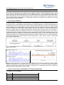
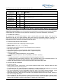
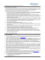
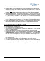
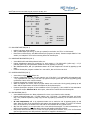
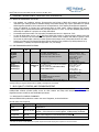
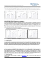
MLPA DNA Protocol version MDP-005; last revised on 22 SEPT 2014 MLPA® General Protocol Instructions For Use General MLPA protocol for the detection and quantification of nucleic acid sequences. To be used in combination with the appropriate MLPA probemix-specific product description. Certain MLPA products from MRC-Holland are registered for In Vitro Diagnostic (IVD) use, but only in specific countries. In all other cases, MLPA products are for Research Use Only (RUO). Information on IVD registration can be found in the appropriate probemix-specific product description. An alternative “two-tube” protocol is available which is only recommended for use on low quality DNA samples (RUO). Separate protocols exist for the detection of DNA methylation status (MS-MLPA®) and RNA expression (RT-MLPA®) These protocols are available on www.mlpa.com Manufacturer: MRC-Holland B.V. Willem Schoutenstraat 1, 1057 DL Amsterdam, the Netherlands Website : www.mlpa.com Phone: +31 888 657 200; E-mail: [email protected] (information & technical questions); [email protected] (for orders); [email protected] (for Coffalyser-related questions). www.mlpa.com page 1 of 12 MLPA DNA Protocol version MDP-005; last revised on 22 SEPT 2014 MLPA General Protocol – Document History Version MDP-005 (22 SEPT 2014) - In section 3.3. Storage and Stability, the sentence “Ligase buffer A should not be exposed to more than 20 freeze-thaw cycles” has been replaced by ”Reagents should not be exposed to more than 25 freeze-thaw cycles”. The word “IVD-certified” has been replaced by “IVD-registered” (p.10) Version MDP-004 (11 JUL 2014) - Information on IVD registration added; 3.5 Precautions & Warnings and 3.6 Limitations of the Procedure added Information presented more concisely Quality Control Flowchart removed 8. Data Analysis section rewritten Version MDP-003 (08 AUG 2013) - Document COMPLETELY rewritten New Quality Control Flowchart added Information presented more concisely Version MDP-002 (23 JAN 2012) - Sentence removed that the new PCR primer mix has a lot no. of F44 or newer. Old PCR primer mix cannot be recognised by the lot no., but by the absence of the MRC-Holland logo on its label. Version MDP-001 (17 JUN 2011) - NEW DOCUMENT. Previous MLPA General Protocol completely rewritten for use with the one-tube protocol. Table of Contents 1. INTENDED USE ...................................................................................................................................... 3 2. MLPA ASSAY PRINCIPLE.......................................................................................................................... 3 3. SALSA® MLPA® ASSAY COMPONENTS & STORAGE CONDITIONS................................................................. 3 4. 3.1. 3.2. 3.3. 3.4. 3.5. 3.6. ASSAY 5. 4.1. SAMPLE TREATMENT .................................................................................................... 5 4.2. SELECTING REFERENCE & OTHER CONTROL SAMPLES ..................................................... 6 MLPA REACTION - DNA DETECTION/QUANTIFICATION .............................................................................. 6 6. 5.1. NOTES TO READ BEFORE YOU START (DAY 1) ................................................................ 6 5.2. THERMOCYCLER PROGRAM FOR THE MLPA REACTION ..................................................... 7 5.3. DNA DENATURATION (DAY 1) ........................................................................................ 7 5.4. HYBRIDISATION REACTION (DAY 1) ............................................................................... 7 5.5. LIGATION REACTION (DAY 2) ........................................................................................ 7 5.6. PCR REACTION (DAY 2) ................................................................................................. 7 FRAGMENT SEPARATION BY CAPILLARY ELECTROPHORESIS ...................................................................... 8 7. 6.1. NOTES TO READ BEFORE YOU START............................................................................. 8 6.2. ELECTROPHORESIS SPECIFICATIONS ............................................................................. 8 MLPA QUALITY CONTROL AND TROUBLESHOOTING .................................................................................. 8 8. 7.1. MLPA QUALITY CONTROL FRAGMENTS ........................................................................... 8 7.2. NO DNA CONTROL ........................................................................................................ 9 7.3. EVAPORATION PROBLEMS ............................................................................................. 9 7.4. TROUBLESHOOTING ..................................................................................................... 9 PRINCIPLE OF MLPA DATA ANALYSIS ..................................................................................................... 10 9. 8.1. COFFALYSER.NET FOR MLPA DATA ANALYSIS................................................................. 10 8.2. PRINCIPLES OF MLPA DATA NORMALISATION I: FROM PEAKS to DOSAGE QUOTIENT (DQ) 10 8.3. PRINCIPLES OF MLPA DATA NORMALISATION II: FROM DOSAGE QUOTIENT TO COPY NUMBER 10 INTERPRETATION OF RESULTS ............................................................................................................. 11 10. MLPA PROTOCOL IN A NUTSHELL .......................................................................................................... 12 REAGENT KIT ITEM NUMBERS ....................................................................................... 3 COMPONENTS PROVIDED PER SALSA MLPA REAGENT KIT ................................................ 4 STORAGE AND STABILITY ............................................................................................. 4 MATERIALS REQUIRED BUT NOT PROVIDED ................................................................... 4 PRECAUTIONS AND WARNINGS ..................................................................................... 4 LIMITATIONS OF THE PROCEDURE ................................................................................ 5 SETUP INSTRUCTIONS ................................................................................................................. 5 www.mlpa.com page 2 of 12 MLPA DNA Protocol version MDP-005; last revised on 22 SEPT 2014 1. INTENDED USE Copy Number Variations (CNVs) are a prominent source of genetic variation in human DNA and play a role in a wide range of disorders. Multiplex Ligation-dependent Probe Amplification (MLPA) is a semi-quantitative technique that is used to determine the relative copy number of up to 60 DNA sequences in a single multiplex PCR-based reaction. MRC-Holland manufactures and sells MLPA reagents and a wide range of MLPA probemixes. Together, these can be used to detect deletions and duplications in a DNA sample. Details on the intended use are specified in the MLPA probemix-specific Product Description. 2. MLPA ASSAY PRINCIPLE As outlined in Figure 1, the principle of MLPA is based on the amplification (by use of a single PCR primer pair) of up to 60 probes, each of which detecting a specific DNA sequence of approximately 60 nt in length. After denaturation of the sample DNA, a mixture of MLPA probes is added to the sample. Each MLPA probe consists of two oligonucleotides that must hybridise to immediately adjacent target sequences in order to be ligated into a single probe. Each probe in an MLPA probemix has a unique amplicon length, typically ranging between 130-500 nt. During the subsequent PCR reaction, all ligated probes are amplified simultaneously using the same PCR primer pair. One PCR primer is fluorescently labelled, enabling the amplification products to be visualised during fragment separation. This is done on a capillary electrophoresis instrument, yielding a specific electropherogram (Figure 2, left). The relative height of each individual probe peak, as compared to the relative probe peak height in various reference DNA samples, reflects the relative copy number of the corresponding target sequence in the sample. A deletion of one or more target sequence thus becomes apparent as a relative decrease in peak height (Figure 2, right), while an increase in relative peak height reflects an amplification. Figure 1. MLPA reaction. B A Figure 2. A: Electropherogram of a test sample (bottom) is compared to that of a reference sample (top) showing a relative decrease of three probes in the test sample (arrows). B: Calculated probe ratios of the same test sample (as displayed by Coffalyser.Net software) after analysis of these two samples: arranging probes by chromosomal location shows a reduced copy number for these three adjacent probes in the test sample. 3. SALSA® MLPA® ASSAY COMPONENTS & STORAGE CONDITIONS 3.1. REAGENT KIT ITEM NUMBERS Item no. Description EK1-FAM EK1-Cy5 EK5-FAM EK5-Cy5 SALSA MLPA SALSA MLPA SALSA MLPA SALSA MLPA www.mlpa.com EK1 EK1 EK5 EK5 reagent kit – 100 reagent kit – 100 reagent kit – 500 reagent kit – 500 rxn rxn rxn rxn - FAM-labelled PCR primer Cy5-labelled PCR primer FAM-labelled PCR primer Cy5-labelled PCR primer page 3 of 12 MLPA DNA Protocol version MDP-005; last revised on 22 SEPT 2014 3.2. COMPONENTS PROVIDED PER SALSA MLPA REAGENT KIT Reagent kit component 1 2 3 4 5 6 7 Volumes EK1 EK5 SALSA MLPA Buffer 180 µl 5×180 µl (yellow cap) SALSA Ligase-65 115 µl 5×115 µl (green cap) Ligase Buffer A 360 µl 5×360 µl (transparent cap) Ligase Buffer B 360 µl 5×360 µl (white cap) SALSA PCR Primer 240 µl 5×240 µl Mix (brown cap) SALSA Polymerase 65 µl 5×65 µl (orange cap) MLPA General protocol Ingredients KCl, Tris-HCl, EDTA, PEG-6000, oligonucleotide Glycerol, EDTA, Beta-Mercaptoethanol, KCl, Tris-HCl, nonionic detergent, Ligase-65 enzyme (bacterial origin) Coenzyme NAD (bacterial origin) Tris-HCl, MgCl2, non-ionic detergent Synthetic oligonucleotides with fluorescent dye (FAM or Cy5), dNTPs, Tris-HCl, KCl, EDTA, nonionic detergent Glycerol, non-ionic detergents, EDTA, DTT, KCl, Tris-HCl, Polymerase enzyme (bacterial origin) None of these ingredients are derived from humans, animals or pathogenic bacteria. Based on the concentrations present, none of the ingredients are hazardous as defined in the Hazard Communication Standard. An MSDS is not required for these products: none of the preparations contain dangerous substances (as per directive 67/548/EEC & amendments) at concentrations requiring distribution of MSDS (as per directives 1999/45/EEC, 2001/58/EEC). 3.3. STORAGE AND STABILITY All components of the SALSA MLPA reagent kit must be stored directly upon arrival between -25°C and -15°C, shielded from light and in the original packaging. When stored under the recommended conditions, a shelf life of at least 1 year is guaranteed, also after opening. See the labels on each vial for the exact expiry date of each reagent. Reagents should not be exposed to more than 25 freeze-thaw cycles 3.4. MATERIALS REQUIRED BUT NOT PROVIDED SALSA MLPA Probemix and corresponding Product Description Ultrapure water TE0.1 (10 mM Tris-HCl pH 8.2 + 0.1 mM EDTA) Thermocycler with heated lid (99-105°C) and standard laboratory equipment PCR tubes, strips or plates High quality formamide (e.g. Hi-Di Formamide, Applied Biosystems 4311320) Capillary Electrophoresis equipment (Beckman Coulter or Applied Biosystems) Labelled size standard o Beckman: CEQ™ DNA Size Standard Kit - 600 (p/n 608095) o Applied Biosystems: GeneScan™ 500 LIZ® (4322682) or 500 ROX™ (4310361) • Polymers o Beckman: GenomeLab™ Linear Polyacrylamide (LPA) denaturing gel (p/n 391438) o Applied Biosystems: preferred POP-4 (4352755) or POP-7 (4352759). POP-6 is not recommended because of its high resolution, though for some older sequencers, POP-6 may be the only option. • Coffalyser.Net analysis software and User manual, and product-specific data analysis sheet • • • • • • • • 3.5. PRECAUTIONS AND WARNINGS • For in vitro use only. • Always consult the most recent version of the relevant probemix-specific product description AND this MLPA General protocol. Strictly follow this protocol. • For professional use only. Assay performance is dependent on operator proficiency and adherence to procedural directions. Assay should be performed by professionals trained in molecular techniques. • The person responsible for result interpretation should be aware of the latest scientific knowledge of the application in question and of known limitations of the MLPA procedure that may lead to incorrect results. • Abnormalities detected by MLPA should be confirmed by an independent technique whenever possible, especially if they concern a single MLPA probe. • Internal validation of each MLPA application is essential; include at least 16 normal DNA samples. Validation should show a standard deviation <0.10 for each probe (unless the relevant Product Description states otherwise). Samples used for validation should be representative of samples used in daily practice. www.mlpa.com page 4 of 12 MLPA DNA Protocol version MDP-005; last revised on 22 SEPT 2014 3.6. LIMITATIONS OF THE PROCEDURE • For most MLPA applications, the major cause of most genetic defects are small (point) mutations, most of which will not be detected by MLPA probemixes. MLPA will not detect most inversions, balanced translocations, nor copy number changes that lie outside (or only partially inside) the sequence detected by an MLPA probe. • False positive or negative results can be caused by various factors, including: a. Impurities in the test or reference DNA samples, including NaCl or KCl (>40 mM) and other salts, phenol, ethanol, heparin, EDTA (>1.5 mM) and Fe. The concentration of impurities will increase due to evaporation of sample DNA or by SpeedVac concentration of sample DNA– do not use this! b. Use of low quality plastics, as these may leak impurities such as releasing agents into the MLPA reaction c. Depurination of sample DNA during the initial 98°C heat treatment. This can occur when the sample has insufficient buffering capacity (sample dissolved in dH2O instead of TE). A minimum of 5 mM Tris pH 8.2 in the sample DNA solution is required d. Sample DNA denaturation problems causing (part of) the DNA template to be unavailable for the MLPA probes. Certain DNA purification methods, e.g. Qiagen EZ1, result in a high salt concentration in DNA samples. Extremely GC-rich regions are not denatured at 98o C when more than 40 mM NaCl or KCl is present e. Use of whole genome amplification (WGA) of sample DNA, due to its amplification bias f. Use of incorrect DNA quantities g. Use of insufficient or unsuitable reference samples h. Improper mixing of enzyme solutions, e.g. by mixing insufficiently or too vigorously i. Deviating probe ratios that are due to (harmless) SNPs, mutations and small indels within the sequence detected by a probe j. Evaporation during o/n hybridisation, causing increased salt concentration and strong sample DNA secondary structure. This may prevent certain probes from binding to their target sequences k. Contamination during the MLPA reaction or electrophoresis with other PCR products l. Problems during capillary electrophoresis, including saturation of the device causing peaks to be off-scale m. Problems during normalisation, including the use of incorrect normalisation algorithms or software n. An incorrect interpretation of results due to insufficient knowledge of the application or gene(s) in question o. Insufficient knowledge of the clinical effect of a found genetic aberration. Not all deletions and duplications detected by MLPA may be pathogenic. 4. ASSAY SETUP INSTRUCTIONS 4.1. SAMPLE TREATMENT 1. 2. 3. 4. 5. 6. 7. 8. 9. Use a total quantity of 50-250 ng (preferably 50-100 ng) of human DNA in a 5 µl volume for each MLPA reaction. If necessary, DNA samples can be concentrated by ethanol precipitation. Glycogen (Roche 901393) can be used as carrier in ethanol precipitations. More info on www.mlpa.com. Dissolve and dilute sample DNA in TE0.1 (10 mM Tris-HCl pH 8.2 + 0.1 mM EDTA). DNA preparations should contain 5-10 mM Tris buffer with a pH of 8.0-8.5 to prevent depurination during initial heat treatment at 98o C. If unknown whether sufficient buffer is present, add Tris-HCl: 4µl sample DNA + 1µl 50mM Tris-HCl pH 8.5. In case of doubts about DNA quality: a) use only 50 ng of sample DNA; b) clean contaminated samples by ethanol precipitation or silica based clean-up kits; c) use the alternative two-tube PCR protocol which uses only part of the ligation reaction for the PCR (see section 5.1). MLPA is more sensitive to contaminants than simple monoplex PCR assays. Contaminants left after DNA extraction (listed in 3.6) may influence MLPA performance. To minimise their effect, ensure the extraction method, tissue type, DNA concentration and treatment are as identical as possible in test and reference samples. EDTA concentration of the samples should not be higher than 1.5 mM. Sample DNA should not be concentrated by evaporation or SpeedVac as this leads to a very high EDTA concentration. Heparinised blood is not preferred for DNA extraction. Heparin is difficult to remove from DNA preparations and can distort the MLPA PCR reaction. Certain DNA purification methods (e.g. Nucleospin gDNA Clean-up XS) are capable of removing heparin contamination. Do not use Qiagen M6, M48 and M96 systems for DNA purification (high salt concentrations). For Qiagen EZ1, only use the QIAGEN Supplementary Protocol for use in Third Wave Invader® assays (see www.mlpa.com). DNA from whole genome amplification reactions (WGA) is not suitable for MLPA because of its amplification bias. MRC-Holland has tested and can recommend the following extraction methods: Qiagen Autopure LS (automated) and QIAamp DNA mini/midi/maxi kit (manual) Promega DNA extraction Wizard (manual) Salting out (manual) www.mlpa.com page 5 of 12 MLPA DNA Protocol version MDP-005; last revised on 22 SEPT 2014 4.2. SELECTING REFERENCE & OTHER CONTROL SAMPLES 1. 2. 3. 4. 5. 6. 7. REFERENCE SAMPLES. Reference samples should be included in each MLPA experiment. Minor differences in experimental execution may affect the MLPA peak pattern. Only compare samples that are a) included in the same MLPA experiment, b) tested with the same probemix lot. MULTIPLE REFERENCE SAMPLES are needed to estimate the reproducibility of each probe within each MLPA run. Minimum is 3 different reference samples. When testing >21 samples: add 1 additional reference per 7 additional test samples. Reference samples should be distributed randomly over the sample plate to minimise variation. SELECTING REFERENCE SAMPLES. Reference samples are DNA samples in which target and reference probe sequences are expected to have a normal copy number, obtained from healthy individuals/tissues. They should be as similar as possible to test samples in all other aspects (see 4.1). For formalin-fixed paraffin-embedded tissue, use reference samples derived from similarly treated healthy tissue. COMMERCIAL DNA. In case of doubts about sample quality, include one or more commercial DNA samples for comparison (recommended: Promega Cat. Nr G1471 male, G1521 female DNA). NO DNA CONTROL. Per MLPA run, include a No DNA Control reaction: replace 5 µl DNA by TE (10 mM Tris-HCl pH 8.2 + 0.1 mM EDTA) to check for contamination of TE, MLPA reagents, electrophoresis reagents or capillaries. POSITIVE CONTROL SAMPLES. Including positive controls is recommended when possible. When using cell line DNA, note that this may have acquired additional copy numbers, incl. complete chromosomal gains/losses. ALIQUOT PRECIOUS REFERENCE/CONTROL SAMPLES and store these at -20°C. Contamination with microorganisms or moulds can deteriorate samples that are stored at 4°C for an extended period. 5. MLPA REACTION - DNA DETECTION/QUANTIFICATION 5.1. NOTES TO READ BEFORE YOU START (DAY 1) • • • • • • • Use a calibrated thermocycler with heated lid (99-105°C). Always vortex thawed buffers and probemix before use. MLPA buffer is typically frozen at -20°C but may remain liquid due to its high salt concentration. Centrifuge all MLPA reagent tubes for a few seconds before use, as drops may have adhered to the lid. Enzyme solutions contain 50% glycerol and remain fluid at the recommended storage temperature. Never vortex enzyme solutions. Master mixes containing enzymes should be mixed by gently pipetting up and down. When the viscous enzyme solution is not mixed properly with the buffers, unreliable results will occur! If the enzyme solution is mixed too vigorously however, enzyme inactivation occurs. When preparing master mixes, always add enzymes last. To minimise sample variation, prepare sufficiently large volumes of master mix solutions. Include a 5-10% volume surplus to allow for pipetting errors. Prepare Ligase-65 master mix and Polymerase master mix <1 hour before use and store on ice. When running a large number of samples, use multi-channel pipettes to avoid excessive evaporation. In this General MLPA protocol, the complete (40 µl) ligation reaction is used for the PCR. The alternative twotube MLPA protocol, which uses only 10 µl of the ligation reaction for the PCR, is available on www.mlpa.com. The two-tube protocol may have advantages when using DNA samples containing impurities such as high EDTA concentrations (>1.5 mM) or PCR inhibitors. A vial of PCR buffer, required for this alternative protocol, can be ordered for free. The two-tube protocol is for research use only (RUO), not for in vitro diagnostic use (IVD). www.mlpa.com page 6 of 12 MLPA DNA Protocol version MDP-005; last revised on 22 SEPT 2014 5.2. THERMOCYCLER PROGRAM FOR THE MLPA REACTION 1. DNA denaturation 1. 98°C 2. 25°C 2. Hybridisation reaction 3. 95°C 4. 60°C 3. Ligation reaction 5. 54°C 6. 54°C 7. 98°C 8. 20°C 4. PCR reaction 9. 35 cycles: 10. 11. 72°C 15°C 5 minutes pause 1 minute pause Pause 15 minutes 5 minutes pause • • • 95°C 60°C 72°C 30 seconds 30 seconds 60 seconds 20 minutes pause 5.3. DNA DENATURATION (DAY 1) • • • Label 0.2 ml tubes, strips or plates. Add 5 µl DNA sample (50-250 ng; 50-100 ng is optimal) to each tube. Use TE for ‘no DNA control’. Place tubes in thermocycler; start MLPA thermocycler program (see above). Denature sample DNA for 5 min at 98°C; cool samples to 25°C before removing tubes from thermocycler. 5.4. HYBRIDISATION REACTION (DAY 1) • • • • Vortex MLPA buffer and MLPA probemix before use. Prepare hybridisation master mix containing, for each reaction: 1.5 µl MLPA buffer (yellow cap) + 1.5 µl probemix (black cap). Mix hybridisation master mix well by pipetting or vortexing. After DNA denaturation, add 3 µl hybridisation master mix to each sample tube. Mix well by pipetting up and down. Continue thermocycler program: incubate for 1 min at 95°C, then 16–20 hours at 60°C. 5.5. LIGATION REACTION (DAY 2) • • • • • Vortex the two Ligase Buffers before use. Prepare a Ligase-65 master mix. For each reaction, mix: 25 µl dH2O + 3 µl Ligase Buffer A (transparent cap) + 3 µl Ligase Buffer B (white cap). Then add 1 µl Ligase-65 enzyme (green cap). Mix well by pipetting gently up and down. Never vortex enzyme solutions. Continue thermocycler program: pause at 54°C. When the samples are at 54°C, add 32 µl ligase master mix to each tube. Mix by gently pipetting up and down. Add the ligase master mix while the samples are IN the thermocycler! Continue thermocycler program: 15 min incubation at 54°C (for ligation); 5 min at 98°C for heat inactivation of Ligase-65 enzyme. Pause at 20°C. At this point, tubes can be removed from the thermocycler. 5.6. PCR REACTION (DAY 2) • • • • • Vortex SALSA PCR primer mix. Warm polymerase for 10 sec in your hand to reduce viscosity. Prepare polymerase master mix. For each reaction, mix: 7.5 µl dH2O + 2 µl SALSA PCR primer mix (brown cap) + 0.5 µl SALSA Polymerase (orange cap). Mix well by pipetting up and down; do not vortex. Store on ice until use. At room temperature, add 10 µl polymerase master mix to each tube. Mix by pipetting gently up and down. Place the tubes in the thermocycler and continue the thermocycler program; 35 cycles of 30 seconds 95°C; 30 seconds 60°C; 60 seconds 72°C. End with 20 min incubation at 72°C; pause at 15°C. After the PCR reaction, do not open tubes in the room with the thermocycler. To avoid contamination, use different micropipettes for performing MLPA reactions and handling MLPA PCR products. PCR product can be stored at 4°C for 1 week. For longer periods, store between -25°C /-15°C. As fluorescent dyes are light-sensitive, store PCR products in dark box or wrapped in aluminium foil. www.mlpa.com page 7 of 12 MLPA DNA Protocol version MDP-005; last revised on 22 SEPT 2014 6. FRAGMENT SEPARATION BY CAPILLARY ELECTROPHORESIS 6.1. NOTES TO READ BEFORE YOU START Size standard, run conditions, polymer, fluorescent dye and volume of MLPA PCR reaction used depend on instrument type. Settings below are standard settings. Instrument settings may require optimisation for optimal MLPA fragment separation; follow instructions of the manufacturer of the capillary electrophoresis apparatus. Using old capillaries or polymer has a detrimental effect on MLPA results. Change capillaries and polymer regularly. Polymer quickly deteriorates after prolonged exposure to >25°C. In case size standard peaks are low and broad, the capillaries or polymer are usually deteriorated. Formamide can become acidic. Use high quality formamide and store it in aliquots at -20°C. In case all MLPA peaks are low, do not add more MLPA PCR product to injection mixture! Adding more PCR product increases the salt concentration in the injection mixture which competes with DNA for injection. When an increase in peak heights is desired, increase injection time/voltage. None of the peaks should be off-scale! Reset the bin settings in the fragment analysis software whenever using a different a) MLPA probemix lot; b) size standard; c) capillary electrophoresis apparatus or d) run settings. 6.2. ELECTROPHORESIS SPECIFICATIONS Primer Dye Capillaries Beckman CEQ-2000 CEQ-8000 CEQ-8800 GeXP Cy5 33 cm ABI-Prism 3100 (Avant) ABI-3130 (xL) * ABI-3500 (xL)* ABI-3730 (xL)* FAM 36, 50 cm ABI-Prism 310 FAM Instrument ABI3500: 50 cm! 47 cm Injection mixture Initial Settings 0.7 µl PCR reaction 0.2 µl CEQ - size standard 600 32 µl HiDi formamide / Beckman SLS Add one drop of high quality mineral oil. run method: Frag capillary temperature: 50°C denaturation: 90°C for 120 sec injection voltage: 1.6 kV injection time: 30 sec run time: 60 min run voltage: 4.8 kV run module: FragmentAnalysis injection voltage: 1.6 kV injection time: 15 sec run voltage: 15 kV run time: 1800 s oven temperature: 60°C polymer: POP4 or POP7 injection voltage: 1.6 kV injection time: 15 sec filter set: D polymer: POP4 0.7 µl PCR reaction 0.3 µl (ROX) or 0.2 µl (LIZ) GS 500 size standard 9 µl HiDi formamide Seal the injection plate, heat 3 min at 86°C, cool for 2 min at 4°C.‡ 0.75 µl PCR reaction 0.75 µl dH2O 0.5 µl GS 500 ROX/LIZ size standard 13.5 µl HiDi formamide Heat 3 min at 86 °C, cool for 2 min at 4°C. ‡ * ABI-3130, 3500 & 3730 have been validated and found suitable to be used with IVD-registered MLPA products. ‡ Briefly heating the injection mixture before capillary electrophoresis is essential. 7. MLPA QUALITY CONTROL AND TROUBLESHOOTING For analysis of MLPA data, use Coffalyser.Net software, which can be freely downloaded from www.mlpa.com. The Coffalyser.Net software provides quality scores for each reaction and helps with interpretation of the control fragments that are included in each SALSA MLPA probemix. 7.1. MLPA QUALITY CONTROL FRAGMENTS Almost all SALSA MLPA probemixes contain nine control fragments, as described below: Internal quality control fragments 1 Name 92 nt benchmark probe Length (nt) Q-fragments 64, 70, 76, 82 D-fragments 88, 96 X & Y fragments 100, 105 92 Interpretation Normal probe; forms a benchmark to compare other quality control fragments to. High when DNA amount is too low or ligation failed. All four Q-fragment signals > 33% of the 92 nt control fragment DNA quantity insufficient. Low in case of poor sample DNA denaturation. Signal < than 40 % of the 92 nt control fragment DNA denaturation problems. Control for sample swapping.1 Rare cases are known of males lacking this Y-specific sequence and females carrying this Y-sequence on an X-chromosome. www.mlpa.com page 8 of 12 MLPA DNA Protocol version MDP-005; last revised on 22 SEPT 2014 QUANTITY CONTROL-FRAGMENTS (Q-FRAGMENTS) The four Quantity Fragments (Q-fragments, at 64-70-76-82 nt) are complete fragments that do not need to hybridise to DNA or be ligated to be amplified during PCR. The more sample DNA is added, the lower they get; see Figure 3. ALL FOUR Q-FRAGMENTS HIGH: INSUFFICIENT SAMPLE DNA USED OR LIGATION REACTION FAILED Figure 3 – Effect of DNA quantity on Q-fragments. The more sample DNA is used, the lower the Q-fragments. MLPA results with A. 5 ng DNA, B. 10 ng, C. 50 ng. DENATURATION CONTROL FRAGMENTS (D-FRAGMENTS) The two DNA Denaturation Fragments (D-fragments, 88 & 96 nt) detect sequences in exceptionally strong CpG islands. CpG islands have a high GC-content and are difficult to denature. When the 88* and 96 nt fragments are low (< than 40 % of the 92 nt control fragment) this indicates denaturation problems of the sample DNA. A poor denaturation can be due to presence of >40 mM salt in the DNA sample. Incomplete sample DNA denaturation can result in false positive deletions! * NOTE: When using ABI POP7 polymer, a non-specific fragment of ~87 nt is usually present which may coincide with the 88 nt D-fragment! D-FRAGMENTS LOW: SAMPLE DNA DENATURATION INCOMPLETE! Figure 4 – Effect of poor denaturation on D-fragments. D-fragments are low when sample DNA denaturation is incomplete (here induced by adding salt). MLPA results on DNA sample containing in A. TE. B. TE + 40 mM NaCl. C. TE + 100 mM NaCl. 7.2. NO DNA CONTROL In a typical No DNA control, only the four Q-fragments are visible. However, MLPA PCR reactions are more prone to non-specific peaks in the No DNA Control than a normal PCR. In some probe mixes, a few peaks may be visible in No DNA Controls. These non-specific peaks should not influence MLPA results: when sufficient sample DNA is used, they should be outcompeted by the MLPA probes, similarly to the Q fragments in figure 3. Notify MRC-Holland in case a non-specific peak in the No DNA control is reproducibly higher than the Q-fragments. 7.3. EVAPORATION PROBLEMS Evaporation may occur during (A) pipetting the ligation reaction at 54°C or (B) overnight hybridization. In case you suspect evaporation problems, the following may help: (A): Reduce handling time by using multi-channel pipettes. (B): Test evaporation by incubating 8 µl H2O overnight at 60o C; >5 µl H2O should remain. To reduce evaporation: 1. ensure heated lid works well; 2. increase/decrease pressure of lid on tubes; 3. try different tubes (e.g. Thermo Fischer ABgene AB-0773, AB-0451); 4. use mineral oil (Vapor-lock, Qiagen 981611): add small drop of oil to DNA sample, just enough to cover it. There is no need to remove oil. After addition of MLPA buffer-probemix mixture or polymerase mix: centrifuge very briefly. After addition of ligase mix: gently pipet up and down. 7.4. TROUBLESHOOTING A detailed Quality Control Flowchart is available on www.mlpa.com and can be used for quality control and troubleshooting. www.mlpa.com page 9 of 12 MLPA DNA Protocol version MDP-005; last revised on 22 SEPT 2014 8. PRINCIPLE OF MLPA DATA ANALYSIS 8.1. COFFALYSER.NET FOR MLPA DATA ANALYSIS Use Coffalyser.Net software for MLPA data analysis. The Coffalyser.Net Manual provides a step-by-step instruction on MLPA data analysis. Both software and manual are freely downloadable on www.mlpa.com. This section describes the basic principles of MLPA analysis. Coffalyser.Net analysis is based on these principles but uses a more robust algorithm. In addition, Coffalyser.Net selects the best analysis method for each MLPA probemix and offers extensive quality control2. For IVD-registered MLPA applications, Coffalyser.net must be used! 8.2. PRINCIPLES OF MLPA DATA NORMALISATION I: FROM PEAKS to DOSAGE QUOTIENT (DQ) The absolute fluorescence measured by capillary electrophoresis cannot be used directly for copy number calculations as it is affected by many variables. First, each probe’s measured fluorescence must be normalised within each sample to get meaningful data. Second, various samples need to be compared to establish which sample has aberrant copy number changes. Therefore, MLPA normalisation consist of 2 steps: intrasample normalisation (comparison of probe peaks WITHIN the sample) and intersample normalisation (comparison or relative probe peaks BETWEEN samples.) 1. Intrasample normalisation. Within each sample, compare each probe peak TO the peaks of the reference probes. Reference probes detect sequences that are expected to have a normal copy number in all samples. Almost all MLPA probemixes contain 8 or more reference probes located on various chromosomes. The relative probe signals determined in step 1 are then used in: 2. Intersample normalisation. Final probe ratios are determined by comparing the relative probe peak in the DNA sample of interest TO all reference samples. Reference DNA samples are expected to have a normal copy number for both the reference and target probes. The MLPA peak pattern of a DNA sample without genomic abnormalities will be identical to that of reference samples: final probe ratios determined in step 2 will be ~1.0. For heterozygous deletions, probe ratios will be ~0.5. This final probe ratio is also called Dosage Quotient (DQ). Coffalyser.Net calculates the DQ for each probe in each sample. Probes should be arranged based on chromosomal location for correct interpretation; this will also aid in detecting subtle changes such as mosaicism. 8.3. PRINCIPLES OF MLPA DATA NORMALISATION II: FROM DOSAGE QUOTIENT TO COPY NUMBER How to discriminate normal, deletion and duplication results? Table 1 gives the relationship between copy number status and the typical distribution of DQs (based on a large number of samples at MRC-Holland). Table 1 – Relation Dosage Quotients and Copy Number (based on normal status of 2 copies) Dosage Quotient Distribution Copy Number Status DQ = 0 0 copies ( homozygous deletion) 0.40 < DQ < 0.65 2 1 copy (heterozygous deletion) 0.80 < DQ < 1.20 NORMAL (identical to reference samples) 1.30 < DQ < 1.65 2 3 copies (heterozygous duplication) 1.75 < DQ < 2.15 2 4 copies (or 12 copies) All other values Ambiguous result In addition, the standard deviation of each probe should be measured to ensure the result is reliable3. Coffalyser.Net software considers a result as statistically significant change if: 1. DQ probe 1,sample A > 2 standard deviations higher/lower than average DQ probe 1, ref sample X, Y, Z Furthermore, to be indicative of a heterozygous deletion, duplication respectively, the following applies: 2. DQprobe 1,sample A below 0.7 or above 1.3 indicates a heterozygous deletion, duplication, respectively4. 2 Coffalyser.Net starts with raw data analysis (baseline correction, peak identification) and extensive quality control (e.g. DNA quantity used; complete DNA denaturation, degree of sloping). Slope correction of the peak pattern is one of the many options. 3 The standard deviation differs per probe and depends on many factors, including 1. quality of DNA samples (purity; sample DNA fragmentation, depurination, other modifications); 2. quantity of sample DNA used; 3. number and quality of reference samples and positive samples; 4. experimental conditions (e.g. pipetting accuracy, thermocycler temperature uniformity, quality of amplicon separation and detection) and 5. method used for data analysis. Coffalyser.Net evaluates ALL of these factors. 4 Coffalyser.Net uses values of 0.7 and 1.3 as cut-off values for a heterozygous deletion or duplication, respectively (when normal status is diploid). Coffalyser.Net thus uses sharp cut-off values, while table 1 gives a substantial ambiguous DQ range www.mlpa.com page 10 of 12 MLPA DNA Protocol version MDP-005; last revised on 22 SEPT 2014 For mosaic and tumour samples, the 0.7/1.3 cut-off lines of Coffalyser.Net are not applicable as the DQs can take on any value: their calculated DQs will depend on a) the magnitude of the copy number change AND b) the percentage of the different cell types in the DNA sample5. As Coffalyser.Net determines the significance of a probe signal change by evaluating the magnitude of the calculated DQ in combination with its statistical significance in the experiment, such a partial copy number change can still be recognised if a) the experiment was performed well and b) a similar DQ was obtained for adjacent probes. 9. INTERPRETATION OF RESULTS The information provided in the MLPA probemix-specific Product Description is essential for a correct interpretation of MLPA results. To judge whether obtained results are reliable and to interpret MLPA results correctly, a good understanding of the MLPA technique and the application tested for is essential. Keep the following in mind: • For most applications, the major cause of genetic defects will be small (point) mutations, most of which will not be detected by MLPA. MLPA will not detect most inversions, translocations, nor copy number changes that lie outside the sequence detected by the MLPA probes. MLPA probemixes cannot detect deletions/duplications lying outside the target sequence of its probes. MLPA probes typically detect a sequence of ~55-80 nt. Most product descriptions mention only partial probe sequences. Complete probe sequences are available on www.mlpa.com. • Sequence changes (SNPs, point mutations) in the probe’s target DNA sequence (even when >10 nt from the ligation site) can cause false deletions: they may lower a probe signal by hampering ligation or by destabilising the binding of the probe oligos to the sample DNA 6. • Copy number changes detected by a single probe always require confirmation. Sequencing of probe target sequences may show that a lowered probe signal is caused by a mutation/polymorphism. The finding of two heterozygous sequences typically indicates that the sample DNA does contain two different alleles. In contrast, note that the finding of only a single rare allele by sequencing does not yet imply that the other, “normal” allele is deleted: the lower probe signal may be caused by a homozygous SNP, even when the SNP is very rare! • Long-range PCR and qPCR are often used to confirm (single) exon deletions. It is also possible to design your own synthetic MLPA probes for confirmation of results (e.g. Hills A. et al., (2010) Mol Cytogenet. 3:19). Information on MLPA probe design is available on www.mlpa.com (for research use only). • Not all deletions and duplications detected by MLPA are pathogenic. MRC-Holland cannot provide information whether a deletion or duplication of a specific exon will result in disease. For many genes, exons are described that are only present in certain transcript variants. For some genes, such as DMD, in-frame deletions resulting in mild, or no disease, have been described7. A duplication of one or more exons may disrupt that copy of the gene resulting in disease, whereas a complete gene duplication may not be pathogenic. Note that duplications that include the first or the last exon of a gene, may leave one functionally intact copy. • Germline copy number variations reported in healthy individuals can be found at http://projects.tcag.ca/variation/. Certain copy number aberrations can be due to somatic alterations. For instance, a somatic trisomy 12 is an early sign of Chronic Lymphocytic Leukemia (CLL). • In case of poor sample DNA denaturation, even the apparent deletion of several probes recognising adjacent genomic targets can be a false positive result! The presence of salt in DNA samples (e.g. >40 mM NaCl) prevents DNA denaturation of GC-rich regions. Sequences in the vicinity of such CpG islands may denature at 98oC but will reanneal immediately upon cooling as the non-denatured CpG island holds the two strands together. Binding of probes to their target sequence in these regions will be hindered, resulting in reduced signals for probes located within several kb from such strong CpG islands. Always examine the D-fragments carefully! • MLPA tests provide the average copy number of the target sequences in the cells from which the DNA sample was extracted. In case several probes targeting adjacent sequences have an unusual value but do not reach the usual threshold values for a deletion/duplication, mosaicism is a possible cause. (0.65<DQ<0.80; 1.20 <DQ<1.30). However, Coffalyser also calculates whether the DQ of a probe is statistically different from that in the reference population. Coffalyser.Net can therefore also be used on tumour samples and can identify some mosaic cases. 5 Example: In Sample A, 30% of its cells contain 3 copies of chromosome 21, 70% contain 2 copies. The DQ found for chr. 21 probes will be: (30% x 3) + (70% x 2) / 2*= 1.15 (2*: number of copies present in reference samples) 6 When designing probes, known SNPs are avoided when possible. However, new SNPs are continuously being discovered. Please notify us when a polymorphism or a frequent pathogenic mutation influences a probe signal. 7 Schwartz M. and Dunø M. (2004). Improved molecular diagnosis of dystrophin gene mutations using the multiplex ligationdependent probe amplification method. Genet Test. 8:361-7. www.mlpa.com page 11 of 12 MLPA DNA Protocol version MDP-005; last revised on 22 SEPT 2014 • • • Most MLPA probemixes contain reference probes. Copy number changes detected by reference probes are unlikely to be related to the condition tested for. The identity of reference probes is available on request. In certain cases, analysis of parental samples might be necessary for correct interpretation of results. Internal MLPA validation in your laboratory is essential. MLPA results are more reliable when: • The overall standard deviation of each probe in the reference samples is low (<10%). • Probes show a decreased or increased signal for adjacent exons (multi-exon deletion or duplication). • The same result is obtained in a new MLPA run using less DNA (if possible) or using different reference samples. When less DNA is used, any possible contaminants which may influence the probe signal are diluted. An MLPA result is unlikely to be reliable when: • Probes for non-neighbouring exons show a decreased or increased signal, e.g. deletion of exon 3 and 17. • In the same sample, one or more reference probes show an abnormal copy number. • Copy number changes are detected with an unusually high frequency in a patient cohort for a certain disease. 10. MLPA PROTOCOL IN A NUTSHELL 1. DNA DENATURATION • Heat a 5 µl DNA sample for 5 minutes at 98°C 2. HYBRIDISATION OF PROBES TO SAMPLE DNA • Cool down to room temperature, open the tubes • Add a mixture of 1.5 µl SALSA probemix and 1.5 µl MLPA buffer and mix • Incubate 1 minute at 95°C + 16 hours hybridisation at 60°C 3. LIGATION OF HYBRIDISED PROBES • Lower thermocycler temperature to 54°C, open tubes • Add 32 µl ligase master mix, incubate 15 minutes at 54°C • Heat to inactivate the ligase enzyme: 5 minutes 98°C 4. PCR AMPLIFICATION OF LIGATED PROBES • Cool down to room temperature, open tubes • Add 10 µl polymerase master mix at room temperature • Start PCR 5. CAPILLARY ELECTROPHORESIS OF PCR PRODUCTS 6. ANALYSE RESULTS • Determine RELATIVE size of the fluorescent peaks within each sample • Compare these results to reference samples Ligase master mix: 3 µl Ligase Buffer A + 3 µl Ligase Buffer B + 25 µl water + 1 µl Ligase-65 Polymerase master mix: 7.5 µl water + 2 µl PCR Primer mix + 0.5 µl SALSA Polymerase Troubleshooting: a detailed Quality Control Flowchart is available on www.mlpa.com. PREVENT FALSE POSITIVE OR NEGATIVE RESULTS READ THE COMPLETE PROTOCOL AND STRICTLY FOLLOW THIS PROTOCOL READ THE MOST RECENT VERSION OF THE PRODUCT DESCRIPTION www.mlpa.com page 12 of 12