1
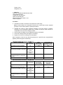
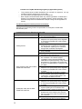
Lab Manual Section 1: Title Page Volume 2: Molecular assays Module name: Single Nucleotide Polymorphism. Author(s) name/affiliation/email: Carmen Alaez PhD, Clara Gorodezky PhD, DSc. Prof. Department of Immnunology and Immunogenetics, Instituto de Diagnóstico y Referencia Epìdemiológicos. MexicoCity .México. [email protected]; [email protected]; [email protected] Date prepared or last revised: 09/15/2014 1 Section 2 : Comparison of Technical Alternatives A broad range of techniques are available for SNP genotyping (Slide 11); they differ in throughput, cost, complexity, equipment, and bioinformatics resources needed etc. Needs are different in academic laboratories performing research studies versus commercial companies or genome centers that require high throughput. The type of project to be performed must also be considered in the selection. For example, genotyping of a small number of SNPs in a large population could be performed using real-time PCR allelic discrimination assays. However, for typing of a large number of SNPs on a limited number of individuals, a high throughput system, such as microarray assays are more adequate. Choosing a SNP typing platform is not easy, and the following factors should be considered: (Slide 12) -Throughput: depending on the study design, the number of SNPs to be typed could vary from one to a few thousand, and the number of individuals from a hundred to a few thousands. There are ongoing efforts to increase throughput. -Simplicity and robustness of the assay: some assays are simple, allow typing of many different SNPs under similar reaction conditions, and require little optimization. Other systems require individual reactions to be optimized and run under different conditions. -Success rate and accuracy: rates of failure or mistyping are variable, and acceptable levels depend upon the aims of the study and the statistical methods used for data analysis. -Track record: Some technologies are new and they do not yet have a well-validated track record. -DNA consumption: most methods require only a few nanograms of DNA to type a SNP. Multiplexing reduces the amount of DNA required, and methods without a PCR step further reduce further the amount of DNA needed. - Cost: Some platforms, such as mass spectrometry, require expensive equipment, while others can be done with standard laboratory equipment. Reagent costs vary greatly depending on the platform. We present here the main features of each methodology. Depending on the SNPs available, commercial kits may be available from various vendors. Allele specific hybridization (PCR-SSOP): Genotypes can be inferred from the hybridization signals of allele-specific oligonucleotide probes. The DNA to be tested (most commonly a PCR product) is fixed to a membrane in a dot or slot format and hybridized with the allele-specific oligonucleotide probe (referred to as a Dot Blot). Radioactive, enzymatic, and chemiluminescent systems have been used for detection of the hybridization patterns (Slide 13). A modification of this format is called reverse dot blot or reverse hybridization, where oligonucleotide probes are immobilized in a solid phase (membrane, plastic beads, glass etc) and the hybridization is performed with the DNA to be tested, enabling a higher throughput to be achieved (Slide 14). High throughput genotyping is the process of identifying the SNP alleles in as many different individual genomes as possible. This has been achieved using filters or glass slides containing very high probe densities. Steps of genotyping involve DNA sample preparation, PCR amplification, and microarray assays. The last improvement in this methodology is the use of DNA chips with extremely high probe densities that allows one to achieve up to 100,000 genotypes/day (Slide 15). High throughput genotyping is used to determine haplotypes for SNP mapping. SNP mapping allows tracking of disease-associated genes and determining the contribution of specific genes to diseases or phenotypes. Luminex Corporation has developed a panel of up to 200 bead sets, each of them with unique fluorescent labels that allow bead identification using a flow 2 analyzer. Each bead can be coupled to a DNA probe, to create a bead-based array for multiplexing genotyping (Slide 16). Allele discrimination performed on sequence detection systems (SDS) using the TaqMan assay chemistry also depends on hybridization of two allele-specific probes (Slide 17). The probe for each allele is labeled with a specific-fluorochrome (FAM or VIC). The oligonucleotide probe also has a generic nonfluorescent quencher attached to its 3’ end that eliminates background fluorescence. If both are linked to the oligonucleotide, the fluorescence of the dye is quenched. During amplification, the 5´ to 3´exonuclease activity of Taq polymerase degrades the probe, but only if it is hybridized to the template DNA. The fluorophore is released from the quencher, and the emitted fluorescence can be monitored on a sequence detection system with a real-time PCR instrument and SDS software. By using two different fluorophores, monitoring can be done in an allele specific way. Ninety-six or 384-well formats are available and assay design, and manufacture service is offered by Life Technologies to facilitate the development of assays. Molecular beacons probes, scorpion probes and dynamic allele-specific hybridization are also assays based on allele-specific hybridization probes developed by different vendors. Allele specific digestion (PCR-RFLP-Restriction Fragment length polymorphisms) (Slide 18): Restriction enzymes recognize and cleave specific DNA sequences. A change in any of the recognition site bases will lead to a failure in the cleavage. Conversely, a mutation in a sequence closely related to that of a restriction site can create a new site. If the SNP to be tested modifies a restriction enzyme site, a PCR product containing the SNP could be digested with the corresponding restriction enzyme. If the PCR product is cut, specific fragments will be generated. After digestion, they are separated, according to their size by gel electrophoresis, and genotypes are determined according to the sizes of the digestion fragments obtained. Each SNP allele is expected to have a different restriction pattern. Heteroduplex analysis (Slide 19): When alleles in a heterozygote are amplified, and then denatured and allowed to re-anneal, double-stranded DNA is formed in which one strand is from one allele, and the complementary strand from the other allele. It thus contains a mismatch at the SNP site, and has abnormal properties of migration when analyzed by non-denaturing polyacrylamide gel electrophoresis. Another version of this methodology is denaturing high performance liquid chromatography (DHPLC) where heteroduplex and homoduplex strands are separated by their altered retention time on chromatography columns under near denaturing conditions. Single Strand conformation polymorphism (Slide 20): This is a popular, simple, and cheap method of mutation detection, in which changed mobilities of single-stranded mutant molecules are detected on non-denaturing gels. This technology is based on the fact that only one change in the DNA sequence is able to produce an altered mobility in electrophoresis, and is not based on heteroduplex formation. Primer extension (Slide 21): An oligonucleotide is used to prime DNA synthesis by a DNA polymerase, as performed in a common PCR reaction. Variations of this method exist. As an example, for allele specific primer extension, two primers can be used, each of them complementary in its 3´ end to one of the SNP alleles. DNA synthesis will proceed only if the 3´end of the primer is perfectly matched to template DNA. In this way, an allele-specific PCR product can be obtained. Allele separation can be achieved using primers labeled with different dyes, or if the PCR product for each allele has a different size. Amplification refractory mutation system is another alternative that also involves primer extension. A modification of this methodology is the single base primer extension (SBE) assay that uses a single extension base primer. The 3’ end of this primer pair is located exactly before the SNP. The DNA polymerase is then used to extend one base incorporating the corresponding ddNTPs. Each of the four ddNTPs is labeled with a different fluorescent dye, allowing detection of the incorporated nucleotide by gel or capillary electrophoresis on a sequencing apparatus. This reaction can be multiplexed to type different SNPs under similar conditions at the same time, reducing cost and increasing the throughput. The different SNPs genotyped simultaneously could also be separated by hybridization to arrayed tags. Alternatively, the primer to be used can be 3 modified near the 3´end with a charged tag to increase sensitivity to mass spectrometry detection, and in this case no labeling is necessary. Oligonucleotide ligation assay (slide 22): Two allele specific oligonucleotides are designed so they join at the position of the polymorphism to be tested. The oligonucleotides are labeled differently, and allele discrimination occurs by the ability of DNA ligase to join only perfectly matched probes; a 3' mismatch in the capture probe will prevent ligation. Detection of the alleles can be performed by colorimetric methods or by capillary electrophoresis. Sequence base typing (Slide 23): Direct sequencing of PCR products allows both typing and identification of new polymorphisms. Pyrosequencing is new sequencing method, based on primer extension, that produces short segments of sequences (up to 20 nucleotides), simultaneously on 96 templates. Once the templates have been prepared, 96 of them can be sequenced in 15 minutes through an automated machine. The method involves sequential addition of dNTPs to an extension reaction. Incorporation of a nucleotide releases pyrophosphates that trigger a luciferase-catalyzed enzymatic cascade. The produced light signal is detected by a CCD camera and is proportional to the number of nucleotides incorporated. Pyrosequencing is particularly suitable for SNP genotyping. DNA sequencing is the “gold standard for SNP discovery because it shows all the variation within a sequence. However some regions are not reliably sequenced because of their high GC content or their secondary structure. Flap endonuclease discrimination (Slide 24): The “invader” assay involves nuclease cleavage of a signal probe when two overlapping oligonucleotides hybridize to a complimentary DNA target. A generic invader probe and an allele-specific primary probe are simultaneously hybridized to the target sequence such that they overlap at the SNP site. The structure that results is recognized and cleaved by a flap endonuclease, releasing a probe-specific tail sequence or “flap”. Flap endonucleases catalyze structure-specific cleavage and are highly sensitive to mismatches. The cleaved fragment may be labeled with a probe specific fluorescent dye, which emits light after probe cleavage due to spatial separation from a quencher. PCR amplicons can be used as the DNA template and the method can also be multiplexed for higher throughput and cost reduction. Next Generation Sequencing (NGS) Slide 25: In this methodology, the whole genome or target regions of the genome, are digested into small fragments. DNA fragments are ligated to platformspecific oligonucleotide adapters that allow laboratories to perform the sequencing biochemistry. The library of fragments is massively parallel sequences, producing hundreds of gigabases of data in a single sequence run. The identified strings of bases, called reads, are either aligned to a reference genome or assembled (de novo sequencing). The full set of aligned reads represents the entire sequence of the studied region or genome. Having aligned the fragments of one or more individuals to a reference genome, dedicated software can identify variable regions and perform an “SNP calling” to determine the genotype for each the individual. NGS has a high error rate that can affect accurate SNP and genotype calling. To reduce uncertainty associated to SNP calling, target regions must be sequenced deeply (20X); however, deeper sequencing will increase costs. Another alternative is using algorithms that have a probabilistic framework which incorporates errors that may have been introduced in base calling, alignment, and assembly, and also uses prior information, such as allele frequencies and patterns of linkage disequilibrium. 4 Section 3 :Specimen Requirements DNA prepared from the samples to be tested. Known DNA is to be used as positive controls for each SNP genotype. Minimum requirements The DNA to be used in SNP genotyping experiments must: • Be extracted from the raw material with an optimized protocol • Not contain PCR inhibitors • Has an A260/280 ratio greater than 1.7 • Be intact as visualized by gel electrophoresis • Not have been heated above 60 °C, which can cause degradation 5 Section 4: Protocols Genotyping SNPs using allele specific hybridization probes labeled using Taq Man 5'-nuclease chemistry and real-time PCR as detection systems Please refer to the TaqMan SNP Genotyping Assays Protocol - Applied Biosystems product insert found here: http://tools.lifetechnologies.com/content/sfs/manuals/cms_042998.pdf Other SNP genotyping assays are also based on allele-specific hybridization probe principles that use different strategies for signal generation. For example: molecular beacons probes, scorpion probes, dynamic allele-specific hybridization assays, FRET probes (Roche Diagnostics GmbH), etc. These are available from different vendors; details can be found in these and other web sites: http://www.molecular-beacons.com / www.sigmaaldrich.com, http://www.genomics.agilent.com. Principle For each assay, a unique pair of fluorescent dye detectors is used. One fluorescent dye detector is a perfect match to the wild type (allele 1) and the other one is a perfect match to the mutation (allele 2). The change in the fluorescence of the dyes associated with each probes is measured during the experiments. The actual quantity of target sequence is not determined. After the automated analysis, the unknown samples are assigned to one of the following groups: • Homozygotes (samples having only allele 1 or allele 2) • Heterozygotes (samples having both, allele 1 and allele 2) Reagents – a) Taqman assay(s) for the desired SNP(s). Note. TaqMan assays for any SNP can be ordered from http://www.appliedbiosystems.com, and then by choosing the TaqMan® Assays-onDemand SNP Genotyping Products. If the assay for the desired SNP is not ready, you can use the Assays-by-Design service. b) TaqMan® genotyping Master Mix. Contains Taq DNA polymerase, dNTPs (with dUTP) and ROX as passive reference to normalize. Normalization is necessary to correct for fluorescence fluctuations caused by changes in concentration or in volume. Consult the vendor manual to choose the master mix according to your assay. c) Genomic DNA to be tested d) Appropriate DNA control for each genotype e) Sterile nuclease-free water Precautions: TaqMan Universal PCR Master Mix may cause eye and skin irritation. Exposure may cause discomfort if swallowed or inhaled. Wear appropriate protective eyewear, clothing, and gloves. SNP Genotyping Assay Mix contains formamide. Exposure causes eye, skin, and respiratory tract irritation. It is a possible developmental and birth defect hazard. Wear appropriate protective eyewear, clothing, and gloves. Equipment – Positive displacement pipette, variable volume: 100-1000L, 0.1-10L, 20-200L (pre PCR) Vortex Centrifuge with adapter for 96-well plates Microcentrifuge UV spectrophotometer for DNA quantification Real time PCR platform 6 Freezer, -20ºC Refrigerator, 4ºC Materials: -Real-time compatible 96-Well Reaction Plate -Optical Adhesive Cover - Pipette tips, with filter plugs -Splash-free 96 well base -Microcentrifuge tubes, sterile 1.5-mL -Power-free gloves Procedure: 1. Calculate the number of reactions to be performed in each assay. Note: Include at least two non-template controls and a control DNA for each expected genotype on each reaction plate for optimal performance. 2. Calculate the volume of each component needed for the total number of reactions according to the corresponding table below. Consider at least two extra reactions to compensate for the loss due to reagent transfers. 3. Swirl the bottle of TaqMan Universal Master Mix gently to resuspend. 4. Vortex and centrifuge briefly the reagents in the assay. 5. Prepare the “PCR mix” according to the calculations. Note: Preparation of the PCR mix should be performed in a pre-PCR area, using dedicated pipettes, powder free gloves and filter tips. Allelic discrimination PCR reaction using 40X mix Reaction Component Final concentration 2X TaqMan® Genotyping Master Mix 40X SNP genotyping Assay Mix (according to the SNP to be typed) 1X 1X 900 nM each primer 250 nM for the probe Genomic DNA diluted in sterile nuclease-free dH2O (1-20ng total amount) Total Genomic DNA diluted in sterile nuclease-free dH2O (1-20ng total amount) Total 7 Total volume μL Example for 100 reactions of 25 μL 1250 0.625 62.5 11.875 NA 25 Allelic discrimination PCR reaction using 80X mix Reaction Component Final concentration 2X TaqMan® Genotyping Master Mix 80X SNP genotyping Assay Mix (according to the SNP to be typed) 25 μL Volume/Well (one reaction) 12.5 1X 1X 900 nM each primer 250 nM for the probe 25 μL Volume/Well (one reaction) 12.5 Total volume μL Ex for 100 reactions 1250 0.3125 31.25 12.1875 NA 25 Note: If a different reaction volume is used, the amounts of each component should be adjusted accordingly. To prepare 5L reaction for 384 well plates, reduce the amount of each component proportionally 6. Vortex and centrifuge the tube with the “PCR mix” briefly to spin down the contents and to eliminate air bubbles. 7. Dispense the appropriate volume of the prepared PCR reaction mix into each well in a 96-well reaction plate (13.1ul for 40X assays and 12.81uL for 80X assays). 8. Add sample DNA (11.88 μL for 40X assays or 12.2 μL for 80X assays) to each well (typically 10ng total amount/per well is used in our lab). Use a calibrated, positive displacement pipette to minimize contamination and error. Change tips between samples to prevent cross-contamination 9. Add the corresponding volume of the control DNA for each genotype, to the designated wells 10. Add the same amount of sterile nuclease-free dH2O to the non template control wells 11. Cover the reaction plate with an optical adhesive cover. 12. Keep the reaction plate on ice until loading in the real-time PCR system Thermal Cycler conditions: Times and Temperatures Initial Steps Pre PCR read Holding Stage (holding stage) 1 min. to 60° C 10 min. to 95° C PCR (each for 40 cycles) Denature Anneal/extend 15 sec. to 95° C 1 min. to 60° C Final Step Post PCR read (holding stage) 1 min. to 60OC Review the user manual for your real-time PCR instrument for instructions for setting up, running, and analyzing plates Interpretation After the run, the software will analyze, convert, and express the raw data in terms of fluorescence signal versus wavelength, to pure dye components using the extracted pure dye standards. After identifying the dye components, the software determines the contribution of each dye in the raw data and plots the results of the allelic discrimination run on a scatter plot of Allele X versus Allele Y. Each well of the 96-well reaction plate is represented with a point on the plot. The clustering of points can vary along the horizontal axis (Allele X), vertical axis (Allele Y), or diagonal (Allele X/Allele Y). This variation is due to differences in the extent of reporter dye fluorescent intensity after PCR amplification. The clustering algorithm used in the analysis software does not call genotypes when only one cluster is present. The correlation between fluorescence signals and sequences in the sample is the basis for results interpretation A substantial increase in… VIC® dye fluorescence only FAM™ dye fluorescence only Both fluorescence signals 8 Indicates… Homozygosity for allele 1 Homozygosity for allele 2 Heterozygosity allele 1-allele 2 Limitations for TaqMan SNP Genotyping Assays (Applied Biosystems). o o o This procedure does not allow identification of a new SNP in a sequence. It is only intended to perform genotyping of a known SNP. The actual quantity of target sequence is not determined. Allelic frequencies between allele SNPs vary between populations. An allele that is frequent in one population could be completely absent in another population. Thus, each SNP assay should be validated with a sample coming from the population of interest. Troubleshooting and common problems encountered. See also the corresponding Power Point presentation for examples of trouble allele discrimination plots Troubleshooting from the allele discrimination plots: Problem POSSIBLE CAUSES Only one or two cluster are present Minor allele frequency is too low for sample size from the tested population SAMPLE PREPARATION PROBLEMS: Samples not in equal quantity due to degraded or incorrectly quantitated DNA, PCR inhibitors in the sample ASSAY PROBLEMS: reagents mishandled or expired. Rox dye not present in PCR master Trailing clusters mix. Evaporation. Pipetting errors. Inefficient mixing and/or insufficient centrifugation INSTRUMENT PROBLEM: Thermal cycler poorly calibrated SOFTWARE PROBLEMS: Rox dye not designated as the reference dye GENETIC REASONS: Individual samples have two null alleles. If an individual sample consistently clusters with NON TEMPLATE CONTROL for a particular assay, it may indicate the presence of a null allele in the sample due to partial or complete deletion of the gene; SNP is tri-allelic SAMPLE PREPARATION PROBLEMS: Some samples cluster with the NON Samples not in equal quantity due to degraded TEMPLATE CONTROLs or incorrectly quantitated DNA, PCR inhibitors in sample ASSAY PROBLEMS: Evaporation. Pipetting errors. Inefficient mixing and/or insufficient centrifugation. Insufficient DNA added to the well, reagent not added to the well INSTRUMENT PROBLEM: Block contaminated SAMPLE PREPARATION PROBLEMS: Samples not in equal quantity due to degraded or incorrectly quantitated DNA, PCR inhibitors in sample ASSAY PROBLEMS: reagents mishandled or expired. Pipetting errors. Insufficient DNA All samples cluster with the NON added to the well, reagent not added to the well. TEMPLATE CONTROL INSTRUMENT PROBLEM: annealing temperatures on the thermal cycler were too high or too low for the primer or probes due to poor calibration AmpliTaq Gold DNA polymerase was not activated efficiently due to incorrect thermal 9 Cloudy of diffuse clusters NON TEMPLATE CONTROL generate high fluorescence signals that cluster with samples rather than close to the origin Sample(s) did not cluster with specific allele Samples not in Hardy-Weinberg equilibrium (expected ratios of each genotypes not seen) Some or all data is missing ( no data shown on the allelic discrimination plot) Some or all alleles not called ( X is shown on the allelic discrimination plot) More than three clusters 10 cycler method. Poor calibration of the thermal cycler. SAMPLE PREPARATION PROBLEMS: Samples not in equal quantity due to degraded or incorrectly quantitated DNA, PCR inhibitors in sample ASSAY PROBLEMS: Rox dye not present in PCR master mix. Evaporation. Pipetting errors SOFTWARE PROBLEMS: Rox dye not designated as the reference dye. If only one cluster is present, allele discrimination plot incorrectly scaled ASSAY PROBLEMS: Reagents mishandled or expired. Contamination due to poor laboratory practices SOFTWARE PROBLEMS: Reporter dye assigned incorrectly INSTRUMENT PROBLEM: Block contaminated GENETIC REASONS: Additional SNPs under the primer. The presence of additional polymorphisms under the primer led to inefficient PCR. SNP is tri or tetra allelic. If a SNP is tri-allelic, six cluster three homozygotes and three heterozygotes may be seen. In tetra allelic the pattern is more complicated. Copy number polymorphisms with different genotypes in each copy. SAMPLE PREPARATION PROBLEMS: Samples not in equal quantity due to degraded or incorrectly quantitated DNA; PCR inhibitors in sample ASSAY PROBLEMS: Contamination. Evaporation. Pipetting errors. More than one sample in the well. Inefficient mixing and/or insufficient centrifugation. INSTRUMENT PROBLEM: Block contaminated GENETIC REASONS: Copy number polymorphism with a different genotype in each copy. SNP is on X chromosome. If the SNP is on X chromosome the number of heterozygotes will be lower than expected. No male will be heterozygous for the SNP because they have only one X chromosome SOFTWARE PROBLEMS: Detectors and markers set up incorrectly SOFTWARE PROBLEMS: No marker assigned to sample. May have checked Omit for the missing well(s) SOFTWARE PROBLEMS: NON TEMPLATE CONTROL task not assigned to NON TEMPLATE CONTROL wells. Auto call option not selected. Sample has only two clusters, but 2-cluster calling option not selected. SDS can´t assign alleles in this case. Sample has only one cluster, SDS can´t assign alleles in this case. Outlier sample too far off scale for alleles to be called for other samples GENETIC REASONS: Additional SNPs under the primer. SNP is tri or tetra-allelic. Copy number polymorphism with a different genotype Vector cluster (sample data has two cluster at the same angle) 11 in each copy. SOFTWARE PROBLEMS: Several assays were run, but only one marker was assigned. GENETIC REASONS: Additional SNPs under the probe SAMPLE PREPARATION PROBLEMS: Samples not in equal quantity due to degraded or incorrectly quantitated DNA; PCR inhibitors in sample Section 6.Quality Control/Assurance/ Validation Purpose: To define quality control measures used to help ensure the accuracy of SNP genotyping Materials: Calibration material recommended for your real-time PCR instrument. Known DNAs to be used as positive control for each SNP genotype Centrifuge with plate adaptor Background plate Powder free gloves Procedure: 1. Control positive DNAs for each genotype should be included in every run 2. A minimum of two non template controls should be included in each run; a higher number of non template controls wells may be used. 3. The concentration of the template DNA should be the same for each well, in order to have the best genotype plot during the analysis; high variability in DNA concentration produces dispersion of the plot and difficulty in the interpretation of the results. 4. The accuracy of genotype calls performed should be reviewed by the operator 5. A positive amplification signal in non template control wells is an indication of reagent contamination or contamination during plate set up; the whole run should be repeated 6. All reagents should be homogenized before use 7. Fluorophores are light sensitive, thus the assays should be kept on ice and protected from light. Recommended Maintenance Schedule for Real-Time PCR instrument Follow vendor recommendations 12 Section 7.Clinical consideration. The methodology presented here may be used to type any SNP in the genome. Clinical consideration and interpretation of the results must be directly related to the purpose of the study and the specific SNP studied. Variations in DNA sequences can affect disease development in different autoimmune, infectious, and chronic diseases where a genetic component has been suggested or an immunological alteration has been documented. Response to pathogens, chemicals, drugs, vaccines, and other clinical settings may also have relationships with different SNPs. Nowadays SNP testing is also used in genetic mapping, pharmacogenetics, pharmacogenomics, genetic testing or functional proteomics. Recently, knowledge of the SNP content of HLA haplotypes has provided a means to estimate possible risks prior to hematopoietic stem cell transplantation. In particular, possible complications may be avoided if donor selection includes matching SNPs in the MHC region. (Petersdorf EW, et al. Mapping MHC haplotype effects in unrelated donor hematopoietic cell transplantation. Blood. 2013 PubMed PMID: 23305741). 13 Section 8.Challenges and future Direction. Over the last 5 years, GWAS (Genome Wide Association Studies) have rapidly and successfully outperformed candidate gene studies as tools for locating SNP loci as risk factors in many chronic, autoimmune, and infectious diseases. Susceptibility loci such as various cytokines and cytokine receptors as well as chemokines, TLRs, TRIMs, and many others have been identified clearly as genuine classes of autoimmune disease risk loci..(Baschal EE et al., Congruence as a measurement of extended haplotype structure across the genome. J Transl Med. 2012 Feb 27; 10:32. doi: (electronic version publication): 10.1186/1479-5876-10-32; New susceptibility loci associated with kidney disease in type 1 diabetes. Sandholm N, et al, PLoS Genet. 2012;8: e1002921. Doi (electronic version publication): 10.1371 A unified approach for allele frequency estimation, SNP detection and association studies based on pooled sequencing data using EM algorithms. Chen Q, Sun F. BMC Genomics. 2013 21;14Suppl 1:S1. Doi (electronic version publication): 10.1186) Improving the prediction of many diseases in the future will be possible, but the question remains what the clinical implications of such predictive risk models would be. The discriminative accuracy that is required in preventive or clinical care depends on the goal of testing, the availability of (preventive) treatment, and the adverse effects of falsepositive and false-negative test results. Although the early results from GWAS studies have not yet been used clinically, at least a partial goal of understanding the genetic basis of these diseases is to investigate the use of these variants to predict disease risk, so that environmental changes or therapeutic interventions can be initiated before the inflammatory process progresses or even starts. Also, by better mapping the genetics of any disease, we hope to improve our understanding of their pathophysiologies. This understanding may help us find better and new therapeutic drugs. By combining family history with a quantitative measure of genetic risk, a screening method might eventually be implemented that could identify clinically silent evidence of disease among first-degree relatives of patients, who have a high risk of developing the disease. Also improving the risk prediction would enable us to distinguish individuals at risk to start early treatment for reducing the accumulation of disability. (Jafari N., Perspectives on the Use of Multiple Sclerosis Risk Genes for Prediction. Plos ONE, 2011: 12: e26493; Vanderbroeck K et al, A cytokine gene screen uncovers SOCS1 as genetic risk factor for Multiple Sclerosis. Genes & Immun., 2012, 13:21) In conclusion, a broad range of techniques are now available for SNP genotyping, although they differ in throughput, cost, complexity, equipment and bioinformatics resources needed. Evolution toward a lower cost, higher simplicity and more powerful analysis software will be necessary in the future, due to the increasing number of applications of SNP typing in research, clinics, microbiology and pharmaceutical industry (High-throughput, high-fidelity HLA genotyping with deep sequencing. Wang C,et al., ProcNatlAcadSci U S A. 2012 109:8676; 14