1
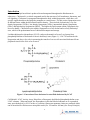
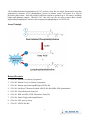
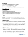
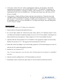
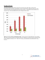
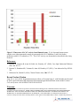
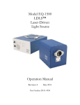
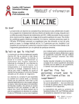
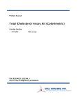
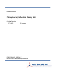
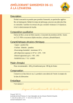
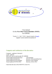
Product Manual Lecithin Cholesterol Acyltransferase (LCAT) Activity Assay Kit (Fluorometric) Catalog Number STA-615 100 assays FOR RESEARCH USE ONLY Not for use in diagnostic procedures Introduction Cholesterol is a lipid sterol that is produced in and transported throughout the bloodstream in eukaryotes. Cholesterol is a critical compound used in the structure of cell membranes, hormones, and cell signaling. Cholesterol is transported throughout the body within lipoproteins, which have cellspecific signals that direct the lipids they transport to certain tissues. For this reason, lipoproteins exist in different forms within the blood based on their density. These include chylomicrons, very-low density lipoproteins (VLDLs), low-density lipoproteins (LDLs), intermediate-density lipoproteins (IDLs), and high-density lipoproteins (HDLs). The higher the lipid content within a lipoprotein, the lower its density. Cholesterol exists within a lipoprotein as a free alcohol and as a fatty cholesteryl ester, which is the predominant form of cholesterol transport and storage. Lecithin:cholesterol acyltransferase (LCAT) catalyzes the transfer of an sn-2 acyl group from phosphatidylcholine to cholesterol to form a cholesteryl ester (Figure 1). LCAT is associated with lipoproteins and plays a key role in promoting the transfer of excess cell-associated cholesterol from peripheral tissues to the liver to be excreted. Figure 1. Conversion of free cholesterol to esterified cholesterol by LCAT Cell Biolabs’ LCAT Activity Assay Kit utilizes a fluorogenic dual-labeled phosphatidycholine as an LCAT substrate. When uncleaved, the fluorophores in the dual-labeled substrate are in a quenched state; upon hydrolysis by LCAT at the sn-2 position, fluorescent monomer chains are produced which can be measured in a fluorescence microplate reader (Ex. 342 nm/Em. 400 nm) (See Assay Principle). 2 The Lecithin:cholesterol acyltransferase (LCAT) Activity Assay Kit is a simple, fluorometric assay that quantitatively measures LCAT phospholipase activity in plasma, serum, and lysates in a 96-well microtiter plate format. Each kit provides sufficient reagents to perform up to 100 assays, including blanks and unknown samples. Besides LCAT, this assay can also be used to detect other calcium independent phospholipase activities such as lipoprotein phospholipase A2 (LP-PLA2). Assay Principle Related Products 1. STA-241: Human Low Density Lipoprotein 2. STA-242: Human Very Low Density Lipoprotein 3. STA-361: Human ApoAI and ApoB Duplex ELISA Kit 4. STA-369: OxiSelect™ Human Oxidized LDL ELISA Kit (MDA-LDL Quantitation) 5. STA-390: Total Cholesterol Assay Kit 6. STA-391: HDL and LDL/VLDL Cholesterol Assay Kit 7. STA-396: Serum Triglyceride Quantification 8. STA-610: LPL Activity Assay 9. STA-611: LPL ELISA Kit 3 Kit Components 1. LCAT Fluorometric Substrate (Part No. 261501): One 50 µL amber vial. 2. 10X LCAT Assay Buffer (Part No. 261502): Two 1.5 mL vials. 3. 2X Stop Solution (Part No. 261503): One 12 mL bottle. 4. 50X LCAT Inhibitor (Part No. 261504): One 100 µL vial of iodoacetic acid (IAA) at 100 mM. Materials Not Supplied 1. Serum, plasma, cell or tissue lysate 2. β-mercaptoethanol 3. 4. 5. 6. 7. 96-well plate suitable for a fluorescence plate reader 10 µL to 1000 µL adjustable single channel micropipettes with disposable tips 50 µL to 300 µL adjustable multichannel micropipette with disposable tips Multichannel micropipette reservoir Fluorescence microplate reader equipped with an excitation filter at 342 nm and an emission filter at 400 nm Storage Upon receipt, store the LCAT Fluorometric Substrate and 50X LCAT Inhibitor at -20ºC. Avoid multiple freeze/thaws by aliquoting. The LCAT Fluorometric Substrate is light sensitive and should be maintained in amber tubes. Store the remainder of the kit at 4ºC. Preparation of Reagents 1X LCAT Assay Buffer: Dilute the 10X LCAT Assay Buffer to 1X with deionized water and add βmercaptoethanol to a final concentration of 4 mM. Stir to homogeneity. LCAT Reaction Reagent: Immediately before use, briefly centrifuge the vial of LCAT Fluorometric Substrate to dislodge any liquid in the container’s cap. Dilute required amount of the LCAT Fluorometric Substrate 1:100 in 1X LCAT Assay Buffer (e.g. for 10 assays, add 5 µL of LCAT Substrate to 495 µL of 1X LCAT Assay Buffer). Briefly vortex to homogeneity. For best results, use within 30 minutes of preparation. Do not store diluted solutions. Preparation of Samples Plasma: Collect blood with an anticoagulant such as heparin, citrate or EDTA and mix by inversion. Centrifuge the blood at 1000 x g at 4°C for 10 minutes. Collect plasma supernatant without disturbing the white buffy layer. Samples should be tested immediately or frozen at -80°C for up to 3 months. Serum: Collect blood in a tube with no anticoagulant. Allow the blood to clot at room temperature for 30 minutes. Centrifuge at 2500 x g for 20 minutes. Remove the yellow serum supernatant without disturbing the white buffy layer. Samples should be tested immediately or frozen at -80°C for up to 3 months. 4 Cell Lysates: Collect 106 to 107 cells by centrifugation at 1000 x g for 10 minutes. Discard the supernatant and resuspend in 1 mL of cold 20 mM Tris, pH 7.5, 150 mM NaCl. Homogenize or sonicate the cell suspension. Centrifuge at 10,000 x g for 10 minutes at 4°C. Carefully collect the supernatant and store on ice for immediate use. For longer term storage, freeze the lysate at -80°C for up to 3 months. Tissue Samples: Weigh out 200 mg of tissue and mince into small pieces. Rinse the tissue with cold PBS to remove red blood cells and clots. Homogenize the minced tissue in 1 mL of cold 20 mM Tris, pH 7.5, 150 mM NaCl. Centrifuge at 10,000 x g for 10 minutes at 4°C. Carefully collect the supernatant and store on ice for immediate use. For longer term storage, freeze the homogenate at -80°C for up to 3 months. Assay Protocol Note: Maintain all samples at 4ºC during assay preparation. 1. Prepare and mix all reagents thoroughly before use. 2. In a 96-well plate suitable for a fluorescence plate reader, add 50 µL of unknown sample to each well. Blank wells should be included by adding 50 µL of 1X LCAT Assay Buffer. Each sample and blank should be assayed in duplicate. Dilute samples in 1X LCAT Assay Buffer if needed. 3. (optional) Add the desired amount of LCAT inhibitor to the appropriate samples and mix briefly. Incubate for one hour at 37ºC. We suggest a final concentration of 2 mM IAA. 4. Initiate the reaction by adding 50 µL of the freshly prepared LCAT Reaction Reagent to each well and mix the well contents thoroughly by pipetting. 5. Incubate for 3 to 18 hours at 37ºC. Note: To avoid evaporation, cover the plate with a lid and perform the incubation in a humidified incubator. 6. Stop the reaction by adding 100 µL of 2X Stop Solution to each well. 7. Immediately read on a fluorescence plate reader at 342 nm excitation and 400 nm emission. 5 Example of Results The following figures demonstrate typical LCAT Activity Assay Kit results. Fluorescence measurement was performed on SpectraMax Gemini XS Fluorometer (Molecular Devices) with a 355/405 nm filter set for the hydrolyzed fluorescent monomer. One should use the data below for reference only. This data should not be used to interpret actual results. Figure 2: LCAT Activity from Human Serum. Various amounts of normal human serum samples were either untreated (control) or pretreated with LCAT Inhibitor (2 mM IAA) before incubation with LCAT substrate for 18 hrs at 37ºC. The LCAT activity was determined as described in the Assay Protocol. 6 Figure 3: Timecourse of LCAT Activity from Human Serum. 25 µL of normal human serum samples were either untreated (control) or pretreated with LCAT Inhibitor (2 mM IAA) before incubation with LCAT substrate at 37ºC. The LCAT activity was determined as described in the Assay Protocol. References 1. Rousset X, Vaisman B, Amar M, Sethi AA, Remaley AT (2009), Curr Opin Endocrinol Diabetes Obes. 16: 183-71. 2. Rousset X, Shamburek R, Vaisman B, Amar M, Remaley AT (2011), Curr Atherosler Rep 13:249256 3. Dobiasova M, Frolich JJ (1999), Clinica Chimica Acta, 286: 257-271 Recent Product Citation Jung, M. A. et al. (2015). Hypocholesterolemic effects of Curcuma longa L. with Nelumbo nucifera leaf in an in vitro model and a high cholesterol diet-induced hypercholesterolemic mouse model. Animal Cells and Systems. doi:10.1080/19768354.2014.992953. Warranty These products are warranted to perform as described in their labeling and in Cell Biolabs literature when used in accordance with their instructions. THERE ARE NO WARRANTIES THAT EXTEND BEYOND THIS EXPRESSED WARRANTY AND CELL BIOLABS DISCLAIMS ANY IMPLIED WARRANTY OF MERCHANTABILITY OR WARRANTY OF FITNESS FOR PARTICULAR PURPOSE. CELL BIOLABS’s sole obligation and purchaser’s exclusive remedy for breach of this warranty shall be, at the option of CELL BIOLABS, to repair or replace the products. In 7 no event shall CELL BIOLABS be liable for any proximate, incidental or consequential damages in connection with the products. Contact Information Cell Biolabs, Inc. 7758 Arjons Drive San Diego, CA 92126 Worldwide: +1 858-271-6500 USA Toll-Free: 1-888-CBL-0505 E-mail: [email protected] www.cellbiolabs.com 2013-2015: Cell Biolabs, Inc. - All rights reserved. No part of these works may be reproduced in any form without permissions in writing. 8