1
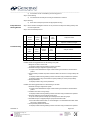
______________________________________________________________________________ Power-StainTM 1.0 Poly HRP DAB Kit for Mouse + Rabbit Cat No. Quantity 52-0017 15 mL 54-0017 100 mL Intended Use For In Vitro Diagnostic Use. This kit is intended for use with Mouse and Rabbit Primary Antibodies and other ancillary reagents supplied by user for qualitative detection of targeted protein (antigen) using immunohistochemistry (IHC) methodology by light microscopy on routine formalin-fixed, paraffin-embedded (FFPE) tissue section. Interpretation of any positive or negative staining shall be supported by implementation of a proper control, and must be made within the context of the patient’s clinical history and other diagnostic test by a qualified pathologist. Summary And Explanation This kit is a non-biotin system and utilizes a Poly HRP (horseradish peroxidase) conjugate to locate where the mouse or rabbit primary antibody is bound to the target antigen. The complex formed between Poly HRP conjugate and the mouse or rabbit primary antibody is observed through the use of a substratechromogen solution, which when added, results in a colored precipitate at the antigen location. The staining location and pattern is easily observable by light microscopy. Reagents Supplied Reagent A: One bottle of ready-to-use Poly HRP Conjugate for Mouse + Rabbit in an enzyme conjugate buffer containing stabilizing proteins and anti-microbial agents. Reagent B1: One bottle of 2X DAB Chromogen Solution Reagent B2: One bottle of 2X DAB Buffer Solution Storage Store at 2-8°C. Do not freeze. All performance claims are void after the kit expiration date. Materials Required But Not Supplied Primary Antibody (Genemed offers prediluted and concentrate Primary Antibodies) Primary Antibody Diluent (Cat No. 10-0001) Reagent Control (Non-immune Mouse IgG Cat No. 60-0045 and Non-Immune Rabbit IgG Cat No. 60-0060) Positive and Negative Control Specimens Microscope Slides, Positively Charged Xylene Ethanol Endogenous Peroxidase Blocking Solution - 3% Hydrogen Peroxide (Cat No. 10-0056) Wash Buffer - 10 mM Phosphate Buffer Saline, pH 7.4; optional with 0.05% Tween 20 Hematoxylin (Cat No. 10-0027, 10-0049) Antigen retrieval reagents (e.g. Cat No. 10-0022 Citrate Buffer pH 6.0 1X; Cat No. 10-0020 Citrate Buffer pH 6.0 20X; Cat No. 10-0021 Tris Buffer pH 9 20X; Cat No. 10-0023 Tris Buffer pH 9 1X; Cat No. 10-0046 Tris EDTA Buffer pH 9 1X; Cat No. 10-0037 Tris EDTA Buffer pH 9 20X; Cat No. 10-0024 Proteinase K; Cat No. 10-0025 Trypsin; Cat No. 10-0050 Ficin) Precautions For professional users only. DAB Chromogen Solution (Reagent B1) is susceptible to contamination from oxidizing agents. To avoid contamination, do not pipette Reagent B1 directly out of the bottle. Proper handling of this product as with any product derived from biological sources should be used according to local and applicable regulations. 30408 Rev. 01 Page 1 of 4 Genemed Biotechnologies, Inc 458 Carlton Ct. South San Francisco, CA 94080, U.S.A. Tel: 650-952-0110 Fax: 650-952-1060 MDSS GmbH Schiffgraben 41 30175 Hannover Germany ______________________________________________________________________________ Risk Statements: DAB Chromogen R40 Procedural Notes Limited evidence of carcinogenic effect. R43 May cause sensitization by skin contact. R68 Possible risk of irreversible effects. The directions accompanying this kit provide step by step instructions for optimal staining. Any change in procedure or incubation times may give erroneous staining results. For optimal results, do not substitute reagents provided in the kit. Reagent A shall be equilibrated to room temperature readily before usage. All incubations should be performed at room temperature in a humid environment. Do not allow the tissue section to dry out at any point in the staining procedure. The reagents are for single use. Preliminary Preparation Of Slides Routine de-paraffinization and rehydration of tissue section. Control Slides Three types of control slides are necessary for proper interpretation. Antigen retrieval as required by the primary antibody. Positive Tissue Control – A tissue containing the desired antigen. Negative Tissue – A tissue that does not contain the desired antigen. Reagent Control – A slide to be treated with a homologous non-immune immunoglobulin. (Cat No. 60-0045 or Cat No. 60-0060) Staining Protocol Step 1: Endogenous Peroxidase Blocking a) Submerge slides in Peroxidase Blocking Solution for 10 minutes. b) Wash slides with Wash Buffer to remove excess Peroxidase Blocking Solution. c) Tap off excess liquid and carefully wipe around tissue. Step 2: Primary Antibody Incubation a) Prepare Primary Antibody to optimum concentration. If necessary, dilute with Primary Antibody Diluent. b) Add 2 drops (100 µL) or as much as needed of Primary Antibody to completely cover each tissue. c) Incubate for 30-60 minutes at room temperature. d) Rinse 3 times with Wash Buffer for 2 minutes each. e) Tap off excess liquid and carefully wipe around tissue. Step 3: Poly HRP Conjugate Incubation (Reagent A) a) Add 2 drops (100 µL) or as much as needed of Enzyme Conjugate to completely cover each tissue. b) Incubate for 15 ± 1 minutes. c) Rinse 3 times with Wash Buffer for 2 minutes each. d) Tap off excess liquid and carefully wipe around tissue. Step 4: Substrate/Chromogen a) Prepare Ready-To-Use DAB substrate solution. Add DAB Chromogen Solution (Reagent B1) to DAB Buffer Solution (Reagent B2) and mix the two solutions in a 1:1 ratio with the volume determined by the number of slides to stain. In general, 200 µL of mixed substrate solution is sufficient to cover one tissue slide. Note: Do not equilibrate the entire bottle of either reagent at Room Temperature. Take out the necessary amount of each solution and allow the aliquoted volumes to equilibrate at Room Temperature before mixing. After mixing in a 1:1 ratio, the resulting Ready-To-Use substrate solution should be used within 2 hours. b) Add substrate solution on slides and incubate for 5-10 minutes at room temperature. c) Rinse slides with tap water to remove excess substrate solution. 30408 Rev. 01 Page 2 of 4 Genemed Biotechnologies, Inc 458 Carlton Ct. South San Francisco, CA 94080, U.S.A. Tel: 650-952-0110 Fax: 650-952-1060 MDSS GmbH Schiffgraben 41 30175 Hannover Germany ______________________________________________________________________________ d) Proceed with normal counterstaining and mounting protocol. Step 5: Counterstaining a) Counterstain with Hematoxylin according to manufacturer’s instruction. Step 6: Mounting a) Interpretation Of Staining Results Mount and coverslip the specimen with appropriate mounting. Step 1: Review Positive and Negative Controls. Do not proceed to next step if the staining intensity does not meet requirements. Step 2: Score the tested specimens. Positive Negative Control Control Reagent Control Test Tissue Analysis of Result Specimen contains the antigen Tissue Tissue 1 + -- -- + 2 + -- -- -- Reagent Control Test Tissue Analysis of Result Troubleshooting Specimen does not contain the antigen Positive Negative Control Control Tissue Tissue 1 -- -- -- -- No staining 2 Weak + -- -- +/- Weak staining 3 + + + + High background staining Possible causes and suggested action for: No staining on any slide 1. Reagents not used in correct order. Repeat procedure following Staining Protocol Instructions. 2. Substrate-Chromogen reagent not prepared properly. Prepare a fresh Substrate-Chromogen solution following the instructions included with the product. 3. Primary antibody incubation steps were omitted or dilution was incorrect or wrong antibody was used. Repeat procedure following Staining Protocol Instructions using incubation times specified. Repeat procedure using correct dilution for primary antibody or correct primary antibody. 4. Wrong Pretreatment. Repeat procedure using correct pretreatment. Possible cause and suggested action for: Weak staining on all slides 1. Substrate-Chromogen reagent has expired. Prepare a fresh Substrate-Chromogen solution following the instructions included with the product. 2. Incubation times were not long enough. Repeat procedure following Staining Protocol Instructions using incubation times specified. 3. Specimen retained too much liquid after rinsing steps. Tap off excess liquid and carefully wipe around specimen after rinsing steps. 4. Peroxidase Enzyme Conjugate (Reagent A) exposed to Sodium Azide. Use buffer without Sodium Azide, or check if Reagent A is contaminated with Sodium Azide during use or aliquot/pipetting. 5. Primary antibody dilution was incorrect. Repeat procedure following Staining Protocol Instructions using incubation times specified. 30408 Rev. 01 Page 3 of 4 Genemed Biotechnologies, Inc 458 Carlton Ct. South San Francisco, CA 94080, U.S.A. Tel: 650-952-0110 Fax: 650-952-1060 MDSS GmbH Schiffgraben 41 30175 Hannover Germany ______________________________________________________________________________ Repeat procedure using correct dilution for primary antibody. 6. Insufficient Pretreatment. Repeat procedure using correct pretreatment. Possible cause and suggested action for: High background staining on all slides 1. Specimens contain high endogenous peroxidase activity. Check preparation of Peroxidase Solution and verify timing of specimens submerged in solution. 2. Inadequate rinsing of slides. Use freshly prepared buffer solutions. Follow rinsing instructions specified. 3. De-paraffinization not complete. Use fresh xylene. Check slides are de-paraffinized before rehydration step. 4. Over-reaction of substrate. Do not incubate substrate longer than specified in procedure. 5. Specimens dry out during staining procedure. Incubate in humid environment. Wipe fewer than 10 slides at a time before adding next solution. 6. Wrong Pretreatment. Repeat procedure using correct pretreatment. Symbols Catalog No. 30408 Rev. 01 Batch No. In Vitro Diagnostic Use Temperature Range Use By Page 4 of 4 Genemed Biotechnologies, Inc 458 Carlton Ct. South San Francisco, CA 94080, U.S.A. Tel: 650-952-0110 Fax: 650-952-1060 MDSS GmbH Schiffgraben 41 30175 Hannover Germany