1
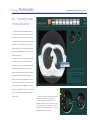
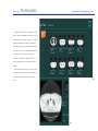
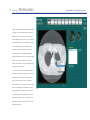
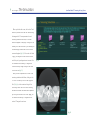
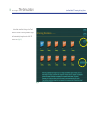
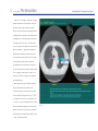
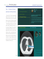
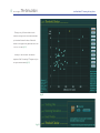
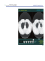
1
ALCA Project
The Simulation
Low-Dose Helical CT Screening for Lung Cancer
Low-Dose Helical CT Screening for Lung Cancer
Anti-Lung Cancer Association Project:"The Simulation"
User's Manual
Introduction
2
Part 1 - Teaching Files
(Atlas of Small Lung Cancers)
4
Part 2 - Screening Simulation
(Practical CT Screening)
7
Part 3 - Small Nodules
(Detection)
11
Part 4 - Threshold Checker
(Detection Self-Assessment)
Acknowledgements
References
14
17
17
fig. 1
2
ALCA Project
The Simulation
Low-Dose Helical CT Screening for Lung Cancer
Introduction
At the request of the Anti-Lung Cancer
Project1~5) we recently developed a teaching
software program entitled "The Simulation" that
runs on both Macintosh and Windows (fig.1).
The software consists of four parts: Part 1,
Teaching Files, an atlas of small lung cancers; Part
2, Screening Simulation, a dataset for practical CT
screening; Part 3, Small Nodules, a dataset for the
detection of small nodules either in retrospective
or prospective chronological order; and Part 4,
Threshold Checker, a dataset for self-assessment
of the threshold of the user's ability to detect small
nodules on screening CT images.
Part 1 consists of two folders each of which
fig. 2
includes eight cases with a single lung cancer
nodule present in each case (fig.2).
In Part 2, 10 randomly chosen cases are
presented in each of 10 screening simulation trial
folders for a total of 100 cases (fig.3).
fig. 3
3
ALCA Project
The Simulation
Low-Dose Helical CT Screening for Lung Cancer
Part 3 includes a number of pitfall cases (fig.4).
The dataset in Part 4 consists of 36 screening CT
images that include retrospective nodules from 10
cases of minute lung cancer. The user's threshold
output is evaluated on a graph based on the sizes
and differences in mean CT number between each
nodule and the surrounding lung field (fig.5).
fig. 4
fig. 5
4
ALCA Project
The Simulation
Low-Dose Helical CT Screening for Lung Cancer
Part 1 - Teaching Files (Atlas
of Small Lung Cancers)
The menu page lists two separate folders for
Part 1. Each folder contains eight cases. Clicking
on either of the folder icons allows you to enter
the test mode. Clicking on the "Go Index" button
takes you to the "Index" page, which lists all 16
cases and allows you to sort them by diagnosis,
extent of ground-glass opacity (GGO), size, and
Noguchi type6) (fig.2).
A single lung cancer nodule is present in each
test mode case. Please try to detect the lung
cancer nodule as you use the toggle and speed
buttons located on the upper right side of the
screen to read through the screening CT images
from top (apex) to bottom (diaphragm) (fig.6, 7).
When you think you have detected the cancer
nodule, click on the location of the nodule, and a
"Select Diagnosis" box will appear on the screen.
Click on "Select Diagnosis", then on "Lung Cancer
fig. 6
fig. 7
Nodule", and you will automatically proceed to
the next case.
Click the left side arrows (small arrows) for manual
upward and downward scrolling control.
Click the right side arrows (large arrows) for
automatic upward and downward scrolling control.
The smaller button is for speed control. Click the
right arrow to increase scrolling speed. Click the left
arrow to decrease scrolling speed.
5
ALCA Project
The Simulation
Low-Dose Helical CT Screening for Lung Cancer
When you finish reading all eight cases, a "Go
Answer" button will appear on the screen. Click on
it to proceed to the "Answer" page (fig.8). Orangebordered boxes on the "Answer" page indicate
that your response was correct for that particular
case. Clicking on any CT image thumbnail allows
you to review the screening CT images and
thin-section CT images for that case (fig.9). You
also can compare your answer with the correct
answer.
After reviewing the screening CT images for the
eight cases, you can return to the menu page by
clicking on the "Menu" button on the "Answer"
fig. 8
page.
fig. 9
6
ALCA Project
The Simulation
Low-Dose Helical CT Screening for Lung Cancer
If you would like to see an index of the cases,
you can proceed to the "Index" page by clicking
on the "Go Index" button. As indicated above,
the cases in the index can be sorted by diagnosis,
extent of ground-glass opacity (GGO), size, and
Noguchi type (fig.10). Clicking on each line allows
you to review the screening CT images and thinsection CT images for each case and compare
your answer with the correct answer.
fig. 10
7
ALCA Project
The Simulation
Low-Dose Helical CT Screening for Lung Cancer
Part 2 - Screening Simulation
(Practical CT Screening)
Click on the "Part 2" line on the menu page to
display the folder icon for Part 2 (fig.11). Clicking
on either the folder icon or the "Start" button
will take you to the main "Screening Simulation"
page, which contains a total of 10 different folders
(fig.12). Clicking on one of the folders enables you
to proceed to the screening simulation trial for
that folder. Ten cases are presented within each
fig. 11
screening simulation trial folder, but only one of
the 10 cases is actually lung cancer (fig.13). In the
other nine cases there are one or more benign
nodules or no nodules at all.
fig. 12
8
ALCA Project
The Simulation
Low-Dose Helical CT Screening for Lung Cancer
After completely reading through the screening
CT images of a case and identifying a location of
interest, click on the location, and a "Diagnosis"
box will appear on the screen. Click on "Diagnosis"
and a pull-down menu of four diagnostic choices
will be displayed for your consideration. If you
suspect a lung cancer nodule at the location,
select "Lung Cancer", remembering that each
screening simulation folder of 10 cases will contain
only one lung cancer nodule. If you want to
change your answer, click on your diagnosis, and
then on the "Delete" button.
If you identify one or more nodules that you do
not suspect of being lung cancer, select "Nodule"
from the diagnostic choices on the pull-down
menu. The circle-cursor on the CT images is 5
mm in diameter. If you identify one or more other
findings, for example, old inflammatory change,
bulla, emphysema, old tuberculosis, etc., select
"Other findings" from the diagnostic choices on
the pull-down menu. If you do not observe any
abnormal findings in a case, select "None" from
the pull-down menu.
fig. 13
9
ALCA Project
The Simulation
Low-Dose Helical CT Screening for Lung Cancer
When you finish with a case, click on the "Save"
button to proceed to the next case. After reading
the images of all 10 cases presented in each
screening simulation trial folder, an "Answer"
button will appear on the page. Clicking on it will
enable you to view the results of your readings on
the answer page, where folder icons of the ten
cases are displayed (fig.14). If the color of a folder
is gray, your diagnosis for that case was correct,
and if it is pink, your diagnosis was incorrect. You
fig. 14
can re-examine the screening CT images and
correct answers by simply clicking on the folder
icon of that case (fig.15).
After you have completed one or more of the
screening simulation trial folders, clicking on the
"Go to List" will take you to the "Index" page for
Part 2 (fig.16), which contains a listing of the
actual lung cancer cases in all of the screening
simulation trial folders that have been completed.
By clicking on each line of the "Index" page, you
can review the screening CT images and thinsection CT images for each case.
fig. 15
10
ALCA Project
The Simulation
Low-Dose Helical CT Screening for Lung Cancer
It should be noted that clicking on the "Reset"
button on the main "Screening Simulation" page
will automatically change the order of all 100
cases in Part 2 (fig.16).
fig. 16
11
ALCA Project
The Simulation
Low-Dose Helical CT Screening for Lung Cancer
Part 3 - Small Nodules
(Detection)
Click on the "Part 3" line on the menu page to
display the folder icon for Part 3 (fig.17). Clicking
on either the folder icon or the "Start" button will
enable you to proceed to the main "Small Nodules"
page (fig.18), which is divided into "Lung Cancer",
at the top and containing 15 separate folders, and
"Inflammation", at the bottom and containing 8
other folders. If you click on any of the folders, you
will proceed to the test mode for that folder.
fig. 17
The screening CT image on which the nodule
was first detected or the nodule increased in size
or opacity will be shown on the left side of the
screen and a screening CT image taken earlier will
be shown on the right side (fig.19). Please try to
detect a new or an enlarged nodule in each of the
lung cancer cases and a new nodule or nodules
in each of the inflammation cases after reading
through the screening CT images from top (apex)
to bottom (diaphragm) in each folder. The buttons
located in the upper center portion of the screen
allow you to move the CT images up towards the
apex or down towards the diaphragm.
fig. 18
12
ALCA Project
The Simulation
Low-Dose Helical CT Screening for Lung Cancer
In part 3, the CT image control button is slightly
different from that in the other parts. When you
move the cursor to the location of the small
arrow, the color of the arrow changes from dark
to bright yellow. By clicking on the small arrow in
the upper part or in the lower part of the button,
CT images move 1 slice up or 1 slice down per
click. By clicking on the large arrow in the middle
part of the button, CT images move upward or
downward continuously. When you move the
cursor outside of the large arrow, the CT images
will stop moving. There are two red striped
rectangular areas on both sides of the arrows. If
you click one side of the red area, it will turn dark
and CT images on that side will not move. This
allows you to move CT images on the opposite
side independently.
When you think you have detected a nodule,
click on the location of the nodule on the
"Detection" image on the left side of the screen,
and you will see a "Diagnosis" box (fig.19). Click
on "Diagnosis" and then on "Small Nodule", and
a "Correct" or "New or Enlarged Nodule?" answer
will be immediately displayed in the lower center
portion of the screen. If you want to change the
location that you selected for the nodule, click on
"Cancel" and start over again.
fig. 19
13
ALCA Project
The Simulation
Low-Dose Helical CT Screening for Lung Cancer
Clicking on the "Go Answer" button will take
you to the answer page (fig. 20). The screening
CT image on which the nodule was first detected
will be shown on the left side, and a screening
CT image taken earlier or thin-section CT image
will be shown on the right side. By clicking on the
CT image thumbnail boxes at the bottom of the
screen, you can compare screening CT images
taken earlier with the screening CT image on
which the nodule was first detected. If you click
on the "Quick Tour" button in the upper center
portion of the screen, "Next" button will appear.
Repeated clicking of the "Next" button will display
the key CT images of the case in sequence.
After completing one or more of the 23 folders,
clicking on the "Go to List" will take you to the
"Index" page for Part 3, which contains a list of the
cases in all of the folders that you have completed.
Clicking on each line of the "Index" page will allow
you to review the screening CT images and thinsection CT images in each case.
fig. 20
14
ALCA Project
The Simulation
Low-Dose Helical CT Screening for Lung Cancer
Part 4 - Threshold Checker
(Detection Self-Assessment)
Click on the "Part 4" line on the menu page to
display the folder icon for Part 4 (fig.21). Clicking
on either the folder icon or the "Start" button will
take you to the first of 36 screening CT images
from 10 different cases presented in sequence.
After identifying and clicking on one location
fig. 21
where a nodule is suspected on each of the
36 screening CT images (fig.22), the result for
your readings will be shown as 36 dots on a
graph, based on the size of the nodules and the
differences in mean CT number between each
nodule and the surrounding lung field (fig.23).
Clicking on any dot on the graph will allow you
to review the CT images corresponding to that
particular case based on the timeline indicated at
the bottom of the screen (fig.25).
fig. 22
15
ALCA Project
The Simulation
Low-Dose Helical CT Screening for Lung Cancer
Clicking on any of the case buttons in the
column at the right side of the screen will enable
you to review the exact location of the actual
nodules on the graph in any particular case in Part
4, or in all 10 cases (fig.23).
Clicking on "new case order" will alter the
sequence of the 36 screening CT images, and you
can try another test mode (fig.24).
fig. 23
fig. 24
16
ALCA Project
The Simulation
Low-Dose Helical CT Screening for Lung Cancer
fig. 25
17
ALCA Project
The Simulation
Low-Dose Helical CT Screening for Lung Cancer
Acknowledgements
This work was supported in part by grants from Scientific Research Expenses for Health and Welfare Programs,
Foundation for the Promotion of Cancer Research, and 2nd-Term Comprehensive 10-year Strategy for Cancer
Control and by a Grant-in-Aid for Cancer Research from the Ministry of Health, Labor and Welfare.
References
1. Kaneko M, Eguchi K, Ohmatsu H, et al. Peripheral lung cancer: screening and detection with low-dose spiral CT
versus radiography. Radiology 1996;201:798-802.
2. Kakinuma R, Ohmatsu H, Kaneko M, et al. Detection failures in spiral CT screening for lung cancer: analysis of
CT findings. Radiology. 1999;212:61-66.
3. Kaneko M, Kusumoto M, Kobayashi T, et al. Computed tomography screening for lung carcinoma in Japan.
Cancer. 2000;89(11 Suppl):2485-2488.
4. Sobue T, Moriyama N, Kaneko M, et al. Screening for lung cancer with low-dose helical computed tomography:
anti-lung cancer association project. J Clin Oncol. 2002;20:911-920.
5. Kakinuma R, Ohmatsu H, Kaneko M, et al. Progression of focal pure ground-glass opacity detected by low-dose
helical computed tomography screening for lung cancer. J Comput Assist Tomogr. 2004;28:17-23.
6. Noguchi M, Morikawa A, Kawasaki M, et al. Small adenocarcinoma of the lung: histologic characteristics and
prognosis. Cancer 1995;75:2844-2852.
© 2000-2005 Ryutaro Kakinuma. All rights reserved.
© 2005 Tokyo Health Service Association
© 2005 The Society of Thoracic CT Screening


![INSTALL GUIDE OEM-AL(RS)-FM6-[ADS-ALCA]-EN](http://vs1.manualzilla.com/store/data/005732796_1-2697dd4b3634b9609d4f115002bbbdbf-150x150.png)