1
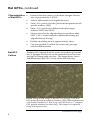
Rat Glial Precursor Cells (GPCs) Catalog no. N7746-100 Rev. date: 14 May 2009 Manual part no. A11232 MAN0001663 Corporate Headquarters Invitrogen Corporation 1600 Faraday Avenue Carlsbad, CA 92008 T: 1 760 603 7200 F: 1 760 602 6500 E: [email protected] For country-specific contact information visit our web site at www.invitrogen.com User Manual ii Contents Contents and Storage........................................................................................... iv Additional Products.............................................................................................. v Introduction ........................................................................................ 1 Rat Glial Precursor Cells (GPCs) .........................................................................1 Methods............................................................................................... 3 Handling Rat GPCs................................................................................................3 Media Requirements..............................................................................................4 Preparing Growth Medium..................................................................................5 Preparing Matrix for Adherent Cell Culture.....................................................6 Thawing and Establishing Cells ..........................................................................8 Expanding Cells....................................................................................................10 Differentiating Rat GPCs ....................................................................................12 Characterizing Phenotype of Rat GPCs............................................................13 Phenotype Marker Expression of Rat GPCs ....................................................15 Troubleshooting ...................................................................................................17 Appendix ........................................................................................... 20 Recipes ...................................................................................................................20 Technical Support ................................................................................................21 Purchaser Notification.........................................................................................22 References..............................................................................................................24 iii Contents and Storage Shipping and Storage Rat Glial Precursor Cells (GPCs) are shipped on dry ice. Upon receipt, store the cells in liquid nitrogen. Contents Amount supplied: One vial containing 1 × 10⁶ cells. Composition: 1 mL of cells in freezing medium.* *Freezing medium: Complete StemPro® NSC SFM with 2 mM GlutaMAX™-I and 10 ng/mL Platelet Derived Growth Factor AA Homodimer (PDGF-AA), plus 10% DMSO. Handle cells as potentially biohazardous material under at least Biosafety Level 1 (BL-1) containment. This product contains Dimethyl Sulfoxide (DMSO), a hazardous material. Review the Material Safety Data Sheet (MSDS) before handling. Material Safety Data Sheets (MSDSs) are available on our website at www.invitrogen.com/msds. iv Additional Products The products listed in this section may be used with Rat Glial Precursor Cells (GPCs). For more information, refer to our website (www.invitrogen.com) or contact Technical Support (see page 20). Additional Products Item Quantity Cat. no. 1 kit A1050901 100 mL 35050-061 500 mL 12660-012 FGF Basic Recombinant Human (bFGF) 10 μg PHG0024 EGF Recombinant Human 10 μg PHG0314 PDGF-AA, Recombinant Human 10 μg PHG0035 Fetal Bovine Serum (FBS), ES Cell-Qualified 100 mL 500 mL 16141-061 16141-079 BSA, 10% Stock Solution 25 mL P2489 Dulbecco’s Phosphate Buffered Saline (D-PBS), containing no calcium, magnesium, or phenol red 500 mL 14190-144 Dulbecco’s Phosphate Buffered Saline (D-PBS), containing calcium and magnesium, but no phenol red 500 mL 14040-133 Dulbecco’s Modified Eagle Medium (D-MEM) (1X), liquid (high glucose) 1000 mL 11995-040 CELLStart™ Defined, Humanized Substrate for Cell Culture 2 mL 10142-01 Natural Mouse Laminin 1 mg 23017-015 StemPro Accutase Cell Dissociation Reagent 100 mL A11105-01 -Mercaptoethanol (1,000X), liquid 50 mL 21985-023 StemPro® NSC SFM (contains KnockOut™ DMEM/F-12, FGF Basic Recombinant Human, EGF Recombinant Human, and StemPro® NSC SFM Supplement) GlutaMAX™-I Supplement ™ KnockOut DMEM/F-12 ® ® Continued on next page v Additional Products, continued Additional Products, continued The products listed in this section may be used with Rat Glial Precursor Cells (GPCs). For more information, refer to our website (www.invitrogen.com) or contact Technical Support (see page 20). Item Trypan Blue Stain LIVE/DEAD® Cell Vitality Assay Kit ™ Countess Automated Cell Counter (includes 50 Countess™ cell counting chamber slides and 2 mL of Trypan Blue Stain) Water, distilled Products for Marker Analysis Quantity Cat. no. 100 mL 15250-061 1000 assays L34951 1 unit C10227 20 100 mL 15230-196 The products listed below may be used for analyzing the phenotype of undifferentiated Rat Glial Precursor Cells (GPCs), as well as neurons, oligodendrocytes, and astrocytes. In addition to the primary antibodies listed below, Invitrogen offers a variety of isotype specific secondary antibodies conjugated with enzymatic and fluorescent indicators, as well as antibody sera and diluents. For more information, refer to our website (www.invitrogen.com) or contact Technical Support (see page 20). Quantity Cat. no. Mouse anti-MAP2 Item 100 μg 13-1500 Rabbit anti-Doublecortin (DCX) 100 μg 48-1200 Mouse anti-A2B5 (105) 100 μg 433110 Rabbit anti-GFAP (Glial Fibrillary Acid Protein) concentrate 1 mL 18-0063 DAPI (4,6-diamidino-2-phenylindole, dihydrochloride) 10 mg D1306 ® 10 mL P36930 ® 10 mL P36931 ProLong Gold Antifade Reagent ProLong Gold Antifade Reagent with DAPI vi Introduction Rat Glial Precursor Cells (GPCs) Introduction Rat Glial Precursor Cells (GPCs), also referred to as Oligodendrocyte Progenitor Cells (OPCs) or Glial Restricted Precursor Cells (GRPCs), are isolated from the cortices of newborn Sprague-Dawley rats at day 2 after birth (PN2). The cells are isolated under sterile conditions using dissociation and magnetic bead separation, and expanded in complete StemPro® NSC SFM containing 10 ng/mL Platelet Derived Growth Factor AA Homodimer (PDGF-AA). The cells are cryopreserved at passage 2 (P2) in 90% complete StemPro® NSC SFM with 2 mM GlutaMAX™-I supplement and 10 ng/mL PDGF-AA, plus 10% DMSO. Each vial of Rat GPCs contains 1 × 10⁶ viable cells that can be expanded in culture for at least one passage. Withdrawal of the growth factors (i.e., PDGF-AA, bFGF, and EGF) from the medium allows the cells to differentiate into a mixed population of cells, a substantial portion of which consists of oligodendrocytes (Mujtaba et al., 1999). Because of their capacity to generate oligodendrocytes, Rat GPCs can be used for neuroscience studies as well as stem cell differentiation, tissue engineering, cell and genetic therapy, and transplantation experiments (Gage, 2000; Kalyani & Rao, 1998; Rao, 1999; Temple, 2001). The ability of GPCs to migrate over large distances in intact brain tissue also make these cells the ideal candidate for delivering drugs or genes in a variety of ganglioside disorders (Rao, 1999). For optimal growth and recovery of your Rat GPCs, we recommend that you use complete GPC growth medium, which consists of complete StemPro® NSC SFM supplemented with 2 mM GlutaMAX™-I and 10 ng/mL PDGF-AA (see page 5 for recipe). Continued on next page 1 Rat GPCs, continued Characteristics of Rat GPCs Rat GPC Culture Isolated from the cortices of newborn Sprague-Dawley rats on postnatal day 2 (PN 2) Able to differentiate into oligodendrocytes Stain ≥ 80% positive for the glial restricted precursor cell specific marker, A2B5 Stain ≤ 10% positive for differentiated phenotype markers MAP2 and GFAP Stain positive for the oligodendrocyte-specific marker, GalC (≥ 30%) when induced to differentiate along the oligodendrocyte lineage Exhibit a doubling time of approximately 4 days Can be expanded in culture for at least one passage without differentiation Primary cells isolated from the cortex of newborn (postnatal day 2) Sprague-Dawley rat can be expanded for at least one passage in culture upon recovery. The image below shows undifferentiated Rat GPCs at day three after thawing. Figure 1. Bright field image of Rat GPCs at passage 3 (P3) that have been cultured in complete StemPro® NSC SFM supplemented with 2 mM GlutaMAX™-I and 10 ng/mL PDGF-AA (i.e., complete GPC growth medium) for three days. The image was captured using 10X objective lens. Continued on next page 2 Methods Handling Rat GPCs As with other mammalian cell lines, when working with Rat GPCs, handle as potentially biohazardous material under at least Biosafety Level 1 (BL-1) containment. For more information on BL-1 guidelines, refer to Biosafety in Microbiological and Biomedical Laboratories, 5th ed., published by the Centers for Disease Control, or see the following website: www.cdc.gov/od/ohs/biosfty/bmbl5/bmbl5toc.htm Guidelines for Culturing Rat GPCs Follow the general guidelines below to grow and maintain Rat GPCs. All solutions and equipment that come in contact with the cells must be sterile. Always use proper aseptic technique and work in a laminar flow hood. For consistent results in your differentiation studies and other experiments, we recommend using cells below passage 4 (P4). If you expand Rat GPCs beyond P3 (i.e., more than a single passage upon recovery), we recommend that you perform another round of characterization prior to further experiments. To keep Rat GPCs undifferentiated, change medium every two days. For general maintenance of Rat GPCs in culture, the growth rate should be in mid-logarithmic phase with 70–80% confluency prior to subculturing. Passage cells at a seeding density of 30,000 cells/cm2. Note: Passaging Rat GPCs at a lower density causes the cells to differentiate. Important When thawing or subculturing cells, transfer cells into pre-warmed medium. Standard physical growth conditions for Rat GPCs are 37°C in a humidified atmosphere of 5% CO2 in air. We recommend that you use Rat GPCs right after recovery. After thawing Rat GPCs, expand the cells once to have a two-fold increase in their number, and harvest them to use in your experiments (e.g., transplantation studies, testing differentiation media). Continued on next page 3 Media Requirements Important Media Requirements 4 It is very important to strictly follow the guidelines for culturing Rat GPCs in this manual to keep them undifferentiated. We recommend using complete GPC growth medium, which consists of complete StemPro® NSC SFM supplemented with 2 mM GlutaMAX™-I and 10 ng/mL PDGF-AA, for optimal growth and expansion of Rat GPCs, and to keep them undifferentiated (for a recipe, see Preparing Growth Medium, page 5). This medium is designed to support isolation and growth of glial precursor cells derived from the cortical tissue of newborn rats, which can be grown as an adherent culture on CELLStart™ or poly-L-ornithine coated tissue culture treated vessels (see page v for ordering information). Prepare your growth medium prior to use. To maintain undifferentiated GPCs, change medium every two days. When thawing or subculturing cells, transfer cells into pre-warmed medium at 37°C. We recommend that you aliquot complete GPC growth medium into required working amounts to avoid exposing it to 37°C multiple times. Complete GPC growth medium is stable for up to two weeks when stored in the dark at 4°C. Do not freeze complete GPC growth medium. You may store the complete StemPro® NSC SFM in the dark at 4°C for up to four weeks. Do not freeze complete StemPro® NSC SFM. You may refreeze unused StemPro® NSC SFM Supplement; however, avoid repeated freeze-thaw cycles. Preparing Growth Medium Preparing Growth Medium Complete GPC growth medium consists of complete StemPro® NSC SFM (KnockOut™ D-MEM/F-12 with StemPro® NSC SFM Supplement, EGF, and bFGF) supplemented with 2 mM GlutaMAX™-I and 10 ng/mL PDGF-AA. Complete GPC growth medium is stable for up to two weeks when stored in the dark at 4°C. To make 100 mL of complete GPC growth medium, aseptically mix the following components: Component KnockOut™ D-MEM/F-12 GlutaMAX™-I Supplement bFGF EGF StemPro® NSC SFM Supplement PDGF-AA Concentration 1X 2 mM 20 ng/mL 20 ng/mL 2% 10 ng/mL Amount 97 mL 1 mL 2 μg 2 μg 2 mL 1 μg Note: You may observe a white precipitate when thawing StemPro® NSC SFM supplement, which will disappear when it is completely thawed or dissolved. 5 Preparing Matrix for Adherent Cell Culture Matrix for Rat GPC Culture You may use CELLStart™ or poly-L-ornithine as a matrix for maintaining your Rat GPCs. The attachment strength of Rat GPCs may be greater for CELLStart™ than poly-L-ornithine, depending upon the tissue culture vessel used. Materials Needed The following materials are required (see pages v–vi for ordering information). Coating Culture Vessels with CELLStart™ Tissue-culture treated flasks, plates, or Petri dishes CELLStart™ or poly-L-ornithine (Sigma, Cat. no. P3655) D-PBS containing calcium and magnesium, but no phenol red Cell culture grade, distilled water (if coating plates with poly-L-ornithine) 37°C incubator with a humidified atmosphere of 5% CO2 Optional: Parafilm (if planning to store coated dishes for future use) Do not freeze, vortex, or expose CELLStart™ to vigorous agitation to prevent potential gel formation. You may coat the plates in advance, and store at 4°C wrapped tightly with Parafilm for up to 2 weeks. Do not remove CELLStart™ solution until just prior to use. Make sure the plates do not dry out. 1. Dilute CELLStart™ 1:100 in D-PBS with calcium and magnesium to prepare the working solution of CELLStart™ (i.e., 50 μL of CELLStart™ into 5 mL of D-PBS). 2. Coat the surface of the culture vessel with the working solution of CELLStart™ (14 mL for T75, 7 mL for T25, 3.5 mL for 60-mm dish, 2 mL for 35-mm dish). 3. Incubate the culture vessel at 37°C in a humidified atmosphere of 5% CO2 in air for 1 hour. 4. Remove the vessel from the incubator and store until use. Immediately before use, aspirate all CELLStart™ solution and replace with complete growth medium (see page 5). Note: You do not need to wash the culture dishes coated with CELLStart™; you may use them directly after aspiration. Continued on next page 6 Preparing Matrix for Adherent Cell Culture, continued Coating Culture Vessels with PolyL-ornithine You may coat the plates with poly-L-ornithine in advance, and store at room temperature wrapped tightly with Parafilm for up to 1 week. Do not remove D-PBS until just prior to use. Make sure the plates do not dry out. 1. Dissolve poly-L-ornithine (Sigma, Cat. no. P3655) in distilled water to make 10 mg/mL stock solution (500X). Aliquot and store at –20°C until use. 2. Dilute poly-L-ornithine stock solution 1:500 in cell culture grade distilled water make 20 μg/mL working solution. 3. Coat the surface of the culture vessel (with or without cover slips) with poly-L-ornithine working solution (14 mL for T75, 7 mL for T25, 3.5 mL for 60-mm dish, 2 mL for 35-mm dish). 4. Incubate the culture vessel overnight at room temperature. 5. Rinse the culture vessel twice with D-PBS without Ca2+ and Mg2+, and store covered with D-PBS until use. Immediately before use, remove all D-PBS and replace with complete growth medium (see page 5). 7 Thawing and Establishing Cells Materials Needed The following materials are required (see pages v–vi for ordering information). Rat GPCs, stored in liquid nitrogen Ethanol or 70% isopropanol Complete growth medium (complete GPC growth medium, see page 5); pre-warmed to 37°C Disposable, sterile 15-mL tubes Flame-polished and autoclaved glass Pasteur pipettes, or plastic Pasteur pipettes pre-rinsed with growth medium 37°C water bath 37°C incubator with a humidified atmosphere of 5% CO2 Microcentrifuge CELLStart™ or poly-L-ornithine coated, tissue-culture treated flasks, plates, or Petri dishes (see pages 6–7) Hemacytometer, cell counter and Trypan Blue, LIVE/DEAD® Cell Vitality Assay Kit, or the Countess™ Automated Cell Counter The Countess™ Automated Cell Counter is a benchtop instrument designed to measure cell count and viability (live, dead, and total cells) accurately and precisely in less than a minute per sample, using the standard Trypan Blue uptake technique (see page vi for ordering information). Using the same amount of sample that you currently use with the hemocytometer, the Countess™ Automated Cell Counter takes less than a minute per sample for a typical cell count and is compatible with a wide variety of eukaryotic cells. Important Rat GPCs readily stick to the plastic used in cell culture dishes and centrifuge tubes. Prior to use, rinse all material that will come in contact with the cells with medium to prevent cells from sticking to the plastic. To thaw and establish Rat GPCs, follow the procedure on the next page. Continued on next page 8 Thawing and Establishing Cells, continued Thawing Procedure 1. Pre-rinse your culture flasks, plates, or Petri dishes with growth medium to coat the plastic surface. Make sure to pre-rinse any other material that will come in contact with the cells to prevent cells from sticking to the plastic. 2. Remove the cells from liquid nitrogen storage, and immediately transfer the cells to a 37°C water bath to prevent crystal formation. 3. Quickly thaw the vial of cells by gently swirling it in the 37°C water bath and removing it when the last bit of ice has melted, typically < 2 minutes. Do not submerge the vial completely. Do not thaw the cells for longer than 2 minutes. Do not introduce bubbles into the cell suspension as it decreases cell viability. 4. When thawed, transfer the tube containing the cells into the laminar flow hood, and wash the outside of the tube with 70% isopropanol. 5. Rinse the pipette tip with media, and very gently transfer the cells into a pre-rinsed 15-mL centrifuge tube. 6. Rinse the vial with 1 mL of growth medium (see page 5), and dropwise add to the cells in the 15-mL centrifuge (one drop/second). Mix by gentle swirling after each drop. 7. Slowly add 2 mL of growth medium to the cell solution, and mix gently. 8. Determine the viable cell count using your method of choice. The viability of thawed cells should be >50%, and the total live cell number should be >1 106. 9. Plate the cells at a seeding density of 3 104–5 104 cells per cm2 on a CELLStart™ or poly-L-ornithine coated, tissue-culture treated culture dish. If necessary, gently add growth medium to the cells to achieve the desired cell concentration and recount the cells. 10. Incubate at 37°C, 5% CO2, and 90% humidity and allow cells to adhere for at least 24 hours. 11. The next day, replace the medium with an equal volume of fresh, pre-warmed complete growth medium. Change the medium every other day, and passage cells when the culture is 75–90% confluent. 9 Expanding Cells Introduction You may expand Rat GPCs as an adherent culture on CELLStart™ or poly-L-ornithine coated, tissue-culture treated flasks, plates or dishes. Harvest your cells when 70–80% confluent, before colonies start contacting each other. Note: We recommend that you use Rat GPCs right after recovery. Upon thawing Rat GPCs, expand the cells once to have a two-fold increase in their number, and harvest them to use in your experiments (e.g., transplantation studies, testing differentiation media). Materials Needed Important The following materials are required for passaging Rat GPCs (pages v–vi for ordering information). Culture vessels containing Rat GPCs (70–80% confluent) CELLStart™ or poly-L-ornithine coated, tissue-culture treated flasks, plates, or Petri dishes (see pages 6–7) Complete growth medium (see page 5), pre-warmed to 37°C Disposable, sterile 15-mL conical tubes, pre-rinsed with medium 37°C incubator with humidified atmosphere of 5% CO2 Dulbecco’s Phosphate Buffered Saline (D-PBS), containing no calcium, magnesium, or phenol red StemPro® Accutase® Cell Dissociation Reagent, prewarmed to 37°C Hemacytometer, cell counter and Trypan Blue, LIVE/DEAD® Cell Vitality Assay Kit, or the Countess™ Automated Cell Counter Rat GPCs readily stick to the plastic used in cell culture dishes and centrifuge tubes. Prior to use, rinse all material that will come in contact with the cells with medium to prevent cells from sticking to the plastic. To expand Rat GPCs, follow the procedure on the next page. Continued on next page 10 Expanding Cells, continued Harvesting Rat GPCs 1. Remove the spent growth medium from the culture dish containing the cells, and store in a 15-mL tube to use as a washing solution. 2. Rinse the surface of the cell layer once with D-PBS without Ca2+ and Mg2+ (approximately 2 mL D-PBS per 10 cm2 culture surface area) by adding the D-PBS to the side of the vessel opposite the attached cell layer, and rocking back and forth several times. 3. Aspirate the D-PBS and discard. 4. To detach the cells, add 3 mL of pre-warmed StemPro® Accutase® Cell Dissociation Reagent per T75 flask; adjust volume accordingly for culture dishes of other sizes. 5. Once you observe cell detachment, gently pipette up and down to break clumps into a single cell suspension. Stop the cell dissociation reaction by adding 7 mL of the spent medium from step 1. Disperse the medium by pipetteting over the cell layer surface several times 6. Transfer the cells to a new 15-mL conical tube, pre-rinsed with medium, and centrifuge at 300 g for 7 minutes at room temperature. Aspirate and discard the medium. 7. Resuspend the cell pellet in a minimal volume of pre-warmed complete growth medium and remove a sample for counting. 8. Determine the total number of cells and percent viability using your method of choice. If necessary, add complete growth medium to the cells to achieve the desired cell concentration and recount the cells. 9. Add complete growth medium to the tube containing cells so that the final viable cell concentration is 1 104 cells per μL. 10. Add the appropriate volume of cells to each CELLStart™ or poly-L-ornithine coated flask, plate, or Petri dish to the correct seeding density of 3 104 cells per cm2. 11. Incubate cells at 37°C, 5% CO2, and 90% humidity, and change growth medium every other day. 11 Differentiating Rat GPCs Introduction One critical hallmark of GPCs is their capacity to differentiate into oligodendrocytes (Rao, 1999). Traditional and modern bioassays are used to demonstrate the ability of Rat GPCs to differentiate along this lineage. This section provides guidelines for spontaneously differentiating Rat GPCs. Note: Spontaneous differentiation of Rat GPCs will result in a mixed population of cells; however, a large proportion of the differentiated cells will be oligodenrocytes. Materials Needed Spontaneous Differentiation Protocol 12 In addition to materials for expanding Rat GPCs (see page 10), the following materials are required: Complete growth medium (complete GPC growth medium) without PDGF-AA, bFGF, or EGF (i.e., complete StemPro® NSC SFM supplemented with 2 mM GlutaMAX™-I, but without PDGF-AA, bFGF, or EGF) and supplemented with 2% Fetal Bovine Serum (FBS) Poly-L-ornithine and laminin coated (i.e., double-coated), tissue-culture treated plate To spontaneously differentiate Rat GPCs into oligodendrocytes, follow the protocol below. 1. Plate Rat GPCs on a poly-L-ornithine and laminin coated, tissue culture-treated plate at 3 104 cells/cm2 following the protocol for expanding Rat GPCs (see page 11). 2. After 2 days, change medium to complete StemPro® NSC SFM supplemented with 2 mM GlutaMAX™-I and 2% FBS, but without PGDF-AA, bFGF, or EGF (i.e., withdraw growth factors from cell culture), and replace with fresh medium every other day. Characterizing Phenotype of Rat GPCs Introduction This section provides information on phenotypic marker expression of Rat GPCs in their undifferentiated state, and after their differentiation into oligodendrocytes. Phenotypic Markers The following table lists the primary antibodies used for classifying undifferentiated Rat GPCs as well as oligodendrocytes, neurons, and astrocytes. See page vi for ordering information. Note: The behavior of the antibodies and their dilution ratio is dependent on their source and concentration. We recommend that you optimize the parameters of your immunocytochemistry experiments (e.g., dilution ratio, incubation time) if you use antibodies from a source other than listed below. Cell Type Antigen Undifferentiated GPCs A2B5 (Invitrogen, Cat. no. 433110) MAP2 (Invitrogen, Cat. no. 13-1500) DCX (Invitrogen, Cat. no. 48-1200) GalC (Millipore, Cat. no. MAB342) GFAP (Invitrogen, Cat. no. 18-0063) Neurons Oligodendrocytes Astrocytes Dilution ratio 1:200 Antibody type Mouse IgG 1:200 IgG1, kappa 1:400 Rabbit IgG 1:200 Mouse IgG 1:200 Rabbit IgG See Figures 2 and 3 on pages 15–16 for examples of fluorescent images showing phenotypic marker expression of Rat GPCs in their undifferentiated state, and after their differentiation into oligodendrocytes. Continued on next page 13 Characterizing Phenotype of Rat GPCs, continued Immunocytochemistry Fixing Cells: 1. Remove culture medium and gently rinse the cells once with D-PBS without dislodging. 2. Fix the cells with 4% fresh Paraformaldehyde Fixing Solution (PFA; see Appendix, page 20 for recipe) at room temperature for 15 minutes. 3. Rinse 3X with D-PBS containing Ca2+ and Mg2+. 4. Check for presence of cells after fixing. 5. Proceed to staining on the next page. You may also store slides for up to 3–4 weeks in D-PBS at 4°C. Do not allow slides to dry. Staining Cells: 1. Incubate cells for 30–60 minutes in blocking buffer (5% serum of the secondary antibody host species, 1% BSA, 0.1% Triton-X in D-PBS with Ca2+ and Mg2+). Note: If you are using a surface antigen such as GalC, omit Triton-X in blocking buffer. 14 2. Remove the blocking buffer and incubate cells overnight at 4°C with primary antibody diluted in 5% serum. Ensure that the cell surfaces are covered uniformly with the antibody solution. 3. Wash the cells 3X for 5 minutes with D-PBS containing Ca2+ and Mg2+ (if using a slide, use a staining dish with a magnetic stirrer). 4. Incubate the cells with fluorescence-labeled secondary antibody (5% serum in D-PBS with Ca2+ and Mg2+) in the dark at 37°C for 30–45 minutes. 5. Wash the cells 3X with D-PBS containing Ca2+ and Mg2+, and in the last wash counter stain with DAPI solution (3 ng/mL) for 5 minutes, and rinse with D-PBS. 6. If desired, mount with 3 drops of ProLong® Gold antifade reagent per slide and seal with the cover slip (see page vi for ordering information). You may store the slides in the dark at 4°C. Phenotype Marker Expression of Rat GPCs Undifferentiated Rat GPCs The presence of Platelet Derived Growth Factor AA Homodimer (PDGF-AA), basic Fibroblast Growth Factor (bFGF), and Epidermal Growth Factor (EGF) in the complete GPC growth medium allows the Rat GPCs to remain undifferentiated. The images below show the phenotype marker expression of undifferentiated Rat GPCs cultured in complete GPC growth medium for three days. Figure 2. Rat GPCs stained by indirect immunofluorescence for the cell-surface marker A2B5 (green) showing ≥ 80% expression. Nuclei were stained with DAPI (blue). The cells were maintained in the undifferentiated state in complete GPC growth medium (complete StemPro® NSC SFM supplemented with 10 ng/mL PDGF-AA) for three days prior to 4% paraformaldehyde fixation and staining. Rat GPCs from the same culture showed ≤ 10% positive staining for the differentiated lineage markers GFAP and MAP2 (data not shown). Scale bar = 200 μm. Continued on next page 15 Phenotype Marker Expression of Rat GPCs, continued Differentiated Rat GPCs Rat GPCs spontaneously differentiate upon withdrawal of growth factors from culture media, and the addition of 2% FBS. The images below show the phenotype marker expression of Rat GPCs after they were cultured in GPC growth medium, and allowed to differentiate spontaneously. Figure 3. Rat GPCs differentiating to oligodendrocytes. The cells were differentiated in growth medium supplemented with 2% FBS and lacking PDGF-AA, bFGF, and EGF for three days prior to fixation with 4% paraformaldehyde and subsequent staining. The cell-surface marker GalC was detected by indirect immunofluorescence (green) in ≥ 30% of the cells in the culture. Nuclei were stained with DAPI (blue). Scale bar = 200 μm. 16 Troubleshooting Culturing Cells The table below lists some potential problems and solutions that help you troubleshoot your cell culture problems. Problem Cause Solution No viable cells after thawing stock Stock not stored correctly Order new stock and store in liquid nitrogen. Keep in liquid nitrogen until thawing. Home-made stock not viable Freeze cells at a density of 1 × 10⁶ viable cells/mL. Use low-passage cells to make your own stocks. Follow procedures in Thawing and Establishing Cells (page 8) exactly. Fast thawing is the key for a healthy culture. Add medium in drop-wise manner (slowly). At time of thawing, thaw quickly and do not expose vial to the air but quickly change from nitrogen tank to 37°C water bath. Obtain new Rat GPCs. Thawing medium not correct Use pre-warmed complete growth medium, prepared as described on page 5. Cells too diluted Generally we recommend a culture density of 3 104–5 104 cells per cm2 at the time of recovery. Cell not handled gently Rat GPCs are fragile; treat your cells gently, do not vortex, bang the flasks to dislodge the cells, or centrifuge the cells at high speeds. Poly-L-ornithine incompletely removed from culture vessel Poly-L-ornithine is toxic to cells. Completely remove poly-L-ornithine from the culture vessel by washing the vessel twice with PBS without Ca2+ and Mg2+. Fewer viable cells than expected after thawing stock Cells sticking to plastic culture vessel Prior to use, rinse all material that will come in contact with the cells with medium to prevent cells from sticking to the plastic. Cells grow slowly Growth medium not correct Use pre-warmed complete growth medium (see page 5). Cells have been passaged more than once. Obtain new, P2 Rat GPCs. Continued on next page 17 Troubleshooting, continued Culturing Cells The table below lists some potential problems and solutions that help you troubleshoot your cell culture problems. Problem Cause Solution Cells differentiated Culture conditions not correct Thaw and culture fresh vial of new Rat GPCs. Follow thawing instructions (page 8) and expansion procedures (page 10) exactly. Do not omit PDGF-AA from the medium. Cell seeding density at the time of plating is too low or too high Cells passaged too sparsely or cells allowed to get too confluent can cause differentiation. Seed cells at a density of 3 104–5 104 cells per cm2. Growth medium does not contain PDGF-AA, bFGF, or EGF Obtain new, P2 Rat GPCs, and do not omit PDGF-AA, bFGF, or EGF from the growth medium. Used D-PBS without Ca2+ and Mg2+ for CELLStart™ Be sure to prepare CELLStart™-coated culture vessels using D-PBS containing Ca2+ and Mg2+ (see page v for ordering information). CELLStart™ too dilute You may increase the concentration of CELLStart™ up to 1:50 for better adhesion. Incubation for poly-L-ornithine too short Make sure you incubate your culture vessel overnight at room temperature after coating it with poly-L-ornithine. Cells not adherent after initial thaw Continued on next page 18 Troubleshooting, continued Differentiating Cells The table below lists some potential problems and solutions that help you troubleshoot your cell culture problems. Problem Cause Solution Cells fail to differentiate Culture medium contains PDGF-AA, bFGF, or EGF Remove PDGF-AA, bFGF, and EGF from culture medium. Cell density too high, and endogenous PDGF-AA and bFGF are preventing differentiation Reduce cell density. Cells have been passaged more than once Obtain new, P2 Rat GPCs. Cells are not sufficiently attached to the culture vessel Use culture vessels double-coated with poly-L-ornithine and laminin. Differentiating oligodendrocytes detach from surface or die 19 Appendix Recipes Paraformaldehyde Solution To prepare 20% paraformaldehyde (PFA) stock solution: 1. Add PBS to 20 g of EM grade paraformaldehyde (Electron Microscopy Services, Cat. no. 19208), and bring the volume up to 100 mL. 2. Add 0.25 mL of 10 N NaOH and heat at 60°C using a magnetic stirrer until completely dissolved. 3. Filter through 0.22 micron filter, and cool on ice. Make sure the pH is 7.5–8.0. 4. Aliquot 2 mL in 15-mL tubes, freeze on dry ice, and store at –20°C. To prepare 4% PFA for fixing: 20 1. Add 8 mL PBS into each 15-mL tube containing 2 mL of 20% PFA, and thaw in a 37°C water bath. 2. Once dissolved, cool on ice. Technical Support Web Resources Contact Us Visit the Invitrogen website at www.invitrogen.com for: Technical resources, including manuals, vector maps and sequences, application notes, MSDSs, FAQs, formulations, citations, handbooks, etc. Complete Technical Support contact information Access to the Invitrogen Online Catalog Additional product information and special offers For more information or technical assistance, call, write, fax, or email. Additional international offices are listed on our website (www.invitrogen.com). Corporate Headquarters: 5791 Van Allen Way Carlsbad, CA 92008 USA Tel: 1 760 603 7200 Tel (Toll Free): 1 800 955 6288 Fax: 1 760 602 6500 E-mail: [email protected] Japanese Headquarters: LOOP-X Bldg. 6F 3-9-15, Kaigan Minato-ku, Tokyo 108-0022 Tel: 81 3 5730 6509 Fax: 81 3 5730 6519 E-mail: [email protected] European Headquarters: Inchinnan Business Park 3 Fountain Drive Paisley PA4 9RF, UK Tel: 44 (0) 141 814 6100 Tech Fax: 44 (0) 141 814 6117 E-mail: [email protected] Material Safety Data Sheets (MSDSs) MSDSs (Material Safety Data Sheets) are available on our website at www.invitrogen.com/msds. Certificate of Analysis The Certificate of Analysis provides detailed quality control information for each product. Certificates of Analysis are available on our website. Go to www.invitrogen.com/support and search for the Certificate of Analysis by product lot number, which is printed on the box. 21 Purchaser Notification Limited Warranty 22 Invitrogen (a part of Life Technologies Corporation) is committed to providing our customers with high-quality goods and services. Our goal is to ensure that every customer is 100% satisfied with our products and our service. If you should have any questions or concerns about an Invitrogen product or service, contact our Technical Support Representatives. All Invitrogen products are warranted to perform according to specifications stated on the certificate of analysis. The Company will replace, free of charge, any product that does not meet those specifications. This warranty limits the Company’s liability to only the price of the product. No warranty is granted for products beyond their listed expiration date. No warranty is applicable unless all product components are stored in accordance with instructions. The Company reserves the right to select the method(s) used to analyze a product unless the Company agrees to a specified method in writing prior to acceptance of the order. Invitrogen makes every effort to ensure the accuracy of its publications, but realizes that the occasional typographical or other error is inevitable. Therefore the Company makes no warranty of any kind regarding the contents of any publications or documentation. If you discover an error in any of our publications, please report it to our Technical Support Representatives. Life Technologies Corporation shall have no responsibility or liability for any special, incidental, indirect or consequential loss or damage whatsoever. The above limited warranty is sole and exclusive. No other warranty is made, whether expressed or implied, including any warranty of merchantability or fitness for a particular purpose. Purchaser Notification, continued Limited Use Label License No. 5: Invitrogen Technology The purchase of this product conveys to the buyer the nontransferable right to use the purchased amount of the product and components of the product in research conducted by the buyer (whether the buyer is an academic or for-profit entity). The buyer cannot sell or otherwise transfer (a) this product (b) its components or (c) materials made using this product or its components to a third party or otherwise use this product or its components or materials made using this product or its components for Commercial Purposes. The buyer may transfer information or materials made through the use of this product to a scientific collaborator, provided that such transfer is not for any Commercial Purpose, and that such collaborator agrees in writing (a) not to transfer such materials to any third party, and (b) to use such transferred materials and/or information solely for research and not for Commercial Purposes. Commercial Purposes means any activity by a party for consideration and may include, but is not limited to: (1) use of the product or its components in manufacturing; (2) use of the product or its components to provide a service, information, or data; (3) use of the product or its components for therapeutic, diagnostic or prophylactic purposes; or (4) resale of the product or its components, whether or not such product or its components are resold for use in research. For products that are subject to multiple limited use label licenses, the terms of the most restrictive limited use label license shall control. Life Technologies Corporation will not assert a claim against the buyer of infringement of patents owned or controlled by Life Technologies Corporation which cover this product based upon the manufacture, use or sale of a therapeutic, clinical diagnostic, vaccine or prophylactic product developed in research by the buyer in which this product or its components was employed, provided that neither this product nor any of its components was used in the manufacture of such product. If the purchaser is not willing to accept the limitations of this limited use statement, Life Technologies is willing to accept return of the product with a full refund. For information on purchasing a license to this product for purposes other than research, contact Licensing Department, Life Technologies Corporation, 5791 Van Allen Way, Carlsbad, California 92008. Phone (760) 603-7200. Fax (760) 602-6500. Email: [email protected]. 23 References Gage, F. H. (2000) Mammalian neural stem cells. Science 287, 1433-1438 Kalyani, A. J., and Rao, M. S. (1998) Cell lineage in the developing neural tube. Biochem Cell Biol 76, 1051-1068 Mujtaba, T., Piper, D. R., Kalyani, A., Groves, A. K., Lucero, M. T., and Rao, M. S. (1999) Lineage-restricted neural precursors can be isolated from both the mouse neural tube and cultured ES cells. Dev Biol 214, 113-127 Rao, M. S. (1999) Multipotent and restricted precursors in the central nervous system. Anat Rec 257, 137-148 Temple, S. (2001) The development of neural stem cells. Nature 414, 112-117 ©2009 Life Technologies Corporation. All rights reserved. For research use only. Not intended for any animal or human therapeutic or diagnostic use. 24 Corporate Headquarters Invitrogen Corporation 5791 Van Allen Way Carlsbad, CA 92008 T: 1 760 603 7200 F: 1 760 602 6500 E: [email protected] For country-specific contact information, visit our web site at www.invitrogen.com User Manual