1
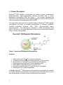
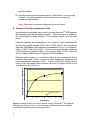
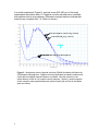
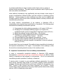
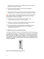
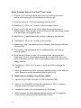
Version 08.30.2012 NanoLink™ 4-FB Magnetic Beads _____________________________________________________ User Manual Catalog# M-1001 Technical Manual I. Product Description 3 II. Applications 4 III. Immobilization of PepLink Microspheres TM A. B. C. D. E. IV. Introduction Washing NanoLink 4FB Magnetic Microspheres Materials Required Peptide Immobilization Protocol Summary Peptide Immobilization Data Immobilization of Antibodies to NanoLink 4FB Magnetic Microspheres A. B. C. D. E. F. G. H. I. J. K. 2 Peptides to NanoLink 4FB Magnetic Introduction Important Considerations Prior to Antibody Immobilization Materials Required Initial Antibody Concentration Concentrating Antibodies Buffer Exchange of Concentrated Antibody Modification of Antibody using S-HyNic Buffer Exchange of HyNic Antibody Antibody Immobilization Washing NanoLink Antibody Magnetic Microspheres Summary Antibody Immobilization Data 4 4 6 6 7 8 10 10 11 12 13 13 15 16 17 17 18 19 I. Product Description NanoLinkTM 4FB magnetic microspheres are uniform, polymer encapsulated, nanometer-sized, super-paramagnetic particles activated with 4 formyl benzamide functionalities (4FB) see Figure 1. The aromatic aldehydes are introduced to the hydrophilic amino-modified surface by reaction with SoluLink’s sulfo-succinymidly 4-formylbenzoate (Sulfo-S-4FB). The high surface area and low non-specific binding of NanoLinkTM 4FB magnetic microspheres are ideal for covalent immobilization of aromatic hydrazinemodified antibodies, peptides, DNA, RNA, oligonucleotides and/or oligosaccharides. The paramagnetic properties of these microspheres allow them to be used for the rapid separation of bound from unbound biomolecules using a suitably strong magnet. Figure 1. NanoLink 4FB Magnetic Microspheres Features: High surface area (788 + 5% nanometer diameter) Consistent aromatic aldehyde loading capacities (30-45 nmol/mg) Encapsulated (no exposed iron) monodisperse microspheres Fast magnetic response time (25 electromagnetic units) 4FB is stable in aqueous solution for months at 4oC Immobilizes approximately 120 ug IgG per mg of solid phase Paramagnetic (no residual magnetisim) % Solids: NanoLinKTM 4FB magnetic microspheres are packaged at nominally 1% solids (10mg/ml) as measured using spectroscopic analysis which is set by their optical density at 600nm against a microsphere size standard. 3 Aromatic Aldehyde (4FB) Functional Capacity: The aromatic aldehyde (4FB) surface capacity of each lot of particles is quantified by measuring the amount of unbound HyNic-peptide (OD280) left in solution versus an unmodified control microsphere after a 2 hr incubation. Refer to Certificate of Analysis for the specific 4FB functional capacity of each lot (nmol/mg). II. Applications NanoLinkTM 4FB magnetic microspheres are a new class of paramagnetic solid phase used to readily immobilize aromatic hydrazine-modified (HyNic) biomolecules. These reactive microspheres are modified with extremely stable aromatic aldehydes called 4FB that specifically react only with aromatic hydrazine moieties (HyNic) attached to the surface of any biomolecule. NanoLinkTM 4FB magnetic microspheres can be used to immobilize: HyNic-activated peptides HyNic-modified proteins (e.g. antibodies and enzymes) The main advantage of SoluLink’s HyNic/4FB chemistry is its specificity and stability. The aromatic aldehyde, 4FB surface reacts only with HyNic-modified biomolecules and is insensitive to reaction with other biological functionalities such as -NH2, -SH, -COOH, or -OH species. The 4FB microspheres exhibit long-term storage stability in aqueous solution and remain reactive to hydrazinemodified molecules for 6 months at 4oC. During reaction of HyNic-modified biomolecules to 4FB functional groups, the 4FB is immune to hydrolysis or other unwanted side-reactions. NanoLinkTM 4FB magnetic microspheres are ideal for automated, high throughput immobilization processes that use 96-well magnets to affect multiplex binding and separation of defined biomolecules. III. Immobilization of PepLinkTM Peptides to NanoLink 4FB Magnetic Microspheres A. Introduction Immobilization of a PepLinkTM peptide to a solid support is as simple as mixing SoluLink’s 4FB magnetic microspheres with a hydrazine-modified peptide (PepLinkTM) in an appropriate conjugation buffer. This process is diagrammed in Figure 2. 4 PepLink TM Linka ble Peptides O N H + N + R N H O N Incubate O H N O Bis aryl hydrazone covalent bond O N C N H O N N H H Figure 2. Diagram illustrating peptide immobilization process on NanoLinkTM 4FB magnetic microspheres. 5 B. Washing NanoLink 4FB Magnetic Microspheres Note- standard peptide immobilization protocol requires 1 mg of microspheres 1.) Re-suspend NanoLinkTM 4FB magnetic microsphere as supplied in their original container by vortexing or pipetting them vigorously to fully disperse the particles. If necessary residual aggregation can be eliminated by brief sonication. (10 min.). 2.) Transfer 1 mg of microspheres to a 1.5 ml microfuge tube. For example, 100 ul is equal to 1 mg of particles. 3.) Place the tube on a suitable magnetic rack for 2 minutes (Figure 3). 4.) Carefully and slowly pipette off the supernatant, leaving the beads undisturbed. 5.) Remove the tube from the magnetic rack and re-suspend the microspheres in 100 ul of 1x Conjugation Buffer containing 0.05% Tween20. Vortex to mix well. 6.) One milligram of NanoLink 4FB microspheres is now ready for coupling to HyNic-modified peptide (i.e. PepLink peptide) Figure 3. Standard commercially available magnetic racks suitable for washing and handling paramagnetic microspheres. On the left is a 2-tube magnetic rack and on the right is a 6-tube magnetic rack (MPC-S) both manufactured by Dynal, Inc. C. Materials Required PepLinkTM peptide (0.5 mg solid) (HyNic-activated peptide @ NH2 terminus) NanoLinkTM 4FB Magnetic Microspheres (SoluLink, Cat.# M-1001-010) Molecular grade water 1L (Ambion, Cat.# 9932 or similar) 6 10% Tween-20 Platform shaker (Titer Shaker, Lab-Line Instruments). Vortex mixer Table-top microcentrifuge Rainin Pipetman pipettes or similar (P-1000, P-200, P-10) Speed VAC (optional for long term-storage of PepLink) Magnetic tube rack (Dynal MPC-S or similar, Invitrogen Corporation) 10x Conjugation Buffer (1M NaH2PO4, 1.5M NaCl, pH 5.0) 1x Conjugation Buffer + 0.05% Tween-20 (0.1M NaH2PO4, 0.15MNaCl, 0.05% Tween-20, pH 6.0) D. Peptide Immobilization Protocol (Microsphere batch size = 1 mg) 1.) Add 50 ul molecular grade water to 0.5 mg PepLinkTM peptide (final HyNicpeptide concentration @ 10 mg/ml) and vortex until the peptide is completely re-suspended. Note-Once the peptide is dissolved @10 mg/ml, it is stable for ~1 week at 4oC. For longer-term storage and use (~3 months), store the 10 mg/ml HyNic-peptide suspension at –20oC or colder. For long-term storage (>1yr) dry the suspension under vacuum using a Speed-vac and store at –80oC as a solid. . 2.) Transfer 100 ul @ 10 mg/ml pre-washed NanoLinkTM 4FB magnetic microspheres (from step B5) into a clean 1.5 ml microfuge tube. 3.) Add 20 ul of PepLinkTM peptide (from step 1 above) to the microspheres. 4.) Place the reaction tube on a platform shaker and allow the immobilization reaction to proceed for 1 hour at room temperature. 5.) Remove the reaction tube from the shaker and place on a suitable magnetic stand for 2 minutes. 6.) Remove the supernatant carefully without disturbing the pellet. 7.) Wash the microspheres using 500 ul 1x Conjugation Buffer + 0.05% Tween-20. 8.) Place the tube on the magnetic stand for 2 minutes, and carefully remove the supernatant without disturbing the pellet. 9.) Repeat step 7 and 8 one additional time. Discard the supernatant 7 between washes. 10.) Re-suspend the final microsphere pellet in 100ul buffer of your choice @ 10mg/ml. Immobilized peptide on the solid phase is now ready for downstream applications. Note- Store the microspheres refrigerated but never freeze! E. Summary of Peptide Immobilization Data Immobilization experiments were carried out using NanoLinkTM 4FB Magnetic Microspheres and PepLink linkable peptides. These were used to determine the immobilization binding kinetics of the activated peptide with the solid phase. Duplicate reactions were assembled in 1.5 ml reaction tubes containing 500 uM PepLink linkable peptide (S-Tag, M.W. 2030) and 0.5 mg pre-washed NanoLink 4FB Magnetic Microspheres re-suspended in 100 uL 1x conjugation buffer + 0.05% Tween 20 (pH 6.0). Control reactions (duplicates) consisted of 500 uM PepLink peptide in 100 uL 1x conjugation buffer + 0.05% Tween 20 (pH 6.0) without beads or with 0.5 mg amino-modified beads. nmol peptide bound per 0.5 mg NanoLink Reactions were carried out on a platform shaker at room temperature. At the indicated time points (Figure 4) solutions were magnetically separated and bead supernatants assayed by OD280. A drop in the OD280 as the peptide bound to the solid phase was converted into nmol of PepLink bound to the solid phase vs. time (Figure 4). 25 20 15 10 5 Negative control 0 0 10 20 30 40 50 60 Time (minutes) Figure 4. Binding kinetics of a PepLink peptide (S-tag) to NanoLinKTM 4FB Magnetic Microspheres. A drop in OD280 vs. time measures immobilization kinetics. Negative control bead supernatants retain their original OD280 8 In a similar experiment (Figure 5), spectral scans (220-420 nm) of the bead supernatants were taken after 1 h. Negative controls included amino–modified microspheres and no microspheres. Extended overnight reactions indicate that reactions are complete after 1 h. (Data not shown). No microsphere control (Neg. control) controls) NanoLink -NH NH (Neg.controls) control) 2 (Neg. 2 TM NanoLink 4FB -Aldehyde Magnetic Microspheres Microspheres Figure 5. Absorbance scans of peptide solutions (500uM) incubated with NanoLink 4FB Magnetic Microspheres. Negative controls (duplicates) included incubation with similar amino-modified magnetic beads or no beads. Note the reduction in the optical density at 280 nm vs. negative control reactions. Similarly, another negative control using the same peptide sequence (without HyNic) did not bind to the beads (data not shown). 9 IV. Immobilization of Antibodies to NanoLinkTM 4FB Magnetic Microspheres A. Introduction Immobilization of an antibody to NanoLink 4FB magnetic microspheres is similar to the process of immobilizing a peptide. This process is illustrated in Figure 6. HyN ic-Antib od y O N H + N + R N H N O Incubate O H N O Bis aryl hydrazone covalent bond O N C N H N N H H Figure 6. Immobilization of HyNic-modified antibody to NanoLink 4FB Magnetic Microspheres. However, in practice antibody immobilizations are more complex because PepLinkTM peptides are small synthetic molecules that come pre-activated with HyNic and are ready for immobilization to NanoLink 4FB magnetic microspheres. 10 In contrast, antibodies are large complex proteins that come in a variety of different initial formulations. They also require modification with S-HyNic (by the end-user) before immobilization. Initial antibody formulations vary significantly and may include a wide range of protein concentrations, different buffers, and the inclusion of interfering protein additives. Carrier proteins (or other additives) are often used to stabilize and increase antibody shelf life but these unwanted additives severely interfere with immobilization procedures. For these reasons, immobilization of an antibody to NanoLink 4FB is considerably more complex than immobilization of a peptide. The following steps summarize the antibody immobilization process: 1. Concentrate the initial antibody preparation to 2.5 mg/ml (15-60 min) 2. Exchange the concentrated antibody solution (2.5 mg/ml) into modification buffer (pH 8.0) using ZebaTM desalt spin column (20 min) 3. Modify the antibody solution with S-HyNic. (90 minutes) 4. Exchange the HyNic-modified antibody into conjugation buffer (pH 6.0) using a ZebaTM Spin Column. (20 min.) 5. Pre-wash NanoLink 4FB magnetic microspheres. (5 min.) 6. Incubate HyNic-modified antibody with microspheres (2-4 h) 7. Wash NanoLink antibody magnetic microspheres to remove unbound antibody (15 min.) A great deal of time can be saved if the antibody being immobilized is already at the required concentration of 2.5 mg/ml. Concentration of the antibody is often the most time consuming (hands-on) step in the overall process. B. Important Considerations Prior to Antibody Immobilization In order to successfully immobilize antibody to NanoLink 4FB magnetic microspheres a minimum of 0.25 mg of carrier-free antibody (no BSA or other carrier) is required per half milligram of microspheres. We do not recommend immobilizing smaller quantities of antibody to NanoLink. If carrier protein is present in your particular antibody preparation, the carrier must first be removed using Protein G or Protein A affinity column purification methods. Several rapid commercial products are available for this purpose (e.g. NAbTM Protein G Spin Column from Pierce Chemical, Cat. # 89953). Protein concentrations can be determined using any suitable protein determination assay such as a BCA or Bradford protein assay. 11 C. Materials Required: ZebaTM Desalting Spin Columns (0.5 ml) (Pierce Chemical, Cat. # 89882) VIVASPIN 500 Diafiltration Spin Filter (Sartorius, Cat. # VS0112) Antibody Required: 0.25 mg (minimum) Molecular grade water 1L (Ambion, Cat. # 9932) 10% Tween-20 S-HyNic (SANH) (5 mg solid, SoluLink, Cat. # S-1002-105) Anhydrous DMF (SoluLink, Cat. # S-4001-015) NanoLink 4FB Magnetic Microspheres (1 mg) (SoluLink, Cat. # M-1001-010) Magnetic tube rack (Dynal MPC-S or similar) BCA Protein Reagents A and B (Pierce Chemical, Cat.# 23223 and 23224) Albumin Standard (2mg/ml) (Pierce Chemical, Cat. #23209) Rainin Pipetman pipettes or similar (P-1000, P-200, P-10) Other Required Buffers: 10x Modification Buffer (1M NaH2PO4, 1.5M NaCl, pH 6.8) (SoluLink 4003-005) 10x Conjugation Buffer (1M NaH2PO4, 1.5M NaCl, pH 5.0) (SoluLink 4002-015) 1x Modification Buffer (0.1M NaH2PO4, 0.15M NaCl, pH 7.3) 1x Conjugation buffer (0.1M NaH2PO4, 0.15M NaCl, pH 6.0) 1x PBS (0.1M Phosphate, 150mM NaCl, pH 7.2) D. Initial Antibody Concentration Immobilization requires a minimum mass of 0.25 mg of antibody at a concentration of 2.5 mg/ml (for each 0.5 mg NanoLink solid phase). Antibody preparations come either as solids or liquids. The instructions that follow indicate how your particular sample should be processed prior to immobilization. Solid Antibody (solid form) If the initial (carrier-free) antibody to be immobilized is lyophilized (solid form), then simply re-suspend the antibody in sufficient 1x Modification Buffer (pH 8.0) to obtain a 2.5 mg/ml solution. This protocol requires a minimum of 0.25 mg antibody per 0.5 mg of NanoLink. A volume of 100 ul @ 2.5 mg/ml is required (0.25 mg) for this immobilization protocol. If the initial antibody is at 2.5 mg/ml in a volume of 100 ul then proceed directly to the Buffer Exchange of Concentrated Antibody (section IV-F). Antibody Solution (initial concentration greater than 2.5 mg/ml) If the initial (carrier-free) antibody is already in solution at a concentration of 2.5 mg/ml or greater, transfer a volume equivalent to 2.5 mg to a 1.5 ml tube and dilute with 1x Modification Buffer pH 8.0 to obtain 100ul @ 2.5 mg/ml. This protocol requires a minimum of 0.25 mg antibody per 0.5 mg of NanoLink. 12 If the initial antibody is at 2.5 mg/ml in a volume of 100 ul then proceed directly to the Buffer Exchange of Concentrated Antibody (section IV-F). Antibody Solution (initial concentration less than 2.5 mg/ml) If the initial (carrier-free) antibody is in solution at less than 2.5 mg/ml, then the antibody solution needs to be concentrated to 2.5 mg/ml using a VIVASPINTM 500 diafiltration spin filter (section IV-E). Note- In our experience, under certain conditions antibodies can irreversibly aggregate on the surface of a VIVASPIN 500 filter membrane while they are being concentrated. Great care must be taken during concentration to avoid this irreversible aggregation of the antibody. The antibody is slowly concentrated w/ frequent and intermittent mixing of the antibody solution between centrifugation steps. Over-concentration of the antibody on the membrane surface is to be avoided. E. Concentrating Antibodies Determine the initial protein concentration of the antibody (if unknown) using BCA protein assay or other suitable assay (e.g. Bradford assay). A minimum of 0.25 mg of total protein is required before concentration can begin. To determine if sufficient total mass of antibody is available (i.e. 0.25 mg), multiply the initial volume of antibody available for immobilization (ml) by the initial antibody concentration (mg/ml) (see Eq.1). Equation 1. (mg/ml) x (ml) > 0.25 mg If the total mass of antibody available is equal to or greater than 0.25 mg but the concentration is less than 2.5 mg/ml, then proceed to concentrate the antibody. Antibody Concentration Protocol Having determined that the total mass of antibody available for immobilization is equal to or greater than 0.25 mg but at a concentration that is less than 2.5 mg/ml, the sample must be concentrated using a VIVASPIN 500 filter unit as illustrated in Figure 7. Note- Dilute antibodies need to be concentrated before HyNic modification because antibodies do not label as efficiently below 2.5 mg/ml. 13 Concentrator body Filtrate tube Figure 7. Spin filter used to concentrated dilute antibody samples. Note- VIVASPIN 500 filter devices are made to contain and process volumes of 500 ul or less. If volumes greater 500 ul need to be concentrated and processed, then multiple loading may be required. 1). Open the lid of a VIVASPIN 500 filter device. 2). Transfer the dilute antibody solution (equivalent to 0.25 mg total mass) into a VIVASPIN 500 filter device. Multiple loadings may be required. Examples: #1 If the antibody is at 1 mg/ml in a volume of 0.25 ml, then transfer 0.25 ml antibody solution into a VIVASPIN 500 filter device and concentrate to a final volume of 100 ul. #2 If the antibody solution is at 0.50 mg/ml in a volume of 1 ml, transfer 0.5 ml into a VIVASPIN 500 filter device and concentrate to a final volume of 100 ul. Note- When transferring or mixing solutions in the concentrator body, make sure never to contact or puncture the membrane in the process. Never bring a pipette tip in contact with the membrane 3). After loading, orient the VIVASPIN 500 spin filter in the centrifuge so that the volume markers on the concentrator body face toward the center of the centrifuge rotor each time. 4). Centrifuge for exactly 3 minutes @ 7,500 x g. 5). Check the volume. Using a pipette, gently and slowly pipette the antibody solution up and down 10-15 times to mix the antibody solution thoroughly within the concentrator body. Avoid foaming. Mixing avoids aggregation of 14 the antibody on the surface of the VIVASPIN membrane. After mixing, replace the cap on the concentrator. 6). Re-orient the spin filter back in the centrifuge and centrifuge for another 3 minutes @ 7,500 x g. 7). Repeat steps 5 and 6 as many times as required until the final volume in the device is 100 ul or less and the sample is concentrated to 2.5 mg/ml. Note- If the final target concentration @ 2.5 mg/ml has been exceeded then add a sufficient volume of 1x modification buffer to the sample to bring the final volume to 100 ul and 2.5 mg/ml. 8). Carefully transfer the concentrated antibody sample from the concentrator body into a new 1.5 ml microfuge tube. 9). Remove a 2 ul aliquot of the concentrated sample (~5ug) and confirm the protein concentration using a BCA protein assay. 10.) Now that the antibody is at the appropriate concentration, proceed to exchange the concentrated antibody solution into modification buffer (pH 8.0) (Section IV-F) F. Buffer Exchange of Concentrated Antibody Concentrated antibody (2.5 mg/ml) must be desalted and buffer exchanged prior to modification with S-HyNic. SoluLink highly recommends the use of ZebaTM Desalt Spin Columns (Figure 8) to desalt proteins. This product is available from Pierce Chemical (Cat. #89882) and is required by our immobilization protocol. This rapid spin column is recommended because it does not significantly dilute antibodies during buffer exchange whereas other methods significantly dilute the sample being desalted. 0.2 ml 0.5 Figure 8. ZebaTM desalt spin column (0.5 ml) 15 Buffer Exchange Using a 0.5 ml ZebaTM Spin Column 1). Prepare a (0.5 ml) ZebaTM desalt spin column by removing the bottom closure and loosening the top red cap (do not remove cap). 2). Place the column in 1.5 ml microcentrifuge collection tubes. 3). Centrifuge at 1,500 x g for 1 minute to remove storage solution. 4). Place a mark on the side of the column where the compacted resin is slanted upward. Place columns in the microfuge with the mark facing outward in all subsequent centrifugation steps. 5). Add 300 ul of 1x modification buffer (pH 8.0) to the top of the resin bed. 6). Centrifuge at 1,500 x g for 1 minute to remove buffer. 7). Repeat step 5 and 6 two additional times, discarding flow-through buffer from collection tube. 8). The column is now equilibrated with 1x modification bffer and ready for antibody loading. 9). Place the spin column in a new 1.5 ml collection tube, remove cap and slowly apply 100 µl of concentrated antibody @ 2.5 mg/ml (0.25 mg) to the center of compact resin bed. Avoid contact with the sides of the column when loading samples. 10). Centrifuge at 1,500 x g for 2 minutes to desalt and buffer exchange the sample. 11). Retain the desalted and buffer exchanged antibody solution in the collection tube. Proceed to modify the antibody with S-HyNic (IV-G). G. Modification of Antibody using S-HyNic (SANH) 1). Dissolve 5 mg S-HyNic (S-HyNic) in 1000 ul anhydrous DMF. 2). Add 2 ul of S-HyNic/DMF solution (20 equivalents) to 100 ul of desalted antibody solution (2.5 mg/ml) and mix well. 3). Incubate the reaction at room temperature for 90 minutes. 4). Buffer exchange the HyNic-labeled antibody reaction into 1x conjugation buffer (pH 6.0) using a new Zeba Desalt Spin Column. 16 H. Buffer Exchange of HyNic-Antibody Reaction 1). Prepare a ZebaTM Spin Column by removing the bottom closure and loosening the top red cap (do not remove cap). 2). Place the column in 1.5 ml microcentrifuge collection tube. 3). Centrifuge at 1,500 x g for 1 minute to remove storage solution. 4). Place a mark on the side of the column where the compacted resin is slanted upward. Place column in the microfuge with the mark facing outward in all subsequent centrifugation steps. 5). Add 300 ul of 1x conjugation buffer (pH 6.0) to the top of the resin bed. 6). Centrifuge at 1,500 x g for 1 minute to remove buffer. 7). Repeat step 5 and 6 two additional times, discarding flow-through buffer from the collection tube each time. 8). The column is now equilibrated with 1x conjugation buffer and ready for antibody loading. 9). Place the equilibrated ZebaTM column into a new 1.5 ml collection tube and remove cap. Slowly apply the entire S-HyNic modification reaction to the column. Avoid contact with the sides of the column when loading. 10). Centrifuge at 1,500 x g for 2 minutes to collect desalted and buffer exchanged HyNic-modified antibody. Retain the eluate (~100 ul). 11). Remove a 2 ul aliquot and determine the protein concentration using a BCA protein assay. 12). The antibody is now ready for immobilization on washed NanoLink 4FB magnetic microspheres. I. Antibody Immobilization 1). Transfer 50 ul @ 10mg/ml of particles to a clean 1.5 ml microfuge tube. 2). Place the 1.5 ml tube on a suitable magnetic tube rack for 2 minutes. 17 3). Carefully and slowly pipette off the supernatant, leaving the beads undisturbed. 4). Remove the tube from the magnetic rack and add 900 ul of 1x Conjugation Buffer + 0.05% Tween-20 to the beads. Microspheres are now ready for coupling to HyNic-modified antibody. 5). Add 100ul HyNic-modified antibody @ 2.5 mg/ml in 1x conjugation buffer to the beads. 6). Incubate the microspheres and antibody on a platform shaker for 2-4 h at room temperature. Note- It is important that agitation be employed to insure efficient immobilization of the antibody and to prevent the microspheres from settling during the coupling procedure. 8). After immobilization, proceed to washing the microspheres. J. Washing NanoLink Antibody Magnetic Microspheres 1). Remove the immobilization reaction from the platform shaker. 2). Place the tube on a suitable magnetic rack for 2 minutes. 3). Carefully and slowly pipette off the supernatant, leaving the beads undisturbed. 4). Add 1 ml of 1x PBS + 0.1% Tween-20 and vortex to wash the microspheres. 5). Repeat steps 2-4, two additional times to thoroughly remove any unbound antibody. 6). Place the tube on a suitable magnetic rack for 2 minutes. 7). Carefully and slowly pipette off the supernatant, leaving the beads undisturbed. 8). Re-suspend the microspheres in 0.5 ml of 1x PBS + 0.1% Tween-20. The antibody is now immobilized and the beads are at a final concentration of 1 mg/ml. 9). Immobilized antibody is now ready for downstream applications. 18 K. Summary Antibody Immobilization Data Immobilization Characteristics of NanoLink 4FB Microspheres are illustrated in Figure 9. Immobilization of HyNic-IgG vs. IgG (NanoLink 4FB Magnetic Microspheres) Antibody binding (ug/0.5mg) 70 60 50 40 30 20 10 0 HyNic-IgG IgG Input Antibody (0.5 mg) Error bars = std. deviation (triplicates) Figure 9. A typical NanoLink 4FB/antibody immobilization reaction will immobilize between 12-20% of input (mass) HyNic-IgG per 0.5 mg beads. In contrast, passive adsorption levels (using unmodified IgG) are typically 3-5 % of input IgG. Approximately 65 ug HyNic-antibody can be immobilized per half milligram of beads (0.5 mg HyNic-IgG input). The products offered here are for research use only. Any commercial application will require a license from Solulink. The Solulink Conjugation System is patented and has multiple patents pending. Please contact Solulink for information regarding licensing information. Solulink products and methods may be covered by one or more of the following United States patents Nos. 6,686,461, 6,800,728, 7,102,024, 7,173,125, 7,462,689 and other pending patent applications. Information in this manual is subject to change without notice and does not constitute a commitment on the part of Solulink, Inc. It is supplied on an “as is” basis without any warranty of any kind, either explicit or implied. Information may be changed or updated in this manual at any time. This document may not be copied, transferred, reproduced, disclosed, or duplicated, in whole or in part, without the prior written consent of Solulink, Inc. This documentation is proprietary information and protected by the copyright laws of the United States and international treaties. The manufacturer of this documentation is Solulink, Inc. 19