1
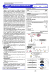
Molecular Detection│ July, 2011 (2nd Edition) ISO 9001 / 14001 Certificated Company e-MycoTM Mycoplasma PCR Detection Kit The Instruction Manual for Mycoplasma Detection Using Gene Specific Primer This kit is covered by patents owned by Abbott Molecular Inc. (US Pat. No. 5,851,767 and its foreign counterparts) (ver. 2.0 for 20 μl rxn) RUO DESCRIPTION Research Use Only REF 25235 48 25236 8 • Mycoplasma are common and serious contaminants of cell cultures. It has been shown that 30% to 87% of cell cultures are infected with mycoplasma. Many mycoplasma contaminations, particularly in continuous cell lines, grow slowly and do not destroy host cells but are still able to affect various parameters, leading to unreliable or false results. These effects include changes in metabolism, growth, viability, DNA, RNA, and protein synthesis, and morphology. Testing for mycoplasma is an essential quality control tool to assure accurate research and reliable biotechnological products. • The e-Myco™ (ver.2.0) product is a set of primers designed to detect the presence of mycoplasma that might contaminate biological materials such as cultured cells. Conventional techniques that are used to detect mycoplasma involve culturing samples on selective media, which needs at least 1 week to obtain results, whereas by performing PCR with this primer set, which is based on conserved 16S rRNA, detection results are obtained in a few hours. Because the presence of contaminant mycoplasma can be easily detected by only verifying the bands of amplified DNA fragments using electrophoresis, there is no need to prepare probes that are labeled with radioisotope, etc. You can determine the species groups of mycoplasma by sequencing analysis using the sequencing primers suggested in this manual. Furthermore, if you want to know the detailed species, you may perform PCR and sequencing from your designed primers. The adopted 8-methoxypsoralen (8-MOP) is used to extinguish the template activity of contaminated DNAs. 8-MOP is known to intercalate into double-stranded nucleic acids and form a covalent interstrand crosslink after photo-activation by incident light at wavelength 320-400 nm. An internal control of this product was constructed to identify false negative results in each reaction. The internal control was designed in such a way that the sample primer pair was used to amplify the internal control and target DNA, which were differentiated by size. Each tube of the e-Myco™ Mycoplasma PCR Detection Kit (ver.2.0) contains all the components for PCR except for template: i-StarTaqTM DNA Polymerase, dNTPs, 10x Buffer, primers, 8-MOP, and internal control for mycoplasma partial gene amplifications. So, you can just add your templates and perform the PCR reaction. CHARACTERISTICS [ Overview of Mycoplasma Detection ] • Premix Type - This e-Myco™ Mycoplasma PCR Detection Kit(ver.2.0) contains all the components for the PCR reaction. You just add template DNA or samples. • Broad Species Detection - You can detect five common cell culture-infecting species of mycoplasma and also other various species of mycoplasma (See Technical Guide). • Species Determination - You can determine the species of mycoplasma by sequencing the amplified PCR products. • Internal Control - Internal control embedded in the product prevents misjudgment that possibly arises from an erroneous PCR test. • Elimination of Carryover Contamination System - 8-MOP solution prevents carryover contamination by PCR products. Sample (culture) PROTOCOL B : Genomic DNA method PROTOCOL A : Cell boiling method APPLICATIONS • The kit is used for the detection of mycoplasma species that are most commonly encountered in cell culture, including M. arginini, M. fermentans, M. hyorhinis, M. orale, and Acholeplasma laidlawii. Furthermore, this kit can detect other various species of mycoplasma (See Technical Guide). KIT CONTENTS Label Contain e-Myco™ Mycoplasma PCR Detection Kit 48T / 8 T Control DNA (Recombinant DNA included partial 16S sequence of M. hyorhinis) DNase/RNase-free Distilled Water i-genomic CTB DNA Extraction Mini Kit (Cat.No. 17341) 95C 10min e- MycoTM Mycoplasma PCR Detection Kit (ver.2.0) 1 tube : 20 μl D.W. (as negative control) 1 tube : 20 μl D.W. (as negative control) 2 tube : 10 μl supernatant + 10ul D.W. 2 tube : 1 μl DNA + 19 ul D.W. 20 μl / 5 μl 1 ml / 0.2 ml Store at -20℃. e-Myco™ Mycoplasma PCR Detection Kit is a novel vacuum-dried premix type. This product has a stable shelf-life of a minimum of 1 year when stored at -20C and unopened container. PCR Reaction Detection on agarose gel Trademarks: iNtRON, DNA-spin™, DNA-midi™, DNA-maxi™, PCRquick-spin™, MEGA-spin™, MEGAquick-spin™, MEGA-bead™, PROBER™, G-DEX™, G-spin™, Viral Gene-spin™, easy-spin™, RNA-spin™, easy-BLUE™, easyRED™, WEST-one™, WEST-ZOL™, PRO-PREP™, SMART™, PRO-MEASURE™, Genelator™, F-Detector™, Broad-Way™, PRO-STAIN™, pLUG, Maxime™, i-Taq™, i-StarTaq™, i-MAX™, i-StarMAX™, RedSafe™, MutaDirect™, e-Myco™, M-Solution™, CENDORI™, VeTeK™, iNNOPLEX™, GxN™, teleFAXgene™, CLP™ and IQeasy™ is a trademark of iNtRON Biotehcnology, Inc. iNtRON kits are intended for research use only. Prior to using them for other purposes, the user must validate the system in compliance with the applicable law, directives, and regulations. The PCR process is covered by patents issued and applicable in certain countries. iNtRON Biotechnology, Inc. does not encourage or support the unauthorized or unlicensed use of the PCR process. Use of this product is recommended for persons that either have a license to perform PCR or are not required to obtain a license. © 2010 iNtRON, all rights reserved. UV irradiation (10 min) Discard PCR product tube [email protected] www.intronbio.com www.intron-innoplex.com Molecular Detection│ July, 2011 (2nd Edition) ISO 9001 / 14001 Certificated Company REACTION TUBE COMPONENT • PCR Reaction volume 7. Transfer an aliquot of the heated supernatant to a fresh tube. This supernatant will be used as the template in the PCR. 8. Add 10 μl of the template to each tube of e-Myco™ Mycoplasma PCR Detection Kit (ver. 2.0), and then resuspend after adding 10 μl of sterile water for a 20-μl PCR reaction volume. 9. Perform PCR reaction as in the following table. Note : We recommend that you perform one negative control reaction by adding 20 μl of sterile water. 20 μL reaction e-MycoTM Mycoplasma PCR Detection Kit (ver. 2.0) DNA Polymerase Chemical Stabilizer Loading Buffer dNTPs Tris-HCl (pH 8.3) KCl MgCl2 Mycoplasma Primers Set Internal Control 8-MOP (dissolved in DMSO) 2.5U 1x 1x 250 mM each 10 mM 50 mM 1.5 mM 35 cycles Dried under iNtRON’s instruction PROTOCOLS You can use this protocol just for detecting the contamination of mycoplasma. However, if you want to perform genotyping for the detailed determination of species, please purify the genomic DNA of suspected mycoplasma-infected cells using our i-genomic CTB DNA Extraction Mini Kit (Cat. No. 17341). You may use simply this protocol or your other general boiling methods. Use clean, disposable gloves when performing the assay and make sure that the work area is clean prior to starting the assay setup. 2. Keep your reagents and PCR mixture tubes on a cold block during reaction setup. 3. Use positive displacement pipettes. 4. The amplification and detection areas should be physically separated; i.e., do not use the same bench area to set up the PCR reactions and run your gels. [ CAUTIONS ] • DO NOT expose to UV irradiation, which activates 8-MOP, if you want to determine the detailed species of mycoplasma by DNA sequencing analysis. • If you want to do genotyping, excise the target band from the agarose gel, then isolate the DNA fragment using a gel extraction kit.(eg. MEGA-spinTM Agarose Gel DNA Exttaction, iNtRON , Cat.No 17181, MEGAquick-spinTM PCR & Agarose Gel DNA Extraction Kit, iNtRON , Cat.No 17281) PROTOCOL A : Using the genomic DNA extraction kit 1. Prepare cell suspensions from the test cell culture in a 1.5 ml tube. Then count cell numbers by general counting methods. You need at least 5x10 4 cells per test. Note 1: Harvest adherent cells with trypsin-EDTA solution using standard techniques. Pipette 1 ml of TE-treated adherent cells. Generally, with suspension cells, such as K562, you need not treat with TE solution. We recommend that you count the cells. You should prepare at least 5x10⁴ cells per test (see Technical Guide, >50,000 cells are needed to complete this protocol). Note 2: Strong mycoplasma infections are detected in as little as 20~100 cells, while weak infections require cells over 50,000 cells. You can dilute the template according to the infection rates you suspect. We recommend that you perform the PCR reaction after preparing serial dilutions of the straight supernatant to obtain optimal results. 2. Transfer the counted cells (over 5x10 4 cells) to a 1.5 ml tube. Spin the tube in a microcentrifuge for 10~15 seconds. Carefully decant the supernatant. 3. Resuspend the cells in 1 ml of sterile PBS or DPBS solution for washing. 4. Spin the tube in a microcentrifuge for 10~15 seconds. Carefully decant the supernatant. Option : Repeat this wash step once more. 5. Resuspend the cell pellets in 100 μl of sterile PBS or DPBS solution. Note : If you want the best result, use of PBS solution is better than Tris (10 mM, pH 8.5), TE (10 mM Tris, 0.1 mM EDTA), or autoclaved DW. 6. Temp. Time 94oC 1 min Denaturation 94oC 30 sec Annealing 60oC 20 sec Extension 72oC 1 min 72oC 5 min Final extension PROTOCOL B : Using genomic DNA as a template 1. Add purified genomic DNA as a template using the i-genomic CTB DNA Extraction Mini Kit (Cat.No. 17431) to each tube of e-MycoTM Mycoplasma PCR Detection Kit (ver. 2.0), and then resuspend after adding sterile water for a 20-μl PCR reaction volume Note: Appropriate amounts of DNA template sample: genomic DNA, 50 ng–100 ng 2. Follow protocol A from step 9. Note: Recommend to perform one negative control reaction by adding 20 μl of sterile water. We recommend to add 0.1 ~ 1μl of control DNA for positive control reaction [ TECHNICAL TIP ] 1. PCR Condition Initial denaturation TROUBLESHOOTING GUIDE 1. No Target band in positive reaction • Check internal control band : If internal control band is seen, PCR has been performed properly; it is not a problem of the product. • Check the quality or concentration of template : If the PCR reaction is inhibited by impurities included in DNA preparation, the use of diluted DNA as a template may be helpful. : Whereas the signals of sample control (app. 570 bp length) and internal control (app. 160 bp length) are shown, if the target band is not shown, it indicates that the sample is not infected by mycoplasma. • Check a PCR machine : The problem can be caused by the PCR machine. Please check the temperature and make sure to check that the machine is working properly. 2. No internal control band • Check template concentration : Competition can occur by using high concentrated DNA template. Please repeat the PCR with a diluted template. If the concentration of template is above 50 ng, the signal of internal control may be disappeared by competition with the template. It does not cause any problem, because the signal of sample control (app. 570 bp length) can function as a internal control. • Check the quality of template (possibility of contamination with PCR inhibitors) : If the PCR reaction is inhibited by impurities included in DNA preparation, the use of diluted DNA as a template may be helpful. If there is no internal control band, please inquire with our technical support staff. • Check the storage condition of product. 3. Presence of amplified product in the negative control • Check contamination of D.W. : D.W. can be contaminated. Perform PCR again with fresh sterile water. • Check contamination of lab instruments and other environments : We recommend that you use filter tips to reduce contamination and that you use a pipette after sterilization. All procedures should be done in sterilized conditions. 4. Poor resolution on agarose gel • We recommend to use a 1.5~2% agarose gel. • We recommend that electrophoresis is performed for 40 min at 100 V/14 cm using a 6 cm long 2% agarose gel. Heat the samples for 10 min, and vortex for 5-10 sec. Then, centrifuge for 2 min at 13,000 rpm with a tabletop centrifuge (at RT). [email protected] www.intronbio.com www.intron-innoplex.com Molecular Detection│ July, 2011 (2nd Edition) ISO 9001 / 14001 Certificated Company SPECIES DETERMINATION BY SEQUENCING ANALYSIS PRINCIPLE OF MYCOPLASMA DETECTION • The newly developed e-Myco™ Mycoplasma PCR Detection Kit (ver.2.0) is a highly sensitive PCR assay that detects various mycoplasma species that may contaminate cell culture samples. The primer sets primarily allow for detection of major mycoplasma species in cell culture contaminations (M. arginini, M. faucium, M. fermentans, M. hyorhinis, M. orale) as well as Acholeplasma laidlawii. Furthermore, you can detect various mycoplasma species with this kit (see below). It is a quick, simple, reliable, and cost-effective method for regularly monitoring cells for mycoplasma detection. • The sequence of amplified PCR products differs slightly from species to species. You can determine approximately the mycoplasma species by sequencing analysis with the following primers. Please refer to the phylogenetic table on the next page. For more detailed species analysis, you should perform additional PCR reactions with your designed primers. • The primer sets used in the kit are derived from a highly conserved region within the 16S rRNA gene and can detect very low levels of contamination. The rRNA gene sequences of prokaryotes, including mycoplasma, are well conserved, whereas the lengths and sequences of the spacer region in the rRNA operon differ from species to species. So, you can determine the species by sequencing analysis. • Sequencing primer sequences : M. species Forward 5’- GAT TAG ATA CCC TGG TAG TC-3’ (20 mer) A. laidlawii Forward 5’- GAT ACC CTG GTA GTC CAC GC-3’ (20 mer) • We list only the Forward primer sequences. Please synthesize the primer, and then analyze by general sequencing methods. [Note] The PCR primers used in this kit differ from the sequencing primers. We do not list the PCR primer sequences contained in this kit. ANALYTIC INFORMAION PARTIAL SEQUENCES OF MAJOR CONTAMINANTS IN CELL CULTURE Origin Type Table 1 shows the broad species of mycoplasma detected by this kit. As shown, this kit can detect a broad range of mycoplasma with high specificity and sensitivity. The name mycoplasma comes from the Greek words mykes (fungus) and plasma (formed) and was proposed in the 1950s. Mycoplasma is a genus of small bacteria that lack cell walls. Many species were purified and characterized from various origins such as cell culture, human, and cows. We classify them briefly by origin. Type Origin Type Origin A Cell culture L Guinea Pig B Human M Squirrels C Avian N Turkey D Porcine O Puma E Bovine P Canine F Ovine Q Primates G Equine R Lion H Murine S Monkey I Insect T Mink J Goat U Hamster K Geese V Iguana Target Primers The target regions in this kit are divided into seven M types (M1~M7) and one A type for detecting the bulk of the species in the genus mycoplasma. The designed primers are sufficient to detect major contaminants in cell cultures such as M. arginini, M. faucium, M. fermentans, M. hyorhinis, M. orale, and A. laidlawii as well as other broad species of mycoplasma. Type Designed Primer* M1 Standard M2 15CT M3 16TC M4 17CT M5 18TC M6 18TC, 20TA M7 8TC A A. laidlawii only *Not revealed primer sequences PCR Product Size Type I II III VI V VI The size of DNA fragments that are amplified by the specific primers in this kit is about 270 bp. However, the sizes of PCR product differ slightly from species to species (268 bp~277 bp). You can confirm by sequencing analysis after T/A vector cloning and other cloning methods. PCR Size 268 bp 269 bp 270 bp 271 bp 272 bp 277 bp Table 1. Mycoplasma Species Detected by e-Myco™ Kit (ver.2.0) M. species A. laidlawii M. adleri M. agalactiae M. alkalescenns M. anseris M. arginini M. arthritidis M. auris M. bovigenitalium M. bovirhinis M. bovis M. buccale M. californicum M. canadense M. caviae M. citelli M. cloacale Origin Type A/E J F E K A/B H F E E E B E E L M N Primer Type A M2 M2 M5 M5 M5 M5 M5 M2 M2 M2 M5 M2 M5 M2 M2 M5 PCR Size I III III III II III III III III IV III II II III III I II M. species M. columbinasale M. columbinum M. equirhinis M. falconis M. faucium M. felifaucium M. fermentans M. gallinarum M. gateae M. hominis M. hyorhinis M. hyosynoviae M. iguanae M. indiense M. iners M. leopharyngis M. maculosum The following sequences are partial sequences of major contaminants in general cell culture. You can classify the species by sequencing analysis. M. arginini M. faucium M. fermentans M. hyorhinis M. orale A. laidlawii 1 1 1 1 1 1 GATTAGATAC GATTAGATAC GATTAGATAC GATTAGATAC GATTAGATAC GATACCCTGG CCTGGTAGTC CCTGGTAGTC CCTGGTAGTC CCTGGTAGTC CCTGGTAGTC TAGTCCACGC cacgccgtaa cacgccgtaa cacgccctaa cacgccgtaa cacgctgtaa cgtaaacgat acgatgatca acgatgatca acgatgatca acgatgatca acgatgatca gagaactaag ttagtcggtg ttagtcggtg ttagctgatg ttagttggtg ttagtcggtg tgttggccaa M. arginini M. faucium M. fermentans M. hyorhinis M. orale A. laidlawii 61 61 61 61 61 61 gagagttcac ggagccactg gggaactcat gaataatttc gaaaactact aaggtcagtg tgacgcagct acgcagctaa cggcgcagct actaacgcag gacgcagcta ctgcagttaa aacgcattaa cgcattaaat aacgcattaa ctaacgcgtt acgcattaaa cgcattaagt atgatccgcc gatccgcctg atgatccgcc aaatgatccg tgatccgcct tctccgcctg tgagtagtat agtagtatgc tgagtagtac cctgaatagt gagtagtatg agtagtacgt M. arginini M. faucium M. fermentans M. hyorhinis M. orale A. laidlawii 101 101 101 101 101 101 gctcgcaaga tcgcaagagt gttcgcaaga atgctcgcaa ctcgcaagag acgcaagtat gtgaaactta gaaacttaaa ataaaactta gagtgaaact tgaaacttaa gaaactcaaa aaggaattga ggaattgacg aaggaattga taaaggaatt aggaattgac ggaattgacg cggggacccg gggacccgca cggggatccg gacgggaacc ggggacccgc ggaccccgca cacaagcggt caagcggtgg cacaagcggt cgcacaagcg acaagcggtg caagcggtgg M. arginini M. faucium M. fermentans M. hyorhinis M. orale A. laidlawii 151 151 151 151 151 151 ggagcatgtg agcatgtggt ggagcatgtg gtggagcatg gagcatgtgg atcatgttgt gtttaatttg ttaatttgaa gtttaatttg tggtttaatt tttaatttga ttaattcgaa aagatacgcg gatacgcgga aagatacgcg tgaagatacg agatacgcgg gatacacgaa gagaacctta gaaccttacc tagaacctta cgtagaacct agaaccttac aaaccttacc cccactcttg cactcttgac cccactcttg tacccactct ccactcttga aggtcttgac M. arginini M. faucium M. fermentans M. hyorhinis M. orale A. laidlawii 201 201 201 201 201 201 acatccttcg atcctttgca acatcttctg tgacatcttc catcccctgc atactctgca caatgctata aagctataga caaagctatg tgcaaagcta aaagctatag aagttcggag Origin Type C C G C A O A C P A A/D P V Q C R P Primer Type M2 M2 M4 M2 M1 M2 M2 M2 M5 M6 M2 M5 M4 M3 M2 M2 M2 PCR Size II III II IV I II III III III II V II III II II II II M. species M. meleagridis M. moatsii M. mustelae M. opalescens M. orale M. oxoniensis M. penetrans M. primatum M. pulmonis M. salivarium M. spermatophilum M. sualvi M. subdolum M. synoviae M. verecundum [email protected] www.intronbio.com www.intron-innoplex.com This sequences are the partial sequence of PCR products Origin Type C S T P A U B Q H A B P G C E Primer Type M2 M2 M2 M2 M3 M2 M7 M2 M5 M5 M2 M2 M5 M2 M2 PCR Size II III I III II I VI III IV II II III III I I Molecular Detection│ July, 2011 (2nd Edition) ISO 9001 / 14001 Certificated Company TECHNICAL INFORMATION EXPERIMENTAL INFORMATION • This e-Myco™ Mycoplasma PCR Detection Kit (ver.2.0) will provide a sensitive means to detect mycoplasma contamination in cell lines. Under optimal conditions, templates derived from supernatants of an infected cell culture will yield a maximum signal in the PCR reaction, whereas an uninfected cell line will yield no PCR products. Undoubtedly, there will be variations in cell numbers, infection amount, and templates that may contribute to signal differences in your experiments. • It is recommended that you use cultured cells that have cultivated for 3~6 days after subculturing as a sample for mycoplasma detection. You may not detect mycoplasma infection efficiently when you use cells that are not or shortly cultivated. • The PCR amplification efficiency varies by mycoplasma infection range. Strong mycoplasma infections are detected in as little as 10~100 cell equivalents, while weak infections require cell equivalents from the 5000~50,000 range. So, we recommend that you plan various cell numbers in preparing PCR templates from the cultured cells by using the boiling method. Please refer to Fig. 2. • If you perform genetic analysis for determining more detailed species, please extract the DNA and apply it to the PCR process. We recommend that you use our i-genomic CTB DNA Extraction Mini Kit (Cat. No. 17341). Mycoplasma Detection limit • K562 cell (M. fermentans-infected ) : small cell numbers, such as 12 cells • K562 gDNA (M. fermentans-infected ) : small quantities, such as 3.25 pg • M. fermentans : small copy numbers, such as 20 cfu/ml 1) Result for the various concentration of template DNA ½ dilution M 1 2 3 4 5 6 7 8 9 10 11 12 13 14 15 IC Mycoplasma Internal control Fig.1. Mycoplasma detection was performed for genomic DNA Genomic DNA was isolated from M. fermentans-infected K562 using the i-genomic CTB DNA Extraction Mini Kit (17341). The isolated gDNA was serially diluted for PCR of mycoplasma detection. These results show that it can be applied to mycoplasma detection with small quantities, such as 3.25 pg of gDNA Lane M, 100bp DNA Marker; lane IC, Internal control ,lane 1, 50 ng; lane 2, 25 ng; lane 3, 12.5 ng; lane 4, 6.3 ng; lane 5, 3.2 ng; lane 6, 1.6 ng; lane 7, 800 pg; lane 8, 400 pg; lane 9, 200 pg; lane 10, 100 pg; lane 11, 50 pg; lane 12, 25 pg; lane 13, 12.5 pg; lane 14, 6.3 pg; lane 15, 3.25 pg 2) Result for the various cell number PHYLOGENETIC ANALYSIS TABLE • The following phylogenetic analysis table shows the classification based on the sequence variations of PCR-amplified products. This cluster can be changed by which sequences are based on. This cluster is just a reference table. • With a suggested sequencing primer, you can approximately determine the species. For example, because the cluster between M. fermentans and M. gallinarum is different, you can simply classify the species with just sequencing analysis. However, there is no difference between M. agalactiae, M. caviae, M. fermentans, and M. opalescens. In this case, you can’t determine the detailed species with only this kit and a suggested sequencing primer. If you want to know the detailed species, you have to synthesize your own PCR primers, and then analyze by sequencing analysis. M. agalactiae M. caviae M. fermentans M. opalescens M 1 2 3 4 5 6 ½ dilution 7 8 9 10 11 12 13 14 15 IC Mycoplasma Internal control Fig.2. Mycoplasma detection was performed using the e-MycoTM Mycoplasma PCR Detection Kit (ver.2.0) method Mycoplasma detection from cell lysates of M. fermentans-infected K562 using the e-MycoTM Mycoplasma Detection Kit (ver.2.0). The M. fermentans-infected K562 cells were serially diluted for PCR of mycoplasma detection and then PCR was performed per the e-MycoTM Kit ‘s protocol. These results show that it can be applied to the mycoplasma detection with small cell numbers, such as 12 cells Lane M, 100bp DNA Marker; lane IC, Internal control; lane1, 2x10⁵; lane 2, 1x10⁵; lane 3, 5x10⁴; lane 4, 2.5x10⁴; lane 5, 1.25x10⁴; lane 6, 6.25x10³; lane 7, 3.125x10³; lane 8, 1.56x10³; lane 9, 7.8x10²; lane 10, 3.9x10²; lane 11, 1.9x10²; lane 12, 96; lane 13, 48; lane 14, 24; lane 15, 12 3) Result for Mycoplasma copy number M. gallinarum ½ dilution M. adleri M. bovigenitalium M 1 2 3 M. californicum M. phocirhinis M. verecundum M. citelli 4 5 6 7 8 9 10 11 12 13 14 15 16 Mycoplasma M. oxoniensis M. synoviae M. mustelae Internal control M. bovirhinis M. moatsii Fig.3. Mycoplasma detection was performed using the e-MycoTM Mycoplasma PCR Detection Kit (ver.2.0) method M. sualvi M. subdolum M. auris M. alkalescens M. anseris M. hominis M. cloacale M. buccale M. equirhinis M. canadense M. arginini M. gateae M. hyosynoviae M. faucium M. arthritidis Mycoplasma detection from fermentans using the e-MycoTM Mycoplasma Detection Kit. M. fermentans were serially diluted for PCR of mycoplasma detection. These results show that it can be applied to mycoplasma detection with small copy numbers, such as 20 cfu/ml Lane M, 100bp DNA Marker; lane1, 6.6x10 5 ; lane 2, 3.3x10 5 ; lane 3, 1.65x10 5 ; lane 4, 8.25x10 4 ; lane 5, 4.12x10⁴; lane 6, 2.06x10⁴; lane 7, 1.0x10⁴ lane 8, 5.1x10³; lane 9, 2.5x10³; lane 10, 1.28x10³; lane 11, 6.4x10²; lane 12, 3.22x10² lane13, 1.61x10²; lane 14, 80; lane 15, 40; lane 16, 20 Elimination of Carryover Contamination ½ dilution 1) 1st PCR 1 M 2 3 4 5 M. salivarium M. indiense M. orale M. falconis M. hyorhinis Mycoplasma Internal control M. iguanae Fig. 4. Mycoplasma detection was performed for genomic DNA Lane M, 100bp DNA Marker; lane 1, Internal control; lane 2, 25pg; lane 3, 12.5pg; lane 4, 6.3pg; lane 5, 3.25pg M. pulmonis M. penetrans M. spermatophilum w/o UV M 1 2 M. felifaucium 3 4 2) 2nd PCR w/ UV 5 6 Fig. 5. UV irradiation (10min) of 1st PCR template Lane w/o UV, without UV irradiation; Lane w/UV, with UV irradiation (10 min); lane M, 100bp DNA Marker ; lane 1, 7 8 M. maculosum M. leopharyngis M. bovis M. iners M. columbinum M. primatum M. lipofaciens M. meleagridis M. columbinasale PCR product (1 μl) used from fiq. 4. lane 2; lane 2, PCR product (1 μl) used from fiq. 4. lane 3; lane 3 , PCR product (1 μl) used from fiq.4. lane 4; lane 4, PCR product (1 μl) used from fiq. 4. lane 5; lane 5, PCR product (1 μl) used from fiq. 4. lane 2; lane 6, PCR product (1 μl) used from fiq. 4. lane 3; lane 7, PCR product (1 μl) used from fiq. 4. lane 4; lane 8, PCR product (1 μl) used from fiq. 4. Lane 5 Internal control [email protected] www.intronbio.com www.intron-innoplex.com