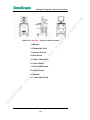
1
SSI-5000™ Diagnostic Ultrasound System rv e d. Regulatory Requirement This product complies with regulatory requirements of the re se following European directive 93/42/EEC concerning medical So no Sc ap e co nf i de n ti al , al l ri gh ts devices. P/N 4701-0063-01A BASIC Operator’s Manual 1 e Sc ap So no de n nf i co , al ti al l ts gh ri re se rv e d. SSI-5000™ Diagnostic Ultrasound System System Safety and Maintenance ........................................1-1 rv e 1 Safety Overview .................................................................... 1-1 WARNING Symbols .............................................................. 1-3 Biological Safety.................................................................... 1-4 Scanning Patients and Education.......................................... 1-5 1.4.1 Safe Scanning Guideline................................................ 1-6 1.4.2 Understanding the MI/TI Display .................................... 1-8 1.5 Environmental Requirements .............................................. 1-13 1.6 Transport and Storage Environmental Requirements.......... 1-14 1.7 Electrical Requirements....................................................... 1-14 1.8 Electrical Safety................................................................... 1-15 1.9 Transducer Maintenance ..................................................... 1-16 1.10 System Positioning/Transporting ......................................... 1-22 1.10.1 Moving the System....................................................... 1-22 1.10.2 Transporting the System .............................................. 1-23 l ri gh ts re se 1.1 1.2 1.3 1.4 ti al , Base System ......................................................................... 2-1 Application and Contraindication ........................................... 2-7 Image Modes......................................................................... 2-7 User Interface Control ........................................................... 2-8 Probes and Accessories ........................................................ 2-9 Physical Specifications ........................................................ 2-11 User Interface Function Keys Description ........................... 2-11 2.7.1 System power up ......................................................... 2-11 2.7.2 System Set-Up ............................................................. 2-12 2.7.2.1 File Manager .................................................. 2-12 2.7.2.1.1 File manager operation ........................... 2-13 2.7.2.1.2 Functions of the File manager ................ 2-14 2.7.2.2 Facility Name and/or Department Display...... 2-16 2.7.2.3 Set Date/Time ................................................ 2-17 2.7.2.4 System Information ........................................ 2-18 2.7.2.5 System Setting............................................... 2-19 2.7.2.5.1 General Setting....................................... 2-19 2.7.2.5.2 Set Printer............................................... 2-20 2.7.2.5.3 Set Calculation Menu.............................. 2-21 2.7.2.5.4 Set Measurement Method....................... 2-22 2.7.2.5.5 Load Default ........................................... 2-23 2.7.2.5.6 Exit.......................................................... 2-23 2.7.2.6 DICOM ........................................................... 2-23 2.7.2.7 Exit................................................................. 2-23 So no Sc ap e co nf i 2.1 2.2 2.3 2.4 2.5 2.6 2.7 al System Specifications.........................................................2-1 de n 2 d. TABLE OF CONTENTS P/N 4701-0063-01A BASIC Operator’s Manual I So no Sc ap e co nf i de n ti al , al l ri gh ts rv e re se 2.7.3 EXAM-Key ....................................................................2-24 2.7.4 Mode Select Key (M, B, THI, CDI, DPI, PW, CW) ........2-24 2.7.5 PATIENT Key / Patient Data Entry window ...................2-27 2.7.6 Save Key ......................................................................2-30 2.7.7 PRINT Key....................................................................2-31 2.7.8 M-Tuning Key ...............................................................2-32 2.7.9 4D Key..........................................................................2-32 2.7.10 B/M/C/D Gain Knob ......................................................2-32 2.7.11 CLR Key .......................................................................2-33 2.7.12 DEL key ........................................................................2-33 2.7.13 ARROW Key.................................................................2-33 2.7.14 BDMK Key (Body Marker Key) .....................................2-34 2.7.15 ANNOT Key..................................................................2-35 2.7.16 ZOOM key ....................................................................2-36 2.7.17 Report key ....................................................................2-36 2.7.18 DIST Key ......................................................................2-37 2.7.19 CALC Key.....................................................................2-38 2.7.20 ELLIPSE Key................................................................2-38 2.7.21 TRACE Key ..................................................................2-38 Functional Manipulation Control ..........................................2-39 2.8 2.8.1 Trackball .......................................................................2-39 2.8.2 Set Key.........................................................................2-40 2.8.3 FREEZE Key ................................................................2-40 2.8.4 UPDATE Key ................................................................2-41 2.8.5 AUDIO Volume Knob....................................................2-41 2.8.6 Time Gain Compensation (TGC) slider.........................2-41 2.8.7 MENU Key....................................................................2-42 2.8.8 QUAD DISPLAY ...........................................................2-42 2.8.9 DUAL Key.....................................................................2-43 2.8.10 L/R Key.........................................................................2-45 2.8.11 UP/DOWN Key .............................................................2-45 2.8.12 PRF Paddle ..................................................................2-45 2.8.13 BASELINE Paddle........................................................2-45 2.8.14 FOCUS Paddle.............................................................2-46 2.8.15 Depth Paddle................................................................2-46 2.9 Cine Playback Mode............................................................2-47 2.10 Soft-Menu Controls - Introduction ........................................2-48 2.10.1 Trapezoid mode............................................................2-48 2.10.2 Display format...............................................................2-48 2.10.3 Real-time triplex............................................................2-48 2.11 User Define Exam Setting ...................................................2-49 2.11.1 Activating the Command ..............................................2-49 2.11.2 Naming the User Exam Icon.........................................2-49 d. SSI-5000™ Diagnostic Ultrasound System P/N 4701-0063-01A BASIC Operator’s Manual II SSI-5000™ Diagnostic Ultrasound System 3 Delete User Defined Exam Icon ................................... 2-51 B Mode..................................................................................3-1 B-mode Operation ................................................................. 3-1 3.1.1 B GAIN ........................................................................... 3-1 3.1.2 DEPTH ........................................................................... 3-2 3.1.3 FOUCS........................................................................... 3-2 3.1.4 TGC................................................................................ 3-3 3.1.5 PANORAMIC Key (in B mode only)(Optional) ................ 3-4 3.2 B-mode Menu........................................................................ 3-6 3.2.1 FOCAL NUMBER........................................................... 3-7 3.2.2 FOCAL SPAN................................................................. 3-7 3.2.3 ECG (optional) ............................................................... 3-7 3.2.4 TISSUE CHARACTERISTICS (TC) INDEX ................... 3-9 3.2.5 DYNAMIC RANGE (DYN) ............................................ 3-10 3.2.6 GSC (also in Freeze state)........................................... 3-11 3.2.7 PERSIST...................................................................... 3-11 3.2.8 CHROMA (also in Freeze state) ................................... 3-11 3.2.9 SEC. WIDTH ................................................................ 3-11 3.2.10 SEC. POS .................................................................... 3-11 3.2.11 LINE DENSITY............................................................. 3-11 3.2.12 BIOPSY GUIDE (only for certain probes) ..................... 3-12 3.2.13 COMPOUND................................................................ 3-13 3.2.14 FREQUENCY............................................................... 3-13 3.2.15 LT←→RT (also in Freeze state) ................................... 3-13 3.2.16 IMAGE PROCESSING................................................. 3-13 3.2.17 μScan(Optional) ........................................................... 3-13 3.2.18 POWER% .................................................................... 3-13 3.2.19 TRAPEZOID (only for linear array probe)..................... 3-14 3.3 THI Mode............................................................................. 3-14 4 nf i de n ti al , al l ri gh ts re se rv e 3.1 M Mode .................................................................................4-1 M Sample-line Position.......................................................... 4-1 Activation of M-mode............................................................. 4-1 M-mode Operation ................................................................ 4-2 4.3.1 GAIN .............................................................................. 4-2 4.3.2 TGC................................................................................ 4-2 4.4 B+M mode Menu ................................................................... 4-3 4.4.1 STEER M(Optional)........................................................ 4-4 4.4.2 DISPLAY FORMAT (also in Freeze state) ...................... 4-4 4.4.3 VIDEO INVERT (M active, also in Freeze state) ............ 4-7 4.4.4 M PROCESS (M active) ................................................. 4-7 4.5 CDI+M mode Menu ............................................................... 4-7 4.5.1 BASELINE...................................................................... 4-8 4.5.2 FLOW INVERT(also in Freeze state) ............................. 4-9 Sc ap e co 4.1 4.2 4.3 So no d. 2.11.3 P/N 4701-0063-01A BASIC Operator’s Manual III SSI-5000™ Diagnostic Ultrasound System PW Sample Volume Adjustment ............................................5-1 Activation of PW/CW Mode ...................................................5-2 PW/CW Mode Operation .......................................................5-3 5.3.1 PW GAIN ........................................................................5-3 5.3.2 BASELINE ......................................................................5-3 5.3.3 FILTER ...........................................................................5-4 5.3.4 PRF ................................................................................5-4 5.3.5 HPRF..............................................................................5-4 5.4 PW/CW Mode Menu ..............................................................5-5 5.4.1 DYN................................................................................5-7 5.4.2 UP/DOWN ......................................................................5-7 5.4.3 2D-REFRESH.................................................................5-7 5.4.4 DISPLAY FORMAT (also in Freeze state) ......................5-7 5.4.5 STEER ANGLE...............................................................5-8 5.4.6 SWEEP SPEED (also in Freeze state) ...........................5-8 5.4.7 BASELINE ......................................................................5-8 5.4.8 ANGLE CORRECT.........................................................5-9 5.4.9 WALL FILTER (WF) ........................................................5-9 5.4.10 PW←→CW.....................................................................5-9 5.5 PW + B + Color Information (Triplex Mode) .........................5-10 CDI Mode ..............................................................................6-1 al 6 , al l ri gh ts re se 5.1 5.2 5.3 d. PW and CW Mode ................................................................5-1 rv e 5 CDI Sample-Box Adjustment .................................................6-1 CDI Mode Operation..............................................................6-2 6.2.1 GAIN...............................................................................6-2 6.2.2 BASELINE ......................................................................6-2 6.2.3 FOCUS ...........................................................................6-2 6.2.4 PRF ................................................................................6-2 6.2.5 DEPTH ...........................................................................6-2 6.2.6 FILTER ...........................................................................6-3 6.3 CDI Mode Menu.....................................................................6-3 6.3.1 PERSIST ........................................................................6-4 6.3.2 C MAP (also in Freeze state)..........................................6-4 6.3.3 B- REJECT (also in Freeze state ) ...............................6-4 6.3.4 FLOW INVERT (also in Freeze state).............................6-4 6.3.5 LINE DENSITY ...............................................................6-4 Sc ap e co nf i de n ti 6.1 6.2 So no 7 8 DPI Mode ..............................................................................7-1 7.1 7.2 DPI Operation ........................................................................7-1 DPI Menu...............................................................................7-1 TDI Mode(Optional)..............................................................8-1 8.1 8.2 8.3 TDI Sample-Box Adjustment..................................................8-2 TDI Operation ........................................................................8-2 TDI Menu...............................................................................8-2 P/N 4701-0063-01A BASIC Operator’s Manual IV SSI-5000™ Diagnostic Ultrasound System FRAME BY FRAME............................................................... 9-1 SWEEP SPEED (Doppler active) .......................................... 9-1 LOOP SPEED (Doppler in-active)....................................... 9-1 VIDEO INVERT (Doppler active) ........................................... 9-2 PLAY/STOP (Doppler in-active)............................................. 9-2 START................................................................................... 9-2 END....................................................................................... 9-2 rv e 9.1 9.2 9.3 9.4 9.5 9.6 9.7 10 3D/4D Mode ........................................................................10-1 Introduction.......................................................................... 10-1 10.1.1 3D Sample-Box Adjustment ......................................... 10-4 10.1.2 4D Cutoff Box/Line ....................................................... 10-4 10.1.3 Change the DEPTH...................................................... 10-6 10.2 Menu Options ...................................................................... 10-7 10.2.1 Clear ROI ..................................................................... 10-7 10.2.2 RESTORE ROI............................................................. 10-7 10.2.3 CROP........................................................................... 10-8 10.2.4 ROI MODE ................................................................... 10-8 10.2.5 HIDE ROI ..................................................................... 10-8 10.2.6 RENDER MODE .......................................................... 10-8 10.2.7 AUTO ROTATE .......................................................... 10-10 10.2.8 TRACE CUT............................................................... 10-11 10.2.9 UNDO CUT ................................................................ 10-13 10.2.10 CLIP PLANE ....................................................... 10-13 10.2.11 OPACITY OFFSET.............................................. 10-13 10.2.12 OPACITY SLOPE................................................ 10-14 10.2.13 SCAN METHOD (only in 3D) .............................. 10-14 10.2.14 Z-SCALE (only in 3D).......................................... 10-15 10.2.15 Z-ANGLE (only in 3D) ......................................... 10-15 10.2.16 SWEEP ANGLE (only in 4D) ............................... 10-16 10.2.17 CINE REVIEW (only in 4D) ................................. 10-16 10.2.18 RESCAN (only in 4D).......................................... 10-17 10.2.19 DUAL DISPLAY................................................... 10-18 10.2.20 QUAD DISPLAY.................................................. 10-19 10.2.21 FULL DISPLAY.................................................... 10-20 10.3 ROI Manipulation............................................................... 10-21 10.4 Sectional View Traversal ................................................... 10-21 10.5 Save Option....................................................................... 10-22 10.6 Saving a screenshot .......................................................... 10-23 10.7 Print ................................................................................... 10-23 So no Sc ap e co nf i de n ti al , al l ri gh ts 10.1 11 The Probe/ Transducer ......................................................11-1 11.1 11.2 d. Freeze Mode Menu Options ................................................9-1 re se 9 General Description and Specifications............................... 11-1 Operating Instructions for the Probes.................................. 11-1 P/N 4701-0063-01A BASIC Operator’s Manual V rv e re se 11.2.1 Inspection ..................................................................... 11-1 11.2.2 WARNING .................................................................... 11-2 11.2.3 Connecting and Disconnecting a Transducer ............... 11-2 11.2.4 Cleaning and Disinfecting ............................................. 11-3 11.2.5 Scanning the Patient .................................................... 11-4 11.3 Operation introduction for 6V1/6V3...................................... 11-4 11.3.1 Cleaning and Disinfecting 6V1/6V3 Probe.................... 11-4 11.3.2 Operating 6V1/6V3 probe ............................................. 11-6 d. SSI-5000™ Diagnostic Ultrasound System 12 System Maintenance .........................................................12-1 General probe cleaning and disinfecting..............................12-1 12.1.1 Cleaning .......................................................................12-1 12.1.2 Disinfecting ...................................................................12-2 12.2 Acoustic Intensity Information..............................................12-2 12.3 System Service method.......................................................12-3 12.4 Service responsibility ...........................................................12-3 12.5 Contact information of the Service Department: ..................12-3 ri gh ts 12.1 So no Sc ap e co nf i de n ti al , al l Appendix A Descriptions of the WARNING signs................ A-1 Appendix B The information of Representatives................. B-1 P/N 4701-0063-01A BASIC Operator’s Manual VI SSI-5000™ Diagnostic Ultrasound System rv e d. 1 System Safety and Maintenance re se 1.1 Safety Overview ri gh ts This section discusses measure to ensure the safety of both the operator and patient. To ensure the safety of both operator and patient, please read the relevant details in this chapter carefully before operating this system. Disregarding the WARNINGs or violation of relevant rules may result in personal injury or even loss of life for operator or patient. al l Users should observe the following preCAUTIONs: This system complies with Type BF general equipment, and the EN60601-1 standard. Please follow Section 1. “System Safety” in the user’s manual to use this system properly. z Do not modify this system in any way. Necessary modifications must be made only by the manufacturer or its designated agents. This system has been fully adjusted at the factory. Do not adjust any fixed adjustable parts. nf i z de n ti al , z co z z The power cable of the system should only be connected to a grounded power socket. Do not remove the ground cable for any reason. z Only connect this system, either electronically or mechanically, with devices that comply with the EN60601-1 standard. Recheck the leakage current and other safety performance indices of the entire system to avoid potential system damage caused by leakage from a current superposition. e Sc ap So no In the event of a malfunction, turn off the system immediately and inform the manufacturer or its designated agents. P/N 4701-0063-01A BASIC Operator’s Manual 1-1 z The system should be installed only by personnel authorized by the manufacturer. Do not attempt to install the system yourself. z Only an authorized service engineer may perform maintenance. z Only a qualified operator, or someone under qualified supervision, should use the system. z Do not use this system in the presence of flammable substances or an explosion may occur. z Do not continuously scan the same part of a patient or expose the patient to prolonged scanning. Doing so may harm the patient. z When using the system for ultrasound testing, use only qualified ultrasound gel that complies with system standards. z Do not use the switch at the back of the unit for normal shut down. Always use the power-on button in the keyboard area z Do not unplug probe when the system is in active operation. Doing so may damage the probe. Always go to EXAM screen when need to remove the probe. z To prevent from arm or neck injury, the operator should not stay at the same position for too long during patient scanning without taking break. ts gh ri l al , al ti de n nf i co Do not put liquid on top of the main unit. e z So no Sc ap ¾ The unit has build-in screen saver to avoid the tic mark on the display. It is not recommended to constantly turning on and off the unit. ¾ To dispose of this product properly, please call your local service department. P/N 4701-0063-01A BASIC Operator’s Manual 1-2 rv e The system does not incorporate any specialized protective measures in the event it is configured with high-frequency operation devices. The operator should use CAUTION in these types of applications. re se z d. SSI-5000™ Diagnostic Ultrasound System SSI-5000™ Diagnostic Ultrasound System rv e d. 1.2 WARNING Symbols re se 1 Type BF Equipment gh ts 2 Dangerous voltage ri 3 Attention al l 4 Off (power: disconnection from the main power) ti al , 5 On (power: connection to the main power) Equi-potentiality 7 Alternating current So no Sc ap e co nf i de n 6 P/N 4701-0063-01A BASIC Operator’s Manual 1-3 SSI-5000™ Diagnostic Ultrasound System re se This product, as with all diagnostic ultrasound equipment, should be used only for valid reasons and should be used both for the shortest period of time and at the lowest power settings necessary (ALARA As Low As Reasonably Achievable) to produce diagnostically acceptable images. ri Approved March 26, 1997 gh Clinical Safety Quoted from AIUM ts The AIUM offers the following guidelines: al l Diagnostic ultrasound has been in use since the late 1950s. Given its known benefits and recognized efficacy for medical diagnosis, including use during human pregnancy, the American Institute of Ultrasound in Medicine herein addresses the clinical safety of such use: nf i de n ti al , There are no confirmed biological effects on patients or instrument operators caused by exposures from present diagnostic ultrasound instruments. Although the possibility exists that such biological effects may be identified in the future, current data indicate that the benefits to patients of the prudent use of diagnostic ultrasound outweigh the risks, if any that may be present. Heating: e co Elevating tissue temperature during obstetrical examinations creates medical concerns. At the embryo development stage, the rise in temperature and the length of time exposed to heat combine to determine potential detrimental effects. Exercise CAUTION particularly during Doppler/Color exams. So no Sc ap The Thermal Index (TI) provides a statistical estimate of the potential temperature elevation (in centigrade) of tissue temperature. Three forms of TI are available: TIS, for soft tissue exposures; TIB, for instances when bone lies near the beam focus; and TIC, for the heating of bone situated close to the transducer. P/N 4701-0063-01A BASIC Operator’s Manual 1-4 rv e d. 1.3 Biological Safety SSI-5000™ Diagnostic Ultrasound System ts re se rv e Cavitation may occur when sound passes through an area that contains a cavity, such as a gas bubble or air pocket (in the lung or intestine, for example). During the process of cavitation, the sound wave may cause the bubble to contract or resonate. This oscillation may cause the bubbles to explode and damage the tissue. The Mechanical Index (MI) has been created to help users accurately evaluate the likelihood of cavitation and the related adverse effects. d. Cavitation: ri gh 1.4 Scanning Patients and Education al l The Track-3 or IEC60601-2-37 output display standard allows users to share the responsibility for the safe use of this ultrasound system. Follow these usage guidelines for safe operation: In order to maintain proper cleanliness of the transducers, always clean them between patients. z Always use a new disinfected sheath on all EV/ER probes during every exam. z Continuously move the probe, rather than staying in a single spot, to avoid elevated temperatures in one part of the patient’s body. al ti de n Move probe away from the patient when not actively scanning. nf i z , z co z Expose the patient to only the very lowest practical transmit powerlevels for the shortest possible time to achieve a satisfactory diagnosis (ALARA - As Low As Reasonably Achievable). So no Sc ap e z Understand the meaning of the TI, TIS, TIB, TIC, and MI output display, as well as the relationship between these parameters and the thermal/cavitation bioeffect to the tissue. P/N 4701-0063-01A BASIC Operator’s Manual 1-5 SSI-5000™ Diagnostic Ultrasound System re se 1. Ultrasound should only be used for medical diagnosis and only by trained medical personnel. rv e d. 1.4.1 Safe Scanning Guideline ts 2. Diagnostic ultrasound procedures should be done only by personnel fully trained in the use of the equipment, in the interpretation of the results and images, and in the safe use of ultrasound (including education as to potential hazards). ri gh 3. Operators should understand the likely influence of the machine controls, the operating mode (e.g. B-mode, color Doppler imaging or spectral Doppler) and probe frequency on thermal and cavitation hazards. al l 4. Select a low setting for each new patient. Output should only be increased during the examination if penetration is still required to achieve a satisfactory result, and after the Gain control has been moved to its maximum value. al , 5. Maintain the shortest examination time necessary to produce a useful diagnostic result. nf i de n ti 6. Do not hold the probe in a fixed position for any longer than is necessary. It should be removed from the patient whenever there is no need for real-time imaging or spectral Doppler acquisition. The freeze frame and Cine loop capabilities allow images to be reviewed and discussed without exposing the patient to continuous scanning. e co 7. Do not use endo-cavity probes if there is noticeable self heating of the probe when operating in the air. Although applicable to any probe, take particular care during trans-vaginal exams during the first eight weeks of gestation. So no Sc ap 8. Take particular care to reduce output and minimize exposure time of an embryo or fetus when the temperature of the mother is already elevated. 9. Take particular care to reduce the risk of thermal hazard during diagnostic ultrasound when exposing: an embryo less than eight weeks after gestation; or the head, brain or spine of any fetus or neonate. P/N 4701-0063-01A BASIC Operator’s Manual 1-6 rv e 10. Operators should continually monitor the on-screen thermal index (TI) and mechanical index (MI) values and use control settings that keep these settings as low as possible while still achieving diagnostically useful results. d. SSI-5000™ Diagnostic Ultrasound System ts re se In obstetric examinations, TIS (soft tissue thermal index) should be monitored during scans carried out in the first eight weeks after gestation, and TIB (bone thermal index) thereafter. In applications where the probe is very close to bone (e.g. trans-cranial applications), TIC (cranial thermal index) should be monitored. There is a possibility of minor damage to neonatal lung or intestine. If such exposure is necessary, reduce the exposure time as much as possible. MI> 0.7 There is a risk of cavitation if an ultrasound contrast agent containing gas micro-spheres is being used. There is a theoretical risk of cavitation without the presence of ultrasound contrast agents. The risk increases with MI values above this threshold. al , al l ri gh MI> 0.3 de n ti TI> 0.7 Maximum exposure time (minutes) 0.7 60 1.0 30 1.5 15 2.0 4 2.5 1 Table 2.4.1 Maximum recommended exposure times for an embryo or fetus So no Sc ap e co nf i TI The overall exposure time of an embryo or fetus should be restricted in accordance with Table 2.4.1 below as a reference: P/N 4701-0063-01A BASIC Operator’s Manual 1-7 re se For equipment of which the safety indices are displayed over their full range of values, the TI should always be less than 0.5 and the MI should always be less than 0.3. Avoid frequent repeated exposure of any subject. rv e 11. Non-diagnostic use of ultrasound equipment is not generally recommended. Examples of non-diagnostic uses of ultrasound equipment include repeated scans for operator training, equipment demonstration using normal subjects, and the production of souvenir pictures or videos of a fetus. d. SSI-5000™ Diagnostic Ultrasound System ri gh ts Scans in the first trimester of pregnancy should not be carried out for the sole purpose of producing souvenir videos or photographs, nor should their production involve increasing the exposure levels or extending the scan times beyond those needed for clinical purposes. al , al l 12. Diagnostic ultrasound has the potential for both false positive and false negative results. Misdiagnosis is far more dangerous than any effect that might result from the ultrasound exposure. Therefore, diagnostic ultrasound should be performed only by those with sufficient training and education. de n ti 1.4.2 Understanding the MI/TI Display So no Sc ap e co nf i Track-3 follows the Output Display Standard for systems which include fetal Doppler applications. The acoustic output will not be evaluated on an application-specific basis, but the global maximum de-rated Ispta must be ≤ 720 mW/cm2 and either the global maximum MI must be ≤ 1.9 or the global maximum de-rated Isppa must be ≤ 190 W/cm2. An exception is for ophthalmic use, in which case the TI = max (TIS_as, TIC) is not to exceed 1.0; Ispta.3 ≤50mW/cm2, and MI ≤ 0.23. Track-3 gives the user the freedom to increase the output acoustic power for a specific exam, and still limit output acoustic power within the global maximum de-rated Ispta ≤ 720 mW/cm2 under an Output Display Standard. For any diagnostic ultrasonic systems, Track-3 provides an Output Indices Display Standard. The diagnostic ultrasound systems and its operator’s manual contain the information regarding an ALARA (As Low As Reasonably Achievable) education program for the clinical end-user and the acoustic output indices, MI and TI. P/N 4701-0063-01A BASIC Operator’s Manual 1-8 rv e The MI describes the likelihood of cavitation, and the TI offers the predicted maximum temperature rise in tissue as a result of the diagnostic examination. d. SSI-5000™ Diagnostic Ultrasound System re se In general, a temperature increase of 2.5oC must be present consistently at one spot for 2 hours to cause fetal abnormalities. Avoiding a local temperature rise above 1oC should ensure that no thermally induced biologic effect occurs. al l ri gh ts When referring to the TI for potential thermal effect, a TI equal to 1 does not mean the temperature will rise 1 degree C. It only means an increased potential for thermal effects can be expected as the TI increases. A high index does not mean that bioeffects are occurring, but only that the potential exists and there is no consideration in the TI for the scan duration, so minimizing the overall scan time will reduce the potential for effects. These operator control and display features shift the safety responsibility from the manufacturer to the user. So it is very important to have the Ultrasound systems display the acoustic output indices correctly and the education of the user to interpret the value appropriately. , RF: De-rating factor So no Sc ap e co nf i de n ti al In Situ intensity and pressure cannot currently be measured. Therefore, the acoustic power measurement is normally done in the water tank, and when soft tissue replaces water along the ultrasound path, a decrease in intensity is expected. The fractional reduction in intensity caused by attenuation is deNOTEd by the de-rating factor (RF), RF = 10 (-0.1 a f z) Where a is the attenuation coefficient in dB cm-1 MHz-1, f is the transducer center frequency, and z is the distance along the beam axis between the source and the point of interest. De-rating factor RF for the various distances and frequencies with attenuation coefficient 0.3dB cm-1 MHz-1 in homogeneous soft tissue is listed in the following table. An example is if the user uses 7.5MHz frequency, the power will be attenuated by .0750 at 5cm, or 0.3x7.5x5=-11.25dB. The De-rated Intensity is also referred to as ‘.3’ at the end (e.g. Ispta.3). P/N 4701-0063-01A BASIC Operator’s Manual 1-9 ri gh ts re se rv e d. SSI-5000™ Diagnostic Ultrasound System Tissue Model: Sc ap e co nf i de n ti al , al l Tissue temperature elevation depends on power, tissue type, beam width, and scanning mode. Six models are developed to mimic possible clinical situations. So no Soft tissue: Describes low fat content tissue that does not contain calcifications or large gas-filled spaces. P/N 4701-0063-01A BASIC Operator’s Manual 1-10 SSI-5000™ Diagnostic Ultrasound System d. Scanned: (auto-scan): rv e Refers to the steering of successive burst through the field of view, e.g. B and color mode. re se UnScanned: Emission of ultrasonic pulses occurs along a single line of sight and is unchanged until the transducer is moved to a new position. For instance, the PW, CW and M mode. ts TI: ri TI = W.3/Wdeg gh TI is defined as the ratio of the In Situ acoustic power (W.3) to the acoustic power required to raise tissue temperature by 1oC (Wdeg), ti al , al l Three TIs corresponding to soft tissue (TIS) for abdominal; bone (TIB) for fetal and neonatal cephalic; and cranial bone (TIC) for pediatric and adult cephalic, have been developed for applications in different exams. An estimate of the acoustic power in milli-watts necessary to produce a 1oC temperature elevation in soft tissue is: Wdeg = 210/fc, de n for model 1 to 4, where fc is the center frequency in MHz. Wdeg = 40 K D nf i for model 5 and 6, where K (beam shape factor) is 1.0, D is the aperture diameter in cm at the depth of interest. Cavitation is more likely to occur at high pressures and low frequencies in pulse ultrasound wave in the tissue, which contains the bubble or air pocket (for instance, the lung, intestine, or scan with gas contrast agents). The threshold under optimum conditions of pulsed ultrasound is predicted by the ratio of the peak pressure to the square root of the frequency. So no Sc ap e co MI: P/N 4701-0063-01A BASIC Operator’s Manual 1-11 SSI-5000™ Diagnostic Ultrasound System ts rv e re se Pr’ is the de-rated (0.3) peak rare-fractional pressure in Mpa at the point where PII is the maximum, and fc is the center frequency in MHz. PII is the Pulse Intensity Integral that the total energy per unit area carried by the wave during the time duration of the pulse. The peak rare-fractional pressure is measured in hydrophone maximum negative voltage normalized by the hydrophone calibration parameter. d. MI = Pr’ / sqrt(fc) gh Display Guideline: ri For different operation modes, different indices must be displayed. However, only ti al , al l one index needs to be shown at a time. Display is not required if maximum MI is less than 1.0 for any setting of the operating mode, or if maximum TI is less than 1.0 for any setting of the operating mode. For TI, if the TIS and TIC are both greater than 1.0, the scanners need not be capable of displaying both indices simultaneously. If the index falls below 0.4, no display is needed. The display increments are no greater than 0.2 for index value less than one and no greater than 1.0 for index values greater than one (e.g. 0.4, 0.6, 0.8, 1, 2, 3). de n Display and Report in Different Mode For B-Scan Mode nf i Only display and report MI, and start from 0.4 if maximum MI > 1.0 Sc ap e co For Color Mode Only display and report TIS or TIB and start from 0.4 if maximum TI > 1.0 So no For Doppler Mode Only display and report TIS or TIB and start from 0.4 if maximum TI > 1.0 P/N 4701-0063-01A BASIC Operator’s Manual 1-12 rv e Below is a simple guideline for the user when TI exceeds one limit exposure time to 4(6-TI) minutes based on the ‘National Council on Radiation Protection. Exposure Criteria for Medical Diagnostic Ultrasound: I. Criteria Based on Thermal Mechanisms. Report No.113 1992’: d. SSI-5000™ Diagnostic Ultrasound System re se Operator Control Features: al l ri gh ts The user should be aware that certain operator controls may affect the acoustic output. It is recommended to use the default (or lowest) output power setting and compensate using Gain control to acquire an image. Other than the output power setting in the soft-menu, which has the most direct impact on the power; the PRF, image sector size, frame rate, depth, and focal position also slightly affect the output power. The default setting is normally around 70% of the allowable power depending on the exam icon. , 1.5 Environmental Requirements ti al The following environmental conditions are within system tolerances for operation: 30%~75% non-condensing de n Relative Humidity: 10℃ ~ 40℃ Atmosphere Pressure: 700~1060hPa nf i Temperature: So no Sc ap e co Strong radiation sources or powerful electromagnetic waves (e.g. electromagnetic waves from radio broadcasting) may result in image ghosting or noise. The system should be isolated from such radiation sources or electromagnetic waves. P/N 4701-0063-01A BASIC Operator’s Manual 1-13 SSI-5000™ Diagnostic Ultrasound System Requirements re se The following environmental transport and storage conditions are within system tolerances: -5℃ ~ 40℃ Relative Humidity: ≤ 80% non-condensing Pressure: 700~1060hPa ri gh ts Temperature: al l 1.7 Electrical Requirements al 100 Volts AC, 3.3 Amps , Power Requirements: 120 Volts AC, 2.7 Amps ti 230 Volts AC, 1.4 Amps de n 250 Volts AC, 1.3 Amps nf i Power Consumption: 330 watts, max co Main unit voltage e Maintain a fluctuation range of less than ±10% or the system may be damaged. Sc ap Grounding So no Before connecting the power cable, connect the attached ground protection cable to a specialized grounding device. P/N 4701-0063-01A BASIC Operator’s Manual 1-14 rv e d. 1.6 Transport and Storage Environmental NOTE: re se rv e 1. Please adhere to the outlined power requirements. Only use power lines that meet the system guidelines—failure to follow these procedures may result in system damage. d. SSI-5000™ Diagnostic Ultrasound System 2. Line power may vary in different geographic locations. Refer to the detailed ratings on the rear panel of the system for detailed information. ri gh ts 3. Use only the power button in the keyboard area to turn on or off the unit. Leave the power switch at the back of the unit on all the time unless unplug the wall outlet of the unit is needed. al l 1.8 Electrical Safety al , Only trained health professionals may operate this system. This equipment meets the following standards: ti Electrical: EN Standard 60601-1 Class IIa, Type BF, continuous operation. de n EMC/EMI: EN60601-1-2 Class A (CE) nf i Harmful liquid protection: IPX0 classification For maximum safety, adhere to these guidelines: Proper grounding of the system is critical to avoid electrical shock. For rotection, ground the chassis with a three-wire cable and plug, and plug the system into a hospital-grade, three-hole outlet. z Do not remove or circumvent the grounding wire. So no Sc ap e co z P/N 4701-0063-01A BASIC Operator’s Manual 1-15 z CAUTION needs to be taken when operate this system in the presence of flammable gases or anesthetics. z All peripheral devices (unless certified as medical grade) that are connected to the system must be powered through the electrical outlet through an optional isolation transformer. ts gh ri 1.9 Transducer Maintenance al l The transducers that come with the system are designed to be durable and dependable. These precision instruments should be inspected daily and handled with care. Please observe the following preCAUTIONs: Do not drop the transducer on hard surface. This can damage the transducer elements and compromise the electrical safety of the transducer. z Avoid kinking or pinching the transducer cable. z Use only approved ultrasonic coupling gels. z Follow the instructions for cleaning and disinfecting that come z with each probe. nf i de n ti al , z co Disinfecting Extracorporeal Transducers Disconnect the transducer from the system. e z z Wipe down all surfaces with isopropyl alcohol and air dry. z Clean all surfaces of the probe and cable with certified wipes. z Allow the transducer to air dry prior to additional handling or scanning. Sc ap So no rv e Do not remove the protective covers on the system. These covers protect users from hazardous voltages. Cabinet panels must remain in place while the system is in use. A qualified electronic technician must make all internal replacements. re se z d. SSI-5000™ Diagnostic Ultrasound System P/N 4701-0063-01A BASIC Operator’s Manual 1-16 The following statement from AIUM outlines cleaning the endocavity transducer: rv e Guidelines for Cleaning and Preparing Endocavitary Ultrasound Transducers between Patients from AIUM re se Approved June 4, 2003 ts The purpose of this document is to provide guidance regarding the cleaning and disinfection of transvaginal and transrectal ultrasound probes. al l ri gh All sterilization/disinfection represents a statistical reduction in the number of microbes present on a surface. Meticulous cleaning of the instrument is the essential key to an initial reduction of the microbial/organic load by at least 99%. This cleaning is followed by a disinfecting procedure to ensure a high degree of protection from infectious disease transmission, even if a disposable barrier covers the instrument during use. de n ti al , Medical instruments fall into different categories with respect to potential for infection transmission. The most critical level of instruments are those that are intended to penetrate skin or mucous membranes. These require sterilization. Less critical instruments (often called "semi-critical" instruments) that simply come into contact with mucous membranes such as fiber optic endoscopes require high-level disinfection rather than sterilization. covers have been observed in recent studies. For maximum safety, one should therefore perform high-level disinfection of the probe between each use and use a probe cover or condom as an aid in keeping the probe clean. There are four generally recognized categories of disinfection and sterilization. So no Sc ap e co nf i Although endocavitary ultrasound probes might be considered even less critical instruments because they are routinely protected by single use disposable probe covers, leakage rates of 0.9% - 2% for condoms and 8%-81% for commercial probe P/N 4701-0063-01A BASIC Operator’s Manual 1-17 d. SSI-5000™ Diagnostic Ultrasound System SSI-5000™ Diagnostic Ultrasound System High-Level Disinfection -Destruction/removal of all microorganisms except bacterial spores. z Mid-Level Disinfection - Inactivation of Mycobacterium Tuberculosis, bacteria, most viruses, fungi, and some bacterial spores. z Low-Level Disinfection - Destruction of most bacteria, some viruses and some fungi. Low-level disinfection will not necessarily inactivate Mycobacterium Tuberculosis or bacterial spores. ri gh ts re se z al l The following specific recommendations are made for the use of Endocavitary ultrasound transducers. de n ti al , Users should also review the Centers for Disease Control and Prevention document on sterilization and disinfection of medical devices to be certain that their procedures conform to the CDC principles for disinfection of patient care equipment. 1. CLEANING After removal of the probe cover, use running water to remove any residual gel or debris from the probe. 2) Use a damp gauze pad or other soft cloth and a small amount of mild non-abrasive liquid soap (household dishwashing liquid is ideal) to thoroughly cleanse the transducer. co nf i 1) So no Sc ap e Consider the use of a small brush especially for crevices and areas of angulation depending on the design of your particular transducer. Rinse the transducer thoroughly with running water, and then dry the transducer with a soft cloth or paper towel. P/N 4701-0063-01A BASIC Operator’s Manual 1-18 rv e Disinfection, the selective removal of microbial life, is divided into three classes: d. Sterilization is the complete elimination of all forms or microbial life including spores and viruses. SSI-5000™ Diagnostic Ultrasound System Cleaning with a detergent/water solution as described above is important as the first step in proper disinfection since chemical disinfectants act more rapidly on clean surfaces. 2) However, the additional use of a high level liquid disinfectant will ensure further statistical reduction in microbial load. Because of the potential disruption of the barrier sheath, additional high level disinfection with chemical agents is necessary. ts re se rv e 1) d. 2. DISINFECTION gh Examples of such high level disinfectants include but are not limited to: 2.4-3.2% glutaraldehyde products (a variety of available proprietary products including "Cidex," "Metricide," or "Procide"). z Non-glutaraldehyde agents including Cidex OPA (o-phthalaldehyde), Cidex PA (hydrogen peroxide & peroxyacetic acid).- 7.5% Hydrogen Peroxide solution. z Common household bleach (5.25% sodium hypochlorite) diluted to yield 500 parts per million chlorine (10 cc in one liter of tap water). This agent is effective, but generally not recommended by probe manufacturers because it can damage metal and plastic parts. de n ti al , al l ri z z Other agents such as quaternary ammonium compounds are not considered high level disinfectants and should not be used. Isopropanol is not a high level disinfectant when used as a wipe and probe manufacturers generally do not recommend soaking probes in the liquid. The FDA has published a list of approved sterilants and high level disinfectants for use in processing reusable medical and dental devices. That list can be consulted to find agents that may be useful for probe disinfection. So no Sc ap e co nf i z P/N 4701-0063-01A BASIC Operator’s Manual 1-19 SSI-5000™ Diagnostic Ultrasound System re se They should also consult instrument manufacturers regarding compatibility of these agents with probes. Many of the chemical disinfectants are potentially toxic and many require adequate preCAUTIONs such as proper ventilation, personal protective devices (gloves, face/eye protection, etc.) and thorough rinsing before reuse of the probe. ts 3. PROBE COVERS The transducer should be covered with a barrier. If the barriers used are condoms, these should be nonlubricated and nonmedicated. 2) Practitioners should be aware that condoms have been shown to be less prone to leakage than commercial probe covers, and have a six-fold enhanced AQL (acceptable quality level) when compared to standard examination gloves. They have an AQL equal to that of surgical gloves. al l ri gh 1) al , 3) Users should be aware of latex-sensitivity issues and have available nonlatex-containing barriers. ti 4. ASEPTIC TECHNIQUE For the protection of the patient and the health care worker, all endocavitary examinations should be performed with the operator properly gloved throughout the procedure. 2) Gloves should be used to remove the condom or other barrier from the transducer and to wash the transducer as outlined above. co nf i de n 1) As the barrier (condom) is removed, care should be taken not to contaminate the probe with secretions from the patient. At the completion of the procedure, hands should be thoroughly washed with soap and water. So no Sc ap e 3) d. Practitioners should consult the labels of proprietary products for specific instructions. rv e 3) P/N 4701-0063-01A BASIC Operator’s Manual 1-20 SSI-5000™ Diagnostic Ultrasound System ¾ rv e ¾ Obvious disruption in condom integrity does NOT require modification of this protocol. d. NOTE: re se These guidelines take into account possible probe contamination due to a disruption in the barrier sheath. gh ts In summary, routine high-level disinfection of the endocavitary probe between patients, plus the use of a probe cover or condom during each examination is required to properly protect patients from infection during endocavitary examinations. ri For all chemical disinfectants, preCAUTIONs must be taken to protect workers and patients from the toxicity of the disinfectant. l Reference: Amis S, Ruddy M, Kibbler CC, Economides DL, MacLean AB. Assessment of condoms as probe covers for transvaginal sonography. J Clin Ultrasound 2000; 28:295-8. z Rooks VJ, Yancey MK, Elg SA, Brueske L. Comparison of probe sheaths for endovaginal sonography. Obstet. Gynecol 1996; 87:27-9. z Milki AA, Fisch JD. Vaginal ultrasound probe cover leakage: implications for patient care. Fertil Steril 1998; 69:409-11. z Hignett M, Claman P. High rates of perforation are found in endovaginal ultrasound probe covers before and after oocyte retrieval for in vitro fertilization-embryo transfer. J Assist Reprod Genet 1995; 12:606-9. z Sterilization and Disinfection of Medical Devices: General Principles. Centers for Disease Control, Division of Healthcare Quality Promotion. http://www.cdc.gov/ncidod/hip/sterile/sterilgp.htm (5-2003). z ODE Device Evaluation Information--FDA Cleared Sterilants and High Level Disinfectants with General Claims for Processing Reusable Medical and Dental Devices, March 2003. http://www.fda.gov/cdrh/ode/germlab.html (5-2003). So no Sc ap e co nf i de n ti al , al z P/N 4701-0063-01A BASIC Operator’s Manual 1-21 SSI-5000™ Diagnostic Ultrasound System rv e re se ¾ These transducers are not designed to withstand heat sterilization methods. Exposure to temperatures in excess of 65o C (150o F) will cause permanent damage. d. CAUTION: ¾ The transducers are not designed to be totally submerged in fluid, as permanent damage will result if the entire transducer is submerged. al l 1.10 System Positioning/Transporting ri gh ts ¾ To clean the transducer, refer to the appropriate transducer operator manual. al , 1.10.1 Moving the System de n ti When moving or transporting the system, take the preCAUTIONs outlined below to ensure maximum safety for personnel, the system, and other equipment. Before moving the system: nf i 1. Completely power down the system. 2. Unplug the power cord (if the system is plugged in). co 3. Disconnect all cables from off-board peripheral devices (external printer, etc.) from the console. Sc ap e 4. To prevent damage to the power cord, DO NOT pulls excessively on the cord or sharply bends the cord while wrapping it. So no 5. Store all probes in their original cases or wrap them in soft cloth or foam to prevent damage. 6. Store gel and other essential accessories in the appropriate storage case. P/N 4701-0063-01A BASIC Operator’s Manual 1-22 CAUTION: re se rv e Always use the handle to move the system. The system weighs around 93 kg. In order to avoid physical harm or equipment damage: d. SSI-5000™ Diagnostic Ultrasound System ¾ Do not let the system strike walls or doorframe. gh ts ¾ Lift and walk slowly and carefully when moving the system. ri 1.10.2 Transporting the System al l Use extra care when transporting the system in a vehicle. After preparing the system as described above, take the following additional preCAUTIONs: al , ¾ Before transporting, place the system in its specialized storage case. ti ¾ Secure the system firmly with straps (or as directed otherwise) within the vehicle to prevent movement during transport. So no Sc ap e co nf i de n ¾ Drive carefully to prevent damage from vibration. Avoid unpaved roads, excessive speeds, and erratic stops or starts. P/N 4701-0063-01A BASIC Operator’s Manual 1-23 e Sc ap So no de n nf i co , al ti al l ts gh ri re se rv e d. SSI-5000™ Diagnostic Ultrasound System rv e d. 2 System Specifications Figure 2-1a: SSI-5000™ System Overview So no Sc ap e co nf i de n ti al , al l ri gh ts re se 2.1 Base System P/N 4701-0063-01A BASIC Operator’s Manual 2-1 ts re se rv e d. SSI-5000™ Diagnostic Ultrasound System 2. Exhaust Air Port 4. Rear Panel al 3. Inhaust Air Port l 1. Monitor ri gh Figure 2-1b: SSI-5000™ System in different views al , 5. Power Cable Hook ti 6. Castor Wheel de n 7. Probe Cable Hook 8. Probe Socket nf i 9. Speaker So no Sc ap e co 10. Foot Switch Port P/N 4701-0063-01A BASIC Operator’s Manual 2-2 SSI-5000™ Diagnostic Ultrasound System 6 re se rv e 5 d. 4 3 2 8 ri gh ts 1 12 11 10 9 al l Figure2-1c: SSI-5000™ Rear Panel 1. Grounding Pole , 2. Power Switch al 3. Line Input With Fuse Box 5. VIDEO OUT 6. S-Video I 7. S-Video II 8. ECG Port (Optional) 9. Printer Port 10. Audio Out 11. USB Port (two) 12. LAN Port(DICOM ) So no Sc ap e co nf i de n ti 4. VGA Out Port P/N 4701-0063-01A BASIC Operator’s Manual 2-3 7 SSI-5000™ Diagnostic Ultrasound System , al l ri gh ts re se Contrast and brightness controls for the LCD monitor are located at the front the monitor housing.Adjust the displayed image on the monitor using the brightness and contrast controls. rv e Adjusting the monitor’s contrast and brightness is one of the most important factors for proper image quality. The figure2-1d is the system monitor control panel d. Display Monitor Controls + _ ti al ENTER de n 3 4 1 2 Figure2-1d: Monitor Control Panel nf i Locating on the bottom of the display, the monitor adjustment buttons are described as following: co 1. LCD Light: Press the monitor power supply to turn on/off the LCD Light. e 2. ENTER: Press the ENTER key to enter the monitor adjustment, at the same time save all of adjusting value. So no Sc ap 3./4. Minus-Key/Plus-Key: Minus-Key and Plus-Key are used to control the brightness and contrast as well as adjusting an item value in display menu. P/N 4701-0063-01A BASIC Operator’s Manual 2-4 SSI-5000™ Diagnostic Ultrasound System d. Key System Features: Full digital transmit and receive beam-former ¾ Full digital demodulation and detection ¾ Wideband pulser receiver ¾ Super low noise TGC with high resolution ADC (12bits) ¾ Progressive dynamic receive focusing ¾ Progressive dynamic aperture opening ¾ Progressive dynamic apodization ¾ Tissue sound velocity compensation ¾ Broadband full digital complex demodulation for tissue and Flow ¾ Digital match filter for color Doppler processing ¾ Minimum 80GB hard drive for in-system image storage ¾ Compound imaging ¾ Tissue Harmonic imaging ¾ Trapezoidal imaging re se ts gh ri l al , al ti Real time triplex imaging Steer CW Doppler in phase array nf i ¾ de n ¾ rv e ¾ Steer M mode ¾ Extended view imaging ¾ High PRF for PW Doppler ¾ Up to 250 fps in 2D imaging ¾ Support dual and quad display format ¾ Color Doppler, color M, TDI-color, TDI-Doppler, directional Power Doppler, Power Doppler, Pulse wave Doppler, CW ¾ Advanced color and Doppler Algorithm to improve flow Sensitivity So no Sc ap e co ¾ P/N 4701-0063-01A BASIC Operator’s Manual 2-5 SSI-5000™ Diagnostic Ultrasound System Flexible hardware and firmware reconfiguration and software upgrading ¾ Reliable Linux operation system ¾ Support Multi-language ¾ Native resolution scan converter for 1024x768 high resolution LCD display ¾ Cine memory: up to 1000 frames depending on the mode and sector size ¾ Up to 10X smart Zoom ¾ Multi-Port active Probe Connection ¾ Support Linear, Curve linear, Phase array, TEE, Bi-plane, ¾ 4D motor probe ¾ Support up to 256 element probe ¾ Support temperature display and control for trans-vaginal and trans-rectum probe ¾ Cardiac, Ob/Gyn, Vascular measurement package ¾ LAN connectivity for PC base review station ¾ Digital Clips saved in system and PC format ¾ Support post measurement and processing for archive images ¾ Build in 3-lead ECG with image acquisition trigger delay Control ts gh ri l al , al ti de n nf i co ¾ Support Foot switch for print and freeze control Biopsy guide display So no Sc ap e ¾ re se rv e d. ¾ ¾ DICOM 3.0 interface with Worklist and Media Saving ¾ Freehand or real time 3D/4D imaging ¾ Real time surface and volume rendering 4D imaging ¾ SVGA, S-video and LAN port output for external image display and peripherals P/N 4701-0063-01A BASIC Operator’s Manual 2-6 SSI-5000™ Diagnostic Ultrasound System USB2.0 flash mobile drive for off-line image storage and retrieving ¾ Stereo forward/reverse Doppler audio separation ¾ Built-in Easy Network™ for direct image accessing from PC ¾ Full function unit designed for general practice and ¾ specialist clinic re se rv e d. ¾ gh ts 2.2 Application and Contraindication ri Application: 1、 Abdominal l 2、 Vascular al 3、 Breast al 5、 Obstetrical , 4、 Thyroid ti 6、 Gynecological de n 7、 Musculo-skeletal 8、 Cardiac nf i 9、 Urology co Contraindication: So no Sc ap e The system is not intended for OPHTHALMIC use or any use that causes the acoustic beam to pass through the eye. 2.3 Image Modes z 2D z 3D z 4D z Multiple screen format P/N 4701-0063-01A BASIC Operator’s Manual 2-7 Color Doppler Imaging z Doppler Power Imaging (Color Power Angio) z Directional Power Doppler z PW Doppler z Steer CW Doppler z Steer M mode z Color-M z TDI-color z TDI-Doppler z Extended view panoramic imaging z Harmonic tissue imaging z Zoom z Dual B and Real time dual color z Real time Duplex z Real time Triplex z Forward/Reverse audio separation de n ti al , al l ri gh ts re se rv e z 2D/3D/4D B gain and Doppler gain co ¾ nf i 2.4 User Interface Control ¾ So no Sc ap e ¾ d. SSI-5000™ Diagnostic Ultrasound System TGC ¾ Depth control ¾ Focal position/span ¾ Dynamic range select ¾ Tissue Harmonic image enable ¾ Audio volume control ¾ Freeze/cine P/N 4701-0063-01A BASIC Operator’s Manual 2-8 Image save ¾ Zoom ¾ Dual B and Dual Real time color ¾ QUAD display ¾ L/R ¾ Persistence ¾ PRF/HPRF ¾ Wall filter select ¾ Steering ¾ Doppler Angle correction ¾ Baseline movement ¾ Time base scrolling speed ¾ Annotation ¾ Patient data input ¾ Color ROI panning ¾ al , al l ri gh ts re se rv e d. ¾ ti SSI-5000™ Diagnostic Ultrasound System de n Image sector width and position control Calculation and measurement package ¾ File management and image archiving ¾ Clip image saving and conversion ¾ DICOM setting ¾ User defined Default Icon setting So no Sc ap e co nf i ¾ 2.5 Probes and Accessories Transducers: z 2P1 Phase Array z 5P1 Phase Array z 6V1 Micro- Curved Array P/N 4701-0063-01A BASIC Operator’s Manual 2-9 SSI-5000™ Diagnostic Ultrasound System 6V3 Micro- Curved Array z C611 Micro- Curved Array z C344 Curved Array z C542 Curved Array z C362 Curved Array z VC6-2 z LAP C9-5 z L541 Linear Array z L741 Linear Array z L742 Linear Array z L743 Linear Array z L752 Linear Array z 10L1 Linear Array z 10I2 Linear Array z BCL10-5 z 7U2 z EC9-5 z MPTEE z MPTEE mini Multi-Plane TEE phase array re se rv e d. z Curved Array al , al l ri gh ts Curved Array ti Bi-plane Curved and Linear Array Micro- Curved Array Multi-Plane TEE phase array co nf i de n Linear Array e Peripherals SVGA/SVIDEO output ¾ LAN port output for color Image and report printer ¾ LAN for DICOM and image review station ¾ USB 2.0 for flash mobile drive ¾ Foot switch So no Sc ap ¾ P/N 4701-0063-01A BASIC Operator’s Manual 2-10 SSI-5000™ Diagnostic Ultrasound System rv e d. 2.6 Physical Specifications approximately 93 kg Dimensions: Height - 1475mm, Width - 540mm, Depth - 840mm re se Weight: co nf i de n ti al , al l ri gh ts 2.7 User Interface Function Keys Description Figure 2-7a: Key Board So no Sc ap e 2.7.1 System power up z Press system power key momentarily to turn on the system power. z Press the same key momentary will turn off the system, if it is on. z Press this key longer than 4 seconds to force the system to shut down in case it locks up. P/N 4701-0063-01A BASIC Operator’s Manual 2-11 SSI-5000™ Diagnostic Ultrasound System 1. Press the <<MENU>>key on the EXAM screen to pop up the “SYSTEM CONFIGURATION” window. de n ti al , al l ri gh ts 3. Press the <<SET>>key to modify the data in the field. re se 2. Use the <<Up/Down ARROW>> keys or <<Trackball>> to move the cursor to the desired field. nf i Figure 2-7-2a: System Configuration Window co 2.7.2.1 File Manager So no Sc ap e Use this function to browse, delete, convert and copy image files stored on the storage media. Consult the File Management section of this manual for further detail. P/N 4701-0063-01A BASIC Operator’s Manual 2-12 rv e d. 2.7.2 System Set-Up mo /mnt/mo/ 0 CD/DVD Rom /mnt/cdrom/ 0 USB Derice usb /mnt/removable 0 ../general 211M re se general ri gh ts General Directory rv e Mo Dvice CD DVD Muliti Device d. SSI-5000™ Diagnostic Ultrasound System l Exit al Figure 2-7-2-1a: File Manager Window To access the file manager, press the <<MENU>>key on the EXAM screen and select the “file manager” item. ti z al , 2.7.2.1.1 File manager operation nf i de n If a patient ID has been entered, and the folder under that patient ID has files already saved, the images in the folder may be displayed on the screen. So no Sc ap e co If a patient ID is not entered, then the saved files will go under General Folder. Loading USB drive requires user to click “load/unload USB” icon or “USB device” directly. USB device can only be accessed after USB drive is ‘loaded’. Click “load/unload USB” icon to unload USB drive when finishing USB device operation, or simply exiting “USB device directory”. USB drive can only be unplugged after it is unloaded. NOTE: Due to the speed limitations of the USB flash drive, the LOAD/UNLOAD operation involving the USB device may decrease system speed. P/N 4701-0063-01A BASIC Operator’s Manual 2-13 SSI-5000™ Diagnostic Ultrasound System re se Functions within a specific patient directory Go back to patient folder ts Copy selected file. ri gh Paste copied file in the current location. al l Delete selected file. A dialog box will be popped up to confirm the deletion before the action is completed. Convert to PC Format al , Display Report Files Only: Display only reports in the current directory. ti Display Image Files Only: Display only images in the current directory. de n Export DCM to Remote SCU nf i Load/unload USB devices Multiple Selections: Select multiple files in current directory. Search: This function used to search a patient by inputting ether name or patient ID. So no Sc ap e co Deletes whole patient folder Read-in CD-Rom P/N 4701-0063-01A BASIC Operator’s Manual 2-14 rv e When entering the File manager interface,there are multiple functional icons on the top of the screen. d. 2.7.2.1.2 Functions of the File manager SSI-5000™ Diagnostic Ultrasound System rv e By default,the system saves .ppm and .cin format.This format supports post saving 2D measurement and can only be read by the system. d. ¾ Convert to PC Format re se To convert cine clip (.cin) to PC format (.wmv, MediaPlayer™ format), open patient folder and select a clip file with .cin format and click “Convert PC format”. ts This will convert system clip format CIN into PC format WMV. The selected clip will have both CIN and WMV formats available. al l ri gh When converting to the PC format, only one cine clip can be converted at a time. If multiple clips are selected, only the first clip file is converted. The conversion takes longer time for clip than for the still image. The user can plug USB drive, load it and copy the converted .wmv clip or .jpg image over, and unload USB, and take it to PC (with MediaPlayer™ ) for review. , WARNINGS: de n ti al DO NOT convert cine clips taken from other system with system software earlier than version 2.0.0.1. Unexpected result may happen ¾ Export DCM to Remote SCU So no Sc ap e co nf i Open patient folder and select image with system format (PPM). Click “Export DICOM” icon at the top of the screen. The selected image will either be exported to the remote station or to the media storage depending on the configuration set under the system menu. Please see DICOM configuration for detail settings. Pressing this key will take you back to File Management. NOTE: This option does not work with General Directory. P/N 4701-0063-01A BASIC Operator’s Manual 2-15 SSI-5000™ Diagnostic Ultrasound System SonoScape rv e re se To enter a facility Name, simply type the new facility name in text box and press the ENTER or SET-key to save the changes. The screen will be updated after exiting SYSTEM CONFIGURATION. d. 2.7.2.2 Facility Name and/or Department Display New Site ts Facility Name gh New site al , al l OK ri Help: change facility name by typing alphanumeric keys. So no Sc ap e co nf i de n ti Figure 2-7-2-2a: File Name Window P/N 4701-0063-01A BASIC Operator’s Manual 2-16 SSI-5000™ Diagnostic Ultrasound System New Site re se SonoScape rv e Adjusts current Time and Date display by entering the current Time and Date on the text line. d. 2.7.2.3 Set Date/Time 09 / 28 / 08 11:11:36 al , al l ri gh mm dd yy hh mm ss Help: change time/date by typing alphanumeric keys. CANCEL OK ts Set Time/Date So no Sc ap e co nf i de n ti Figure 2-7-2-3a: Set Date/Time Window P/N 4701-0063-01A BASIC Operator’s Manual 2-17 SSI-5000™ Diagnostic Ultrasound System New Site re se SonoScape system lnformation ri al , al l CANCEL EXIT gh ts Control Number: 200044 Software Version: 1 2.0.0.15 SEQ Rev: 1.0 TFC Rev: 1.0 DFC Rev: 0.1 SMR Rev: 0.1 FIFO Rev: 2.1 MXR Rev: 0.0 DET Rev: 0.0 DLY Rev: 0.0 EC Rev: 2.1 ECG Dev: N/A ti Figure 2-7-2-4a: System Information Window de n System Information-DIAGNO So no Sc ap e co nf i Display system configuration and runs in-system diagnostic program. This function is only available to service personnel. P/N 4701-0063-01A BASIC Operator’s Manual 2-18 rv e Displays system number, hardware, software version and Diagnostic entry. d. 2.7.2.4 System Information SSI-5000™ Diagnostic Ultrasound System al , al l ri gh ts re se rv e Pre-sets current language, calculation method, trackball sensitivity, clip saving format, screen saver on/off, and body surface Area (BSA) setting. d. 2.7.2.5 System Setting ti Figure 2-7-2-5a: System Setting Window de n 2.7.2.5.1 General Setting Language: Right now supports English and Simplified Chinese. z Screen Saver: Turn screen saver on/off, will be triggered on after idle for 5 minutes. z Trackball Sensitive: Sensitivity of the trackball movement. z Clip Format: When saving a cine clip user can either choose to save in PC format or System Format. So no Sc ap e co nf i z P/N 4701-0063-01A BASIC Operator’s Manual 2-19 SSI-5000™ Diagnostic Ultrasound System ts Date Format: Format for the date to be entered. Three types of date format are supported right now: mm/dd/yyyy, yyyy/mm/dd, dd/mm/yyyy. 2.7.2.5.2 Set Printer gh z Printer Driver: Supports HP printer driver. z Video Invert: To save printer ink, turn this option on. It will convert black background to white and white data image to black. z Insert Driver: To insert new printer driver, use this option. , New Site al SonoScape al l ri z Set Printer Set Calculation Menu Printer Driver ti Genral Setting Default HP Deskjet Vidco invert OFF Insert Driver Click de n Set Measurement Method Load Default e co nf i Exit Figure 2-7-2-5-2a: Set Printer Window So no Sc ap rv e re se Do not select PC clip format here, it will slow down the system. Instead, use system format and convert it to PC format in the File Manager. Refer to File Manager section for detail. d. NOTE: P/N 4701-0063-01A BASIC Operator’s Manual 2-20 SSI-5000™ Diagnostic Ultrasound System re se rv e This option lets you set calculation menu for 2D、PW or M mode Select the type of calculation to be used in an exam. User can select and de-select a particular method, by clicking on the box next to the name of the method. d. 2.7.2.5.3 Set Calculation Menu When there is a check in the box next to a certain method name,then this method will be included in the exam calculation. z Where the box next to the method name is empty,then this method will not be included in the exam calculation. New Site ri SonoScape gh ts z Genral Setting 2D Set Calculation Menu Load Default PW M REPORT ANGLE VOLUME VOLUME LXWXH DOPPLER AREA COLOR FLOW + VASCULAR > + OB/GYN > + LEFT VENTRIC > + UROLOGIC > Mitral valve Diam LV utflow Diam PUL.valve Diam [UPDATE]:ON/OFF [MENU]:OPEN SUBMENU Figure 2-7-2-5-3a: Set Calculation Men Window So no Sc ap e co nf i de n ti al , Exit al Set Measurement Method l Set Printer P/N 4701-0063-01A BASIC Operator’s Manual 2-21 SSI-5000™ Diagnostic Ultrasound System BPD Method: Hadlock, Jeanty z FL Method: Hadlock, Hohler, Jeanty z z CRL Method: Robinson, Hadlock, Nelson EFW Method: WEI/SAB HC,AC,FL; Shepard AC, BPD; Hadlock1 AC,FL; Hansmann AC,FL,HC; Tokyo BPD,APTD,TTD,FL; Hadlock2 HC,AC,FL; Hadlock3 BPD,AC,FL; Hadlock4 HC,AC; Hadlock5 BPD,HC,AC,FL; Shinozuka BPD,AC,FL; Warsof FL,AC; Cambell AC; z BSA Setting: Western, Eastern z Measure Method: Trace, Ellipse z Package: For each exam, this option lets the user to choose whether to be able to take all measurements or just measurements depends on the particular test the user is in. Icon Driven, the calculation menu for each exam, will list measurements only apply to that certain test. All Package, the calculation menu for each exam will list all measurements for all tests. So no Sc ap e co nf i de n ti al , al l ri gh ts re se z Figure 2-7-2-5-4a: Set Measurement Method Window P/N 4701-0063-01A BASIC Operator’s Manual 2-22 rv e This option lets you set calculation method for each of the following measurement: d. 2.7.2.5.4 Set Measurement Method SSI-5000™ Diagnostic Ultrasound System d. 2.7.2.5.5 Load Default rv e Loads all settings back to factory default. re se 2.7.2.5.6 Exit Exiting the System Setting. Take user back to the Main Menu. ts 2.7.2.6 DICOM SonoScape ri gh Pre-sets local network, SCU’s title, IP address, and port number.Consult the section on DICOM interface for detailed information. New Site l DICOM INTERFACE , al Exoport-USB Export-Remote System Remote IP 192.168.254.104 9000 Port No AE_Title start server 192.168.254.25 Wk.IP 9000 Wk.Port Wk_Title wk list server Delete Database PING ti al ezNetwork Dcfault Set DHCP Set fix IP address Static IP NetMask Gateway Printer IP 192.168.254.183 AE_Title Localsite ECHO co nf i de n Save Setting Exit Figure 2-7-2-6a: DICOM Window Returns user to the EXAM screen. So no Sc ap e 2.7.2.7 Exit P/N 4701-0063-01A BASIC Operator’s Manual 2-23 SSI-5000™ Diagnostic Ultrasound System Use trackball to move the cursor to the probe highlights the <<EXAM>> icons supported for that probe. z Use trackball again to move the cursor to the desired <<EXAM>> icon and press <<SET>>key to start the Exam in B mode. gh ts re se z l ri Each probe can display multiple EXAM icons on the screen simultaneously. al 2.7.4 Mode Select Key (M, B, THI, CDI, DPI, PW, CW) al , M-key Press the <<M>>key to enter duplex M-mode if the current mode is B. z Press the <<M>>key again to exit the M-mode and return to B mode. de n ti z co nf i For phase array probe in cardiac exam, user can choose color-M if the current mode is Color Doppler Imaging (CDI). Sc ap e When entering M-mode, the M-mode cursor appears at a default position on the 2D image. So no rv e Press this key to display all available probes connected in the system. d. 2.7.3 EXAM-Key z Move the <<Trackball>> to change the M cursor position. z Press the <<UPDATE>>key to activate a simultaneous M-mode image display. z Press the <<UPDATE>>key a second time to toggle the display back to duplex M or Color-M mode. P/N 4701-0063-01A BASIC Operator’s Manual 2-24 SSI-5000™ Diagnostic Ultrasound System Press the <<B>>key to activate B-mode imaging. rv e z d. B-key re se The system will stay in B mode if the current state is B, or return to B-mode if the current state is other than B (for example M, color, duplex Doppler, Triplex color). THI-key ts Press the <<THI>>key to toggle the on/off for the Tissue Harmonic Image mode if the icon supports it. gh z ri This function can be activated in any 2-D image mode including color and Doppler. al l CDI-key Press the <<CDI>>key to turn on the Color Doppler Imaging (CDI) mode if the system’s current state is B; and Color Triplex if the system’s current state is Duplex Doppler, and color-M if the current state is M. z Moving the<< Trackball>> will change the CROI (Color Region of Interest) position. So no Sc ap e co nf i de n ti al , z z Press the <<SET>>key to toggle the Trackball function for CROI re-sizing. z Press the <<SET>>key a second time to toggle the Trackball function back to CROI position. z Press the <<CDI>>key a second time to turn off color and return to the previous mode (either B-mode or Duplex Doppler). z In CDI-Freeze mode, press the <<CDI>>key to toggle the system onto or off of the color region of interest and view the frozen B only image. z This function also applies to cine play back mode. z Press the <<FREEZE>>key in freeze mode and return to CDI active mode, regardless of the toggle state. P/N 4701-0063-01A BASIC Operator’s Manual 2-25 SSI-5000™ Diagnostic Ultrasound System z Move the <<Trackball>> to change the CROI position. Press the <<SET>>key to toggle the <<Trackball>>for CROI re-sizing. z Press the <<SET>>key a second time to toggle the <<Trackball>>function back to the CROI position. z Press the <<DPI>>key a second time to turn off DPI and return to the previous mode (either B-mode or Duplex Doppler). ts gh ri al l PW/CW-key Press the <<PW/CW>>key to turn on the Doppler duplex mode if the current state is B, and triplex if the current state is CDI or DPI. z Press <<PW/CW>>key a second time to turn the Spectral Doppler off and return to the previous mode. de n ti al , z Sc ap e co nf i When entering the Duplex (or Triplex) Doppler mode for the first time, the Spectral Doppler is not active, but the Doppler Range Gate appears at the default position, and the B image or 2-D (either B or Color) modes are active. So no rv e Press the<<DPI>>key to turn on the Doppler Power Imaging (also called Color Power Angio CPA) mode if the system is in B mode; and DPI Triplex if the system is in Duplex Doppler. re se z d. DPI-key z Moving the <<Trackball>> will change the Range Gate (RG) position. z Press the <<SET>>key to toggle the <<Trackball>> function for RG resizing. z Press the <<SET>>key a second time to toggle the Trackball function back to the RG positioning. z Press the <<UPDATE>>key after the Range Gate is defined to activate the Spectral Doppler mode. P/N 4701-0063-01A BASIC Operator’s Manual 2-26 Press the <<UPDATE>>key a second time to toggle back to 2-D (B or Color) update and deactivate the Spectral Doppler. rv e z d. SSI-5000™ Diagnostic Ultrasound System gh ts re se When the Range Gate is moved while the Spectral Doppler is active, the system automatically activates the 2-D update until the Range Gate remains stationary for approximately 0.5 second. After that, it automatically returns to Doppler active mode. Refer to Section 3 Soft-Menu for using the auto 2-D refresh mode (which automatically refreshes 2-D images in real time Duplex or real time Triplex mode). ri NOTE: 1. The CW mode is with phase array probe only. , al l 2. If the probe supports CW,press<<PW←→CW>>menu starts CW mode.The CW control operates in the same manner as the PW,and the CW mode indicator is located next to the “Scroll Rate” display. ti Use the <<PATIENT>>key to start a new patient record, edit a current patient’s data, or select a previous patient’s exam data. de n z al 2.7.5 PATIENT Key / Patient Data Entry window Press the <<PATIENT>>key to activate the Patient Data window. NEW PATIENT—Use this option to enter new patient information. co ¾ nf i z 2. Use <<ENTER>>key or <<Trackball>> to move through all the files, I.D. (max of 15 alphanumeric characters), DOB, Name (any text up to 31 characters), ex, LMP, Age,Weight, Height, BSA,EDD,GA,Acc# (maximum of 6 digits number). So no Sc ap e 1. Click the “RESET” button in the patient Data Entry window to clear all entries. P/N 4701-0063-01A BASIC Operator’s Manual 2-27 SSI-5000™ Diagnostic Ultrasound System New Site d. SonoScape PATIENT INFORNIATION Height: Weight: Unit: kg/cm BSA: DOB: mmddyy LMP: mmddyy Age: EDD: Sex: re se GA: Acc#: Search ID: rv e Name: History Name ID Acc# ID DOB Sex Worklist RESET SAVE ri CANCEL gh ts Name al l Figure 2-7-5a:Patient Data Entry window , 3. Fill in the appropriate fields; click on “SAVE”button with <<SET>>key to save information into database. ti al Patient information will be saved in the system and scanning can be continued from the imaging screen. Click “Cancel” to abort the current entry. Any images or clips saved from now on will be in the folder under the entered patient name. nf i de n 4. WARNING So no Sc ap e co In order to ensure that the default setting of scan parameter is used, the system will exit into the exam menu when a new patient ID is entered. This prevents the unknown patient images or data from the previous scan to be saved into the new patient record. It is up to the user to overwrite this protection. P/N 4701-0063-01A BASIC Operator’s Manual 2-28 ¾ SELECT PATIENT INFORMATION FROM THE DATABASE—Enter alphanumeric characters of a patient’s name—a Use the <<Up /Down ARROW>> keys to select a patient name and then press the <<Set>>key to access that patient record (except LMP). z Edit the patient data and then move the cursor to the “OK”button and press the <<Set>>key to finish. ts re se z z gh EDIT CURRENT PATIENT—Use this method to change or edit existing patient information in the database. Press the <<PATIENT>>key to display and modify the current patient data in each field of the patient data window. ri ¾ rv e small dialog will display any matching names in the system’s patient database. al l Input a name that is different from the current name to create a new patient record. Change data within any field to replace the content in the patient record in the database. When finishing with modifications, move the cursor over the <<SAVE>> button and press the <<SET>>key to confirm the change or click the “Cancel”button to abort it. ¾ ti al , z SAVE– Returns the user to the Exam menu screen and stores the de n patient data into the system database. ¾ CANCEL – Discards any changes made by the user and restores e co nf i previous settings. CAUTION: Verify patient data for accuracy prior to saving measurements or images to create a diagnostic record in the system. Failure to do so may cause data or images to be saved to the wrong patient record. So no Sc ap d. SSI-5000™ Diagnostic Ultrasound System P/N 4701-0063-01A BASIC Operator’s Manual 2-29 SSI-5000™ Diagnostic Ultrasound System re se the <<SAVE>> key. rv e In freeze mode, the system can save a image or a clip to the system hard drive.The following dialoge will display on the screen when press d. 2.7.6 Save Key In 3D mode In 4D mode ts In 2D/Doppler mode Save a frozen image to the system hard drive for later review by pressing <<SAVE>>key. ri z gh Save Images: User can change the file name before press <<SET>>or <<ENTER>>key. , z al l The Save function names the image file with the current date/time and patient ID and saves it to the patient folder. nf i de n ti al Files are saved in the JPEG format for still images which may be displayed in the ultrasound system or PC. The saved images can be transferred to a USB mobile drive for off-line viewing on a PC, or to be converted into DICOM format and transferred to SCU or Media storage. If a patient ID is not specified, the system will save image to the General Folder. Save Clips: So no Sc ap e co To save a clip to the system, press the <<SAVE>>key and select option ‘Save Clips’ on the menu. The save name for clip can be changed by the user. It will save into patient folder in order to be review by File Manager. If a patient ID is not specified, the system will save the clip to the General Folder. Clips may be copied into an external USB mobile drive or deleted using the File Manager menu. The File Management Control allows clips to be converted and transferred to a USB 2.0 mobile drive for off-line viewing on PC with Microsoft Media Player™. P/N 4701-0063-01A BASIC Operator’s Manual 2-30 SSI-5000™ Diagnostic Ultrasound System rv e d. 2.7.7 PRINT Key re se The <<PRINT>>key sends on-screen images to a picture quality color printer attached to the system LAN port or through a hub. The Print command is valid in Freeze or File Manager modes. ts z In freeze mode, press the <<PRINT>>key to print the full screen image. ri gh In File Manager mode, only full screen images (not slide size) can be printed. Refer to the Operator Manual of the printer to select the correct paper size. ti NOTE: al , al l For any thermal printer connected to the composite (or S-Video) output, the user can press the ‘print’ key on the thermal printer at any time to print the screen. For different print size or mode, please refer to the operator’s manual comes with the thermal printer. So no Sc ap e co nf i de n The <<Save>> and <<Print>> keys are only in Cine mode after the image is frozen. P/N 4701-0063-01A BASIC Operator’s Manual 2-31 SSI-5000™ Diagnostic Ultrasound System re se During image scanning, pressing this key will optimize the image for a better quality in resolution. ts 2.7.9 4D Key ri gh The Volume Visualization, or 3D, mode allows for the scanning a volume by hand with a regular 2D probe, and the subsequent display of the volume as a three dimensional object. al l In Real-Time, or 4D, mode, a motor-driven probe is used to acquire the volume data, and the volume display is updated every fractional seconds. ti al , Both modes are accessible by pressing the 4D key while in B-mode or Freeze mode. The user may return to B-mode or Freeze mode at any time by pressing the 4D key again. See the section on 3D/4D for more details. de n NOTE: nf i Most of the menu items in 3D and 4D are the same, and provide the same functionality. co The menu items which are only available in 3D, or only in 4D, will be stated as such in their description. Sc ap e 2.7.10 B/M/C/D Gain Knob So no rv e d. 2.7.8 M-Tuning Key Turn the knob to change the overall B gain throughout the image in B mode, or M gain when M-mode is active, or the overall Doppler or Color gain in the activated mode for PW (CW) or Color (DPI) modes. P/N 4701-0063-01A BASIC Operator’s Manual 2-32 SSI-5000™ Diagnostic Ultrasound System re se If a measurement is active, this command will return the system to Cine mode. rv e Press this key to clear all texts, calipers and BODY MARK from the imaging screen in Cine mode. z d. 2.7.11 CLR Key 2.7.12 DEL key ts This key provides a number of capabilities: Delete and edit the most recent annotation and arrow. z Delete 10 pixels at a time while tracing a drawing measurement in 2-D and Doppler mode. z Edit point positions for auto points and auto trace measurements in Doppler freeze mode. 2.7.13 ARROW Key , al l ri gh z Press the <<ARROW>>key to create a new arrow pointer at the center of the image area. ti al z de n This command adds a tail to the cursor’s arrowhead when the <<Trackball>>is moved. So no Sc ap e co nf i z Press the <<LEFT>>key or <<RIGHT>>key to change the arrow direction,and press <<SET>>key to anchor the arrow. z Press <<SET>>key to return to Cine mode. z Press the <<ARROW>>key instead of the<<SET>>key to abort the previous function. Repeat the above procedures to create an additional arrow pointer. Do not confuse the up, down, left and right <<ARROW>>keys in the key pad with the <<ARROW>>key, which is for pointing in a display. Text and arrows are constrained to the Annotation Area (within the Image Area). A maximum 20 arrows can be created on a screen. P/N 4701-0063-01A BASIC Operator’s Manual 2-33 SSI-5000™ Diagnostic Ultrasound System Press the <<BDMK>>key in Cine mode to bring up the entire sets of available Body Marker Icons associated with the current EXAM. Move the <<Trackball>> to change the position and use <<LEFT/RIGHT>> key to change orientation of the Probe bar on a specific BDMK graphic, and press the<<SET>>key to anchor the probe bar. gh ts z re se The probe bar will be highlighted and will appear in the body marker area. l ri The BDMK graphic and the probe bar will be moved to the lower left corner of the screen. The system returns to Cine mode after the body marker is anchored. al Press the <<BDMK>>key again prior to anchoring the probe bar to abort the Body Marker entry and return to the Cine mode. , z 0 5 co 1.0 75 20 2/50 70 2.4 Jul-23-2008 16:08:16 de n 20 3/1 9 3/30 70 3-4.8 16.5cm C344 10 Sc ap e PRF WF GN C/P PWR FRQ New Site nf i FPS D/G GN I/P PWR FRQ D ti al To change body mark or the probe bar orientation, press the BDMK-key and follow the process for entering body marks. SonoScape 38 15 100 So no 0 rv e z d. 2.7.14 BDMK Key (Body Marker Key) P/N 4701-0063-01A BASIC Operator’s Manual 2-34 SSI-5000™ Diagnostic Ultrasound System rv e Press the <<ANNOT>>key to bring up the “|” beam cursor on the screen. Entered alphanumeric text will appear at the cursor. z Use the <<Trackball>> to relocate the beam cursor and enter alphanumeric text or annotations. z After finishing the annotation entry, press the <<SET>>key to anchor it and return to Cine mode. ts re se z ri gh Repeat the procedure to enter additional annotations. The system supports a maximum of 20 annotations and 32 characters each on the screen. To abort an annotation entry, press the <<ANNOT>>key prior to pressing the <<SET>>key. al l z New Site SonoScape C344 Jul-23-2008 16:08:16 D GAIN P.1/32 Ctrl+ GAIN 1 ABDOMINAL CIRCUMFERENCE 2 AC 3 ACA 4 ADNEXA 5 ADRENAL GLAND 6 AMNIOTIC FLUID al , 0 ti 5 de n 7 AMNIOTIC FLUID 8 ANGLE REGION 9 ANT.COMMUNICATION BR. co nf i 10 38 15 100 So no Sc ap e 0 d. 2.7.15 ANNOT Key NOTE: ¾ Press the <<ANNOT>> key in freeze or real-time to activate the annotations. ¾ Turn the<< Gain>>knob to choose the glossary;turn the<<D Gain>>knob can go to the next page. P/N 4701-0063-01A BASIC Operator’s Manual 2-35 SSI-5000™ Diagnostic Ultrasound System Use the <<Trackball>> to pan the ZROI on the 1x image, or press the <<SET>>key to toggle the Trackball function between the ZROI positioning to the ZROI size adjustment. ts ● ri gh Magnification of the original image is limited to 2cm, while maximum zoom ratio is limited to half of the original image depth. The system provides up to a 10x zoom factor with a 20 cm depth on the original image and a 2cm ZROI size. al l z After panning the ZROI on the desired spot, press the <<ZOOM>>key again to magnify full-screen ZROI based upon the new center of ZROI. al , z Press the <<ZOOM>>key again to return to the 1x image and exit ZOOM mode. co nf i de n ti In the ZOOM mode, the Trackball provides real-time image panning. The system will momentarily switch back to the original image size to show the relative position of the ZROI box for easy panning. The system can pan only within the boarder of the original image. ZOOM mode is only available in Color and B modes (including B only, B under M and Doppler mode). So no Sc ap e 2.7.17 Report key During any exam, in freeze mode, after the measurement calculations have been done. z rv e re se This key expands Zoom ROI (ZROI) over the entire image.The ZOOM function can be applied in the B, Color, CPA,and M modes. Press the <<ZOOM>>key to access the ZROI. d. 2.7.16 ZOOM key Pressing <<Report>>key, a report with the measurements will be automatically generated for you. Refer to REPORT section for more detail. P/N 4701-0063-01A BASIC Operator’s Manual 2-36 SSI-5000™ Diagnostic Ultrasound System In B Cine mode, press the <<DIST>>key to bring up the yellow plus sign (+”) cursor. z Use the <<Trackball>> and the <<SET>>key to select and lock the starting point of the desired distance to be measured. z A second yellow plus sign cursor will automatically appear.The system will update the measurement distance in real time in the box on the right side of the screen. z Press the <<SET>>key again to lock the end point of the measurement. z When two points have been defined, a green plus sign cursor will pop up. Repeat the same steps to create other distance pairs. The first pair can be modified when the maximum number of pairs is reached. z Press the<<DIST>>key again to abort the distance function and return to Cine mode. ti al , al l ri gh ts re se rv e z NOTE: e co nf i de n In Doppler and M mode, the DIST-key provides additional functionality. Please refer to Doppler and M measurement section for further detail. NOTE: Before setting the end point of the measurement, user can still go back to change the start point by pressing <<UPDATE >>key. So no Sc ap d. 2.7.18 DIST Key P/N 4701-0063-01A BASIC Operator’s Manual 2-37 SSI-5000™ Diagnostic Ultrasound System rv e re se Use this key to activate calculation packages under a different EXAM. This feature supports the optional OB/GYN, Vascular, Urology, Cardiac and General Calculation packages. Please refer to the Measurement section for further detail. d. 2.7.19 CALC Key 2.7.20 ELLIPSE Key ts The <<ELLIPSE>>key measures an ellipse area in B mode. gh Press the <<ELLIPSE>>key again to abort the ellipse area measurement and return to Cine mode. al l ri Please refer to the 2-D Measurement section for a detailed description. This key can also be used to calculate PI and RI automatically in Doppler mode.Please refer to the Doppler Measurement section for detail. 2.7.21 TRACE Key ti al , This key measures the Area/Perimeter by tracing in B mode. Please refer to the 2-D Measurement section for a detailed description. So no Sc ap e co nf i de n In Doppler mode, pressing <<TRACE>>key allows the user to trace spectra Doppler manually, and pressing <<SET>>key initializes the PI and RI calculation. Please refer to the Doppler Measurement section for detail. P/N 4701-0063-01A BASIC Operator’s Manual 2-38 2.8 Functional Manipulation Control 2.8.1 Trackball re se rv e The keyboard is designed to ergonomically arrange the Primary User Controls. Controls include: z 3. Positions “_” beam when in TEXT. z Positions the M-Mode cursor. z Selects entry in soft-menu. z Selects the EXAM icon. z Positions the PW/CW Doppler cursor and range gate. z Positions and adjusts the size of the Color Doppler Region of Interest (CROI). z Controls Digital Cine Review frames. z Positions and adjusts the size of the ZOOM Region of Interest (ZROI). So no Sc ap e co nf i de n ti al , al l ri gh ts z Positions calipers in measurement. 1. Positions 2. ‘arrow’ cursor for icon selection. z d. SSI-5000™ Diagnostic Ultrasound System P/N 4701-0063-01A BASIC Operator’s Manual 2-39 SSI-5000™ Diagnostic Ultrasound System d. 2.8.2 Set Key Located next to the Trackball. z Confirms the <<Trackball>> function for the command entry. z Confirms <<EXAM>> icon and menu setting. z Confirms caliper and measurement setting. z Switches between “RESIZING”and “REPOSITIONING”for the CROI, ZROI, and Doppler Range Gate (RG). z In 3D/4D mode, pressing this key will rotate the 3D volume along z-axis. z In 3D/4D mode, this key is also used to toggle between the four different view sections. z In 3D/4D mode, when ROI is off, pressing this key in z a certain sectional view, then moving the trackball will traverse through the view. Refer later sections for more detail. z In 3D/4D mode, when Trace Cut is on, holding down this key and moving the trackball to draw an outline for the desired section to cut out. re se ts gh ri nf i de n ti al , al l · rv e z Located on the lower right corner of the keyboard, Freeze/Un-Freeze the ultrasound image, and enter/exit the Cine mode automatically. So no Sc ap e co 2.8.3 FREEZE Key P/N 4701-0063-01A BASIC Operator’s Manual 2-40 SSI-5000™ Diagnostic Ultrasound System rv e Toggle in between Doppler and 2-D update mode. d. 2.8.4 UPDATE Key In 3D/4D mode, pressing this key will rotate the 3D volume along X and Y axis. z In 3D/4D mode, pressing this key will toggle in between ROI positioning and resizing. z In 3D/4D mode, when Clip Plane is turned on, pressing this key will toggle in between regular cursor movement and rotating the clip plane. gh ts re se z ri 2.8.5 AUDIO Volume Knob ti NOTE: al , al l Adjust the audible Doppler audio volume in Doppler mode. de n This knob is used to control the screen brightness in B mode. 2.8.6 Time Gain Compensation (TGC) slider z Move the slider bar left or right to decrease or increase B gain for the desired section in the B mode only. The TGC graphics will disappear from the screen when the slider has been inactive for two seconds. So no Sc ap e co nf i Manipulate the TGC with 8 pairs of sliders. Alter any of these sliders to display the TGC graphic on the screen. P/N 4701-0063-01A BASIC Operator’s Manual 2-41 SSI-5000™ Diagnostic Ultrasound System z Use the <<Up/Down ARROW >>keys or <<Trackball>> to select the menu items in the soft menu. z Use the << Left / Right ARROW>> keys to change menu items’ value. Use the <<SET>>key to confirm the entry. rv e Pressing the <<MENU>>key under different Mode activates the corresponding Soft-Menu dialog box for that particular mode. gh ts re se z d. 2.8.7 MENU Key ri NOTE: al l Press the <<MENU>>key to activate the Soft-Menu control at any time, in case of not finding the control key on the keyboard for the active mode. 2.8.8 QUAD DISPLAY In active B-mode,press the<<4>>key on the alphanumeric keyboard to display a frozen B mode image(at 25 percent of the original size)at the right side of the screen. z Use the <<L/R>>key three times to pop up the quad display. z Press the <<4>>key again to return the previous mode. So no Sc ap e co nf i de n ti al , z P/N 4701-0063-01A BASIC Operator’s Manual 2-42 SSI-5000™ Diagnostic Ultrasound System C344 Jul-23-2008 16:08:16 20 3/1 9 3/30 70 3-4.8 16.5cm 0 ts 0 gh FPS D/G GN I/P PWR FRQ D New Site 5 5 ri SonoScape re se rv e This key splits the imaging screen for a side-by-side image comparison. It may also be used to combine both an active and a frozen image in order to form an extended image field for viewing with a flat probe. This key also allows one image running in B mode, and the other in color in real time. d. 2.8.9 DUAL Key 10 al l 10 15 ti al , 15 de n Figure 2-8-9a: Dual display New Site SonoScape 20 3/1 9 3/30 70 3-4.8 16.5cm So no Sc ap e co nf i FPS D/G GN I/P PWR FRQ D C344 Jul-23-2008 16:08:16 15 10 5 0 0 5 10 15 Figure 2-8-9b: Dual display P/N 4701-0063-01A BASIC Operator’s Manual 2-43 SSI-5000™ Diagnostic Ultrasound System z Use the <<L/R>>key to switch between frozen/active mode between the left and right images z Use the Soft-Menu <<L/R>>button to flip the left/right orientation of the active image and create an extended viewing image for the flat probe. z Use the <<UP/DOWN>>key to flip the up/down orientation of both active image. z Press the <<Dual>>key again to return to B mode. z In summary, <<L/R>> hard key controls the active image, and the L/R soft-button sets the orientation. ts gh ri l al In active color mode(Dual Real time B+color) press the <<Dual>>key to display a frozen Color mode image (at 50 percent of the original size) at the left side of the screen and active Color mode image at the right side of the screen. z Use the <<L/R>>key to switch between frozen/active mode between the left and right images. z Use the Soft-Menu <<L/R>>button to flip the left/right orientation of the active image and create an extended viewing image for the flat probe. co nf i de n ti al , z Press the<<Dual>>key again enter the color split mode to display an active color flow or color power image at the left side of the screen and active B mode image at the right side of screen in order to view better B image under the CROI. Both images on the screen will be active. So no Sc ap e z z Use the <<L/R>>key to flip the left/right orientation of the images. z Use the <<UP/DOWN>>key to flip the up/down orientation of both active images. z Press the<<Dual>>key again to return to the normal color mode. P/N 4701-0063-01A BASIC Operator’s Manual 2-44 rv e press the <<Dual>>key to display a frozen B mode image (at 50 percent of the original size) at the left side of the screen and active B mode image at the right side of the screen. re se z d. In active B-mode(Dual B) SSI-5000™ Diagnostic Ultrasound System d. NOTE: rv e Only One image is active at a time. re se NOTE: When in DUAL mode, switch between the B and color modes by pressing the <<B>>key or<<CDI/DPI>>keys respectively ts 2.8.10 L/R Key ri gh In single image mode, use <<L/R>>key to flip the image between the left and right orientations. In dual mode, use it to set the active image for the display as described in the <<Dual>>key description. al l 2.8.11 UP/DOWN Key de n ti 2.8.12 PRF Paddle al , Reverse the 2-D (B or color) image orientation 180 degrees. nf i Use these keys to increase (UP) or decrease (DOWN) the PRF setting in the color or Spectra Doppler modes. co In CW mode, the PRF paddle changes the spectrum scale. Use these keys to control the zero velocity Baseline shifting. In Color mode, the maximum detectable velocity is stretched. In Spectra Doppler mode, the spectrum is wrapped around. So no Sc ap e 2.8.13 BASELINE Paddle P/N 4701-0063-01A BASIC Operator’s Manual 2-45 SSI-5000™ Diagnostic Ultrasound System d. 2.8.14 FOCUS Paddle ts The transmitted focal position remains at the center of the range gate in spectral Doppler mode, and at the center of the CROI in color mode. re se The small red triangle at the right of the screen next to the depth scale indicates the current focal position. rv e Use these keys to move the transmitted focal position up or down in any mode while B is active. ri gh The <<FOCUS>>keys do not affect receive focal position since it uses the progressive dynamic receive focusing at all times. l 2.8.15 Depth Paddle So no Sc ap e co nf i de n ti al , al Press these keys down to increase the image depth of view, and up to decrease the image depth of view. P/N 4701-0063-01A BASIC Operator’s Manual 2-46 2.9 Cine Playback Mode re se rv e The system does not need or have a key to activate the Cine mode. It enters the Cine mode automatically when in Freeze mode, and displays the Cine bar in the lower corner of the screen. d. SSI-5000™ Diagnostic Ultrasound System Move the <<Trackball>> left or right to review Cine frame by frame. z Move the <<Trackball>> quickly and constantly in one direction (right only) to enter a continuous loop playback mode in the Cine review. z Touch the <<Trackball>> to exit the continuous playback mode. ri gh ts z l All measurements are available on the Cine still image. Refer to the graphic box in the right side of the screen for measurement status. al In color cine play back, pressing the Color key can remove the color off the B image, and pressing it one more time to turns it back on. ti NOTE: al , Many of the measurement and calculation functions are supported in the cine mode. So no Sc ap e co nf i de n Please refer to PLAY/STOP, LOOP SPEED functionalities in the CINE REVIEW Special Function Key sections for more information. P/N 4701-0063-01A BASIC Operator’s Manual 2-47 SSI-5000™ Diagnostic Ultrasound System rv e re se The Soft-Menu is activated depending on the current active mode. The Soft-Menu will provide a second level control to set the parameters in the system. The default setting is EXAM dependent. d. 2.10 Soft-Menu Controls - Introduction Soft-Menu provides the user with an easy and flexible approach to accessing additional system controls. The system will display the appropriate menus for the selected Mode and functions. All Soft-Keys are manipulated by the 4 Arrow keys around the <<MENU>>key or the <<Trackball>>. z Use the <<Trackball>> or <<Up/Down ARROW>> keys to select the appropriate parameter, and the left/right ARROW keys to change the value of the parameter. l ri gh ts z al 2.10.1 Trapezoid mode de n 2.10.2 Display format ti al , Use Soft-Menu to turn on the Trapezoid display mode for the Linear Array probe. co nf i Use Soft-menu to configure different screen aspect ratio and display format. 2.10.3 Real-time triplex So no Sc ap e Use Soft-Menu to turn on and off the real time triplex mode. For further details review:section 5.5 PW + B + Color Information (Triplex Mode). P/N 4701-0063-01A BASIC Operator’s Manual 2-48 2.11 User Define Exam Setting re se rv e Allow the user to add a customized User <<Exam>>Icon to the EXAM screen in order to save system settings for the next usage. d. SSI-5000™ Diagnostic Ultrasound System WARNING: ts User defined Exam Settings may be deleted automatically during a system software upgrade. gh 2.11.1 Activating the Command l ri Before activating the User Setting Command, scan the image under a normal Exam Icon selection, optimize the image by adjusting the settings, and press the <<SAVE>>key in real time mode and select ‘going to Pre-set user menu’. al , al When the User Defined Exam Setting Menu appears, enter the desired name for the new user Exam Icon. This menu is only available in real time mode, and only when the probe and exam type have been defined. de n ti The system allows up to 12 Exam Icons (including the existing system exam) to be saved. When the maximum (12) has been reached, one icon must be deleted before another Exam can be saved. nf i 2.11.2 Naming the User Exam Icon So no Sc ap e co z Select “Create New Exam” from the User define exam icon menu screen. z Enter the name of the desired Exam (maximum 13 characters) and then click “OK”to create the new Exam. z Click “CANCEL” to return to the previous menu and abort the action. z Saved exams will appear as a new icon on the Exam Menu screen. P/N 4701-0063-01A BASIC Operator’s Manual 2-49 ri gh ts re se rv e d. SSI-5000™ Diagnostic Ultrasound System Figure 2-11-2b: Name for the new Exam Window So no Sc ap e co nf i de n ti al , al l Figure 2-11-2a: Create New Exam Window P/N 4701-0063-01A BASIC Operator’s Manual 2-50 SSI-5000™ Diagnostic Ultrasound System Select “Delete Exam” from the user define exam icon menu screen. z The system will display the existing user defined exams. z Select an exam icon using the <<Up and Down ARROW>> keys and click”OK” to delete the Exam. z Click “CANCEL” to return to the previous menu and abort the action. The system will ask for user confirmation at each deletion. ts re se rv e z So no Sc ap e co nf i de n ti al , al l ri gh User Exam settings are saved in the system, and can be activated through the normal Exam selection. Figure 2-11-3a: Delete Exam Window P/N 4701-0063-01A BASIC Operator’s Manual 2-51 d. 2.11.3 Delete User Defined Exam Icon e Sc ap So no de n nf i co , al ti al l ts gh ri re se rv e d. SSI-5000™ Diagnostic Ultrasound System rv e d. 3 B Mode SonoScape C344 Jul-23-2008 16:08:16 20 3/1 9 3/30 70 3-4.8 16.5cm ts 0 5 10 15 al , al l ri gh FPS D/G GN I/P PWR FRQ D New Site re se Press <<B>> key to change from another mode to B mode display. ti Figure 3a : Enter B Mode de n 3.1 B-mode Operation nf i 3.1.1 B GAIN So no Sc ap e co With the “Gain” control the overall brightness of the B image is adjusted. The adjustment of the gain control determines the amount of amplification applied to the received echoes. All incoming echoes are amplified with the same gain value regardless of depth. Rotate it to adjust the brightness of the entire image. When the GAIN control is turned clockwise the entire image becomes brighter. When the GAIN control is turned counterclockwise the entire image becomes less bright. P/N 4701-0063-01A BASIC Operator’s Manual 3-1 SSI-5000™ Diagnostic Ultrasound System ¾ The actual gain value is displayed on the screen. re se ¾ To change the B Gain is only possible with B mode on and real time mode active. 3.1.2 DEPTH ri gh ts With this function the depth range of the ultrasound image for the region of interest is adjusted. The number of image lines and the frame rate are automatically optimized. Changing of the depth is only possible in real time . al l When the Depth control is flipped downwards, the depth range of the B image is enhanced and the display size of the image is reduced to view the entire depth range. al , When the Depth control is flipped upwards, the depth range of the B image is reduced and the display size of the image is magnified. ti NOTE: The maximum and minimum depth depends on the selected probe. The actual depth in [cm] is displayed in left side of the sceen. z Freeze mode: The displayed B image is positioned again on the monitor without change in the depth range. co nf i de n z 3.1.3 FOUCS Sc ap e The selected focal zone determines the depth range of optimized sharpness of the ultrasound beam. The marker red-triangle mark the position of the focal zone(s). So no rv e d. NOTE: Flip the Depth control downwards or upwards to select the depth position of the actual focus zone The marker red-triangle mark the depth position of the focal zone. P/N 4701-0063-01A BASIC Operator’s Manual 3-2 SSI-5000™ Diagnostic Ultrasound System re se rv e The “TGC slider controls” vary the gain in certain depths of the 2D image to allow an exact compensation for the attenuation of the echoes over depth. d. 3.1.4 TGC TGC slide controls to selectively adjust the brightness in depth. ts Slide a slide control to the left to decrease the gain in the corresponding specific B depth. ri gh Slide a slide control to the right to increase the gain in the corresponding specific B depth. al l NOTE: The standard adjustment of the sliders is the middle position because of the preset time gain compensation for each scanhead. z The setting of the sliders is not stored in an user program, because of the absolute position of the sliders. z When slide the TGC slide controls,the TGC curve will appear on the right side of the image; The TGC curve will disappear automatically If you do not slid the TGC slide controls for 1~3 seconds. So no Sc ap e co nf i de n ti al , z P/N 4701-0063-01A BASIC Operator’s Manual 3-3 SSI-5000™ Diagnostic Ultrasound System rv e re se The Panoramic mode allows the operator to view a series of B-mode frames combined together, to display the entire dataset as a comprehensive, single image. This is accomplished by taking each successive frame, and determining its relative position to the previous frame, by evaluating the overlapping data. Any new data is combined, or “stitched” together with the previous data, forming a new image. d. 3.1.5 PANORAMIC Key (in B mode only)(Optional) ri gh ts The final panoramic image is displayed on the screen. Since the stitching of multiple frames may result in a panoramic image larger than the screen, a Picture-In-Picture (PIP) window is used to display a reduced version of the image. al l Inside the PIP window will be a Region of Interest (ROI) window, which represents the portion of the full panoramic image currently displayed on the screen. If the entire panoramic image can fit in the screen, the ROI will not be displayed. In freeze B-mode,press the<<P>>key on the alphanumeric keyboard,there displays ” Please wait……”on the screen, then the dialog “Press CLR to Cancel”appears on the screen, at that time ,press the <<CLR>>key can stop the initialization of the “PANORAMIC”. So no Sc ap e co nf i de n z ti al , The operator should now select the portion of the cine date to be used for Panoramic mode. This can be done by moving the trackball to display the desired starting frame. P/N 4701-0063-01A BASIC Operator’s Manual 3-4 SSI-5000™ Diagnostic Ultrasound System C344 d. Jul-23-2008 16:08:16 54 3/1 255 3/30 70 3-4.8 16.5cm 0 5 re se FPS D/G GN I/P PWR FRQ D New Site rv e SonoScape (press CLR to cancel) ts 10 38 100 ri 0 gh 15 C344 l New Site Jul-23-2008 16:08:16 al SonoScape , 0 ti al 5 nf i de n 10 38 100 Press FREEZE to cine mode Press the<<FREEZE>> key again to exit the“PANORAMIC” and return to the cine mode. So no Sc ap e co 0 15 P/N 4701-0063-01A BASIC Operator’s Manual 3-5 SSI-5000™ Diagnostic Ultrasound System Use the <<left/right ARROW>> keys to change among the predefined values for each option that’s selected. FOCAL SPAN ←→ → TC.INDEX 20 al l ECG DYN al ti CHROMA , GSC PERSIST 0 1 ←→ de n SEC.POS nf i High OFF ON co COMPOUND 2.2-4 FREQUENCY e 1 ←→ BIOSY Sc ap 2 SEC.WIDTH LINE DENSITY So no gh 4 ri FOCAL NUMBER ts The following shows the menu for the B mode, when press the <<MENU>>key.. LT←→RT OFF IMAGE PROCESS 2 μScan OFF The Trapezoid POWER% 30% available with TRAPEZOID OFF linear probes. mode is only Figure 3-2a : B Mode Menu(in real-time state) P/N 4701-0063-01A BASIC Operator’s Manual 3-6 rv e re se Activate and deactivate B-mode Soft Key controls by pressing the <<MENU>>key in B mode and using the <<Up/Down ARROW >>keys or <<Trackball>> to choose the Menu items. d. 3.2 B-mode Menu CHROMA 1 LT←→RT ←→ PLAY/STOP ∣→ LOOP SPEED ←→ START ∣← re se 1 gh ts GSC rv e The following shows the Soft-key Control menu for the display in B mode when press the <<FREEZE>>key. d. SSI-5000™ Diagnostic Ultrasound System →∣ ri END FRAME BY FRAME v1/2… 3.2.1 FOCAL NUMBER al l Figure 3-2b : B Mode Menu(in Freeze state) de n ti al , Press <<left/right ARROW>>key to increase or decrease the number of the transmitted focal zone over the depth. A red small triangle on the depth scale at each focal zone indicates the area of transmit that has been co nf i 3.2.2 FOCAL SPAN Press <<left/right ARROW>>key to set the focal span over the number of currently defined focal zones. So no Sc ap e 3.2.3 ECG (Optional) Available only when the ECG module is installed. Allow the user to set up ECG trace gain, position, trigger delay and inversion. Refer ECG Control for more detail. The ECG module is an optional device which provides the 3 lead ECG signal acquisition for cardiac application. P/N 4701-0063-01A BASIC Operator’s Manual 3-7 rv e re se In the cardiac application, the ECG trace is display on the lower portion of the screen. For echo-stress or Cardiac 4D, the R-wave triggering is used to gate or synchronize the image acquisition. The user can define time delay after R-wave. The ECG has three leads; LL (left leg, RED), LA (left arm, BLACK), RA (right arm, WHITE). LL is for reference, which usually provides a bias voltage from the ECG module, and the RA, LA are the two signals from the body and going to the differential input of the ECG isolation amplifier. d. SSI-5000™ Diagnostic Ultrasound System ts NOTE: ri gh The ECG module is only used for the 3 lead ECG signal acquisition and display,and can not replace ECG monitor/Electrocardiograph. de n ti al , al l The ECG control is in the soft-menu available for the cardiac probe, it allows the user to set up the following control: nf i MAIN MENU: Return to main submenu ECG ON/OFF: Turn on/off the ECG trace co ECG GAIN: Increase or decrease the ECG gain ECG POSITION: Set the ECG trace position e ECG INVERT: Invert ECG trace So no Sc ap R-TRIGGER: This will turn on the ECG detection for R peak in the PQRST wave. P/N 4701-0063-01A BASIC Operator’s Manual 3-8 TRIGGER DELAY: This sets a delay to R-Trigger, so the image that will be captured is the image after a user specified time delay after the R peak. FRAME COUNT: Number of images to be captured after the R peak. FRAME INTERVAL: Sets the interval for each frame. z DELAY: R wave to 2D update delay (for Echo stress or Cardiac 3D option) z INTERVAL: 2D update interval (for Echo stress or Cardiac 3D option) ri gh ts re se rv e NOTE: al l Only the phase array probes support the R-TRIGGLE. , 3.2.4 TISSUE CHARACTERISTICS (TC) INDEX Adjust the Tissue Characteristics Index to achieve the appropriate beam focus and measurement calculation. This function may be used to create a reference related to the tissue (i.e. liver) hardness. As tissue hardness increases so does acoustic impedance and, in turn, sound velocity. Increased sound velocity changes the speed that the sound moves through the tissue, which is used in the beam forming calculation. al ti de n nf i co e Sc ap So no d. SSI-5000™ Diagnostic Ultrasound System ¾ Default 0 corresponds to a sound speed of 1540 m/s. ¾ The +10 setting represents 1540+10=1550m/s, and –10 setting represents 1540-10=1530 m/s. The new sound speed will also affect the distance measurement. NOTE: Verify your TC index setting in order to ensure accurate measurement. The following table is for reference only. P/N 4701-0063-01A BASIC Operator’s Manual 3-9 SSI-5000™ Diagnostic Ultrasound System 330 Soft tissue average 1540 Bone, skull 2770 +/- 185 Brain Fresh 1460 Breast in vivo 1510 +/- 5 Breast Fat 1420 Breast Mass 1600 Fat, fresh 1450 Kidney 1560 Liver, fresh 1570 Blood 1570 Lung, fresh 658 Muscle 1580 Uterus 1630 al , al l ri gh ts re se Air d. Phase Velocity (m/s) rv e Tissue Type Tendon 1750 1675 de n Water (20 degree C) ti Collagen 1480 Press <<left/right arrow>> key to increase or decrease the system Dynamic Range and contrast resolution. So no Sc ap e co nf i 3.2.5 DYNAMIC RANGE (DYN) P/N 4701-0063-01A BASIC Operator’s Manual 3-10 SSI-5000™ Diagnostic Ultrasound System re se rv e Change the Gray Scale Curve (GSC) setting for the current image display. Use the Gray Scale distribution to match different display monitors. d. 3.2.6 GSC (also in Freeze state) 3.2.7 PERSIST Change the amount of temporal filtering or persistence. ri gh ts Lowering persistence improves the temporal resolution; while increasing persistence reduces the temporal noise and smoothes the image. 3.2.8 CHROMA (also in Freeze state) al l Select a color other than Gray Scale for the displayed B-mode image. , 3.2.9 SEC. WIDTH de n ti al Control the B image width of both sector and flat probes. A smaller sector width increases the frame rate. Set the lateral position of the reduced sector width in B image. co nf i 3.2.10 SEC. POS So no Sc ap e 3.2.11 LINE DENSITY Select line density (half pitch, full pitch or double pitch) for optimal frame rate and image quality. Half pitch (which provides highest line density/lateral resolution and lowest frame rate) is defined as acquiring vector per every half-pitch space of the transducer element. P/N 4701-0063-01A BASIC Operator’s Manual 3-11 SSI-5000™ Diagnostic Ultrasound System gh ts re se This function has no action when biopsy is not supported. The soft-menu has settings to calibrate the offset and angle of the biopsy guide. The user needs to follow the procedures described in each probe specification for calibration alignment each time the biopsy bracket is installed. rv e Turn on and off the <<Biopsy >> guide for abdominal and Endo cavity probes. d. 3.2.12 BIOPSY GUIDE (only for certain probes) 12.2 BIOPSY ANGLE l C344 Jul-23-2008 16:08:16 4 ←→ → 0 2 1 0 1 ←→ ←→ High OFF ON 2.1 OFF 2 de n ti al 46 3/1 255 3/30 70 3-4.8 17.0cm 0 5 10 15 co FOCAL NUMBER FOCAL SPAN ECG TCJNDEX DYN GSC PERSIT CHROMA SEC.WIDTH BIOPSY BLOPSY LFFSET BLOPSY ANGLE COMPOUND FREQUENCY LT ←→RT IMAGE PROCESS ¦Μ Scan POWER % 42 al New Site nf i FPS D/G GN I/P PWR FRQ D BIOPSY OFFSET , SonoScape ri when turning on the <<Biopsy >> guide,the submenu and the two Biopsy parallel will display on the screen automatically. Sc ap e 30% So no Adjust the “BIOPSY OFFSET” and “BIOPSY ANGLE” according to the actual situation. P/N 4701-0063-01A BASIC Operator’s Manual 3-12 SSI-5000™ Diagnostic Ultrasound System rv e Press left/right arrow key to turn compound image on or off for display. re se 3.2.14 FREQUENCY gh 3.2.15 LT←→RT (also in Freeze state) ts Select a center frequency and bandwidth of the echo signal for the display image. al 3.2.16 IMAGE PROCESSING l ri Select the orientation of the image. (Refer to thesection for L/R hard-key for more detail.) al , Choose different image processing (such as image smoothing, edge enhancement, etc.) depending upon the image quality presentation. de n ti 3.2.17 μScan(Optional) Choose different μScan value (such as 2, 3,7,11 etc) depending upon the different probe. e co nf i 3.2.18 POWER% Press left/right arrow key to Increase or decrease the acoustic output power in each mode. Changes are made in 10 percent increments and are displayed as a percentage of full power. Always refer to the MI and TIS when changing the acoustic power output. So no Sc ap d. 3.2.13 COMPOUND P/N 4701-0063-01A BASIC Operator’s Manual 3-13 SSI-5000™ Diagnostic Ultrasound System Advantage of the Trapezoid mode: ts The scan area is increased in relation to the linear display by steering the ultrasound lines in the border of the probe. gh 3.3 THI Mode al l ri Tissue Harmonic Imaging is a 2D Mode in which the transducer receives at twice the transmit frequency. Tissue Harmonic Imaging clears clutter from the image and can be helpful when imaging areas that are filled with fluid, such as the gallbladder or heart chambers. , In real-time B/CFM/DPI mode, Press the <<THI>>key to enter the THI mode de n ti al When entering the THI mode,the THI icon appears on the lower left corner of the imaging screen NOTE: So no Sc ap e co nf i Tissue Harmonic Imaging is only available with harmonics-capable transducers. P/N 4701-0063-01A BASIC Operator’s Manual 3-14 rv e re se The Trapezoid mode is only available with linear probes.Press left/right arrow key to turn on/off trapezoid feature. d. 3.2.19 TRAPEZOID (only for linear array probe) 4 M Mode re se rv e By pressing the <<M>> key the M mode is switched on in the preparation mode(B/PW/CW mode), only the M sample-line appears on the active B image. d. SSI-5000™ Diagnostic Ultrasound System 4.1 M Sample-line Position ts When you enter the M mode at the first time,the M Sample-line is in the default position. ri gh After pressing the <<M>> key adjust the M sample-line using the trackball in the B single image. l 4.2 Activation of M-mode al Press the<<UPDATE>> key and B mode and M mode trace are active. ti al , The screen is divided asymmetrically. In the image above the B image appears. In the image below the M mode trace starts running. de n The <<Freeze>>key stops the B image and M mode trace. press the <<Freeze>>key again to return to the previous mode. New Site SonoScape Jul-23-2008 16:08:16 47 3/1 12 3/30 70 3-4.8 16.5cm 0 co nf i FPS D/G GN I/P PWR FRQ D C344 10 PEK 4 7 70 15 0 So no Sc ap e MPR SR GN PWR 5 5 10 15 Figure 4-2a: Enter M mode (in Update state) P/N 4701-0063-01A BASIC Operator’s Manual 4-1 SSI-5000™ Diagnostic Ultrasound System re se 4.3 M-mode Operation rv e Switch between B and M modes in the real-time mode by pressing the <<UPDATE>>key. d. NOTE: ts 4.3.1 GAIN ri gh In B+M mode,please refer to section 3.1.1 B GAIN. al l In CDI+M mode, please refer to section 3.1.1 B GAIN. ti al , 4.3.2 TGC So no Sc ap e co nf i de n TGC setting review: section 3.1.4 TGC P/N 4701-0063-01A BASIC Operator’s Manual 4-2 SSI-5000™ Diagnostic Ultrasound System ri gh ts ti Figure 4-4a: M Mode B active Menu 4 70 1 10 OFF Peak → v1/2 Figure 4-4b: M Mode Menu (in Update state) So no Sc ap e co nf i de n The following shows the Soft-key assignment for the display in M-mode M active when pressed <<Update>>key. SWEEP SPEED POWER % CHORMA FREQUENCY VIDEO INVERT M PROCESS ECG DISPLAY FORMAT P/N 4701-0063-01A BASIC Operator’s Manual 4-3 rv e 4 ←→ → 2 1 0 1 ←→ ←→ High 2.1 OFF OFF 70 OFF v1/2 al , al l FOCAL NUMBER FOCAL SPAN ECG DYN GSC PERSIST CHROMA SEC.WIDTH SEC.POS LINE DENSITY FREQUENCY UP↑↓DOWN LT←→RT POWER % STEER M DISPLAY FORMAT re se The following shows the menu for the M-mode B active. d. 4.4 B+M mode Menu SSI-5000™ Diagnostic Ultrasound System re se rv e 4 1 OFF v1/2 ∣← →∣ … gh Figure 4-4c: M Mode Menu (in Freeze state) ts SWEEP SPEED CHROMA VIDEO INVERT DISPLAY FORMAT START END FRAME BY FRAME d. The following shows the Soft-key assignment for the display in M-mode M active when pressed <<FREEZE>>key. ri 4.4.1 STEER M (Optional) Turn on or off the “STEER M” on the display. z Press the <<UPDATE>>key and move the trackball <<Left/Right arrow>> to adjust M sample-line. z Press the <<Left/Right arrow>>key to rotate t the M sample-line. al , al l z ti 4.4.2 DISPLAY FORMAT (also in Freeze state) SonoScape Jul-23-2008 16:08:16 0 nf i 47 3/1 12 3/30 70 3-4.8 16.5cm C344 5 co FPS D/G GN I/P PWR FRQ D New Site de n Allow the user to configure the screen for different display format. 70 1 10 OFF Peak 15 0 V1/2 Sc ap e SWEEP SPEED POWER% CHROMA FREQUENCY VIDEO INVERT M PROCESS ECG DISPLAY FORMAT 10 4 5 10 So no 15 Figure 4-4-2a:v1/2 Display Format P/N 4701-0063-01A BASIC Operator’s Manual 4-4 SSI-5000™ Diagnostic Ultrasound System C344 47 3/1 12 3/30 70 3-4.8 16.5cm SWEEP SPEED POWER% CHROMA FREQUENCY VIDEO INVERT M PROCESS ECG DISPLAY FORMAT d. Jul-23-2008 16:08:16 0 rv e FPS D/G GN I/P PWR FRQ D New Site 5 4 re se SonoScape 10 70 1 10 OFF Peak 15 V2/3 0 ts 5 10 l ri gh 15 4 SWEEP SPEED POWER% CHROMA FREQUENCY VIDEO INVERT M PROCESS ECG DISPLAY FORMAT Jul-23-2008 16:08:16 0 , 47 3/1 12 3/30 70 3-4.8 16.5cm C344 5 al FPS D/G GN I/P PWR FRQ D New Site 10 15 ti SonoScape al Figure 4-4-2b:v2/3 Display Format 0 de n 70 1 10 OFF Peak 5 nf i V1/3 10 Figure 4-4-2c:v1/3 Display Format So no Sc ap e co 15 P/N 4701-0063-01A BASIC Operator’s Manual 4-5 SSI-5000™ Diagnostic Ultrasound System C344 4 5 5 10 10 15 15 rv e 0 70 1 10 OFF Peak H1/2 l ri gh SWEEP SPEED POWER% CHROMA FREQUENCY VIDEO INVERT M PROCESS ECG DISPLAY FORMAT 0 C344 , Jul-23-2008 16:08:16 de n ti 0 4 70 1 10 OFF Peak H1/4 0 5 5 10 10 15 15 Figure 4-4-2e:h1/4 Display Format So no Sc ap e co SWEEP SPEED POWER% CHROMA FREQUENCY VIDEO INVERT M PROCESS ECG DISPLAY FORMAT al 47 3/1 12 3/30 70 3-4.8 16.5cm nf i FPS D/G GN I/P PWR FRQ D New Site al Figure 4-4-2d:h1/2 Display Format SonoScape P/N 4701-0063-01A BASIC Operator’s Manual 4-6 d. Jul-23-2008 16:08:16 47 3/1 12 3/30 70 3-4.8 16.5cm re se FPS D/G GN I/P PWR FRQ D New Site ts SonoScape SSI-5000™ Diagnostic Ultrasound System re se rv e Reverse video black and white on the M display. d. 4.4.3 VIDEO INVERT (M active, also in Freeze state) 4.4.4 M PROCESS (M active) ts Switch Average or Peak detection processing for the M vector display. ri gh 4.5 CDI+M mode Menu Press the <<M>>key and <<CDI>>key to activate the CDI+M mode. l Press the <<MENU>>key to pop up the CDI+M menu. → 0 70 1 ←→ ←→ 255 2.6 ←→ OFF High Figure 4-5a: CDI+M menu So no Sc ap e co nf i de n ti al , al ECG PERSIST C MAP POWER % BASELINE SEC.WIDTH SEC.POS B REJECT FREQUENCY LT←→RT FLOW INVERT LINE DECSITY P/N 4701-0063-01A BASIC Operator’s Manual 4-7 SSI-5000™ Diagnostic Ultrasound System re se rv e 4 80 1 2.6 OFF Peak → v1/2 gh Figure 4-5b: CDI+M menu(in Update state) ts SWEEP SPEED POWER % CHROMA FREQUENCY VIDEO INVERT M PROCESS ECG DISPLAY FORMAT ri The following shows the submenu in CDI+M mode M active when press the <<FREEZE>>key. l 4 1 OFF v1/2 ∣← →∣ … ti al , al SWEEP SPEED CHROMA VIDEO INVERT DISPLAY FORMAT START END FRAME BY FRAME Select different Baseline settings of the CDI color map by using the <<L/R>>key. Control Baseline shifting and replicate that functionality of the BASELINE hot key paddle. So no Sc ap e co nf i de n Figure 4-5c: CDI+M menu(in Freeze state) 4.5.1 BASELINE P/N 4701-0063-01A BASIC Operator’s Manual 4-8 d. The following shows the submenu in CDI+M mode M active when press the <<UPDATE>>key. SSI-5000™ Diagnostic Ultrasound System re se rv e Allow the user to invert the flow direction(red become blue) of the CDI color map.The color map changes accordingly. d. 4.5.2 FLOW INVERT(also in Freeze state) NOTE: So no Sc ap e co nf i de n ti al , al l ri gh ts The introduction of other function menus, please refer to Section 3.2 B-mode Menu. P/N 4701-0063-01A BASIC Operator’s Manual 4-9 e Sc ap So no de n nf i co , al ti al l ts gh ri re se rv e d. SSI-5000™ Diagnostic Ultrasound System re se rv e Press the <<PW/CW>>key to activate the PW mode in the preparation mode. First only the PW sample-line appears on the active B image. d. 5 PW and CW Mode 5.1 PW Sample Volume Adjustment ts Position gh 1. The location of the PW Sample Volume can be changed with the trackball; l ri 2. Press the <<SET>>key to locate the PW Sample Volume,and use the trackball to change from position of the PW Sample Volume to size of the PW Sample Volume. al Angle , 1. In the Update state,press the <<MENU>>key to pop up the menu,set the “ANGLE CORRECT”item as “ON”; So no Sc ap e co nf i de n ti al 2. Use the <<Left/Right>>key to change the angle of the PW sample-line. P/N 4701-0063-01A BASIC Operator’s Manual 5-1 SSI-5000™ Diagnostic Ultrasound System d. 5.2 Activation of PW/CW Mode Press the <<FREEZE>>key to stop the PW/CW Doppler.. New Site C344 ts SonoScape Jul-23-2008 16:08:16 3/8 15 3/0 80 4.5 16.5cm 10 de n ti al , al l 6.0 450 53 2.2 70 3 ri 5 15 PRF WF GN FRQ PWR DYN gh 0 FPS D/G GN I/P PWR FRQ D So no Sc ap e co nf i Figure 5-2a: Enter PW Mode (in Update state) P/N 4701-0063-01A BASIC Operator’s Manual 5-2 re se The screen is divided asymmetrically. The B image appears above The PW/CW spectrum appears below. rv e Press the<<UPDATE>> key to activate the Spectral Doppler SSI-5000™ Diagnostic Ultrasound System d. 5.3 PW/CW Mode Operation rv e 5.3.1 PW GAIN re se PW Gain controls the amplification of the incoming Doppler signals. The Doppler gain should be adjusted to a level that fills in the grayscale of the spectral analysis waveform without creating a noise. gh ts In B+PW mode,please refer to section 3.1.1 B GAIN. l ri In CDI+PW mode,please refer to section 3.1.1 B GAIN. al 5.3.2 BASELINE ti al , The PW spectrum baseline shift enlarges the velocity range in one direction. The displayed velocities or frequencieson the upper and lower edge of the screen (scale, white borderline) mark the maximum velocity (maximum measuring range). NOTE: Adjusting the baseline is possible in real-time and freeze mode. So no Sc ap e co nf i de n Shift the BASELINE paddle up or down to change the position of the BASELINE. P/N 4701-0063-01A BASIC Operator’s Manual 5-3 SSI-5000™ Diagnostic Ultrasound System d. 5.3.3 FILTER re se rv e Use the << FILTER >>flip switch to select the filter desired. Switch up to increase, switch down to decrease the filter. 5.3.4 PRF gh ts The velocity range on display is governed by the pulse repetition frequency (PRF). ri The PRF rang is 1.0KHZ~24.0KHZ;According to different probes and diagnostic mode,the rang is different. al l Switch up for increasing the PRF; , Switch down to decrease the PRF. NOTE: de n ti al When exceeding this maximum by further increasing the PRF, the HPRF mode is switched on. co 5.3.5 HPRF nf i According to different velocity to adjust the PRF range desired. Sc ap e Press the <<PRF>>key,when the maximum is exceeded, the HPRF mode is automatically switched on. So no Virtual gates are being displayed and the << HPRF >> appears on the monitor. NOTE: The HPRF mode is impossible with linear array probes. P/N 4701-0063-01A BASIC Operator’s Manual 5-4 5.4 PW/CW Mode Menu 4 FOCAL SPAN ←→ ECG → 0 ts TC.INDEX DYN 2 1 gh GSC ri PERSIST CHROMA SEC.WIDTH 0 1 l ←→ al SEC.POS LINE DENSITY , FREQUENCY ←→ High 2.1 OFF LT←→RT ←→ POWER% 30% 2D REFRESH OFF DISPLAY FORMAT V1/2 de n ti al UP↑↓DOWN Figure 5-4a: PW/CW Mode Menu So no Sc ap e co nf i re se FOCAL NUMBER rv e Press the <<Menu >>key, following submenu will be displayed on the left side of the screen. (Doppler not active). P/N 4701-0063-01A BASIC Operator’s Manual 5-5 d. SSI-5000™ Diagnostic Ultrasound System SSI-5000™ Diagnostic Ultrasound System ts probe only. “PW←→CW ”is with al l some probes only. , PW←→CW al Figure 5-4b: PW Mode Menu (in Update state) ti The following shows the sumenu in CW mode when press the <<UPDATE>>key. 1 0 70 2 1 OFF v1/2 400 CW Figure 5-4c: CW Mode Menu (in Update state) So no Sc ap e co nf i de n SWEEP SPEED BASELINE POWER % DYN CHROMA VIDEO INVERT DISPLAY FORMAT WF PW←→CW P/N 4701-0063-01A BASIC Operator’s Manual 5-6 re se with linear array gh 16 → 2.6 2 4 70 ON 3 1 ON v1/2 450 PW → ri STEER ANGLE ECG FREQUENCY SWEEP SPEED BASELINE POWER % ANGLE CORRECT DYN CHROMA VIDEO INVERT DISPLAY FORMAT WF rv e “STEER ANGLE”is d. While in PW mode pressing the <<Update>> key will activate Doppler, and following submenu will be displayed at the left side of the screen. (Doppler active). re se 8 1 OFF v1/2 ∣← →∣ … ts SWEEP SPEED CHROMA VIDEO INVERT DISPLAY FORMAT START END FRAME BY FRAME rv e The following shows the Soft-key assignment for the display in PW mode Doppler active when pressed <<FREEZE>>key. gh Figure 5-4d: PW/CW Mode Menu (in Freeze state) ri 5.4.1 DYN al l Change the Dynamic Range of the Spectral Doppler output display. 5.4.2 UP/DOWN ti al , Select the orientation of the image. (Refer to the section for Up/Down hard key for more details.) The 2D soft-menu controls the 2D refresh on/off. Turn simultaneous triplex mode either on or off in Spectral Doppler mode. co nf i de n 5.4.3 2D-REFRESH Allow the user to configure the screen for different display format. So no Sc ap e 5.4.4 DISPLAY FORMAT (also in Freeze state) P/N 4701-0063-01A BASIC Operator’s Manual 5-7 d. SSI-5000™ Diagnostic Ultrasound System SSI-5000™ Diagnostic Ultrasound System re se selection. The steering angle is probe dependent and the default is 16 degrees. ts For linear array probe, the function can be directly fulfilled by pressing<<UP/DOWN>> arrow key in active current mode gh NOTE: ri The sector probe does not have a steering angle. 5.4.6 SWEEP SPEED (also in Freeze state) al l Set the sweeping speed for the PW/CW Doppler time motion. al , The system offers a choice of a 2, 4, 6 or 8 second sweeping rate over the screen. Allow the user to move the baseline up or down for spectral Doppler. Function replicable by the BASELINE hot key paddle. So no Sc ap e co nf i de n ti The time mark will be displayed at the bottom of the Doppler spectrum. 5.4.7 BASELINE P/N 4701-0063-01A BASIC Operator’s Manual 5-8 rv e Change PW cursor steering directions in the flat probe, using the left, and right and center d. 5.4.5 STEER ANGLE SSI-5000™ Diagnostic Ultrasound System re se rv e Turn on or off the angle correction cursor on the display. d. 5.4.8 ANGLE CORRECT 5.4.9 WALL FILTER (WF) ts Press the left /right arrow key to select different wall filter. ri gh It is used to eliminate Doppler “noise” that is caused by vessel wall or cardiac wall motion and that is low in frequency but high in intensity. Use a wall filter that is high enough to remove the audible thumping of the cardiac walls. al l 5.4.10 PW←→CW So no Sc ap e co nf i de n ti al , Press <<Menu>>the key and use the <<L/R >> arrow key to convert between PW and CW. P/N 4701-0063-01A BASIC Operator’s Manual 5-9 SSI-5000™ Diagnostic Ultrasound System d. 5.5 PW + B + Color Information (Triplex Mode) 1. PW + B mode + Color Doppler mode (CFM) re se There are two possibilities to combine Pulsed/ Continuous Wave Doppler (PW/CW) with Color Information: rv e Triplex mode is the simultaneous real time display of B mode, Spectral Doppler and Color Doppler. ri gh ts If setting the “2D REFRESH”item as “ON” in real-time CFM mode,then you can enter the PW + B+CFM triplex mode(simultaneous mode) by pressing the <<PW/CW>>key and the<<UPDATE>>key in turn. 2. PW + B mode + DPI , al l If setting the “2D REFRESH”item as “ON” in real-time DPI mode,then you can enter the PW + B+DPI triplex mode(simultaneous mode) by pressing the <<PW/CW>>key and the<<UPDATE>>key in turn. al NOTE: de n ti The introduction of other function menus, please refer to Section 3.2B-mode Menu. So no Sc ap e co nf i . P/N 4701-0063-01A BASIC Operator’s Manual 5-10 SSI-5000™ Diagnostic Ultrasound System rv e d. 6 CDI Mode New Site C344 Jul-23-2008 16:08:16 20 2/1 9 3/30 80 4.3 15.4cm PRF WF GN C/P PWR FRQ 1.0 85 22 1/50 70 2.2 0 5 10 15 ti al , al l ri gh FPS D/G GN I/P PWR FRQ D ts SonoScape re se Pressing the<<CDI>>key to activates the CDI mode; the CDI sample-box appears in the active B image de n Figure 6a: Enter CDI Mode nf i 6.1 CDI Sample-Box Adjustment co 1. The location of the CDI sample-box can be changed with the trackball;The box is adjustable within the entire B image area. So no Sc ap e 2. Press the <<SET>>key to change from postion of the CDI sample box to size of the CDI sample box. 3. Press the <<SET>>key once more and it returns to the position change. NOTE: Using <<UP/DOWN>>key to change the angle of the CDI sample-box when with the line array probe. P/N 4701-0063-01A BASIC Operator’s Manual 6-1 SSI-5000™ Diagnostic Ultrasound System d. 6.2 CDI Mode Operation re se rv e 6.2.1 GAIN Please refer to section 3.1.1 B GAIN. gh ts 6.2.2 BASELINE ri Please refer to section 5.3.2 BASELINE. al l 6.2.3 FOCUS ti al , Please refer to section 3.1.3 FOCUS. nf i de n 6.2.4 PRF co Please refer to section 3.1.3 FOCUS. Please refer to section 3.1.2 DEPTH. So no Sc ap e 6.2.5 DEPTH P/N 4701-0063-01A BASIC Operator’s Manual 6-2 SSI-5000™ Diagnostic Ultrasound System rv e d. 6.2.6 FILTER re se Please refer to section 5.3.3 FILTER. ts 6.3 CDI Mode Menu → 0 ri de n ti al , al l ECG PERSIST C MAP POWER % BASELINE SEC.WIDTH SEC.POS B REJECT FREQUENCY LT←→RT FLOW INVERT LINE DENSITY gh The following Soft-Menu control assignments are displayed in Color Flow or Color Doppler (CDI) Modes. 70 1 ←→ ←→ 255 2 ←→ OFF High nf i Figure 6-3a:CDI Menu(in real-time state) So no Sc ap e co The following shows the Soft-key Control menu for the display in CDI mode when press the <<FREEZE>>key. C MAP B REJECT FLOW INVERT LOOP SPEED START END FRAME BY FRAME 3 252 ON ←→ ∣← →∣ … Figure 6-3b:CDI Menu (in Freeze state) P/N 4701-0063-01A BASIC Operator’s Manual 6-3 SSI-5000™ Diagnostic Ultrasound System ts 6.3.2 C MAP (also in Freeze state) ri gh Use the trackball to select different color mapping/DIRECT.D on the screen for Color Flow or Color Power, including Directional Doppler Power Imaging (DDPI). al l There’s indicator on the left side of the image if Directional DPI is selected in DPI mode. 6.3.3 B- REJECT (also in Freeze state ) ti al , Set the Gray Scale display priority between Color and B. A higher B priority will reject more Color against B on the screen. nf i de n 6.3.4 FLOW INVERT (also in Freeze state) Allow the user to invert the flow direction (red becomes blue) of the CFM color map. The color map changes accordingly. Change line density for B image in color mode to optimize for frame rate versus B image quality. Sc ap e co 6.3.5 LINE DENSITY So no NOTE: The introduction of other function menus, please refer to Section 3.2 B-mode Menu. P/N 4701-0063-01A BASIC Operator’s Manual 6-4 rv e Decrease the persistence to improve the color temporal resolution, and increase to improve visibility. re se Select different Persistence settings of color and Color Doppler presentation. d. 6.3.1 PERSIST SSI-5000™ Diagnostic Ultrasound System rv e re se Pressing the<<DPI>>key to activates the DPI mode. d. 7 DPI Mode Adjust color mapping/DIRECT.D to enter Directional Doppler Power Imaging (DDPI) mode. gh ts 7.1 DPI Operation ri Please refer to section 6.2 CDI Operation. l 7.2 DPI Menu So no Sc ap e co nf i de n ti al , al Please refer to section 6.3 CDI Menu. P/N 4701-0063-01A BASIC Operator’s Manual 7-1 e Sc ap So no de n nf i co , al ti al l ts gh ri re se rv e d. SSI-5000™ Diagnostic Ultrasound System rv e d. 8 TDI Mode(Optional) The TDI(Tissue Doppler Imaging)mode is with phase array probes re se only. Tissue-Doppler imaging generates a Color image by using the Doppler principle. ts This Color image is overlaid onto the 2D image. The Tissue image provides information about tissue motion direction and velocity. gh The Tissue-Doppler captures low flow but high amplitude signals associated with wall motion and create a color-coded tissue image. Figure 8 : Enter TDI Mode(in Freeze state) So no Sc ap e co nf i de n ti al , al l ri In real-time B mode,press the <<T>>key on the alphanumeric keyboard to activate TDI mode. See the following picture: P/N 4701-0063-01A BASIC Operator’s Manual 8-1 d. SSI-5000™ Diagnostic Ultrasound System 8.1 TDI Sample-Box Adjustment ts Please refer to section 6.1 CFM-Sample Box Adjustment. re se The TDI(Tissue Doppler Imaging)mode is with phase array probes only. al l 8.3 TDI Menu ri Please refer to section 6.2 CFM Operation. gh 8.2 TDI Operation So no Sc ap e co nf i de n ti al , Please refer to section 6.3 CFM Menu. P/N 4701-0063-01A BASIC Operator’s Manual 8-2 rv e NOTE: 9 Freeze Mode Menu Options During any examination, pressing <<FREEZE>> button will freeze the current image on the screen; it will also stop scanning in real time. ¾ Then you can use the <<Trackball>> to access previous image frames that you have scanned already. re se rv e ¾ d. SSI-5000™ Diagnostic Ultrasound System ts 9.1 FRAME BY FRAME ri gh Allow user move through the image frames one by one on the frame line. 9.2 SWEEP SPEED (Doppler active) al l Set the seeping speed for the PW/CW Dopplertime motion. ti al , The system offers a choice of a 2, 4, 6 or 8 second sweeping rate over the screen. (Doppler in-active) Allow the user to change the speed for playing the image frames. There are 4 values, ranging from 1 to 4, 1 as the fastest and 4 been the slowest. So no Sc ap e co nf i de n 9.3 LOOP SPEED The time mark will be displayed at the bottom of the Doppler spectrum. P/N 4701-0063-01A BASIC Operator’s Manual 9-1 SSI-5000™ Diagnostic Ultrasound System 9.5 PLAY/STOP (Doppler in-active) gh ts Allow the user to play through all the image frames that have already been scanned so far. ri 9.6 START al l Allows the user to move the frame mark to the beginning of the image frame line. , 9.7 END So no Sc ap e co nf i de n ti al Allow user move the frame mark to the end of the image frame line. P/N 4701-0063-01A BASIC Operator’s Manual 9-2 rv e re se Reverse the video display on the Spectral Doppler. d. 9.4 VIDEO INVERT (Doppler active) SSI-5000™ Diagnostic Ultrasound System rv e d. 10 3D/4D Mode 10.1 Introduction re se The Volume Visualization, or 3D, mode allows for the scanning a volume by hand, and the subsequent display of the volume as a three dimensional object. In Real-Time, or 4D, mode, a motor-driven probe is used to acquire the volume data, and the volume display is updated every few seconds. Both modes are accessible by pressing the <<4D>> key while in B-mode or Freeze mode. z The user may return to B-mode or Freeze mode at any time by pressing the<< 4D>> key again. Alternatively, the user may return to the Exam mode by pressing the <<EXAM>>key. l ri gh ts z al 3D Mode al , To access 3D mode, the user must be in Freeze mode, and have selected the cine data to be processed. ti 1. To accomplish this, press the <<FREEZE>> key while in B-mode, to enter the Freeze mode. de n 2. ·A slide bar will appear on the screen, which represents the cine data. nf i 3. Move the <<Trackball>> left and right to cycle through and display each frame of the cine data. So no Sc ap e co 4. Now the user must select the portion of the cine to be used for the volume. 5. This can be done by moving the <<Trackball>> to display the desired starting frame. 6. Press the <<SET>> key to set a start marker. 7. Then move the <<Trackball>> to display the desired ending frame, and press <<SET>> again to set an end marker. 8. The cine data between the two markers will be used in computing the volume data. P/N 4701-0063-01A BASIC Operator’s Manual 10-1 SSI-5000™ Diagnostic Ultrasound System al l ri gh ts rv e re se 10. This will display the cine data as a rendered volume, and views of the volume as slices along the X, Y, and Z-axis. d. 9. Once the starting and ending markers have been set, press the <<4D>> key to enter 3D mode. al , Figure 10-1a: Enter 3D Mode(in Freeze state) de n 4D Mode(Optional) ti 11. To return to Freeze mode, press the<<4D>> key at any time. co nf i To access 4D mode, press the <<4D>> key while in B-mode. Initially, a 2D display will be displayed, along with a cutoff box and cutoff line, which allows the user to specify the region to be rendered (see Cutoff Box/Line for more information). Once the user has positioned the probe appropriately in 2D, the user has two options: Sc ap e 1. Press <<FREEZE>> to do a single, high-quality scan. The motor will automatically be started and stopped in this case. So no 2. Press <<DUAL>>, <<QUAD>> or <<FULL 3D DISP>> to continuously run the motor, and display the acquired data in real time. The motor will not be automatically stopped in this case. In either case, it is important to keep the probe steady during the data acquisition, to prevent the rendered volume from being distorted. P/N 4701-0063-01A BASIC Operator’s Manual 10-2 While the motor is running, most of the menu options and volume manipulation functions will be disabled. To enable the menu options and volume manipulation functionality, press the <<FREEZE>> key to stop the motor and the data acquisition. z To restart the motor and data acquisition, press the <<FREEZE>> key. ti al , al l ri gh ts re se rv e z de n Figure 10-1b: Enter 4D Mode To return to B-mode, press the<<4D>>key at any time. So no Sc ap e co nf i z d. SSI-5000™ Diagnostic Ultrasound System P/N 4701-0063-01A BASIC Operator’s Manual 10-3 SSI-5000™ Diagnostic Ultrasound System d. 10.1.1 3D Sample-Box Adjustment al , al l ri gh ts re se rv e Please refer to section 6.1 CDI Sample-Box Adjustment. ti Figure 10-1-1a: 3D Mode de n 10.1.2 4D Cutoff Box/Line So no Sc ap e co nf i In 4D mode, the user can enter the FULL DISP. 2D screen to specify a cutoff box or cutoff line. This box is used to render only the data inside the box, during acquisition. P/N 4701-0063-01A BASIC Operator’s Manual 10-4 So no Sc ap e co nf i de n ti al , al l ri gh ts re se rv e d. SSI-5000™ Diagnostic Ultrasound System P/N 4701-0063-01A BASIC Operator’s Manual 10-5 SSI-5000™ Diagnostic Ultrasound System re se 2. Pressing the <<SET>> key will toggle between changing the size, and changing the position. rv e 1. Move the <<Trackball>> to change the size and position of the box;. d. The cutoff box is displayed in light blue; data outside of this box is discarded during rendering. gh 1.Press the <<UPDATE>> key to control the cutoff line. ts The cutoff line is displayed in yellow, with a control point displayed in magenta; data below the line is discarded during rendering. ri 2.Move the<<Trackball>>l to change the position of the cutoff line or the position of the control point. al l 3.Pressing the<<UPDATE>>key will toggle between changing the line position or the control point position. al , NOTE: de n ti Changes made in this mode will not take effect until the motor is restarted. 10.1.3 Change the DEPTH nf i In 4D mode, the user is allowed to change the depth setting. co To increase or decrease the depth, the user may use the <<DEPTH>> toggle switch. z If the motor is not running, the new depth will go into effect after the motor is restarted. If the motor is running, the new depth will go into effect immediately, and will reflect in subsequent volume acquisitions. So no Sc ap e z P/N 4701-0063-01A BASIC Operator’s Manual 10-6 SSI-5000™ Diagnostic Ultrasound System 4D Menu(Optional) This function uses the ROI to erase unwanted artifacts fro the dataset, by deleting all volume data enclosed by the ROI. ● Be sure to leave CROP mode before selecting CLEAR ROI, otherwise a blank volume view will be displayed. co nf i de n ti 10.2.1 Clear ROI al , al l ri gh ts re se 3D Menu rv e d. 10.2 Menu Options So no Sc ap e 10.2.2 RESTORE ROI The Clear ROI can be undone by selecting RESTORE ROI, which will restore the last set of data deleted by a CLEAR ROI. NOTE : There is only one level of restore; in the case of multiple clears, only the most recently deleted data can be restored. P/N 4701-0063-01A BASIC Operator’s Manual 10-7 SSI-5000™ Diagnostic Ultrasound System 10.2.4 ROI MODE If ROI MODE is on, the ROI can be manipulated as described in the section “ROI Manipulation”. ● If ROI MODE is off, pressing the <<SET>> key and moving the trackball in a sectional view will traverse through the view as described in the section “Sectional View Traversal”. al l ri gh ts ● If HIDE ROI is on, the ROI box will not be displayed. ti ● al , 10.2.5 HIDE ROI Turning this on will automatically set ROI MODE to off. de n ● Sc ap e co nf i 10.2.6 RENDER MODE So no rv e re se If CROP mode is turned on, only the volume data inside the ROI is displayed. The ROI can still be dynamically resized and moved while in this mode, with the volume rendering reflecting the changes. d. 10.2.3 CROP This will set the render method for the volume view. By default, the rendering mode is Volume rendering (“Vol”). Other available rendering modes are: a. MaxIP – Selecting this mode will render the volume using Maximum Intensity Projection, which displays only the samples with the highest density along the ray path. P/N 4701-0063-01A BASIC Operator’s Manual 10-8 X-ray – Selecting this mode will render the volume using a technique which displays the average of values along the ray path. al l ri gh ts re se rv e b. d. SSI-5000™ Diagnostic Ultrasound System Figure 10-2-6b: MaxIP Mode So no Sc ap e co nf i de n ti al , Figure 10-2-6a: Vol Mode P/N 4701-0063-01A BASIC Operator’s Manual 10-9 Figure 10-2-6c: X-ray Mode al l 10.2.7 AUTO ROTATE ri gh ts re se rv e d. SSI-5000™ Diagnostic Ultrasound System When AUTO ROTATE is on, the volume rendering will be constantly rotated along an arbitrary X-y axis. This axis can be changed by the user, by manually doing a Volume Rotation. The auto rotate will simple continue rotating in the direction last specified by the user. ● The initial default setting will rotate around the Y-axis. So no Sc ap e co nf i de n ti ● al , AUTO ROTATE is useful for demonstration purposes. P/N 4701-0063-01A BASIC Operator’s Manual 10-10 SSI-5000™ Diagnostic Ultrasound System rv e TRACE CUT can be useful in removing areas d. 10.2.8 TRACE CUT re se which may be obstructing interesting parts of the volume. 1. Turning on TRACE CUT will enter cut mode, and allow the user to erase parts of the rendered volume. In this mode, select a start point in the volume view by positioning the cursor and pressing the <<SET>> key. 3. Move the <<Trackball>> to draw an outline of the section to be erased. 4. Pressing the <<SET>>key again will automatically close off the outline and erase everything contained inside. Figure 10-2-8a: Trace cut So no Sc ap e co nf i de n ti al , al l ri gh ts 2. P/N 4701-0063-01A BASIC Operator’s Manual 10-11 l Figure 10-2-8b: Trace cut ri gh ts re se rv e d. SSI-5000™ Diagnostic Ultrasound System , al While in cut mode, all other menu options are disabled, except for UNDO CUT. Any changes made in cut mode will not be reflected in the sectional slice views. al NOTE: nf i NOTE: de n ti Turning off TRACE CUT will leave cut mode and restore the rendered volume. All changes made in cut mode will be lost. So no Sc ap e co The cut mode will only record up to a maximum number of vertices. Once this maximum in reached, the trace will be automatically closed and erased. P/N 4701-0063-01A BASIC Operator’s Manual 10-12 SSI-5000™ Diagnostic Ultrasound System re se rv e UNDO CUT is enabled when TRACE CUT is on. Selecting UNDO CUT will restore the image prior to the last cut made. There is currently one level of undo available. d. 10.2.9 UNDO CUT 10.2.10 CLIP PLANE ri gh ts CLIP PLANE enables an arbitrary clipping plane, which will allow the user to “slice” the volume at an angle. Everything in front of the clipping plane will not be shown. To rotate the clipping plane, click on the volume using the <<UPDATE>> key and move the <<Trackball.>> z Click on the volume again using the <<UPDATE>> key to return to normal cursor movement. z To move the clipping plane forwards or backwards, click on the volume using the<< ZOOM>>key to return to normal cursor movement. z Pressing the <<SPACE >>key will toggle the appropriate keys between controlling the clipping plane and the volume render. de n ti al , al l z Changing this value will modify the value in the volume rendering calculations. So no Sc ap e co nf i 10.2.11 OPACITY OFFSET P/N 4701-0063-01A BASIC Operator’s Manual 10-13 SSI-5000™ Diagnostic Ultrasound System 10.2.13 SCAN METHOD (only in 3D) de n ti al , al l ri gh ts Select the method used to acquire the volume data (either through linear “Lin” or tilting “Sec” motion. So no Sc ap e co nf i Figure 10-2-13a: Line Method P/N 4701-0063-01A BASIC Operator’s Manual 10-14 rv e re se Changing this value will modify the value in the volume rendering calculations. d. 10.2.12 OPACITY SLOPE ri gh ts re se rv e d. SSI-5000™ Diagnostic Ultrasound System al 10.2.14 Z-SCALE (only in 3D) l Figure 10-2-13b: Sector Method The Z-Scale value is only available for scans This value will increase (stretch) or decrease (compress) the frames in the scan direction. de n ti al , acquired through linear motion (SCAN METHOD is set to “Lin”). The Z-Angle value is only available for scans acquired through a tilting motion (SCAN METHOD is set to “Sec”). Changing this value will modify the volume scan conversion calculations based on the new angle. So no Sc ap e co nf i 10.2.15 Z-ANGLE (only in 3D) P/N 4701-0063-01A BASIC Operator’s Manual 10-15 SSI-5000™ Diagnostic Ultrasound System the sweep angle for the motor. re se Any changes will take effect the next time the motor is started. 10.2.17 CINE REVIEW (only in 4D) ts The CINE REVIEW option allows the user to gh review the previous 127 volume renderings. l ri A scale and preview bar will be shown on the bottom of the screen, to indicate the rendering being displayed. The <<LEFT/RIGHT>> arrow keys may be used to traverse the renderings, as well as moving the trackball left and right. Figure 10-2-17a: CINE REVIEW So no Sc ap e co nf i de n ti al , al z P/N 4701-0063-01A BASIC Operator’s Manual 10-16 rv e The SWEEP Angle allows the user to specify d. 10.2.16 SWEEP ANGLE (only in 4D) To automatically play back (traverse) the renderings, move the <<Trackball>> left or right in a large, quick motion. The renderings will be displayed at the same rate they were originally acquired. z The MAIN MENU is disabled while in CINE REVIEW mode. z To leave CINE REVIEW mode, press the <<FREEZE>> key. re se rv e z d. SSI-5000™ Diagnostic Ultrasound System ts 10.2.18 RESCAN (only in 4D) ri gh The RESCAN option affects the next step after the user presses the <<FREEZE>> key to stop the motor. If RESCAN is off, the application will simply display the most recently acquired data. z If RESCAN is on, the application will reconfigure the motor and attempt to rescan the volume at a higher resolution before displaying the data. z If RESCAN is on, the probe should be held steady until the rescan is complete. ti al , al l z So no Sc ap e co nf i de n This option can be used to allow fast scanning at a lower resolution, in order to survey a location of interest. Once this location is found, the higher resolution volume can then be used for diagnostic purposes. P/N 4701-0063-01A BASIC Operator’s Manual 10-17 SSI-5000™ Diagnostic Ultrasound System d. 10.2.19 DUAL DISPLAY al , al l ri gh ts re se rv e Press the <<2>>key on he alphanumeric keyboard,then this will display view A and the volume view. So no Sc ap e co nf i de n ti Figure 10-2-19a: DUAL DISPLAY P/N 4701-0063-01A BASIC Operator’s Manual 10-18 SSI-5000™ Diagnostic Ultrasound System ti al , al l ri gh ts re se Press the <<4>>key on he alphanumeric keyboard,then this will display views A, B, C and the volume view. rv e d. 10.2.20 QUAD DISPLAY de n Figure 10-2-20a: QUAD DISPLAY View A (red) – displays the longitudinal plane. nf i View B (green) – display the transverse plane. View Volume – display the rendered volume data. So no Sc ap e co View C (blue) – display the horizontal plane. P/N 4701-0063-01A BASIC Operator’s Manual 10-19 SSI-5000™ Diagnostic Ultrasound System d. 10.2.21 FULL DISPLAY al , al l ri gh ts re se rv e Press the <<1>>key on he alphanumeric keyboard,then this will enlarge the volume view to the full screen. So no Sc ap e co nf i de n ti Figure 10-2-21a: FULL DISPLAY P/N 4701-0063-01A BASIC Operator’s Manual 10-20 10.3 ROI Manipulation re se rv e The Region of Interest, or ROI, is represented by the yellow box. The ROI is intended to be used in conjunction with the following menu buttons: Crop, Clear ROI, Restore ROI, ROI Mode and Hide ROI, in order to facilitate the viewing of a particular region within the volume. d. SSI-5000™ Diagnostic Ultrasound System The ROI can be moved and/or resized by the user. To do so, be sure the ROI Mode button is selected (it is selected by default). gh ts 1. Press the <<SET>> key on any of the sectional views (A, B or C) to set a view as active. ri 2. Press the <<SET>>key on the same sectional view will unset its active status. al l 3. If a view is active, moving the trackball will either move, or resize the ROI in the active view. , 4. Pressing the <<UPDATE>> key will toggle between moving and resizing. de n ti al The ROI shown in the rendered volume window will automatically update itself to reflect any position or size changes made in the sectional views. 10.4 Sectional View Traversal nf i The user may traverse through any of the sectional views. This is accomplished by first turning off ROI MODE in the Main Menu, or by turning on HIDE ROI in the Main Menu (turning on HIDE ROI will automatically turn off ROI MODE as well). Press the <<SET>> key on any of the sectional views, to set it as active, and move the <<Trackball>>up and down to traverse through the view. So no Sc ap e co z P/N 4701-0063-01A BASIC Operator’s Manual 10-21 SSI-5000™ Diagnostic Ultrasound System When the motor is not running, press the <<SAVE>> key to display the available save options. z Press a number key to select an option, or the 0 key to cancel saving. z If a save option is selected, a default file name will be displayed, and may be changed by using the <<BACKSPACE>> or the <<DELET>>key to delete characters. ts re se z rv e d. 10.5 Save Option ri gh Characters may be inserted by pressing the A-Z keys or 0-9 keys. There is a maximum file name length of 15 characters, and a minimum of 1 character. Press the <<ENTER>>key to save with the displayed file name. An extension will automatically be appended to the file name, depending on the file type. al l z ti al , The default name is based on the current date and time. The format is month-date-year-hour-minute-second (e.g. jul17207113320 would mean the file was saved on July 17, 2007, at 11:33:20). If the file already exists, it will be overwritten SAVE IMAGE – A screenshot will be saved as portable pixmap file (PPM), and a .ppm extension will be appended to the file name. ¾ SAVE VOLUME – This will save the current volume data as a nf i de n ¾ e co FLT file, and a .flt extension will be appended to the file name. Saving a volume will also remove the data that can be restored by RESTORE ROI. Sc ap NOTE: So no If CROP MODE is on, saving the volume will only save the data in the ROI. P/N 4701-0063-01A BASIC Operator’s Manual 10-22 SSI-5000™ Diagnostic Ultrasound System d. 10.6 Saving a screenshot To save a screenshot of the current display, press the <<SAVE>>key. 2. Press 1 to select “Save Image”, and enter a file name (a default file name will already be shown). 3. Press <<ENTER>> to complete the save procedure. re se rv e 1. ts Tips ri gh The algorithm is sensitive to large shifts in data. It is important to ensure a successive frame has at least 50% overlapping data with the previous frame, by scanning at a low or moderate speed. al l The algorithm tries to get a better match by thresholding, or “throwing out”, low-level data during the matching process (although the data is kept in the final image). If the brightness is too low, there may not be enough data after thresholding to perform a match. If this is the case, try increasing the gain of the B-mode image. al , 10.7 Print So no Sc ap e co nf i de n ti Pressing the <<PRINT>> key will bring up a print menu. Press “Y” to send a screen shoot to the attached printer, or press N to cancel. P/N 4701-0063-01A BASIC Operator’s Manual 10-23 e Sc ap So no de n nf i co , al ti al l ts gh ri re se rv e d. SSI-5000™ Diagnostic Ultrasound System , al l ri gh ts re se 11.1 General Description and Specifications rv e d. 11 The Probe/ Transducer al Figure 11-1a: The probe/ Transducer of SSI-5000™system nf i de n ti The Linear Array Ultrasound Transducers provide high spatial and contrast ultrasound imaging of frequencies from 2.0 MHz to 13.0 MHz. These transducers operate by pulsing sound waves into the body and listening to the returning echoes to produce high-resolution brightness mode, and a real time display. The field of view and active elements for the transducers are: co 11.2 Operating Instructions for the Probes Before using the transducer, carefully examine the transducer housing, array module, cable, and connector for damage. If damage is suspected, contact your authorized Service Representative. So no Sc ap e 11.2.1 Inspection P/N 4701-0063-01A BASIC Operator’s Manual 11-1 SSI-5000™ Diagnostic Ultrasound System rv e re se This transducer is designed to be used only with the ultrasound systems designed. Use of this transducer on any other system or a non-qualified probe may cause electrical shock or otherwise damage on the system. d. 11.2.2 WARNING gh 11.2.3 Connecting and Disconnecting a Transducer ts The use of market cleared probe sheaths are recommended for clinical applications. Reference FDA March 29, 1991 "Medical Alert on Latex Products". Figure 11-2-3a: connecting and disconnecting probe/ Transducer So no Sc ap e co nf i de n ti al , al l ri One centrally located transducer ports provide easy access for changing transducers. P/N 4701-0063-01A BASIC Operator’s Manual 11-2 SSI-5000™ Diagnostic Ultrasound System re se rv e The Linear Array transducers are provided in non-sterile condition, but can be disinfected between each use. d. 11.2.4 Cleaning and Disinfecting WARNING: ts To avoid electrical shock, always turn off the system and disconnect the transducer before cleaning the transducer. gh CAUTION: l ri 1. Under no circumstances should the transducer be subjected to heat sterilization methods. Exposure to temperatures of 66º C (150º F) will cause permanent damage to the transducer. ti al , al 2. The transducer is not designed to be totally submerged in fluid. Permanent damage will result if the entire transducer is submerged. The immersed part shall not exceed the transducer binding line. de n CAUTION: Gloves should be worn during the following procedure. 1. Turn off the system. 2. Disconnect the transducer from the system. 3. Follow the disinfectant manufacturer’s instructions for disinfecting the transducer. So no Sc ap e co nf i Use only market-cleared disinfectants. Cidex 7 or Metricide 28 solutions have been tested for compatibility with the transducers. Please read the agent manufacturer's instructions for disinfecting. P/N 4701-0063-01A BASIC Operator’s Manual 11-3 SSI-5000™ Diagnostic Ultrasound System rv e re se Acoustic coupling must be established between the transducer and the patient. This is accomplished by using an approved ultrasonic coupling gel. The gel may be applied either to the physical area of the patient being scanned and/or the transducer face. d. 11.2.5 Scanning the Patient After the examination is complete, follow the cleaning, disinfecting, or sterilizing procedures as appropriate. gh ts 11.3 Operation introduction for 6V1/6V3 ri Like MPTEE probe, the transvaginal probe 6V1/6V3 is also an endo-cavity probe, for the operation safety, please refer to Probe operation safety section for cleaning and disinfection. al l 6V1/6V3 operation mode: B/M, THI, PW, CFM B, CFM THI B mode. , 11.3.1 Cleaning and Disinfecting 6V1/6V3 Probe de n Cleaning ti al Every time before and after each exam, please clean the probe handle and disinfect the 6V1/6V3 probe using liquid chemical germicides. You can clean the 6V1/6V3 Probe to remove all coupling gel by wiping with a soft cloth and rinsing with flowing water. ¾ Then wash the probe with mild soap in lukewarm water. co Scrub the probe as needed and use a soft cloth to remove all visible residues from the 6V1/6V3 probe surface e ¾ nf i ¾ Rinse the probe with enough clean potable water to remove all visible soap residues and let the probe air dry. So no Sc ap ¾ P/N 4701-0063-01A BASIC Operator’s Manual 11-4 CAUTION: re se rv e 1. Please remove the cover (if any) before cleaning the probe.(The cover like condom is one time usable) ; d. SSI-5000™ Diagnostic Ultrasound System ts 2. When cleaning the 6V1/6V3 probe, it is important to be sure that all surfaces are thoroughly cleaned. gh Disinfecting ri Glutaraldehyde-based solutions have been shown to be very effective for this purpose. Cidex is the only germicide that has been evaluated for compatibility with the material used to construct the probes. , al l To keep the effectiveness of the disinfection solutions, a thoroughly cleaning must be done to the probe before the disinfecting, make sure no residues remain on the probe. Bellows are the steps to disinfect the probe: ti al ¾ Following all preCAUTIONs for storage, use and disposal, prepare the germicide solution according to the manufacturer’s instructions. ¾ After removing from the germicide, rinse the probe according to the germicide manufacturer’s rinsing instructions. ¾ Flush all visible germicide residues from the probe and allow to air dry. So no Sc ap e co nf i de n ¾ Place the cleaned and dried probe to in contact with the germicide, being careful not to let the probe drop to the bottom of the container and thus damage the probe. ¾ After placing/immersing, rotate and shake the probe while it is below the surface of the germicide to eliminate air pockets. Allow the germicide to remain in contact with the fully immersed probe. For high level disinfection, follow the manufacturer’s recommended time. P/N 4701-0063-01A BASIC Operator’s Manual 11-5 SSI-5000™ Diagnostic Ultrasound System CAUTION: ri gh ts 1. Some patients may be allergic to natural rubber or medical device with rubber contains. FDA suggests that the user to identify these patients and be prepared to treat allergic reactions promptly before scanning. al l 2. Only water-solvable solutions or gel can be used. Petroleum or mineral oil-based materials may harm the cover. , The following is the 6V1/6V3 operation procedure: al 1. Put on medical sterile glove ti 2. Get the condom for the package de n 3. Unfolded the condom 4. Load some ultrasound gel into condom nf i 5. Take the condom with one hand, and put the probe head into the condom e co 6. Fasten the condom on the end of the probe handle 7. Confirm the integrity of the condom, and repeat the above steps to replace the condom if any damage to the condom is found. So no Sc ap rv e re se 6V1/6V3 transvaginal probe should be used with FDA approved condom or probe cover. Below are the instructions to put the probe into the condom: d. 11.3.2 Operating 6V1/6V3 probe P/N 4701-0063-01A BASIC Operator’s Manual 11-6 SSI-5000™ Diagnostic Ultrasound System rv e re se The system is a precise electrical device. To ensure the best performance and operation of the system, observe proper maintenance procedures. d. 12 System Maintenance The system may be cleaned with a damp cloth soaked in mild soapy water. ¾ Avoid dripping any liquid into the system. ts ¾ gh NOTE: ri Do not use acetone/alcohol or abrasives on painted or plastic surfaces. al WARNING: , al l 12.1 General probe cleaning and disinfecting To avoid electrical shock, always turn off the system and disconnect the transducer before cleaning and disinfecting. z When cleaning and disinfecting, do not immerse the whole transducer in liquid. Immerse the probe only to the transducer binding line. nf i de n ti z 12.1.1 Cleaning co ¾ Wipe down all surfaces with Isopropyl Alcohol and air dry. So no Sc ap e ¾ Clean all surfaces of the probe and cable. P/N 4701-0063-01A BASIC Operator’s Manual 12-1 SSI-5000™ Diagnostic Ultrasound System rv e re se 1. The transducer shall be disinfected with chemical liquid disinfector approved by FDA, e.g. CIDEX. Such solutions shall be mixed, stored and used following product instructions from the manufacturers. d. 12.1.2 Disinfecting ts 2. Immerse the transducer head in the disinfector for more than 20 minutes, but not less than 1 hour. Do not immerse the transducer head past the transducer binding line. ri gh 3. After disinfecting, remove the transducer, rinse thoroughly with clean water, and then dry with a clean dry cloth. CAUTION: al l 1. Do not expose the transducer to high-pressure steam or ethane oxide. Never disinfect with a heating method. de n ti al , 2. The transducer may be damaged if the temperature is above 150º F (66º C). Do not immerse completely in liquid. If the whole transducer is immersed in the liquid, permanent damage may occur. Do not immerse past the transducer binding line. 12.2 Acoustic Intensity Information nf i Acoustic output co See appendix A of ADVANCE OPERATOR’S MANUAL for detailed information. e Explanation of "In Situ" So no Sc ap For any specified spatial peak intensity in water, the estimated "In Situ" spatial peak intensity at the same distance from the transducer can be made from the following equation: P/N 4701-0063-01A BASIC Operator’s Manual 12-2 SSI-5000™ Diagnostic Ultrasound System re se rv e Where It is the estimated "In Situ" intensity, Iw is the measured intensity in water, f is the ultrasonic frequency (or the center frequency of the pulse) in MHz and z is the distance from the face of the transducer assembly to the point of measurement in cm. d. It = Iw exp (-0.069 fz), 12.3 System Service method ri gh ts When any abnormality occurs, shut off the power supply and inform the customer service center. A service engineer will be sent to provide service. Make detailed NOTEs of any abnormal phenomena to help service personnel and shorten service time. al 12.4 Service responsibility l If fuse 50T-T3.15AL 250V fails, it can be replaced with the fuse came with this unit. Additional fuses can be purchased from the retailers. ti al , The system is a precise electronic system. Only an authorized service contractor should replace defective parts. Failures caused by unauthorized service are not the responsibility of the manufacturer. de n 12.5 Contact information of the Service Department: nf i 9/F, Yizhe Building, Yuquan Road, Nanshan District, Shenzhen City, Guangdong Province, China co Post Code: 518051 Fax: 86-755-26722850 Email: [email protected] So no Sc ap e Tel: 400-678-8019 P/N 4701-0063-01A BASIC Operator’s Manual 12-3 e Sc ap So no de n nf i co , al ti al l ts gh ri re se rv e d. Signs TITLE DESCRIPTION rv e Appendix A Descriptions of the WARNING signs Equi-potentiality To eliminate potential problems, connect the port with the housing, which should be connected with the corresponding ports of linked equipment. Shelter Auxiliary protective ground Type CF CF: Isolation from ground; max. Patient leakage current: normal situation ≤10µA, with one defect ≤50 µA al l ri gh ts re se Attention To ensure safety, please refer to the attached user’s manual when this symbol appears. de n ti Type BF Type B BF: Tremble-free protection B: Max. Patient leakage current: normal situation ≤100 µA, with one defect ≤ 500 µA To protect against dust and immersion as per requirements in IEC529. On/Off Switch of the primary power supply Non-ionization radiation Ultrasound scanner transmits acoustic waves. Sterile/ Disinfecting The system is in sterile state. nf i Enclosed So no Sc ap e co IP BF: Isolation from ground; max. Patient leakage current: normal situation ≤100 µA, with one defect ≤ 500 µA , al Type BF P/N 4701-0063-01A BASIC Operator’s Manual A-1 d. SSI-5000™ Diagnostic Ultrasound System e Sc ap So no de n nf i co , al ti al l ts gh ri re se rv e d. The information of Representatives rv e Appendix B Tel: +39-06-5082160 Fax: +39-06-5084752 So no Sc ap e co nf i de n ti al , al l ri gh ts Http: www.sonomed.com re se SONOMED Via Luigino Tandura, 74-00128 Rome, Italy P/N 4701-0063-01A BASIC Operator’s Manual B-1 d. SSI-5000™ Diagnostic Ultrasound System