1
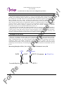
Quantitative test kit for NAD+/NADH
On
ly!
NAD+/NADH Colorimetric Assay Kit
User’s Manual
For Research Use Only, Not for use in diagnostic procedures
CycLex NAD+/NADH Colorimetric
Assay Kit
For 100 Assays
ce
Pu
Intended Use................................................1
Storage.........................................................1
Introduction..................................................2
Principle of the Assay..................................2
Materials Provided.......................................3
Materials Required but not Provided...........3
Precautions...................................................4
Detailed Protocol..........................................5-9
Evaluation of Results...................................10
Cautions........................................................11
Assay Characteristics...................................11
Troubleshooting...........................................11
Reagent Stability..........................................11
Example of Test Results...............................12-15
References....................................................16
Related Products...........................................17
rp
os
e
Cat# CY-1253
Intended Use
en
The CycLex Research Product CycLex NAD+/NADH Colorimetric Assay Kit provides a
convenient method for sensitive measurement of NAD+, NADH and their ratio. This kit is designed for
specific detection of NAD+ and NADH by an enzyme cycling reaction.
NAD+ and NADH extraction and neutralization buffers are NOT provided in this kit.
er
Applications for this kit include:
1) Measuring NAD+ and NADH concentrations in cell or tissue extracts.
2) Measuring NAD+/NADH ratio.
3) Detecting effects of pharmacological agents on cellular level of NAD+ and NADH.
ef
This assay kit is for research use only and not for use in diagnostic or therapeutic procedures.
Storage
rR
• Upon receipt store all components at -70°C.
• Don’t expose reagents to excessive light.
Fo
Cat#: CY-1253
1
Version#: 130823
On
ly!
NAD+/NADH Colorimetric Assay Kit
User’s Manual
For Research Use Only, Not for use in diagnostic procedures
Introduction
rp
os
e
Nicotinamide adenine dinucleotide (NAD+) as well as nicotinamide adenine dinucleotide phosphate
(NADP+) is an important cofactor found in cells. NADH is the reduced form of NAD+, and NAD+ is
the oxidized form of NADH. It has been reported that NAD+ metabolism regulates important biological
effects including life span. NAD+, through poly-ADP-ribosyl polymerase (PARP), mono-ADPribosyltransferase (ARTs) and recently characterized sirtuin enzymes, exerts potential biological effects.
These enzymes modify proteins to regulate their function via ADP-ribosylation or deacetylation in the
presence of NAD+. These enzymes are involved in several pathways including apoptosis, DNA repair,
senescence and endocrine signaling, suggesting that either the enzymes could be an important
therapeutical target for cancer, diabetes atherosclerosis and so on.
The traditional NAD+/NADH and NADP+/NADPH assays are done by monitoring of NADH or
NADPH absorption at 340 nm. This method suffers low sensitivity and high interference since the assay
is done in the UV range that requires expensive quartz microplate. CycLex NAD+/NADH Colorimetric
Assay Kit employs an enzyme cycling reaction, which significantly increases detection sensitivity, and
provides a convenient method for sensitive detection of NAD+, NADH and their ratio.
Principle of the Assay
Pu
Since it is very simple to measure and it can be performed at a low price, the measurement of NAD+
and NADH concentrations in most laboratories is possible if they are equipped with a microtiter plate
reader. Considering that the use of fully automatic apparatus to monitor the absorbance has become
widespread, NAD+ and NADH concentration measurement is now possible with the CycLex
NAD+/NADH Colorimetric Assay Kit using the same equipment. This method of measurement should
dramatically raise the efficiency of measuring NAD+ and NADH concentrations in mammalian cells and
tissues.
ADH
Diapharase
NAD+
WST-1-formazan
er
en
EtOH
ce
Measuring Principle of The CycLex NAD+/NADH Colorimetric Assay Kit
NADH
WST-1
rR
ef
Acetoaldehyde
Read A450
Fo
Cat#: CY-1253
2
Version#: 130823
Materials Provided
Each kit contains
Materials
Quantity
1.5 mL x 1
200 µL x 1
③10X Standard Dilution Buffer
④50X WST-1
⑤50X ADH (alcohol dehydrogenase)
⑥50X Diaphorase
⑦10X EtOH Solution
⑧Instruction manual
2 mL x 1
200 µL x 1
200 µL x 1
200 µL x 1
1 mL x 1
1
Materials Required but not Provided
Storage
-70°C
-70°C
-70°C
-70°C
-70°C
-70°C
rp
os
e
①10X NAD+/NADH Assay Buffer
②400 µM NADH
On
ly!
NAD+/NADH Colorimetric Assay Kit
User’s Manual
For Research Use Only, Not for use in diagnostic procedures
-70°C
Room temp.
• NAD+ Extraction Buffer (See page 5, section “Detailed Protocol”.)
Pu
• NADH extraction Buffer (See page 5, section “Detailed Protocol”.)
• Neutralization Buffer for NAD+ extraction (See page 5, section “Detailed Protocol”.)
• Neutralization Buffer for NADH extraction (See page 5, section “Detailed Protocol”.)
• Water bath or heating block
ce
• Microplate for ELISA
en
• Plate reader capable of measuring absorbance in 96-well plates at dual wavelengths of 450 nm/540
nm. Dual wavelengths of 450/550 or 450/595 nm can also be used. The plate can also be read at a
single wavelength of 450 nm, which will give a somewhat higher reading.
• Pipettors: 2-20 µL, 20-200 µL and 200-1000 µL precision pipettors with disposable tips.
• Multi-channel pipette
er
• Microplate shaker
* Deionized water of the highest quality
ef
• Reagent reservoirs
rR
• FK866 (APO866): FK866 is available from Axon Medchem Cat# Axon 1279 or Cayman Cat.#
13287. Make 2.5 mM stock solution in DMSO (optional)
• 1 N H2SO4: Stop solution (optional)
Fo
Cat#: CY-1253
3
Version#: 130823
On
ly!
NAD+/NADH Colorimetric Assay Kit
User’s Manual
For Research Use Only, Not for use in diagnostic procedures
Precautions
• Please thaw all reagents in crushed ice before use.
• Please keep ADH (alcohol dehydrogenase) and diaphorase in this kit on ice and return them
immediately to -70°C after use. There is a possibility that the enzyme activity may be inactivated.
• Please avoid mixing of any reagents containing SH group like DTT or reduced glutathione, or
alkyl amine in the sample that will interfere this assay.
rp
os
e
• Do not use kit components beyond the indicated kit expiration date.
• Rinse all detergent residue from glassware.
• Use deionized water of the highest quality.
• Do not mix reagents from different kits.
• Do not mouth pipette or ingest any of the reagents.
Pu
• Do not smoke, eat, or drink when performing the assay or in areas where samples or reagents are
handled.
• Biological samples may be contaminated with infectious agents. Do not ingest, expose to open
wounds or breathe aerosols. Wear protective gloves and dispose of biological samples properly.
rR
ef
er
en
ce
• CAUTION: NAD+ Extraction Buffer and NADH extraction Buffer (Not provided in this kit) are
a strong acid and alkaline, respectively. Wear disposable gloves and eye protection
when handling Stop Solution (1 N H2SO4).
Fo
Cat#: CY-1253
4
Version#: 130823
Detailed Protocol
On
ly!
NAD+/NADH Colorimetric Assay Kit
User’s Manual
For Research Use Only, Not for use in diagnostic procedures
CycLex NAD+/NADH Colorimetric Assay Kit can measure NAD+ and NADH concentrations by
enzyme cycling reaction using alcohol dehydrogenase (ADH), diaphorase and WST-1. Since the reaction
is not stopped, it is necessary to monitor absorbance of WST-1-formazan at 450 nm at regular intervals
after the reaction is initiated, and to determine appropriate reaction time.
1. Preparation of Assay Reagents
rp
os
e
1. Prepare Extraction Buffers and Neutralization Buffers: Not provided in this kit
* NAD+ Extraction Buffer: 0.5 M perchloric acid (HClO4)
* NADH Extraction Buffer: 50 mM NaOH and 1mM EDTA
* Neutralization Buffer for NAD+ extraction: 0.55 M K2CO3
* Neutralization Buffer for NADH extraction: 0.3 M potassium phosphate buffer (pH 7.4)
2. Thaw ①10X NAD+/NADH Assay Buffer at room temperature in water bath. Stand all other
reagents on ice to thaw. Use them after they thaw completely.
Pu
3. Prepare a working solution of Standard Dilution Buffer by diluting ③10X Standard Dilution
Buffer 1:10 with distilled water. Mix well. Store at 4°C for two weeks or -20°C for long term
storage.
4. Prepare NADH Standards as follows:
Use ②400 µM NADH to produce a serial dilution series (below). Mix each tube thoroughly before
the next transfer. The 4,000 nM standard (Std.1) serves as the high standard. The NADH Standard
Dilution Buffer serves as the zero standard (Blank).
en
10 µL of ②400 µM NADH
100 µL of Std. 1 (4,000 nM)
100 µL of Std. 2 (2,000 nM)
100 µL of Std. 3 (1,000 nM)
100 µL of Std. 4 (500 nM)
100 µL of Std. 5 (250 nM)
100 µL of Std. 6 (125 nM)
er
Std.1
Std.2
Std.3
Std.4
Std.5
Std.6
Std.7
Blank
ce
Volume of Standard
-
Standard Dilution
Buffer
990 µL
100 µL
100 µL
100 µL
100 µL
100 µL
100 µL
100 µL
Concentration
4,000 nM
2,000 nM
1,000 nM
500 nM
250 nM
125 nM
62.5 nM
0 ng/mL
ef
Note-1: Do not use a Repeating pipette. Change tips for every dilution. Wet tip with Standard before
dispensing. Unused portions of NADH Standards should be discarded.
rR
Note-2: Since NAD+ is converted to NADH in enzyme cycling reaction and relatively labile than
NADH, CycLex only provides NADH Standard.
Fo
Cat#: CY-1253
5
Version#: 130823
On
ly!
NAD+/NADH Colorimetric Assay Kit
User’s Manual
For Research Use Only, Not for use in diagnostic procedures
5. Prepare NAD+/NADH Reaction Buffer (Quantity Required: 75 µL/assay)
・Mix following reagents and put in ice.
・Please use within 30 min after prepared this NAD+/NADH Reaction Buffer. Discard any unused
NAD+/NADH Reaction Buffer after use.
Assay reagents
Volume
①10X NAD+/NADH Assay Buffer
④50X WST-1
10 µL
rp
os
e
2 µL
⑤50X ADH
2 µL
⑥50X Diaphorase
2 µL
⑦10X EtOH Solution
10 µL
dH2O
49 µL
75 µL
rR
ef
er
en
ce
Pu
Total
Fo
Cat#: CY-1253
6
Version#: 130823
On
ly!
NAD+/NADH Colorimetric Assay Kit
User’s Manual
For Research Use Only, Not for use in diagnostic procedures
2. Preparation of Sample
Numerous extraction methods can be used to isolate NAD+ and NADH. The following protocols for
NAD+ and NADH have been shown to work with a number of different mammalian cell lines are
provided as examples of suitable methods. If desired, you can employ other methods for extraction of
NAD+ and NADH. All extraction and neutralization buffers are not provided in this kit.
rp
os
e
Determination of NAD+ and NADH requires two separate samples (acid extract for NAD+ and
alkaline extract for NADH), utilizing the character of NAD+; resistant to acidic condition and heat
labile and NADH: resistant to alkaline condition and relatively heat stable.
Preparation of Cell Extract
1. For adherent cells, after washing with PBS, trypsinize, harvest and transfer the cells (1-5 x 10⁶ cells)
to microcentrifuge tubes followed by centrifugation at 2,000 rpm for 5 min.
Note: Each investigator should optimize the number of cells used per test.
or
Pu
For non-adherent cells, harvest and transfer the cells (1-5 x 10⁶ cells) to microcentrifuge tubes
followed by centrifugation at 2,000 rpm for 5 min.
Note: Each investigator should optimize the number of cells used per test.
2. Wash the cells twice with cold PBS by centrifugation at 2,000 rpm for 5 min.
3. Spin down the cells by microcentrifuge at 10,000 rpm for 1 min. Aspirate the supernatant; remove as
much of the PBS as possible.
ce
For NAD+ measurement
4. Vortex the cell pellet gently. Extract the cells with 100 µL of NAD+ Extraction Buffer (See page 5)
by vortexing 3-4 times for 1 min each with same time intervals or by homogenization using standard
techniques (i.e. sonicate 4 times for 5 sec each on ice.). Then stand for 30 min on ice.
en
5. Add 100 µL of Neutralization Buffer for NAD+ extraction (See page 5) to the acid extract and mix
well by vortexing for neutralization.
er
6. Spin the neutralized cell extract above at 15,000 rpm for 5 min at 4°C. Transfer a supernatant to new
microcentrifuge tube. (The final pH of the supernatant should be 7.5–8.5. Please make sure that the
pH is within this range. If not, adjust pH 7.5–8.5 using either Neutralization Buffers for NAD+
extraction or for NADH extraction, see page 5.).
ef
7. Keep the tube of the cell extract for NAD+ measurement (refer to as an acid extract: “AcE”) on ice,
which is ready to go to “step 8” for NAD+ measurement.
rR
8. Measure protein concentration, for example using the BCA protein assay, data can be normalized and
expressed as pmol NAD+ /mg of protein.
Fo
Cat#: CY-1253
7
Version#: 130823
On
ly!
NAD+/NADH Colorimetric Assay Kit
User’s Manual
For Research Use Only, Not for use in diagnostic procedures
For NADH measurement
4’. Vortex the cell pellet gently. Extract the cells with 100 µL of NADH Extraction Buffer (See page 5)
by vortexing 2-3 times for 1 min each with same time intervals or by homogenization using standard
techniques (i.e. sonicate 4 times for 5 sec each on ice.). Then incubate at 60 °C for 30 min to reduce
the viscosity of the samples.
rp
os
e
5’. Add 100 µL of Neutralization Buffer for NADH extraction (See page 5) to the alkaline extract and
mix well by vortexing for neutralization, then stand on ice for at least 5 min.
6’. Spin the neutralized cell extract at 15,000 rpm for 5 min at 4°C. Transfer a supernatant to new
microcentrifuge tube (The final pH of the supernatant should be 7.5–8.5. Please make sure that the
pH is within this range. If not, adjust pH 7.5–8.5 using either Neutralization Buffers for NAD+
extraction or for NADH extraction, see page 5.).
7’. Keep the tube of the cell extract for NADH measurement (refer to as an alkaline extract: “AlE”)
on ice, which is ready to go to “step 8” for NADH measurement.
8’. Measure protein concentration, for example using the BCA protein assay, data can be normalized and
expressed as pmol NADH /mg of protein.
Pu
Note-1: If necessary, the cell extract can be stored at -70°C. Avoid multiple freeze/thaw cycles. After
thaw the cell extract, centrifuge at 15,000 rpm for 15 minutes at 4°C again since the cell
extract should be clear of any sediments or particulate matters. However this may result in
some loss of NAD/NADH.
ce
Note-2: Although this protocol has been successfully applied to many mammalian cell lines, users
should optimize the cell extraction procedure for their own applications.
en
Note-3: Although we suggest to conduct experiments as outlined above, the optimal experimental
conditions will vary depending on the parameters being investigated, and must be determined
by the individual user. Especially, please make sure the final preparations of cell extract are in
neutral pH, hopefully, pH 7.5-8.5.
rR
ef
er
CycLex does NOT recommend to use sodium carbonate-based buffer as a NAD+/NADH
extraction buffer instead of acid extraction buffer and alkaline extraction buffer for NAD+ and
NADH extraction, respectively, because the extracts prepared by using sodium carbonate-based
buffer give you an inappropriate NAD+ and NADH concentrations and ratio (See Fig. 6 and 7 at
page 14 and 15).
Fo
Cat#: CY-1253
8
Version#: 130823
3. NAD+/NADH Assay Procedures
On
ly!
NAD+/NADH Colorimetric Assay Kit
User’s Manual
For Research Use Only, Not for use in diagnostic procedures
Assay reagents
Test sample
NADH
Standard
NAD+/NADH Reaction Buffer (see page 6)
75 µL
75 µL
-
25 µL
25 µL
-
NADH Standards
rp
os
e
Your sample (Test sample)
1. Following the above table, add 25 µL of NADH Standards (Std.1-Std.7 and Blank: Standard Dilution
Buffer) and Your sample to each well of microplate. Next, initiate reaction by adding 75 µL of
NAD+/NADH Reaction Buffer to the each well and mix thoroughly. Incubate at room temperature
(ca.25°C).
2. Monitor the absorbance at 450 nm for 30 to 90 minutes at 10 min intervals using microplate reader.
2’. Alternatively, after 60 min or appropriate time incubation, the reactions can be stopped by adding 50
µL of Stop Solution 1 N H2SO4 (not provided in this kit) into each well and mix well. Take a reading
with the absorbance at 450 nm.
Pu
Table. Layout of NADH standards and test samples (AcE and AlE) in 96-well microplate:
2
3
4
A
Blank
Blank
AcE-1
AcE-1
B
Std.7
Std.7
AlE-1
AlE-1
C
Std.6
Std.6
AcE-2
AcE-2
D
Std.5
Std.5
AlE-2
AlE-2
E
Std.4
Std.4
AcE-3
AcE-3
F
Std.3
Std.3
AlE-3
AlE-3
G
Std.2
Std.2
AcE-4
AcE-4
H
Std.1
Std.1
AlE-4
AlE-4
5
6
7
8
9
10
11
12
er
en
ce
1
AcE: Acid Extract
AlE: Alkaline Extract
rR
ef
Note: The NADH standards and test samples should be run in duplicate.
Fo
Cat#: CY-1253
9
Version#: 130823
Evaluation of Results
1. Run reactions as described in the Detailed Protocol.
2. Subtract A450 at the 0 time from all reaction time points.
3. Plot A450 versus reaction time.
4. Determine the reaction time range in which the increase in A450 is linear.
5. Fix an appropriate reaction time (usually 60 min).
6. Take a reading with the absorbance at 450 nm.
On
ly!
NAD+/NADH Colorimetric Assay Kit
User’s Manual
For Research Use Only, Not for use in diagnostic procedures
rp
os
e
Average the duplicate readings for each standard, control, and sample and subtract the average zero
standard (Std.7) optical density. Plot the optical density for the standards versus the concentration of the
standards and draw the best curve. The data can be linearized by using log/log paper and regression
analysis may be applied to the log transformation. To determine the NAD+ or NADH concentration of
each sample, first find the absorbance value on the y-axis and extend a horizontal line to the standard
curve. At the point of intersection, extend a vertical line to the x-axis and read the corresponding NAD+
or NADH concentration. If the samples have been diluted, the concentration read from the standard
curve must be multiplied by the dilution factor (See Fig. 2. NADH Standard curve at page 12).
1. The results of unknown samples can be calculated with any computer program having a 5-parameter
logistic function. It is important to make an appropriate mathematical adjustment to accommodate for
the dilution factor.
Pu
2. Most microplate readers perform automatic calculations of analyte concentration. The calibration
curve is constructed by plotting the absorbance (Y) of calibrators versus log of the known
concentration (X) of calibrators, using the four-parameter function.
ce
3. The concentration of NAD+ or NADH in the sample can be calculated, then divide the NAD+ or
NADH concentration by the sample amount (e.g. cell number or extract protein amount) you added
into the sample wells. The concentration of NAD+ or NADH can be expressed in pmol/106 cells or
pmol/mg protein.
NAD+ concentration of AcE (nM) x 2 x 10⁵ /cell number
NADH concentration (pmol/106 cells) =
NADH concentration of AlE (nM) x 2 x 10⁵ /cell number
en
NAD+ concentration (pmol/106 cells) =
er
Total volume of AcE = 200 µL. Volume of AcE for assay = 25 µL.
Total volume of AlE = 200 µL. Volume of AlE for assay = 25 µL.
NAD+/NADH Ratio =
NAD+ concentration (pmol/106 cells)
NADH concentration (pmol/106 cells)
ef
Note-1: The absorbance background increases with time, thus it is important to subtract the absorbance
value of the zero standard (Std.7) wells for each data point.
rR
Note-2: Usually, an appropriate reaction time is from 60 to 90 min. This value is variable depending on
reaction conditions and preparation of cell extract. Decreasing the amount of cell extract in the
assay may help to lengthen the time range.
Fo
Cat#: CY-1253
10
Version#: 130823
Cautions
On
ly!
NAD+/NADH Colorimetric Assay Kit
User’s Manual
For Research Use Only, Not for use in diagnostic procedures
1. Since this kit is based on enzyme cycling reaction, it is not possible to measure NAD+ and NADH
concentration in conventional crude cell extract without special extraction and treatment for NAD+
and NADH. Please follow the section “Preparation of Sample” at page 7-8.
2. Please avoid mixing of any reagents containing SH group like DTT or reduced glutathione, or
alkyl amine in the sample that will interfere this assay.
rp
os
e
For research use only, not for use in diagnostic or therapeutic procedures.
Assay Characteristics
The CycLex Research Product CycLex NAD+/NADH Assay kit has been shown to measure NAD+
and NADH concentrations or NAD+/NADH ratio crude cell extract using enzyme cycling reaction. The
assay shows good linearity of sample response.
Troubleshooting
1. When test sample have not adjusted to neutral pH, Enzyme cycling reaction might be inhibited, so that
NAD+ and NADH concentration cannot be measured.
Pu
2. The test samples should be run in duplicate, using the protocol described in the Detailed Protocol.
Incubation times or temperatures significantly different from those specified may give erroneous
results.
Reagent Stability
ce
3. Poor duplicates indicate inaccurate dispensing. If all instructions in the Detailed Protocol were
followed accurately, such results indicate a need for multi-channel pipettor maintenance.
rR
ef
er
en
All of the reagents included in the CycLex Research Product CycLex NAD+/NADH Assay kit have
been tested for stability. Reagents should not be used beyond the stated expiration date. Upon receipt, all
kit reagents should be stored at -70°C.
Fo
Cat#: CY-1253
11
Version#: 130823
On
ly!
NAD+/NADH Colorimetric Assay Kit
User’s Manual
For Research Use Only, Not for use in diagnostic procedures
Example of Test Results
Fig.1 Time course
3.0
4000 nM
2000 nM
1000 nM
500 nM
250 nM
125 nM
62.5 nM
0 nM
2.5
rp
os
e
A450
2.0
1.5
1.0
0.5
0.0
0
10
20
30
40
50
60
Pu
Reaction time (min.)
Fig.2 NADH Standard curve
NADH Standard curve
(Reaction time 30min.)
ce
3.0
2.5
A450
en
2.0
1.5
er
1.0
rR
ef
0.5
Fo
Cat#: CY-1253
0.0
0
1,000
2,000
3,000
4,000
NADH conc. (nM)
12
Version#: 130823
On
ly!
NAD+/NADH Colorimetric Assay Kit
User’s Manual
For Research Use Only, Not for use in diagnostic procedures
Fig.3 Specificity of CycLex NAD+/NADH Colorimetric Assay Kit (As low as 62.5 nM of NADH can be
detected with 60 min incubation time (n=2), there is no response to NADPH)
2.0
NADH
NADPH
rp
os
e
A450
1.5
1.0
0.5
0.0
0
1,000
2,000
3,000
4,000
Pu
NADH/NADPH conc. (nM)
Fig.4 NAD+ and NADH concentration in the cell extracts of Jurkat and SW480 cells
en
NAD+ or NADH conc. (pmol/10
6
200
NAD+
NADH
ce
cells)
250
150
rR
ef
er
100
Fo
Cat#: CY-1253
50
0
SW480
Jurkat
13
Version#: 130823
On
ly!
NAD+/NADH Colorimetric Assay Kit
User’s Manual
For Research Use Only, Not for use in diagnostic procedures
Fig.5 NAD+ and NADH concentration in the cell extracts of Jurkat cells that have been treated with
Nampt specific inhibitor FK866 at indicated concentrations
250
NAD+
200
NADH
150
100
50
0
3.3
10
33
Pu
0
rp
os
e
NAD+ or NADH conc. (pmol/10
6
cells)
NAD+ and NADH concentration
FK866 conc. (nM)
ce
Fig.6 Comparisons of NAD+ and NADH concentrations in SW480 cell extract measured by extraction
methods and two kits, “CycLex NAD+/NADH Assay kit” and Company B’s “NAD/NADH
Quantitation Kit”
150
er
100
NAD+
NADH
en
200
50
ef
NAD+ or NADH conc. (pmol/10
6
cells)
250
rR
0
Fo
Cat#: CY-1253
CycLex Kit
(Conventional extraction)
CycLex Kit (Sodium
carbonate-extraction)
14
Company B Kit
Version#: 130823
On
ly!
NAD+/NADH Colorimetric Assay Kit
User’s Manual
For Research Use Only, Not for use in diagnostic procedures
Fig.7 Comparisons of NAD+/NADH ratio in SW480 cell extract measured by extraction methods and
two kits, “CycLex NAD+/NADH Assay kit” and Company B’s “NAD/NADH Quantitation
Kit”
NAD+/NADH ratio
18
16
rp
os
e
NAD+/NADH ratio
14
12
10
8
6
4
2
0
Pu
CycLex Kit
(Sodium carbonateextraction)
Company B Kit
rR
ef
er
en
ce
CycLex Kit
(Conventional
extraction)
Fo
Cat#: CY-1253
15
Version#: 130823
On
ly!
NAD+/NADH Colorimetric Assay Kit
User’s Manual
For Research Use Only, Not for use in diagnostic procedures
References
1. Ziegenhorn J, Senn M, Bucher T. (1976) Molar absorptivities of beta-NADH and beta-NADPH. Clin
Chem. 22: 151.
2. Matsumura, H. and Miyachi S (1980) Cycling assay for nicotinamide adenine dinucleotides. Methods
Enzymol. 69: 465-470.
rp
os
e
3. Zerez CR, Lee SJ, Tanaka KR (1987) Spectrophotometric determination of oxidized and reduced
pyridine nucleotides in erythrocytes using a single extraction procedure. Anal Biochem. 164: 367-73
4. Zhao, Z, Hu, X and Ross CW (1987). Comparison of Tissue Preparation Methods for Assay of
Nicotinamide Coenzymes. Plant Physiol. 84: 987-988.
5 Umemura, K and Kimura, H (2005) Determination of oxidized and reduced nicotinamide adenine
dinucleotide in cell monolayers using a single extraction procedure and a spectrophotometric assay.
Anal Biochem. 338: 131-5.
6. Hasmann, M and Schemainda, I (2003) FK866, a Highly Specific Noncompetitive Inhibitor of
Nicotinamide Phosphoribosyltransferase, Represents a Novel Mechanism for Induction of Tumor Cell
Apoptosis. Cancer Research 63: 7436-7442, 2003
Pu
7.Vilcheze, C et al. (2005). Altered NADH/NAD+ Ratio Mediates Coresistance to Isoniazid and
Ethionamide in Mycobacteria. Antimicrobial Agents and Chemotherapy. 49: 708-720.
8. Kimura N, Fukuwatari T, Sasaki R, Shibata K. (2006) Comparison of metabolic fates of nicotinamide,
NAD+ and NADH administered orally and intraperitoneally; characterization of oral NADH. J Nutr
Sci Vitaminol. (Tokyo) 52: 142.
ce
9. O'Donnell JM, et al. (2004) Limited transfer of cytosolic NADH into mitochondria at high cardiac
workload. Am J Physiol Heart Circ Physiol. 286: H2237.
en
10. Richard A. et al. (2008) Characterization of NAD Uptake in Mammalian Cells J. Biol. Chem. 283:
6367-6374.
11. Rongvaux, A et al. (2008) Nicotinamide Phosphoribosyl Transferase/Pre-B Cell Colony-Enhancing
Factor/Visfatin Is Required for Lymphocyte Development and Cellular Resistance to Genotoxic
Stress J. Immunol. 181: 4685-4695
rR
ef
er
12. Pogrebniak, A et al. (2006) Chemopotentiating effects of a novel NAD biosynthesis inhibitor, FK866,
in combination with antineoplastic agents. Eur J Med Res. 11: 313-21.
Fo
Cat#: CY-1253
16
Version#: 130823
On
ly!
NAD+/NADH Colorimetric Assay Kit
User’s Manual
For Research Use Only, Not for use in diagnostic procedures
Related Products
Pu
rp
os
e
*CycLex NAMPT Colorimetric Assay Kit: Cat# CY-1251
*CycLex NMNAT Colorimetric Assay kit: Cat# CY-1252
*CycLex NAD+/NADH Colorimetric Assay Kit: Cat# CY-1253
*NAMPT (Nicotinamide Phosphoribosyltransferase): Cat# CY-E1251
*NMNAT1 (Nicotinamide Mononucleotide Adenylyltransferase 1): Cat# CY-E1252-1
*NMNAT2 (Nicotinamide Mononucleotide Adenylyltransferase 1): Cat# CY-E1252-2
*CycLex SIRT1/Sir2 Deacetylase Fluorometric Assay Kit: Cat# CY-1151
*CycLex SIRT2 Deacetylase Fluorometric Assay Kit: Cat# CY-1152
*CycLex SIRT3 Deacetylase Fluorometric Assay Kit: Cat# CY-1153
*CycLex SIRT6 Deacetylase Fluorometric Assay Kit: Cat# CY-1156
*NAD(+)-Dependent Deacetylase SIRT1: Cat# CY-E1151
*NAD(+)-Dependent Deacetylase SIRT2: Cat# CY-E1152
*NAD(+)-Dependent Deacetylase SIRT3: Cat# CY-E1153
*NAD(+)-Dependent Deacetylase SIRT6: Cat# CY-E1156
CycLex Co., Ltd.
1063-103 Terasawaoka
Ina, Nagano 396-0002
Japan
Fax: +81-265-76-7618
e-mail: [email protected]
URL: http://www.cyclex.co.jp
ce
PRODUCED BY
rR
ef
er
en
CycLex/CircuLex products are supplied for research use only. CycLex/CircuLex products and
components thereof may not be resold, modified for resale, or used to manufacture commercial
products without prior written approval from CycLex Co., Ltd.. To inquire about licensing for
such commercial use, please contact us via email.
Fo
Cat#: CY-1253
17
Version#: 130823