1
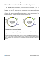
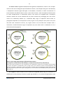
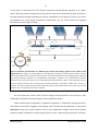
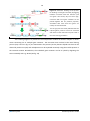
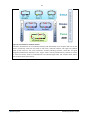
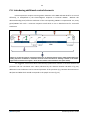
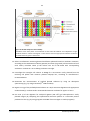
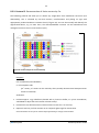
Toggle Expression System Series flEXi Coli Multi-Protein Expression in E.coli User Manual Version 1. 1 V1.1 January 15, 2014 1 TABLE OF CONTENTS A. flEXi Coli kit contents 3 Reagents to be supplied by user (see also section D. Protocols) 4 B. Escherichia coli – a premier laboratory workhorse 5 B.1. Introduction 5 C. A Novel Approach to Multigene Applications 7 C.1. The Toggle Concept – a Primer 7 C.2. Transfer vectors: Acceptor-Donor recombineering system. 10 C.3. Generating multi-gene expression cassettes 15 C.3.1. Creating individual gene expression cassettes 16 C3.1.1. Standard Approach 16 C3.1.2 Inserts with Other Unique Restriction Sites 17 C.3.2. Multi-gene construction via Toggle multiplication 18 C.3.3. Multi-gene construction using Cre-Lox recombination 20 C.4. Introducing additional control elements 22 D. Protocols 23 D.0 Introductory remarks 23 D.1 Cloning into TogColi transfer vectors 23 D.2 Conversion of donors into conditional donors 24 D 2.1 Protocol 1. Conversion into conditional oris 24 D.3 Concatenation of genes (gene cassettes) by Toggling 24 D 3.1 Protocol 2. Adding gene cassettes through toggling 25 D.4 Deconstruction of gene cassette assemblies 28 D 4.1 Protocol 3. Disassembling multi-gene construct to regenerate individual expression boxes D.5 Cre-LoxP reaction of Acceptors and Donors 28 30 D 5.1. Protocol 4: Cre-LoxP fusion of Acceptors and Donors 30 D 5.2. Protocol 5. Deconstruction of fusion vectors by Cre 33 D.6. Expression of multi-gene constructs E. Appendix E.1. Preparing chemically competent cells flEXiColi System 36 37 37 ATG:biosynthetics GmbH 2 E.2. Preparing bacterial stocks from agar stabs 38 E.3. flEXiColi vectors: maps, sequences, restriction 39 E.3.1 Acceptor vectors 40 E.3.1.1 TogColiAccA1.5 & A1.6: 2572 bp / 2623 bp 40 E.3.2.1 TogColiDonA1.5: 2774 bp (KnR), 2619 bp (CmR) 41 E.3.2.2 TogColiDonA1.6: 2864 bp (KnR), 2693 bp (CmR) 42 E.3.3. Analytical restriction digest patterns 43 E.3.4. Sequencing primers 43 F. References 45 NOTES: 46 G. Purchaser Notification 47 flEXiColi System ATG:biosynthetics GmbH 3 A. flEXi Coli kit contents Plasmid acceptor transfer vectors TogColiA1.5GmR; TogColiA1.6GmR; approx. 3-4 µg DNA per vial (in buffer solution) keep at 4°C for short-term storage and in a freezer at -20°C or lower for medium- and long-term storage (take care to avoid repeated freeze-thaw cycles, e.g. by aliquotting DNA prior to freezing) Plasmid donor vectors TogColiA1.5KnR & TogColiA1.5CmR; TogColiA1.6KnR & TogColiA1.6CmR; approx. 3-4 µg DNA per vial (in buffer solution) keep at 4°C for short-term storage and in a freezer at -20°C or lower for medium- and long-term storage (take care to avoid repeated freeze-thaw cycles, e.g. by aliquotting DNA prior to freezing) E. coli strains as agar stabs (opt-in component) a) transformed with acceptor and donor vectors (6 vials) For streaking bacteria on plates as a starting point for plasmid preparation † b) [optional, in some configurations] pirHC, pirLC cells For propagation and amplification of donor vectors, donor multi-gene expression constructs or donor-donor fusions Keep agar stabs at 4°C or at RT; do not freeze! We recommend to immediately prepare stocks from streaked bacterial colonies (see p. 38). † E. coli strains expressing the pir gene for propagation of donor vectors (any other strain + with pir background can be used as well). LC: low copy number propagation, HC: high copy number propagation of plasmids with R6K origin. flEXiColi System ATG:biosynthetics GmbH 4 Reagents to be supplied by user (see also section D. Protocols) Restriction endonucleases and other DNA-modifying enzymes T4 or any other suitable DNA ligase Cre recombinase Standard E. coli strains for cloning (such as TOP10, DH5, HB101 etc.) Standard laboratory buffers, solutions, media and equipment for bacterial cell culture, transformation etc. Commercially available transfection reagents, e.g. FuGENE® (Roche), jetPEI™ (Polyplus transfection), GeneJuice, etc.; alternatively, equipment for electroporation Antibiotics, chemicals (e.g IPTG, X-Gal, etc.) (pir+ cells, depending on the kit configuration) flEXiColi System ATG:biosynthetics GmbH 5 B. Escherichia coli – a premier laboratory workhorse B.1. Introduction Protein complexes are the heart and soul of many crucial cellular processes (Robinson et al., 2007; Charbonnier et al., 2008). Some researchers go as far as describing the cell as “a collection of protein machines” (Albert, 1998) which in essence are multi-protein complexes. Whether you think of replication, transcription (Van Hijum et al., 2009), translation (Estrozi et al., 2011), DNA repair, the processing, import, trafficking as well as export of proteins or other biomolecules, or the maintenance of the structural stability and integrity of any cell, multi-subunit protein assemblies play an important role in all these biological phenomena. In addition, other processes, e.g. entry of viruses into human cells, also critically hinge on the co-operation of proteins or protein complexes (Bhattacharya, 2009). Various prokaryotic microorganisms - E.coli being the prototypical workhorse - are harnessed to express heterologous proteins and protein complexes but also to cost-efficiently produce known or novel compounds by means of metabolic engineering (Chamler and Koffas, 2008; Chou, 2007; Lee et al., 2008). Scientists wishing to study these processes in functional and structural detail, often require significant amounts of the protein complexes under investigation. While obtaining bulk protein usually is not a problem for protein complexes that are abundant in a steady-state or a developing / differentiating cell, this becomes more difficult for complexes that are transient in nature, appear only periodically in cells or simply occur only in low amounts. In such cases, systems come in handy that allow homo- or heterologous expression of these complexes in large amounts. Such a large-scale protein production approach by using recombinant technologies makes analysis of the structurefunction relationships in multi-protein complexes more likely. While various methods and systems have been developed to address this problem, most of them are of little use for intense research efforts directed at generating and investigating scores of protein complexes in parallel, i.e. in an automated fashion. Such a system should be robust and easy-to-install in terms of manipulation operations and protocols and/or components used in the process (Hunt, 2005). In addition, they should provide enough flexibility to rapidly alter the multiprotein components, which is a prerequisite for revising expression studies. The flEXi Coli system addresses these needs and avoids some problems that occur with in-vitro reconstitution of protein complexes from individually or co-expressed protein components. flEXiColi System ATG:biosynthetics GmbH 6 Figure 1: Applications of E.coli multi-protein expression systems. Some of these are closely intertwined, e.g. pathway design, metabolic engineering, and strain development. Escherichia coli is your first choice for expressing proteins and protein complexes that do not require extensive complex posttranslational modifications. E.coli has proved its worth time and again as the go-to organism for initial investigations. While E.coli has some disadvantages such as the limits in protein sizes one can express, the problem of protein being sequestered into inclusion bodies or the inablity to handle complex glysosylation, there are many advantages that make it the favorite workhorse for innumerable expression projects. These include the wide range of gene-engineering tools that allow its genetic modification, the ease of culturing Escherichia coli, various induction protocols, rapid growth, etc. flEXiColi System ATG:biosynthetics GmbH 7 C. A Novel Approach to Multigene Applications C.1. The Toggle Concept – a Primer TogTec Vectors represent a novel, stringent vector design that aims at clearly trimmed vector backbones that still provide all required functionality. This can be achieved by stripping all elements to the bare minimum necessary for proper function. Toggle systems consists of four boxes that are reserved for and harbor different modular functions as exemplified by the pBoX vector in figure 2. It represents the archetypical configuration from which all other designs are derived. Figure 2: Schematic representation of the fundamental pBoX lay-out. Toggle vectors consist of four defined constructional elements (boxes): application (A-Box), selector (S-Box), as well as target (T-Box) and maintenance (M-Box) functions. All Boxes are separated by unique restriction sites (RSs). A-boxes usually contain a pair of inner and outer RSs (R0 and R0’; R1 and R1’). Genes and genes cassettes are cloned into the A-Box which may also host other functions; standard oris for plasmid propagation in E.coli can be inserted / exchanged in the maintenance box, whereas the T box will carry oris for specific applications or target organisms; the S box hosts the selector functions, most commonly antibiotic resistance markers. The TogTEC systems are characterized by a unique constallation of restriction sites (RSs) that demarcate the four Boxes (figure 3). Figure 3: Implementation of pBoX in TogTEC vectors. All TogTec vectors share a common lay-out where the A-Box is bracketed by AbsI and AscI at the 5’-end and MauBI / SgrDI at the 3’end. The S-Box is demarcated by SgrDI and AsiSI, while the T-Box and M-Box are defined by AsiSI-PacI and PacI-AbsI, respectively. The functional role of the individual Boxes is identical to pBoX. Along with this slender make-up, the vectors are designed to allow highly flexible generation of multigene assemblies with a built-in option for easy disassembly and substitution of sequence bits and parts. This is called ACDC-SC (assembly cloning–disassembly cloning substitution-cloning) design. flEXiColi System ATG:biosynthetics GmbH 8 Figure 4: Schematic representation of a gene cassette plus flanking regions to illustrate the ACDC-SD principle – as applied to E.coli. By design, the coding sequence is bracketed by 5’-NdeI and 3’-BclI sites that contain the start ATG and stop codon TGA, respectively. Both restriction sites are eliminated from the coding sequence (CDS) and any relevant flanking sequences via computational optimization. Likewise, the constructionally relevant restriction sites X, R1, R2 R3, R4, R5 and Y are computationally eliminated from a gene as well (although some of them are optional) as they serve to swap genetic elements (promoters, terminators, leaders, etc.). X and Y, in particular, are vector-specific restriction sites that demarcate the constructive borders of the A box, e.g. AscI and MauBI or their corresponding core sequence BssHII. X and Y in figure 4 can theoretically be designed freely but the outer restriction site pair of the A box (AbsI/XhoI, SgrDI/SalI) is suitable for performing the higher-order concatenation of (multi-)gene cassettes (see fig. 5). Figure 5: Close-up of the A box showing the outer (AbsI, SgrDI) and inner (AscI, MauBI) restriction site pair for toggle cloning. The core sequences within pairs are fully compatible with one another. The inner pair can be used for deconstructing Toggle assemblies whereas the outer pair can be used to build higher-order multi-gene constructs. The spacer between AbsI/AscI and MauBI/SgrDI, respectively, can be used to integrate additional functions for recombination, cloning, etc. This manual presents a new system with a set of novel transfer vectors, specifically designed for synthetic biology applications but flexible enough to accommodate standard protocols. The design of the vectors adheres to a stringent Toggle vector design (see fig. 2 and flEXiColi System 5) that is common to all ATG:biosynthetics GmbH 9 cloning/expression vectors of this series. Toggle vectors combine established, time-tested elements with a new approach to the construction and expression of multigene plasmid constructs. flEXiColi System ATG:biosynthetics GmbH 10 C.2. Transfer vectors: Acceptor-Donor recombineering system. The Acceptor vectors TogColiA1.5GmR and TogColiA1.6GmR host time-tested T7 and lac promoters and T7 polyA signal sequences (see fig. 6). A multiplication module M – defined by the AscI and MauBI (see figure 6) – allows integration of multiple gene cassettes (ORFs and associated regulatory regions; as schematized in fig. 4) as TogTec vectors do not contain a classical multiple cloning site (see appendix) in their A boxes although required or desired restriction sites can be introduced on demand. Acceptors come with a gentamycin resistance marker. a b Figure 6: Circle map representation of flexiColi acceptor vectors (a) TogColi A1.5 (2572 bp), (b) TogColi A1.6 (2623 bp) Both vectors carry a modified ColE1 origin of replication for maintenance of a high plasmid copy number. Acceptor vectors host T7 (TogColi A1.5) and lac (TogColi A1.6) promoters, T7 terminators, loxP sites (red circle) and a gentamycin resistance marker (blue). Genes of interest are cloned into the unique restriction sites (NdeI, BclI) in the A box. The multiplication module (for Toggle assembly) is defined by AbsI/AscI and MauBI/SgrDI, respectively (see fig. 5). Genes or gene cassettes can also be recombined via Cre-Lox recombination making use of the LoxP sites hosted on the vectors. Open reading frames can be inserted into the CDS insertion site framed by NdeI and BclI (see fig. 4). flEXiColi System ATG:biosynthetics GmbH 11 The Donor vectors TogColiA1.5KnR/GmR and TogColiA1.6 KnR/GmR are similar to the acceptor vectors in their over-all design but provide different selectors. The multiple cloning site is bracketed by a multiplication element (again AbsI-SgrDI or AscI-MauBI, as desired) to enable concatenation of inserts between the different donor vectors. Vectors also contain a LoxP imcomplete inverted repeat to create acceptor-donor or donor-donor fusions. The vectors are equipped with a kanamycin resistance marker but can be outfitted with any other resistance by exchanging the S box. Donor vectors are traditionally defined by a conditional R6K origin of replication which makes its propagation dependent on the expression of the pir gene in the prokaryotic host (such as the pirLC and pirHC cells contained in the kit). Yet, TogTec vectors can be turned from acceptors with a constitutive, standard bacterial origin of replication (ori) into donors with a conditional ori via a conversion protocol (page 24). a b C d Figure 7: Circle map representation of flexiColi donor vectors a, c) TogColi A1.5 (CmR, 2462 bp; KnR, 2584 bp); b, d) TogColi A1.6 (CmR, 2513 bp; KnR, 2635 bp). Circle maps show promoters (T7, lac), terminators (T7), multiplication module, the incomplete inverted repeat for cre-lox site-specific recombination (LoxP, orange circle) and resistance markers (chloroamphenicol and kanamycin/neomycin, respectively). Genes of interest are cloned into the multiplication module (CDS insertion site) using unique restriction sites. Open reading frames can be inserted into the NdeI-BclI sites (not shown but refer to fig. 4). The ToggleTec system vectors do not contain pre-integrated affinity tags for purification or solubilization of the protein(s) of interest. Typical tags would be C- or N-terminal oligohistidine, FLAG flEXiColi System ATG:biosynthetics GmbH 12 or myc tags, or novel tags such as the stimulus-responsive RTX precipitation tag (Shur et al., 2013), with or without protease cleavage sites for tag removal. These can be introduced via gene synthesis or through specifically designed PCR primers used for amplification of the genes of interest. Just follow the guidelines on critical design parameters. Alternatively, you can order ready-to-use adapted fluorescence or other tags from ATG. Figure 8: Schematic representation of a ACDC-SD gene cassette plus flanking regions in the context of the TogTec vectors. By design, the coding sequence is bracketed by 5’-NdeI and 3’-BclI sites that contain the start ATG and stop codon TGA, respectively. Both restriction sites are eliminated from the coding sequence (CDS) and any relevant flanking sequences via computational optimization. Likewise, the constructionally relevant restriction sites X, R1, R2 R3, R4 and Y are computationally eliminated from a gene as well (although some of them are optional) as they serve to swap genetic elements (promoters, terminators, leaders, etc.). X and Y, in particular, are vector-specific restriction sites that demarcate the constructive borders of the A box, e.g. AscI and MauBI or their corresponding core sequence BssHII. This E.coli expression system offers a few re-designs and improvements that will help in fully exploiting the potential of this heterologous protein production system: 1. New transfer vectors (TogColiA1.5; TogColiA1.6; TogColiA1.5; TogColiA1.6; see figures 6 and 7) that adhere to the uniform ToggleTec vector design. Gene concatenation/multiplication is achieved via directional cloning into unique restriction sites of the multiplication module. These vectors greatly facilitate modular combination of heterologous genes (in their respective gene expression cassettes) flEXiColi System ATG:biosynthetics GmbH 13 with a minimum requirement for unique restriction sites. Strong bacterial promoters (currently T7 and lac very late prmoters) can be exchanged for other promoter sequences (constiutive and inducible) if desired. Likewise, terminator sequences (currently T7) can be substituted as required (as schematically shown in fig. 4). 2. Leaders for T7 and lac promoters possess a uniform length. This obviates the inconsistencies produced by multiple cloning sites that add a variable number of nucleotides to the leader/promoter. 3. Established protocol for rapid generation of multigene expression constructs via Cre-LoxP recombineering. The resulting multigene fusions can be transformed into specific expression host strains. This protocol can be used to integrate multigene cassettes with coding sequences for multiprotein complex subunits into flexiColi, but also to integrate specific enzymes (kinases, acetylases etc.) for modifying the proteins under investigation. 4. Individual gene cassettes used in the Toggle system can, in fact, be re-cycled and re-used to build other multi-gene constructs. This will facilitate building multigene expression constructs with permutated composition or shuffling the order of expression cassettes to probe any positional effects. flEXiColi System ATG:biosynthetics GmbH 14 Figure 9: Schematic overview of the workflow of the flexiColi system. Genes of interest are assembled into multigene expression cassettes using either gene synthesis, the Toggle multiplication module present on the donor and acceptor vectors or any of several other methods the ToggleTec vectors support. Acceptor-donor multigene fusions can also be generated by Cre-LoxP recombination. In this case, the multigene fusions contain one Acceptor and one to several Donor vectors, each with one or several genes of interest (here A-F). Desired AcceptorDonor combinations are transformed into E.coli, subsequently selected by antibiotic challenge and verified by restriction, PCR or DNA sequencing. Correct constructs can then be amplified and isolated to transform suitable E.coli expression strains. LoxP sites in the acceptor-donor fusions have been omitted for reasons of clarity. flEXiColi System ATG:biosynthetics GmbH 15 C.3. Generating multi-gene expression cassettes To create your expression constructs (see illustration 3), introduce your gene or genes of interest - carrying any additional modifications such as purification or reporter tags - using your method of choice (conventional restriction-ligation cloning, LIC or SLIC) into any of the acceptor or donor vectors. You can then create acceptor-donor fusions with the help of Cre recombinase. Note that you need at least one acceptor vector if you wish to amplify the multi-gene constructs in standard laboratory strains. Select your multi-vector multi-gene fusions by subjecting transformed bacteria to multiple antibiotic selection on agar and/or multi-well plates. You will then have to extract the plasmid construct from your host strains since the expression strain will most likely be different (see chapter C.5). Figure 10: Schematic representation of process for generating multi-gene expression constructs. If, for example, your requirements for an antibiotic resistance marker change, you can transfer entire expression cassettes (including promoters and terminators) within acceptors and donors and between acceptors and donors by using the AbsI-SgrDI (XhoI-SallI) or AscI-MauBI (BssHII) module (see fig. 17). For expression in E.coli, the flEXi Coli multi-gene expression vector fusions with appropriate promoters or terminators are transformed into the appropriate expression host of choice. The basic version (T7 and lac promoter elements) should support expression in most currently available expression strains. If particular expression strains already contain helper plasmids with DNA encoding chaperones, lysozyme or other factors of interest, the design of the multi-gene fusion should ideally be such that the flEXi Coli vector containing the resistance marker that is also present on the helper flEXiColi System ATG:biosynthetics GmbH 16 plasmid is not included in multi-gene vector construction to avoid issues with plasmid incompatibility (although this is probably not essential). Alternatively, the issue can be resolved by creating new versions of the flEXi Coli vectors containing resistance markers that circumvent the conflict. This can be easily performed by PCR amplifying the vectors minus the resistance marker, and combine the resulting fragments with a PCR amplified resistance marker by recombination (SLIC) or blunt-end ligation (using 5’phosphorylated primers). Note that resistance markers can also be exchanged in between flEXi Coli vectors by restriction digestion with SgrDI and AsiSI. C.3.1. Creating individual gene expression cassettes C3.1.1. Standard Approach The acceptor and donor vectors are suited for generating multi-gene expression constructs from individual gene expression cassettes (complete with regulatory regions such as promoter and terminator) via Toggle multiplication. All flexiColi vectors contain identical A box-flanking rare cutter restriction sites. One initial basic design condition is that none of the following restriction sites – that are virtually never used for standard cloning strategies - may be present in the gene cassette: AscI, AbsI, MauBI, SgrDI, PacI, AsiSI, NdeI, BclI, ApaLI. As a logical consequence, XhoI, BssHII, SalI and PvuI are off the list as well as these constitute core sequences of the 8-cutters. ATG offers a suitable gene design service that considers all these requirements. The Toggle system is designed for integrating synthetic genes but can also accommodate other sequences as long as they adhere to certain design principles to fully exploit the advantages of the system. ! Avoid AscI, AbsI, MauBI, SgrDI, PacI, AsiSI, PvuI, NdeI, BclI, ApaLI, XhoI, BssHII, SalI in your gene design! ATG performs all the necessary adaptations to and the relevant biocomputational calculations for the Toggle design of your gene cassettes! Please note that BclI can only be cleaved if the plasmid DNA is amplified in dam-negative strains of E.coli! (In some configurations, adequate transformed strains are included.) flEXiColi System ATG:biosynthetics GmbH 17 C3.1.2 Inserts with Other Unique Restriction Sites If you wish to use your own set of restriction sites, the Toggle design supports this as well but mandates an intermediate step for retrofitting the design. For a detailed protocol, please inquire at [email protected]. Alternatively, you may try ligation-independent procedures to introduce non-standard restriction sites. flEXiColi System ATG:biosynthetics GmbH 18 C.3.2. Multi-gene construction via Toggle multiplication To toggle in a second gene cassette, the receiving vector (which can be an acceptor or donor) is restricted with MauBI and AsiSI to release the selector (Step 1). In parallel, the donating vector is restricted with AscI-AsiSI (Step 2) to release the A box / S box combo which contains the second gene expression cassette. The fragments can be mixed and appropriate ligation events selected by virtue of the newly introduced selector (Step 3 and 4). The need for fragment purification can be obviated by dephosphorylating and additionally cleaving the donating vector with ApaLI which will destroy the ori. STEP1 STEP2 STEP3 STEP4 Figure 11: Schematic of gene cassette concatenation by toggling via the inner pair of restriction sites. The strategy and de facto restriction enzymes are detailed in the text. The resulting two-gene assembly is immune to disassembly with AscI-MauBI – as indicated by the no sign in step 4 - but the individual gene cassettes can be retrieved for re-use by BssHII restriction (as described later, see fig. 18). By the same logic, more gene cassettes can be added and in theory a mere two selectors are required (see figure 12). These are used alternatingly, thus you toggle back and forth between the selectors. flEXiColi System ATG:biosynthetics GmbH 19 Figure 12: Schematic drawing of the process for building a three-gene assembly by toggling between constructs with two selectors (red and green solid arrow). By the same logic, constructs with more gene cassettes can be pieced together but for practical purposes, assemblies with more than four genes are usually not recommended. The no sign (red circle-slash) indicates that the hybrid ligation juncture cannot be restricted with either of the restriction enzymes used in the initial cloning procedure. The resulting fragment is placed into the multiplication module of another acceptor or donor vector containing one or multiple gene cassettes. The restriction sites involved in the initial cloning process (step 1 and 2 in fig. 11) are eliminated in the process (as they become hybrid sites that can be cleaved by neither enzyme) and multiplication can be repeated iteratively using the module present in the inserted cassette. Nonetheless, the individual gene cassettes can be re-cycled by digesting the entire assembly with e.g. BssHII (see fig. 18) flEXiColi System ATG:biosynthetics GmbH 20 C.3.3. Multi-gene construction using Cre-Lox recombination Cre recombinase is a member of the integrase family (Type I topoisomerase from bacteriophage P1). It recombines a 34 bp loxP site in the absence of accessory protein or auxiliary DNA sequence. The loxP site is comprised of two 13 bp recombinase-binding elements arranged as inverted repeats which flank an 8 bp central region where cleavage and the ligation reaction occur. The site-specific recombination mediated by Cre recombinase involves the formation of a Holliday junction (HJ). The recombination events catalyzed by Cre recombinase depend on the location and relative orientation of the loxP sites. Two DNA molecules, for example an acceptor and a donor plasmid, containing single loxP sites will be fused (translocation). The Cre recombination is an equilibrium reaction with 20-30% efficiency in recombination. This provides useful options for multigene combinations for multi-protein complex expression. 13bp 8bp 5’…ATAACTTCGTATA 3’…TATTGAAGCATAT GCATACAT CGTATGTA inverted repeat Spacer 13bp TATACGAAGTTAT…3’ ATATGCTTCAATA…5’ inverted repeat Figure 13: LoxP imperfect inverted repeat In a reaction where several DNA molecules such as donors and acceptors are incubated with Cre recombinase, the fusion/excision activity of the enzyme will result in an equilibrium state where single vectors (educt vectors) and all possible fusions coexist. Donor vectors can be used with acceptors and/or donors, and vice versa. Higher-order fusions are also generated where more than two vectors are fused. This is shown schematically in Illustration 14. The fact that Donors contain a conditional origin of replication (R6K) that depends on a pir+ (pir positive) background now allows for selecting out from this reaction mix all desired AcceptorDonor(s) combinations. For this, the reaction mix is used to transform pir negative strains (TOP10, DH5, HB101 or other common laboratory cloning strains). Then, Donor vectors will act as suicide vectors when plated out on agar containing the antibiotic corresponding to the Donor encoded resistance marker, unless fused with an Acceptor. By using agar with the appropriate combinations of antibiotics, all desired Acceptor-Donor fusions can be selected for. flEXiColi System ATG:biosynthetics GmbH 21 Figure 14: Cre and De-Cre reaction cascade Schematic representation of Cre-mediated assembly and disassembly of one acceptor with one or two donors, respectively. LoxP sites are shown as red circles, resistance markers and origins are labelled. White arrows stand for the entire expression cassette (including promoter, terminator and gene integration/multiplication elements) in the TogTec vectors. Not all possible fusion products are shown for reasons of clarity. Levels of multiresistance are indicated (right side). AD: acceptor-donor fusion; ADD: acceptor-donor-donor-fusion. flEXiColi System ATG:biosynthetics GmbH 22 C.4. Introducing additional control elements Protein expression requires certain genetic elements at the DNA and RNA level for it to work efficiently, as exemplified by the Shine-Dalgarno sequence in bacterial mRNAs. Without this ribosome-binding motif, efficient translation of the corresponding mRNAs is compromised. Yet, many genes/mRNAs lack such a consensus sequence motif which is not an absolute must for successful translation. Figure 15: Constructive layout of expression boxes for use with the flexiColi system. NdeI and BclI define the coding sequence. R4 defines a restriction site that can be introduced to exchange variants in the leader region that can extend from approx. -40 to +40 nts relative to the translation start (start codon). The ToggleTec vectors have been constructively optimized to maintain a constant distance the promoter and the translation start codon (indicated by the distance between R3-NdeI in fig. 15). Additional control elements can be inserted upstream of the promoter (e.g. between AbsI and AscI or AbsI/AscI and NdeI which would correspond to the purple arrow in fig. 15). flEXiColi System ATG:biosynthetics GmbH 23 D. Protocols D.0 Introductory remarks ! Please note that the bacteria in the agar stabs have not been made competent for transformation! If you wish to use them to transform your constructs, you will have to prepare competent cells from the agar stabs in the kit (if included in your configuration). This applies specifically to the pirHC and pirLC strains used to maintain and amplify donor constructs. You may follow your preferred protocol for preparing chemically or electrocompetent cells, e.g. Inoue et al. (1990) or variations of this protocol, or standard protocols as described in Current Protocols in Molecular Biology or Sambrook and Russell: Molecular Cloning (3rd edition, 2001, or older versions). D.1 Cloning into TogColi transfer vectors Reagents: Restriction endonucleases NdeI, BclI (caution: restricts only DNA from dam- E.coli strains) DNA-modifying enzymes: DNA Phosphatase, DNA ligase E. coli competent cells Antibiotics: Chloramphenicol, Gentamycin, Kanamycin The genes of choice can be either provided as a) fully synthetic constructs with 5’-NdeI and 3’-BclI restriciton sites or b) amplified from existing sources with PCR primers hosting a NdeI (for the 5’terminus of the forward primer) and a BclI (for the 3’-terminus of the reverse primer) restriction site while making sure that the ORF does not contain any of the restriction sites listed on page 13. Ligation reactions for TogColi acceptor vectors are transformed into standard E. coli cells for cloning (such as TOP10, DH5, HB101) and plated on agar containing gentamycin (7 µg/ml). Ligation reactions for TogColi donor vectors are transformed into a) standard E. coli cells or b) into those expressing the pir gene if the conversion of the ori has been carried out. The strain may be pirHC and pirLC from this kit – in this case you will need to make the cells electro- or chemically competent first; or any other commercially available strains, e.g. BW23473, BW23474). Next, plate putative transformants on agar containing chloramphenicol (25 µg/ml) or kanamycin (50 µg/ml). Correct clones are selected based on specific restriction digestion patterns and DNA sequencing of the inserts. flEXiColi System ATG:biosynthetics GmbH 24 D.2 Conversion of donors into conditional donors Donor vectors TogColiA1.5 and TogColiA1.6 host split origins of replication that in their default state are ColE1-derived oris. If desired and required, they can be converted into conditional pir-dependent oris by following this conversion protocol. D 2.1 Protocol 1. Conversion into conditional oris 1. Cleave the donor vector with ApaLI 2. Dephosphorylate the cleaved vector 3. Test successful dephosphorylation via transformation (no colonies should appear) 4. Repeat until no empty vector (religation product) remains 5. Cleave the donor with SapI 6. Perform ligation following standard procedures 7. Transform re-ligated vector into pir+ E.coli strains 8. Plate and select on LB-agar plates contaning kanamycin or chloroamphenicol D.3 Concatenation of genes (gene cassettes) by Toggling As indicated in figure 11 (page 18), gene cassettes can be added to flEXi Coli vectors by replacing the S box in the receiving plasmid with a gene cassettes plus selector (A and S boxes) from a donating plasmid. In principle, two antibiotic resistance markers will be sufficient to alternate between selectors while building multigene assemblies and, as this occurs in an alternating fashion, the process is called toggling. flEXiColi System ATG:biosynthetics GmbH 25 Figure 16: Adding gene cassettes via toggling One gene cassette is added with every restriction-ligation cycle. In its most basic application, toggling can be accomplished with only two selectors (here: red and green) that are alternately introduced into the growing multi-gene (cassette) assembly. Correct integration is selected by virtue of the introduced selector. D 3.1 Protocol 2. Adding gene cassettes through toggling Reagents required: Restriction enzymes AscI/XhoI, MauBI/BssHII, AsiSI/PvuI, (and enzyme buffers) DNA phosphatase of choice T4 DNA ligase (and buffer) E. coli competent cells Antibiotics Step 1: Prepare vector backbone and insert(s) Restriction reactions are carried out in 25 µl reaction volumes but can be scaled to any desired volume. Enzymes are used according to the manufacturer’s recommendations. Accepting plasmid ( ≥ 1 µg) in ddH2O/buffer N x restriction enzyme buffer (10 mM BSA, if required) x µl 25/N µl 2.5 µl) MauBI (BssHII) 1 µl ca. 10 U AsiSI (PvuI) 1 µl ca. 10 U ddH2O flEXiColi System ad 25 µl ATG:biosynthetics GmbH 26 Digest at 37°C (or any other recommended temperature) for at least 1 to 2 hours. If your design does not allow using the six-cutters SalI and/or PvuI, use the eight-cutters SgrDI and AsiSI instead after checking your sequences for these restriction sites. Donating plasmid ( ≥ 1 µg) in ddH2O/buffer N x restriction enzyme buffer (10 mM BSA, if required) x µl 25/N µl 2.5 µl) AbsI (XhoI) 1 µl ca. 10 U AsiSI (PvuI) 1 µl ca. 10 U ddH2O ad 25 µl You can separate the vector backbone (receiving plasmid) and the insert (donating plasmid) on an agarose gel, excise the desired bands and extract the DNA using commercial kits (Qiagen, MachereyNagel etc) or any alternative protocol established in your lab. Elution of the extracted DNA in the minimal volume defined by the manufacturer is recommended. The vector backbone should be treated with DNA phosphatase (alkaline calf intestinal, shrimp alkaline, antarctic) according to the supplier’s recommendations. Dephosphorylation is performed to minimize vector re-annealing and to increase integration of the insert. Vector backbone DNA in ddH2O/buffer N x phosphatase buffer DNA phosphatase ddH2O x µl 25/N µl 1 µl ca. 10 U Ad 25 µl Alternatively, you can omit the agarose gel electrophoresis and purification step by simply precipitating the DNA from both restriction digestions and then dephosphorylating the vector backbone (receiving plasmid). In this case, antarctic phosphatase may be your best choice as it is reliably inactivated at 65°C after 5 minutes. Residual phosphatase activity is believed to interfere with the ligation reaction. As an additional measure, you can digest the donating plasmid with ApaLI which will destroy the ColEI ori in the residual plasmid backbone. If the backbone should reassemble during cloning, chances are good it will be as a conditional R6K ori which is selected against in standard subcloning strains of E.coli. You can then simply mix (appropriate ratios of) DNA from both reactions and perform the ligation reaction. The cloning efficiency will likely be reduced but this approach should generate a sufficient number of clones as any incorrect ligation events, e.g. re-ligation of the donating plasmid, will be selected against by virtue of the “new” selector. Step 2: Ligation Ligation reactions are carried out in 20 µl reaction volumes. Please note that the values given below or any approximations and that success will critically depend on the correct molar ratios of flEXiColi System ATG:biosynthetics GmbH 27 vector and insert. Generally, a 3:1 ration insert to vector is recommended but optimum ratios may have to be determined experimentally. Phosphatase treated vector x µl DNA insert y µl N x DNA Ligase buffer T4 DNA Ligase ddH2O 20/N µl 0.5 µl 150 - 200 c.e. U or 2-4 Weiss U ad 20 µl Ligation reactions are performed at 25°C for 1-2 h or at 16°C overnight. Step 3: Transformation Mixtures are next transformed into competent cells following standard transformation procedures. Ligation reactions are transformed into standard E. coli cells for cloning (such as TOP10, DH5, HB101) and, after recovery, are plated on agar containing the antibiotic for which the resistance has been introduced (gentamycin, kanamycin, chloroamphenicol). Step 4: Plasmid analysis Plasmids are cultured and correct clones selected based on specific restriction digestion and/or DNA sequencing of the inserts. flEXiColi System ATG:biosynthetics GmbH 28 D.4 Deconstruction of gene cassette assemblies One major advantage of the Toggle system design is that you can release gene cassettes and gene assemblies for use in other multi-gene cloning projects by simple restriction digestions. As the restriction sites are compatible with the entire design you can use them to build a different design. D 4.1 Protocol 3. Disassembling multi-gene construct to regenerate individual expression boxes Expression cassettes hosted in A boxes can be regenerated by: 1. digesting the multi-gene construct with AscI-SgrDI / XhoI-SalI (complete A box including LoxP site, fig. 17A) or AscI-MauBI (multigene cassette including promoter, gene of interest, and terminators; fig. 17B) to release the entire assembly. The overhangs created by either pair of restriction enzymes are identical, i.e. apolar (see figure 17). Restricting with AscI-SgrDI on the other hand will create incompatible overhangs that confer polarity onto the fragment (fig. 17C). B A C D E Figure 17: Releasing individual genes or gene assemblies from TogTec vectors. General lay-out of TogColi donor vectors (A) serves for clearer illustration as they lack the Tn7L and Tn7R sequences. Note that you have an outer pari of restriction sites (AbsI-SgrDI) and an inner pair (AscI-MauBI) which are compatible in terms of yielding identical overhangs. Restriction digest with B) AbsI-MauBDI, C) AscI-MauBI, D) AbsI-SgrDI, and E) AscI-SgrDI yield the results described in the main text. While AbsI-SgrDI and AscI-MauBI generate “apolar” overhangs, AbsI-MauBI and AscI-SgrDI produce “polar” overhangs (as indicated by the arrows) that can be used for directional cloning. flEXiColi System ATG:biosynthetics GmbH 29 2. digesting with BssHII (fig. 17C and 18) to release all individual expression cassettes from the entire multigene assembly. Such a complete disassembly is possible if the external restriction sites have not excluded from toggling. The BssHII digest will give you a mixture of all individual cassettes. The individual expression cassettes possess identical ends and thus are not polar in terms of the overhangs created by endonuclease restriction. Subsequent re-insertion into any design will therefore require either diagnostic digest or sequencing to ascertain a desired orientation. Figure 18: Releasing individual gene cassettes from multi-gene assemblies via BssHII restriction. While you cannot cut the hybrid restriction sites (no sign) with either AscI or MauBI, you can release the individual gene cassettes by restricting with BssHII which is the 6.mer core sequence of both AscI and MauBI. flEXiColi System ATG:biosynthetics GmbH 30 D.5 Cre-LoxP reaction of Acceptors and Donors D 5.1. Protocol 4: Cre-LoxP fusion of Acceptors and Donors This protocol is designed for generating multigene fusions from Donors and Acceptors by way of the Cre-LoxP reaction. Reagents: Cre recombinase (from NEB or self made) Standard E. coli competent cells (pir- strain) Antibiotics 96well microtiter plates (if required) 12 well tissue-culture plates (or petri dishes) w. agar/antibiotics LB medium 1. For a 20µl Cre reaction, mix 1-2 µg of each educt in approximately equal amounts. Add ddH2O to adjust the total volume to 16-17 µl, then add 2 µl 10x Cre buffer and 1-2µl Cre recombinase (1-2 U). 2. Incubate Cre reaction at 37°C (or 30°C) for 1 hour. 3. Optional: load 2-5 µl of Cre reaction on an analytical agarose gel for examination. Heat inactivation at 70°C for 10 minutes before gel loading is strongly recommended. 4. For chemical transformation, mix 10-15µl Cre reaction with 200 µl chemically competent cells. Incubate the mixture on ice for 15-30 minutes. Then perform heat shock at 42°C for 45-60 s. Up to 20 µl Cre reaction (0.1 volumes of the chemically competent cell suspension) can be directly transformed into 200 µl chemically competent cells. For electrotransformation, up to 2 µl Cre reaction can be directly mixed with 100 µl electrocompetent cells, and transformed by using an electroporator (e.g. BIORAD E. coli Pulser) at 1.8-2.0 kV. Larger volumes of Cre reaction must be desalted by ethanol precipitation or via PCR purification columns before electrotransformation. The desalted Cre reaction mix should not exceed 0.1 volumes of the electrocompetent cell suspension. The cell/DNA mixture can be immediately used for electrotransformation without prolonged incubation on ice. 5. Add up to 400 µl of LB (or SOC) medium per 100 µl of cell/DNA suspension immediately after the transformation (heat shock or electroporation). flEXiColi System ATG:biosynthetics GmbH 31 6. Incubate the suspension in a 37°C shaking incubator overnight but for at least 4 hours (recovery period). To recover multifusion plasmid containing more than 2 resistance markers, it is strongly recommended to incubate the suspension at 37°C overnight. 7. Plate out the recovered cell suspension on agar containing the desired combination of antibiotics. Incubate at 37°C overnight. 8. Clones from colonies present after overnight incubation can be verified by restriction digestion at this stage (refer to steps 12-16). This quality control step should be carried out especially when only one specific multifusion plasmid is desired. For further selection by single antibiotic challenges on a 96 well microtiter plate, continue with step 9. Several to many different multifusion plasmid combinations can be processed and selected in parallel on one 96 well microtiter plate. 9. For 96 well antibiotic tests, inoculate four colonies from each agar plate with different antibiotic combinations into approx. 500 µl LB medium without antibiotics. Incubate the cell cultures in a 37°C shaking incubator for 1-2 hours. 10. While incubating the colonies, fill a 96 well microtiter plate with 150 µl antibiotic-containing LB medium (following Illustration 7). It is recommended to add coloured dye (positional marker) in the wells indicated. A typical arrangement of the solutions, which is used for parallel selections of multifusion plasmids, is shown in Illustration 10. The concept behind the 96 well plate experiment is that every cell suspension from single colonies needs to be challenged by all four single antibiotics for unambiguous interpretation. 11. Add 1 µl aliquots of pre-incubated cell culture (Step 9) to the corresponding wells. Then incubate the inoculated 96 well microtiter plate in a 37°C shaking incubator overnight at 180200 rpm. Recommended: use parafilm to wrap the plate to avoid drying out. The remainder of the pre-incubated cell cultures can be kept at 4°C for further inoculations if necessary. flEXiColi System ATG:biosynthetics GmbH 32 Figure 19: 96 well analysis of Cre assembly Inidividual clones from plates are transferred to wells with LB+antibiotic and subjected to singleantibiotic selection. Clones should grow in each well for which the appropriate antibiotic resistance is hosted but fail to grow in other wells. 12. Select transformants containing desired multifusion plasmids based on antibiotic resistance, according to the combination of dense (positive) and clear (no growth) cell microcultures from each colony. Inoculate 10-20 µl cell culture into 10 ml LB media with corresponding antibiotics. Incubate in a 37°C shaking incubator overnight. 13. Centrifuge the overnight cell cultures at 4000g for 5-10 minutes. Purify plasmid from the resulting cell pellets with common plasmid miniprep kits, according to manufacturers’ recommendation. 14. Determine the concentrations of purified plasmid solutions by using UV absorption spectroscopy (e.g. by using a NanoDropTM 1000 machine). 15. Digest 0.5-1 µg of the purified plasmid solution in a 20 µl restriction digestion with appropriate endonuclease(s). Incubate under recommended reaction condition for approx. 2 hours. 16. Use 5-10 µl of the digestion for analytical agarose (0.8-1.2%) gel electrophoresis. Verify plasmid integrity by comparing the experimental restriction pattern to a restriction pattern predicted in silico (e.g. by using program VectorNTI from Invitrogen or similar programs). flEXiColi System ATG:biosynthetics GmbH 33 D 5.2. Protocol 5. Deconstruction of fusion vectors by Cre The following protocol will allow you to release any single educt from multifusion constructs (Cre disassembly). This is achieved by Cre-LoxP reaction, transformation and plating on agar with appropriately reduced antibiotic resistance level (cf. figure 14). You can then modify and diversify the regenerated educts, e.g. via PCR. Then, the altered/modified construct can be reintroduced into multigene constructs via Cre-LoxP reaction (C3.1). Figure 20: 96 well analysis of Cre de-assembly Reagents: Cre recombinase (and 10x Buffer) E. coli competent cells (pir+ strains, pir- strains can be used only when partially deconstructed Acceptor-Donor fusions are desired). Antibiotics 1. Incubate approx. 1 µg multifusion plasmid with 2 µl 10x Cre buffer, 1-2 µl Cre recombinase, add ddH2O to adjust the total reaction volume to 20 µl. 2. Incubate this Cre deconstruction reaction mixture at 30°C for 1 to 4 hour(s). 3. Optional: load 2-5 µl of the reaction on an analytical agarose gel for examination. Heat inactivation at 70°C for 10 minutes before gel loading is strongly recommended. flEXiColi System ATG:biosynthetics GmbH 34 4. For chemical transformation, mix 10-15µl De-Cre reaction with 200 µl chemically competent cells. Incubate the mixture on ice for 15-30 minutes. Then perform heat shock at 42°C for 4560 s. Up to 20 µl De-Cre reaction (0.1 volumes of the chemical competent cell suspension) can be directly transformed into 200 µl chemically competent cells. For electrotransformation, up to 2 µl De-Cre reaction can be directly mixed with 100 µl electrocompetent cells, and transformed by using an electroporator (e.g. BIORAD E. coli Pulser) at 1.8-2.0 kV. Larger volume of De-Cre reaction must be desalted by ethanol precipitation or PCR purification column prior to electrotransformation. The desalted De-Cre reaction mix should not exceed 0.1 volumes of the electrocompetent cell suspension. The cell/DNA mixture can be immediately used for electrotransformation without prior incubation on ice. 5. Add up to 400 µl of LB media (or SOC media) per 100 µl of cell/DNA suspension immediately after the transformation (heat shock or electroporation). 6. Incubate the suspension in a 37°C shaking incubator (recovery). For recovery of partially deconstructed double/triple fusions, incubate the suspension in a 37°C shaking incubator for 1 to 2 hours. For recovery of individual educts, incubate the suspension in a 37°C shaking incubator overnight or for at least 4 hours. 7. Plate out the recovered cell suspension on agar containing the desired (combination of) antibiotic(s). Incubate at 37°C overnight. 8. Colonies after overnight incubation can be verified directly by restriction digestion at this stage (refer to steps 12-16). This is especially recommended in cases where only a single educt or partially deconstructed multifusion plasmid is desired. For further selection by single antibiotic challenge on a 96 well microtiter plate, continue with step 9. Several different single educts/partially deconstructed multifusion plasmids can be processed and selected in parallel on one 96 well microtiter plate. 9. For 96 well analysis, inoculate four colonies each from agar plates containing a defined set of antibiotics into approx. 500 µl LB medium without antibiotics. Incubate the cell cultures in a 37°C shaking incubator for 1-2 hours. 10. While incubating the colonies, fill a 96 well microtiter plate with 150 µl antibiotic-containing LB medium or dye (positional marker) in the corresponding wells. Refer to figures 10 and 11 for the arrangement of the solutions in the wells, which are used for parallel selection of single educts or partially deconstructed multifusion plasmids. The concept is that every cell suspension from a single colony needs to be challenged by all four antibiotics separately for unambiguous interpretation. flEXiColi System ATG:biosynthetics GmbH 35 11. Add 1 µl aliquots from the pre-incubated cell cultures (Step 9) into the corresponding wells. Incubate the 96 well microtiter plate in a 37°C shaking incubator overnight at 180-200 rpm. Recommended: use parafilm to wrap the plate to prevent desiccation. The remainder of the pre-incubated cell cultures can be kept at 4°C in a refrigerator for further inoculations if necessary. 12. Select transformants containing desired single educts or partially deconstructed multifusion plasmids according to the combination of dense (growth) and clear (no growth) cell cultures from each colony. Inoculate 10-20 µl from the cell cultures into 10 ml LB media with corresponding antibiotic(s). Incubate in a 37°C shaking incubator overnight. 13. The next day, centrifuge the overnight cell cultures at 4000g for 5-10 minutes. Purify plasmid from cell pellets with common plasmid miniprep kits, according to manufacturers’ protocols. 14. Determine the concentrations of purified plasmid solutions by using UV absorption spectroscopy (e.g. NanoDropTM 1000). 15. Digest 0.5-1 µg of the purified plasmid solution in a 20 µl restriction digestion (with 5-10 units endonuclease). Incubate under recommended reaction condition for approx. 2 hours. 16. Use 5-10 µl of the digestion for analytical agarose gel (0.8-1.2%) electrophoresis. Verify plasmid integrity by comparing the de facto restriction pattern to the in silico predicted restriction pattern (e.g. by using VectorNTI, Invitrogen, or any other similar program). 17. Optional: Occasionally, a deconstruction reaction is not complete but yields partially deconstructed fusions which still retain entities to be eliminated. In this case, we recommend to pick these partially deconstructed fusions containing and perform a second round of Cre deconstruction reaction (repeat steps 1-8) by using this construct as starting material. flEXiColi System ATG:biosynthetics GmbH 36 D.6. Expression of multi-gene constructs This will not be covered in the manual as we assume that experimenters have a working knowledge of suitable strains and growth / induction conditions. The Current Protocols in Molecular Biology (Ausubel et al.) and the article by Tolia and JoshuaTor (2006) should be excellent starting points and references for protein expression in bacterial systems. Specific strains and applications are presented and discussed, among others, in Valdez-Cruz et al., 2010; Lobstein et al., 2012. flEXiColi System ATG:biosynthetics GmbH 37 E. Appendix E.1. Preparing chemically competent cells Aside from the protocol provided below, there are numerous protocols for preparing competent cells for heat-shock transformation, e.g. the classical CaCl2 method, the TSS protocol (originally by Chung et al., 1989, PNAS USA, Vol. 86, pp. 2172-2175), etc. You will find various protocols online, e.g. http://francois.schweisguth.free.fr/protocols/TSS_competent_cell_preparation.pdf; http://www.personal.psu.edu/dsg11/labmanual/DNA_manipulations/Comp_bact_by_TSS.htm; http://openwetware.org/wiki/Preparing_chemically_competent_cells or in Sambrook and Russell. Preparing competent E.coli (TOP10) cells with the CaCl2 method 1. Grow E.coli strain by streaking on LB agar plates; incubate o/n at 37 ° C 2. Grow a few colonies in 20ml LB at 37 ° C o/n (to OD 600=1.8) 3. Culture 13ml into 100ml freshly prepared LB (to OD 600 = 0.25) 4. Incubate at 37 ° C on a shaker (180 rpm) for 1 hour (OD600=0.5-0.6, depending on the strain) 5. Distribute the culture into 50ml centrifuge tubes and centrifuge at 8000 rpm, 4 ° C for 5 min (or, alternatively, in 50 ml Falcon tubes, spin at -4000rpm for 6 min) 6. Discard the supernatant completely; then add 10ml of ice cold 0.1M CaCl2 (keep the cells on ice and resuspend gently) 7. Incubate the solution on ice for 15 min, then centrifuge at 8000 rpm at 4 ° C for 5 min (or, alternatively, in 50 ml Falcon tubes, spin at -4000rpm for 6 min) 8. Remove the supernatant completely and let the pellet air dry 9. Add 2.8ml ice cold 0.1M CaCl2 and mix gently. Keep on ice till use OR 10. Add 1.2 ml of sterile ice cold 80% glycerol to a tube with 2.8ml cells each and mix gently; transfer 100 μl aliquots into sterile 1.5 ml Eppendorf microcentrifuge tubes (use gloves and Bunsen burner flame); keep the Eppendorfs in ice until snap-freezing the bacterial cells in liquid N2; store at -80°C. Alternatively, transfer 100 μl aliquots into sterile 1.5 ml Eppendorfs, add 32 μl of 80% glycerol, freeze in dry ice/EtOH bath or liquid N2 and store at -80°C. flEXiColi System ATG:biosynthetics GmbH 38 E.2. Preparing bacterial stocks from agar stabs We recommend preparing your personal bacterial stock from the agar stabs you received in the kit or transforming your laboratory strain of choice with the vectors (please note that for the donor vectors this needs to be a pir+ strain). This is advisable since agar stabs only have a limited shelf life (3-12 months depending on storage conditions). Permanent cultures in glycerol or DMSO, on the other side, should yield viable colonies even after 1 year and beyond if properly stored. To generate your bacterial stock for long-term storage, streak bacteria from the agar stab onto an appropriate selective plate (refer to the vector maps for acceptor and donor vectors) or plates without antibiotics (pirHC and pirLC strains; we recommend to test these strains against a panel of antibiotics to be on the safe side; no growth of colonies should be observed under conditions of antibiotic selection). Incubate the plates over night at 37°C and then proceed to prepare stocks from individual colonies for long-term storage according to your protocol of choice (glycerol, DMSO, etc.), as decribed, for example, in Inoue et al. (1990), Molecular Cloning (Sambrook and Russell, 2000), Current Protocols in Molecular Biology (Ausubel et al., 1994), etc. flEXiColi System ATG:biosynthetics GmbH 39 E.3. flEXiColi vectors: maps, sequences, restriction Note: All acceptor and donor vector sequences can be provided in electronic format, as files compatible with the ApE (A plasmid Editor, currently (July 2013) v2.0.46) sequence analysis and presentation software. These sequences contain all relevant information such as unique restriction sites, oris, resistance markers, etc. that is also shown in the circle maps. Request your set of vector files and accompanying files from ATG at [email protected]. In the next section, the acceptor and donor vectors are presented as circle maps and, in addition, the multiple cloning site (MCS) of each vector is shown featuring important unique restriction sites. Moreover, you will find, for the purposes of designing a restriction strategy, a non-exhaustive list of restriction endonucleases that cut once, twice or not at all. Additional restriction sites can be identified with any sequence analysis software, e.g. VectorNTI, ApE, etc. or by using online tools such as WebCutter 2.0 (http://rna.lundberg.gu.se/cutter2) or the NEB cutter V2.0 (http://tools.neb.com/NEBcutter2/). Finally, for verifying vector identity, representative digestion patterns are listed in a table. flEXiColi System ATG:biosynthetics GmbH 40 E.3.1 Acceptor vectors E.3.1.1 TogColiAccA1.5 & A1.6: 2572 bp / 2623 bp TogColi A1.5-GmR TogColi A1.6-GmR Features T7 and lacT promoter, respectively T7 polyA signal (terminator) loxP recombination signal sequence Gentamycin resistance marker Split ColE1 / R6K origin of replication Sequence Please request from [email protected] flEXiColi System ATG:biosynthetics GmbH 41 E.3.2 Donor vectors E.3.2.1 TogColiDonA1.5: 2774 bp (KnR), 2619 bp (CmR) TogColi A1.5-KanR TogColi A1.5-ChlR Features T7 promoter, T7 polyA signal (terminator) loxP recombination signal sequence Kanamycin / chloroamphenicol resistance marker Split ColE1 / R6K origin of replication Sequence Please request from [email protected] flEXiColi System ATG:biosynthetics GmbH 42 E.3.2.2 TogColiDonA1.6: 2864 bp (KnR), 2693 bp (CmR) TogColi A1.6-KanR TogColi A1.6-ChlR Features lacZ promoter, T7 polyA signal (terminator) loxP recombination signal sequence Kanamycin / chloroamphenicol resistance marker Split ColE1 / R6K origin of replication Sequence Please request from [email protected] flEXiColi System ATG:biosynthetics GmbH 43 E.3.3. Analytical restriction digest patterns All FlexiColi acceptor and donor vectors yield characteristic restriction digest patterns when plasmid DNA is restricted with HindIII and/or HinfI or a combination of both. Vector Length [bp] Restriction Fragment(s) [bp] TogColiA1.5GmR 2572 HinfI 1177, 883, 267, 245 HinfI + PacI 883, 616, 561, 267, 245 TogColiA1.6GmR 2513 HinfI + PacI 1179, 616, 561, 267 TogColiA1.5KnR 2584 EarI / SapI 1135/36, 734/33, 505, 210 TogColiA1.6KnR 2625 EarI / SapI 1186/87, 734/33, 505, 210 TogColiA1.5CmR 2462 HinfI 965, 831, 666 TogColiA1.6CmR 2693 HinfI 1682, 831 HinfI + SnaBI 1137, 831, 545 E.3.4. Sequencing primers To analyze integrity of your inserted genes of interest, you may either use internal primers to sequence inside-out or use the following primers to sequence outside-in. Please note that these primers have originally been designed for quality control of the native transfer vectors which is why there is only one for acceptors and donors, respectively. Toggle Universal Sequencing Primers (TUP and TUSPs) for sequencing inserts TUP-1 fwd 29 nt 5’- CAA CAG GTT GAA CTG CTG ATC TTC AGA TC -3' Binds 5’ of AbsI and should identify the A box from the 5’-end Sequencing Primers to verify sequence identity of vectors (if required) TUSP-1R rev 19 nt 5’- CAC TTA ACG GCT GAC ATG G -3’ Binds 3’ of the (antibiotic )resistance marker TUSP-1F fwd 20 nt 5’-CGA TAG TTA CCG GAT AAG GC -3’ Binds centrally in the split ori TUSP-2R rev 19 nt 5’- CAC GCT GTA GGT ATC TCA G -3’ Binds centrally in the split ori flEXiColi System ATG:biosynthetics GmbH 44 flEXiColi System ATG:biosynthetics GmbH 45 F. References 1. Alberts B (1998). The cell as a collection of protein machines: preparing the next generation of molecular biologists. Cell 92: 291-294. 2. Ausubel F, Brent R, et al. (eds., 1994). Current Protocols in Molecular Biology. John Wiley & Sons, New York, electronic version DOI 10.1002/0471142727 3. Bhattacharya A (2009). Protein structure: Structures of desire. Nature 459: 24-27. 4. Charbonnier S, Gallego O, Gavin AC (2008). The social network of a cell: Recent advances in interactome mapping. Biotechnology Annual Review 14: 1-28. 5. Chemler JA and Koffas MAG (2008). Metabolic engineering for plant natural product biosynthesis in microbes. Current Opinion in Biotechnology 19: 597-605. 6. Chou CP (2007). Engineering cell physiology to enhance recombinant protein production in Escherichia coli. Applied Microbiology and Biotechnology 76: 521-532. 7. Estrozi LF, Boehringer D, Shan SO, Ban N, Schaffitzel C (2011). Cryo-EM structure of the E. coli translating ribosome in complex with SRP and its receptor. Nature Structural and Molecular Biology 18: 88-90. 8. Gaiser F, Tan S, and Richmond TJ (2000). Novel dimerization fold of RAP30/RAP74 in human TFIIF at 1.7 Å resolution. Journal of Molecular Biology 302: 1119-1127. 9. Hunt I (2005). From gene to protein: a review of new and enabling technologies for multi-parallel protein expression. Protein Expression & Purification 40: 1-22. 10. Inoue H, Nojima H and Okayama H (1990). High efficiency transformation of Escherichia coli with plasmids. Gene 96: 23-28. 11. Lee SK, Chou H, Ham TS, Lee TS, and Keasling JD (2008). Metabolic engineering of microorganisms for biofuels production: from bugs to synthetic biology to fuels. Current Opinion in Biotechnology 19: 556-563. 12. Li M and Elledge SJ (2007). Harnessing homologous recombination in vitro to generate recombinant DNA via SLIC. Nature Methods 4: 251-256. 13. Lobstein J, Emrich CA, Jeans C, Faulkner M, Riggs P, and Merkman M (2012). SHuffle, a novel Escherichia coli protein expression strain capable of correctly folding disulfide bonded proteins in its cytoplasm. Microbial Cell Factories 11:56 14. Robinson CV, Sali A, and Baumeister W (2007). The molecular sociology of the cell. Nature 450: 973-982. 15. Sambrook J and Russell DW (2000). Molecular Cloning, A Laboratory Manual, 3rd edition. Cold Spring Harbor Laboratory Press, Cold Spring Harbor, available online at http://cshprotocols.cshlp.org 16. Tan S, Kern RC, and Selleck W (2005). The pST44 polycistronic expression system for producing protein complexes in Escherichia coli. Protein Expression and Purification 40: 385-395. 17. Tolia NH and Joshua-Tor L (2006). Strategies for protein coexpression in Escherichia coli. Nature Methods 3: 55-64. 18. Valdez-Cruz NA, Caspeta L, Pérez NO, Ramírez OT, Trujillo-Roldán MA (2010). Production of recombinant proteins in E. coli by the heat inducible expression system based on the phage lambda pL and/or pR promoters. Microbial Cell Factories 9:18 19. Van Hijum SA, Medema MH, and Kuipers OP (2009). Mechanisms and evolution of control logic in prokaryotic transcriptional regulation. Microbiology and Molecular Biology Reviews 73: 481-509. flEXiColi System ATG:biosynthetics GmbH 46 NOTES: flEXiColi System ATG:biosynthetics GmbH 47 G. Purchaser Notification Limited Use Label License No. : EX-FC-1-ATG:biosynthetics technology The purchase of this product conveys to the buyer the non-transferable right to use the purchased amount of the product and components of the product in research conducted by the buyer (whether the buyer is an academic or for-profit entity). The buyer cannot sell or otherwise transfer (a) this product (b) its components or (c) materials made using this product or its components to a third party or otherwise use this product or its components or materials made using this product or its components for Commercial Purposes. The buyer may transfer information or materials made through the use of this product to a scientific collaborator, provided that such transfer is not for any Commercial Purpose, and that such collaborator agrees in writing (a) not to transfer such materials to any third party, and (b) to use such transferred materials and/or information solely for research and not for Commercial Purposes. Commercial Purposes means any activity by a party for consideration and may include, but is not limited to: (1) use of the product or its components in manufacturing; (2) inclusion of the product as part of another product; (3) use of the product or its components to provide a service, information, or data; (4) use of the product or its components for ex vivo or in vivo therapeutic, diagnostic, prophylactic or other unauthorized commercial purposes; (5) use in foods, drugs, devices or cosmetics of any kind, or for consumption by or use in connection with or administration or application to humans or animals; (6) resale of the product or its components, whether or not such product or its components are resold for use in research, or (7) any use other than research use, e.g. any formalized in-house screening programs that are not for ealuation purposes. For-profit / Industry Customers: As detailed in a separate agreement, ATG will grant to the Licensee a nonexclusive limited term (usually one-year) license for evaluation (research and development) purposes only, within its research facilities, to make and use but not to sell the Materials or the Licensed Products and products and processes encompassed within the scope of a claim in the Licensed Patent Rights. The Licensee agrees that any commercial or industrial use or sale of any such products or processes, including, other than for evaluation purposes, shall be made only pursuant to the terms of a commercialization license to be negotiated in good faith by the parties. For products that are subject to multiple limited use label licenses, the terms of the most restrictive limited use label license shall control. ATG will not assert a claim against the buyer of infringement of patents owned or controlled by ATG which cover this product based upon the manufacture, use or sale of a therapeutic, clinical diagnostic, vaccine or prophylactic product developed in research by the buyer in which this product or its components was employed, provided that neither this product nor any of its components was used in the manufacture of such product. If the purchaser does not agree with the terms or limitations of this limited use agreement, the customer can return the product at their own costs but for a full refund (excluding shipping, tax and handling fees). For information about purchasing a license to use this product or the technology embedded in it for any use other than for research use please contact Out Licensing, ATG:biosynthetics GmbH, D-79249 Merzhausen, Weberstr. 40, Germany or [email protected]. Note: However, in accepting ATG's material transfer/limited label use agreement, buyers are encouraged to disseminate the material to their colleagues if the recipient is willing to sign this agreement prior to transfer of the material combined with its regular purchase from ATG. TOGTEC vectors are for research purposes only. By signing the order form and on receipt of the TOGTEC vectors you agree not to distribute TOGTEC vectors yourself and that you will only use the vectors in your individual lab environment. For all other purposes a specific and separate license needs to be acquired. Third parties who come into possession of the TOGTEC vectors through the purchaser’s distributing them need to sign the same agreement, formally order the vectors at ATG and reimburse ATG for permission to use them. flEXiColi System ATG:biosynthetics GmbH