1
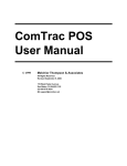
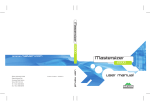
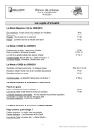
User Manual
LDH Cytotoxicity Detection Kit
User Manual
United States/Canada
800.662.2566
Asia Pacific
+1.650.919.7300
Europe
+33.(0)1.3904.6880
Japan
+81.(0)77.543.6116
Clontech Laboratories, Inc.
A Takara Bio Company
1290 Terra Bella Ave.
Mountain View, CA 94043
Technical Support (US)
E-mail: [email protected]
www.clontech.com
Cat. No. 630117
PT3947-1 (PR6Y2138)
Published 17 January 2007
LDH Cytotoxicity Detection Kit
Table of Contents
I. Introduction............................................................................................................................... 2
II. List of Components................................................................................................................... 4
III.Additional Materials Required.................................................................................................. 5
IV.LDH Cytotoxicity Detection Kit Protocol Overview................................................................ 6
A. Protocol Overview................................................................................................................................. 6
B. Experiment Controls............................................................................................................................. 6
V. Sample Preparation and Experiment Setup........................................................................... 7
A. Protocol: Preparing Working Solutions.................................................................................................. 7
B. Protocol: Determining the Optimal Cell Concentration........................................................................ 7
VI.Measuring Cytotoxicity............................................................................................................. 9
A. Protocol: Preparing Cell Suspensions to Measure Cytotoxicity Due to Soluble Compounds.................. 9
B. Protocol: Preparing Adherent Cells to Measure Cytotoxicity Due to Soluble Compounds................... 10
C. Protocol: Preparing Samples to Measure Cell-Mediated Cytotoxicity................................................... 11
D. Protocol: Assaying Cell Death in Fermentation Cultures..................................................................... 11
E. Protocol: Measuring LDH Release....................................................................................................... 12
F. Protocol: Calculating Cytotoxicity Due to Soluble Substances.............................................................. 12
G. Protocol: Calculating Percent Cell-Mediated Cytotoxicity................................................................... 12
VII.Troubleshooting...................................................................................................................... 13
VIII. References............................................................................................................................. 14
Appendix A: Examples of LDH Cytotoxicity Detection Kit Results.......................................... 15
A. Optimizing Cell Concentration........................................................................................................... 15
B. Measuring Detergent Cytotoxicity....................................................................................................... 15
C. Measuring Cell-Mediated Toxicity....................................................................................................... 16
List of Figures
Figure 1. A two-step enzymatic reaction quantifies cell lysis and cell death................................................. 2
Figure 2. Absorbance spectra of the LDH Cytotoxicity Detection Kit Reaction Mixture............................ 2
Figure 3. Optimizing cell concentration................................................................................................... 15
Figure 4. Measuring detergent cytotoxicity............................................................................................... 15
Figure 5. Measuring the cytolytic activity of allogen-stimulated, cytotoxic T lymphocytes........................ 16
List of Tables
Table I: Protocol Overview......................................................................................................................... 6
Table II: Working Solutions....................................................................................................................... 7
Table III: Plate Layout to Optimize Cell Concentration............................................................................. 7
Table IV: Plate Layout to Measure Cytotoxicity Due to Soluble Compounds........................................... 10
Table V: Plate Layout to Measure Cell-Mediated Cytotoxicity.................................................................. 11
Table VI: Troubleshooting the LDH Cytotoxicity Detection Kit.............................................................. 13
Protocol No. PT3947-1
www.clontech.com
Version No. PR6Y2138
Clontech Laboratories, Inc.
A Takara Bio Company
LDH Cytotoxicity Detection Kit
I. Introduction
Cell death is typically assayed by quantifying plasma membrane damage. Many standard methods are based on the uptake
or exclusion of vital dyes (Cook & Mitchell, 1989; Yuhas et al 1974; Parks, et al. 1979; Jones & Senft, 1985), or on
the release of radioactive isotopes, fluorescent dyes, or calcein-AM from prelabeled target cells (Oldham et al, 1977;
Leibold & Bridge, 1979; Kolber et al, 1988; Danks et al, 1992). Other assays measure cytoplasmic enzymes released by
damaged cells, where the amount of enzyme activity detected in the culture supernatant correlates with the proportion
of lysed cells (Decker & Lohmann-Mathes, 1988; Szekeres et al, 1981; Masanet et al, 1988; Martin & Clynes, 1991).
Enzyme release assays have been described for alkaline and acid phosphatase, glutamate-oxalacetate transaminase, glutamate
pyruvate transaminase and arginosuccinate lyase. However, their use has been hampered by the low amount of these
enzymes present in many cells, and by the elaborate kinetic assays required to quantitate most enzyme activities.
In contrast, lactate dehydrogenase (LDH) is a stable cytoplasmic enzyme which is present in all cells. When the plasma
membrane is damaged, LDH is rapidly released into the culture supernatant. The LDH Cytotoxicity Detection Kit
provides a simple and precise colorimetric method to measure LDH activity. Cell-free culture supernatant is collected
and incubated with the reaction mixture from the kit. LDH activity is determined by a colorimetric assay: In the first
step, NAD+ is reduced to NADH/H+ by the LDH-catalyzed conversion of lactate to pyruvate. In the second step, a
catalyst included in the reaction mixture (diaphorase) transfers H/H+ from NADH/H+ to the tetrazolium salt INT,
which is reduced to a formazan dye (Figure 1).
LDH
Lactic acid
NAD+
Formazan
(red)
Pyruvic acid
NADH H+
Diaphorase
Tetrazolium
(yellow)
Figure 1. A two-step enzymatic reaction quantifies cell lysis and cell death.
An increase in the number of dead or plasma membrane-damaged cells leads to increased LDH activity in the culture
supernatant, which directly correlates with the amount of formazan produced in a defined time period. Therefore, the
amount of dye produced is proportional to the number of lysed (dead or plasma membrane-damaged) cells. The red
formazan dye product is water soluble and shows a broad absorbance maximum around 500 nm, while the tetrazolium
salt INT shows little absorbance at this wavelength (Figure 2).
3.0
2.5
absorbance
2.0
1.5
1.0
0.5
0
400 450 500 550 600 650 700
wavelength [nm]
Figure 2. Absorbance spectra of the LDH Cytotoxicity Detection Kit Reaction Mixture. The reaction mixture from the LDH Cytotoxicity
Detection Kit was added to RPMI 1640 media with 1% BSA and the absorbance spectra was measured in the absence (...) and presence
(---) of LDH.
Clontech Laboratories, Inc. www.clontech.com
A Takara Bio Company
Protocol No. PT3947–1
Version No. PR6Y2138
LDH Cytotoxicity Detection Kit
I. Introduction continued
Proven uses of LDH assays include:
• Determination of the cytotoxic potential of compounds (Dubar et al., 1993; Kondo, et al., 1993; Murphy
et al., 1993; Courjault et al., 1993; Shrivastava et al., 1992; Gelderblom et al., 1993; Thomas et al., 1993;
Sasaki et al. 1992).
• Detection and quantification of cell-mediated cytotoxicity induced by cytotoxic T-lymphocytes (CTL), natural killer (NK) cells, lymphokine activated killer (LAK) cells or monocytes (Decker & Lohmann-Matthes,
1988; Korzeniewski & Callewaert, 1983).
• Determination of mediator-induced cytolysis (Decker & Lohmann-Matthes, 1988).
• Measurement of antibody-dependent cellular cytotoxicity and complement-mediated cytolysis.
• Determination of cell death in bioreactors (Goergen et al., 1993; Legrand et al., 1992; Racher et al., 1990).
The LDH Cytotoxicity Detection Kit provides several advantages over other cell proliferation assay reagents:
• Safety: No radioactive isotopes are used.
• Accuracy: Assay results obtained with this kit strongly correlate with the number of damaged cells. Furthermore, there is a good correlation between results from the LDH Cytotoxicity Detection Kit assay and the
[51Cr] release assay (Decker & Lohmann-Matthes, 1988; Korzeniewski & Callewaert, 1983).
• High sensitivity: Low numbers of dead cells can be detected (e.g., 2 x 10³–2 x 10⁴ cells/well, in a 96-well
plate).
• Speed: The assay takes only 0.5–1 hr, including harvesting supernatants and performing and measuring the
enzymatic reaction. Large numbers of samples can be processed simultaneously with a multiwell plate reader.
• Simple procedure: No need for prelabeling or washing steps. Cleanup and waste disposal are simplified since
this kit does not employ radioactive isotopes.
Protocol No. PT3947-1
www.clontech.com
Version No. PR6Y2138
Clontech Laboratories, Inc.
A Takara Bio Company
LDH Cytotoxicity Detection Kit
II. List of Components
Store unopened LDH Cytotoxicity Detection Kit reagents at –20°C. Dissolved catalyst and thawed dye solution can
be stored for several weeks at 4°C.
The LDH Cytotoxicity Detection Kit contains 5 bottles of Catalyst (lyophilized) and 5 bottles of Dye Solution, enough
for 2,000 reactions.
LDH Cytotoxicity Detection Kit (Cat. No. 630117)
• 5 bottles Catalyst (lyophilized)
• 5 bottles Dye Solution (45 ml each)
Supporting Documents
• LDH Cytotoxicity Detection Kit User Manual (PT3947-1)
Visit our web site at www.clontech.com for a current list of Cell Signaling/Apoptosis products.
Clontech Laboratories, Inc. www.clontech.com
A Takara Bio Company
Protocol No. PT3947–1
Version No. PR6Y2138
LDH Cytotoxicity Detection Kit
III. Additional Materials Required
The following materials are required but not supplied:
• Incubator (37°C)
• Centrifuge with rotor for 96-well plates
• Multiwell plate reader with a 490 or 492 nm filter. If a reference wavelength is to be subtracted, an additional
filter above 600 nm is required.
• Multichannel pipettor (100 µl)
• Sterile pipette tips
• 96-well plates (sterilized, cell culture quality) for measurements of cell-mediated lysis and for the analysis of
cytotoxic compounds:
• For suspension cells: round or V-bottom 96-well plates
• For adherent cells: flat-bottom 96-well plates
• For color development in all assays: optically clear, flat-bottom 96-well plates
• Assay Medium (e.g., medium containing 1% serum or 1% bovine serum albumin). Both human and animal
sera contain LDH, which may increase the background absorbance of the assay. Therefore, we recommend
performing the assay in the presence of low serum concentrations (e. g., 1%) or replacing the serum with
1% bovine serum albumin (w/v).
• Triton X-100 lysing solution (2% Triton X-100 in assay medium). The maximum LDH activity is determined by lysing the cells with Triton X-100 (final concentration 1%). At this concentration, Triton X-100
does not interfere with LDH activity.
• HCl stop solution (1 N). The reaction product can be measured without adding a stop solution.
Alternatively, the enzyme reaction can be stopped by adding 50 µl of 1 N HCl to each well (final concentration: 0.2 N HCl).
• LDH standard solution. If the released LDH activity must be calculated as Units/ml instead of percent cytotoxicity, we recommend that you prepare a standard curve using an LDH standard solution.
• Microscope
• Hemacytometer
Protocol No. PT3947-1
www.clontech.com
Version No. PR6Y2138
Clontech Laboratories, Inc.
A Takara Bio Company
LDH Cytotoxicity Detection Kit
IV. LDH Cytotoxicity Detection Kit Protocol Overview
Please read all protocols in their entirety before beginning.
Successfully implementing the LDH Cytotoxicity Detection Kit consists of performing the
steps listed below, all of which are included in this user manual.
A. Protocol Overview
The LDH Cytotoxicity Detection Kit is a simple colorimetric assay to quantitate cytotoxicity/cytolysis, and is based
on the measurement of LDH activity released from damaged cells into the supernatant (Table I). The cell-free culture
supernatant is collected and incubated with the Reaction Mixture to determine LDH activity and quantify cell death.
Specific protocols are included for four different types of experiments:
• Measuring cytotoxicity caused by soluble compounds for suspended cells.
• Measuring cytotoxicity caused by soluble compounds for adherent cells.
• Measuring cell-mediated cytotoxicity caused by effector cells.
• Measuring cell death in fermentation cultures.
Table I: Protocol Overview
Step
Procedure
Time
Optimizing Experimental Conditions
1.
Prepare working solutions
15 min
2.
Optimize cell concentration
Incubation time +
approx. 45 min
Performing the Experiment
1.
Incubate cells with test substance or cytotoxic effector cells
2–24 hr
2.
Prepare working solutions
15 min
3.
Centrifuge the 96-well plate containing the cells
10 min
4.
Transfer cell-free culture supernatant to optically clear, flat-bottom 96-well plate
5.
Incubate the supernatant with freshly prepared Reaction Mixture containing the
tetrazolium salt, INT
6.
(Optional) Stop the reaction by adding 1 N HCl to each well
7.
Measure absorbance at 490 or 492 nm (reference wavelength 690 nm) and calculate percent cytotoxicity
approx. 10–30 min
B. Experiment Controls
To calculate the percent cytotoxicity, you must perform the following three controls in every experiment:
• Background Control: Measures the LDH activity present in the assay medium. The background absorbance
must be subtracted from all other absorbance measurements.
• Low Control: Measures the level of spontaneous LDH release from untreated cells.
• High Control: Measures the maximum LDH activity that can be released from the 100% dead cells (in
response to Triton X-100).
In addition, the following two controls may be required to perform your experiment or for troubleshooting:
• Substance Control I: Measures the LDH activity contained in the test substance. Alternatively, if cell-mediated toxicity is measured, this control provides information on the LDH activity released from the effector
cells (e.g., NK cells, LAK cells, CTLs).
• Substance Control II: Determines whether the test substance itself interferes with LDH activity.
Note: Perform all assays, including controls, in triplicate.
Clontech Laboratories, Inc. www.clontech.com
A Takara Bio Company
Protocol No. PT3947–1
Version No. PR6Y2138
LDH Cytotoxicity Detection Kit
V. Sample Preparation and Experiment Setup
A. Protocol: Preparing Working Solutions
Prepare working solutions as directed in Table II.
Table II: Working Solutions
Recipes
Protocol
Incubation time,
plus 1 hr
Solution
Preparation
Storage and Stability
Catalyst (bottle 1;
blue cap)
Reconstitute the lyophilate in 1 ml distilled water
and mix thoroughly for 10 min.
Several weeks at 4° C.
Dye Solution (bottle 2;
red cap)
Thawed INT dye solution is ready to use.
Several weeks at 4° C.
Reaction Mixture
For 100 tests: Shortly before use, mix 250 µl of
Catalyst (bottle 1) with 11.25 ml of Dye Solution
(bottle 2).
For 400 tests: Shortly before use, mix the total
volume (1 ml) of one bottle of Catalyst (bottle 1)
with the total volume (45 ml) of one bottle of Dye
Solution (bottle 2).
Attention
Prepare immediately
before use. The reaction
mixture cannot be stored.
B. Protocol: Determining the Optimal Cell Concentration
LDH release varies among cell types. Therefore, you must determine the optimal cell concentration (where the difference between the High and Low Controls is at a maximum) for each cell type. The optimal cell concentration for most
cell lines is between 5 x 10³ and 2 x 10⁴ cells/well in 200 µl of media (2.5 x 10⁴–1 x 10⁵ cells/ml).
1. Fill the entire 96-well plate with 100 µl/well assay medium.
2. Wash the cells with assay medium, and then adjust the cell suspension to a concentration of 2 x 10⁶ cells/ml
with assay medium.
3. Table III outlines the plate setup: the 96-well plate is divided in half, with identical dilutions of the cell
suspension in wells B1–H6 and B7–H12. Titrate the cells by twofold serial dilutions in assay medium, using
a multichannel pipette:
a. Transfer 100 µl/well cell suspension into wells B1–B3 and B7–B9 (Dilution 1).
b. Transfer 100 µl of the diluted cell suspension from these wells into C1–C3 and C7–C9 (Dilution 2).
Repeat this step 12 times to prepare all the cell suspension dilutions.
Table III: Plate Layout to Optimize Cell Concentration
1
2
3
4
5
6
8
9
10
11
12
Background Control
B
1:1 cells:medium
2-7:1 cells:medium
1:1 cells:Triton X-100
2-7:1 cells:Triton X-100
C
0.5:1 cells:medium
2-8:1 cells:medium
0.5:1 cells:Triton X-100
2-8:1 cells:Triton X-100
D
0.25:1 cells:medium
2 :1 cells:medium
0.25:1 cells:Triton X-100
2-9:1 cells:Triton X-100
E
2-3:1 cells:medium
2-10:1 cells:medium
2-3:1 cells:Triton X-100
2-10:1 cells:Triton X-100
F
2-4:1 cells:medium
2-11:1 cells:medium
2-4:1 cells:Triton X-100
2-11:1 cells:Triton X-100
G
2 :1 cells:medium
2 :1 cells:medium
2 :1 cells:Triton X-100
2-12:1 cells:Triton X-100
H
2-6:1 cells:medium
2-13:1 cells:medium
2-6:1 cells:Triton X-100
2-13:1 cells:Triton X-100
-5
-9
-12
Low Controls
Protocol No. PT3947-1
www.clontech.com
Version No. PR6Y2138
7
A
-5
High Controls
Clontech Laboratories, Inc.
A Takara Bio Company
LDH Cytotoxicity Detection Kit
V. Sample Preparation and Experiment Setup continued
4. Prepare the following controls:
a. Background control: Add 100 µl assay medium to wells A1–A3.
b. Low Controls for spontaneous LDH release: Add 100 µl/well assay medium to Rows B–H, Columns 1–6.
c. High Controls for maximal LDH release: Add 100 µl/well Triton X-100 to Rows B–H, Columns 7–12.
5. Incubate the plate in a humidified atmosphere (37˚C, 5% CO2, 90% humidity) for the time required to assay the test substances in your experiment (typically in the range of 2–24 hr)
Note: Each well contains a total volume of 200 µl: 100 µl of assay medium, plus either 100 µl of cell suspension
(steps 2–3) or 100 µl of Control substance (step 4).
6. Centrifuge the plate at 250 x g for 10 min.
7. Remove 100 µl/well supernatant carefully, without disturbing the cell pellet. Transfer the supernatant into the
corresponding wells of an optically clear 96-well flat-bottom plate.
8. Add 100 µl Reaction Mixture (freshly prepared; Part V. A) to each well and incubate at room temperature for
30 min, protected from light.
9. Measure the absorbance of the samples at 490 or 492 nm. The reference wavelength should be greater than
600 nm.
10.Calculate the average absorbance value of each triplicate, and subtract the average value for the Background
Control from the triplicate average. Compare the Low and High Control values at each cell concentration.
The cell concentration with the greatest difference between the absorbance of the Low and High Controls is
the optimal cell concentration for your experiment (see Appendix A, part A for an example).
Clontech Laboratories, Inc. www.clontech.com
A Takara Bio Company
Protocol No. PT3947–1
Version No. PR6Y2138
LDH Cytotoxicity Detection Kit
VI. Measuring Cytotoxicity
Once you have optimized the cell concentration for your experiment (Part V. B), the assay steps are similar in each
case, although the sample preparation varies (Protocols A–D below). In brief, the assay steps are:
1. Incubate the optimal concentration of cells in a 96-well plate with the test substance or effector cells
(at varying concentrations).
2. Centrifuge the 96-well plate.
3. Transfer cell-free supernatant to an optically clear, flat-bottom 96-well plate.
4. Add freshly prepared Reaction Mixture and incubate to allow the enzymatic reaction to take place (Figure 1).
5. (Optional) Add 1 N HCl (not included in the kit) to stop the reaction.
6. Measure absorbance at 490 or 492 nm (reference wavelength greater than 600 nm)
All assays must be performed in triplicate.
A. Protocol: Preparing Cell Suspensions to Measure Cytotoxicity Due to Soluble Compounds
1. Titrate test substance (in triplicate) in assay medium into wells B1–G6 of a sterile 96-well plate by twofold
serial dilution (final volume: 100 µl/well).
Protocol
30 min
Note: Do not add test substance to the Control wells (A1–A6 and H1–H6; see Table IV).
2. Wash the cells in assay medium and dilute the cell suspension to the optimal concentration determined in
Part V. B. Add 100 µl/well cell suspension to the dilutions of the test substance.
Note: Do not add cells to the Control wells (A1–A6 and H1–H6; see Table IV).
3. Prepare the following controls on the plate. (See Table IV)
a. Background Control: Add 200 µl assay medium to triplicate wells A1–A3.
b. Substance Control I: Add 100 µl test substance (at the maximum concentration used in the experiment)
and 100 µl assay medium to triplicate wells A4–A6.
c. Low Control for spontaneous LDH release: Add 100 µl cell suspension and 100 µl assay medium to
triplicate wells H1–H3.
d. High Control for maximum LDH release: Add 100 µl cell suspension and 100 µl Triton X-100 solution
to triplicate wells H4–H6.
4. Proceed to Protocol E.
Protocol No. PT3947-1
www.clontech.com
Version No. PR6Y2138
Clontech Laboratories, Inc.
A Takara Bio Company
LDH Cytotoxicity Detection Kit
VI. Measuring Cytotoxicity continued
B. Protocol: Preparing Adherent Cells to Measure Cytotoxicity Due to Soluble Compounds
1. Plate 100 µl cell suspension (in culture medium, at the optimal concentration determined in Part V. B) into
wells B1–H6 of a sterile 96-well plate (see Table IV).
Protocol
overnight,
plus 30 min
Note: Do not add cells to the Background Control or Substance Control I wells (A1–A6; see Table IV).
2. Incubate the cells overnight (e.g. 37°C, 5% CO2, 90% humidity) to allow the cells to adhere tightly.
3. Immediately before use, titrate the test substance in assay medium in wells B1–G6 of a separate 96-well plate
by twofold serial dilution (final volume of 200 µl/well).
Note: Do not add the test substance to the wells corresponding to the Control wells (A1–A6 and H1-H6; see
Table IV).
4. Remove the culture medium from the adherent cells (to remove LDH released from the cells during the
overnight incubation). Add 100 µl fresh assay medium to each well.
5. Transfer 100 µl of the test substance dilutions into the corresponding wells containing adherent cells.
Note: Do not add the test substance to the Control wells (A1–A6 and H1-H6; see Table IV).
6. Prepare the following controls on the plate (see Table IV).
a. Background Control: Add 200 µl assay medium to triplicate wells A1–A3.
b. Substance Control I: Add 100 µl test substance (at the maximum concentration used in the experiment)
and 100 µl/well assay medium to triplicate wells A4–A6.
c. Low Control for spontaneous LDH release: Add 200 µl assay medium to triplicate wells H1–H3.
d. High Control for maximum LDH release: Add 100 µl assay medium and 100 µl Triton X-100 solution to
triplicate wells H4–H6.
7. Proceed to Protocol E.
Table IV: Plate Layout to Measure Cytotoxicity Due to Soluble Compounds
1
2
3
4
5
6
A
Background Control
Substance Control I
B
1:1 test substance:cells
2-6:1 test substance:cells
C
0.5:1 test substance:cells
2-7:1 test substance:cells
D
0.25:1 test substance:cells
2-8:1 test substance:cells
E
2-3:1 test substance:cells
2-9:1 test substance:cells
F
2 :1 test substance:cells
2-10:1 test substance:cells
G
2 :1 test substance:cells
2-11:1 test substance:cells
H
Low Control
High Control
-4
-5
7
8
9
10
11
12
Table IV. 96-well plate layout to assay cytotoxicity (Columns 1–6). Note that a second test substance could be assayed in columns 7–12.
Clontech Laboratories, Inc. www.clontech.com
A Takara Bio Company
Protocol No. PT3947–1
Version No. PR6Y2138
10
LDH Cytotoxicity Detection Kit
VI. Measuring Cytotoxicity continued
Protocol
30 min
C. Protocol: Preparing Samples to Measure Cell-Mediated Cytotoxicity
1. Table V outlines the plate setup: the 96-well plate is divided in half, with identical dilutions of the effector
cell suspension in wells B1–G6 and B7–G12. Titrate the effector cells (in triplicate) by twofold serial dilutions in assay medium (final volume 100 µl/well), using a multichannel pipette.
Note: Do not add effector cells to the Control wells (Rows A & H; see Table V).
2. Wash the test cells in assay medium, and dilute them to their optimal concentration (Part V. B).
3. Add 100 µl/well test cell suspension to the effector cell dilutions in wells B1–G6 (see Table V).
Note: Do not add test cells to the Control wells (A1–A6 and H1–H6; see Table V) or to the effector-cell only wells
(B7–G12).
4. Prepare the following controls on the plate (see Table V).
a. Background Control: Add 200 µl assay medium to triplicate wells A1–A3.
b. Low Control for spontaneous LDH release: Add 100 µl test cell suspension and 100 µl assay medium to
triplicate wells H1–H3.
c. High Control for maximum LDH release: Add 100 µl test cell suspension and 100 µl Triton X-100 solution to triplicate wells H4–H6.
d. Spontaneous LDH release for each effector cell concentration: Add 100 µl assay medium to wells B7–
G12.
Note: Spontaneous LDH release must be determined for each effector cell concentration used in the assay.
5. Proceed to Protocol E.
Table V: Plate Layout to Measure Cell-Mediated Cytotoxicity
1
2
3
4
5
6
7
8
9
10
11
12
A
Background Control
B
1:1 effector:test cells
2-6:1 effector:test cells
1:1 effector cells:medium
2-6:1 effector cells:medium
C
0.5:1 effector:test cells
2-7:1 effector:test cells
0.5:1 effector cells:medium
2-7:1 effector cells:medium
D
0.25:1 effector:test cells
2 :1 effector:test cells
0.25:1 effector cells:medium
2-8:1 effector cells:medium
E
2-3:1 effector:test cells
2-9:1 effector:test cells
2-3:1 effector cells:medium
2-9:1 effector cells:medium
F
2-4:1 effector:test cells
2-10:1 effector:test cells
2-4:1 effector cells:medium
2-10:1 effector cells:medium
G
2-5:1 effector:test cells
2-11:1 effector:test cells
2-5:1 effector cells:medium
2-11:1 effector cells:medium
H
Low Control
High Control
-8
D. Protocol: Assaying Cell Death in Fermentation Cultures
1. Collect samples from the cell culture (0.5–1 ml) at regular intervals of 12–24 hr.
Protocol
Culture time,
then 30 min
2. Centrifuge each sample and carefully remove the culture supernatant without disturbing the cell pellet.
Note: The cell-free supernatant can be stored at 4°C for several days without loss of LDH enzyme activity.
3. Titrate the culture supernatants with culture medium by serial dilutions into an optically clear 96-well flatbottom plate (final volume 100 µl/well).
4. Proceed to Protocol E, step 4.
Protocol No. PT3947-1
www.clontech.com
Version No. PR6Y2138
11
Clontech Laboratories, Inc.
A Takara Bio Company
LDH Cytotoxicity Detection Kit
VI. Measuring Cytotoxicity continued
E. Protocol: Measuring LDH Release
1. Incubate the plate in an incubator (e.g. 37°C, 5% CO2, 90% humidity) for the time required to assay the test
substance or effector cells (2–24 hr).
Protocol
Incubation time,
plus 45 min
2. After incubation, centrifuge the plate at 250 x g for 10 min.
3. Carefully remove 100 µl of supernatant from each well (do not disturb the cell pellet) and transfer into the
corresponding wells of an optically clear 96-well flat-bottom plate.
4. Add 100 µl freshly prepared Reaction Mixture (Part V.A) to each well and incubate for up to 30 min at room
temperature, protected from light.
5. Measure the absorbance of the samples at 490 or 492 nm using a multiwell plate reader. The reference wavelength should be more than 600 nm.
6. Calculate the percent cytotoxicity according to Protocol F or Protocol G, as appropriate.
F. Protocol: Calculating Cytotoxicity Due to Soluble Substances
Use this formula to calculate cytotoxicity due to the addition of a potentially cytotoxic substance (based on the results
of Part VI, Protocols A, B, or D).
1. Calculate the average absorbance value for each triplicate. Subtract the average Background Control value
from the average absorbance value for each triplicate to determine absorbance due to cell lysis (or other
factors, in the case of Control triplicates).
2. Substitute the resulting values into the following equation:
Cytotoxicity (%) =
Triplicate Absorbance - Low Control
× 100
High Control - Low Control
G. Protocol: Calculating Percent Cell-Mediated Cytotoxicity
Use this formula to calculate cytotoxicity due to effector cells (based on the results of Part VI, Protocol C).
1. Calculate the average absorbance value for each triplicate. Subtract the average Background Control value
from the average absorbance value for each triplicate to determine absorbance due to cell lysis (or other
factors, in the case of Control triplicates).
2. Substitute the resulting values into the following equation, using the Effector: Test Cell Mix and Effector Cell
Control values for a given concentration of effector cells.
Cytotoxicity (%) =
(Effector:Test Cell Mix - Effector Cell Control) - Low Control
High Control - Low Control
Clontech Laboratories, Inc. www.clontech.com
A Takara Bio Company
× 100
Protocol No. PT3947–1
Version No. PR6Y2138
12
LDH Cytotoxicity Detection Kit
VII.Troubleshooting
Table VI: Troubleshooting the LDH Cytotoxicity Detection Kit
Description of Problem Explanation
Solution
No color reaction
Cell concentration may be too
low
Check cell concentration; titrate if necessary.
Test substance or assay
medium inhibit LDH activity
Use Substance Control II to measure test substance
and/or assay medium for intrinsic LDH inhibition.
Avoid using media which includes pyruvate (an LDH
inhibitor)
Strong color reaction in
Low Controls
Cell concentration may be too
high
Check cell concentration; titrate if necessary.
Test substance or assay
medium have LDH activity
Use Substance Control I to measure test substance
and/or assay medium for intrinsic LDH activity.
Spontaneous LDH release
caused by cell death due to
assay conditions
Check culture conditions; some cell lines do not survive in serum free media, even for short incubation
times. Increase serum concentration to about 1–5%.
Strong color reaction, but
low absorbance values
Test substance or assay
medium have LDH activity
Use Substance Control I to measure test substance
and/or assay medium for intrinsic LDH activity.
Strong color reaction in
Effector Cell Controls
Spontaneous LDH release
caused by effector cell damage
or death, due to culture conditions or isolation method
Improve cell culture conditions.
Separate viable from dead effector cells by density
gradient centrifugation.
• Substance Control I: Measures the LDH activity contained in the test substance or effector cells. The procedure to perform Substance Control I is described in detail in the Part VI Protocols.
• Substance Control II: Determines whether the test substance itself interferes with LDH activity. To perform
this control proceed as follows:
a. Add 50 µl test substance solution (diluted in assay medium), 50 µl LDH solution (0.05 U/ml) and 100 µl
freshly prepared Reaction Mixture (Part V.A) to triplicate wells.
b. Add 50 µl assay medium, 50 µl LDH solution (0.05 U/ml) and 100 µl freshly prepared Reaction Mixture
to a second set of triplicate wells (e.g., the control triplicate).
c. Incubate for up to 30 min at room temperature, protected from light.
d. Measure the absorbance of the samples at 490 or 492 nm using a multiwell plate reader. The reference
wavelength should be more than 600 nm.
e. Compare the average absorbance value of the triplicate containing the test substance with the average
absorbance value of the control triplicate.
Protocol No. PT3947-1
www.clontech.com
Version No. PR6Y2138
13
Clontech Laboratories, Inc.
A Takara Bio Company
LDH Cytotoxicity Detection Kit
VIII. References
Cook, J. A. & Mitchell, J. B. (1989) Viability measurements in mammalian cell systems. Anal. Biochem. 179:1-7.
Courjault, F., Leroy, D., Coquery, L. & Toutain, H. (1993) Platinum complex-induced dysfunction of cultured renal proximal tubule cells. A comparative study of
carboplatin and transplatin with cisplatin. Arch. Toxicol. 67:338-346.
Danks, A.M., Hammond, D.N., Wainer, B.H., Van Buskirk, R.G. & Isaacson, R.L. (1992) Cellular alterations produced by the experimental increase in intracellular
calcium and the nature of protective effects from pretreatment with nimodipine. Brain Res. Mol. Brain Res. 16:168-172.
Decker, T. & Lohmann-Matthes, M.-L. (1988) A quick and simple method for the quantitation of lactate dehydrogenase release in measurements of cellular cytotoxicity and tumor necrosis factor (TNF) activity. J. Immunol. Meth. 115:61–69.
Dubar, V., Gosset, P., Aerts, C., Voisin, C., Wallaert, B. & Tonnel, A.B. (1993) In vitro acute effects of tobacco smoke on tumor necrosis factor alpha and
interleukin-6 production by alveolar macrophages. Exp. Lung Res. 19:345-359.
Gelderblom, W.C., Cawood, M.E., Snyman, S.D., Vleggaar, R. & Marasas, W.F. (1993) Structure-activity relationships of fumonisins in short-term carcinogenesis
and cytotoxicity assays. Food Chem. Toxicol. 31:407-414.
Goergen, J.-L., Marc, A. & Engasser, J.-M. (1993) Determination of cell lysis and death kinetics in continuous hybridoma cultures from the measurement of lactate
dehydrogenase release. Cytotechnol. 11:189-195.
Jones, K. H. & Senft, J. A. (1985) An improved method to determine cell viability by simultaneous staining with fluorescein diacetate-propidium iodide. J. Histochem. Cytochem. 33:77-79.
Kolber, M.A., Quinones, R.R., Gress, R.E. & Henkart, P.A. (1988) Measurement of cytotoxicity by target cell release and retention of the fluorescent dye bis-carboxyethyl-carboxyfluorescein (BCECF). J. Immunol. Meth. 108:255-264.
Kondo, T., Takahashi, S., Sato, H., Yamada, M., Kikuchi T. & Furuya, K. (1993) Cytotoxicity of size-density fractionated coal fly ash in rat alveolar macrophages
cultured in vitro. Toxicol. in Vitro 7:61-67.
Korzeniewski, C. & Callewaert, D. M. (1983) An enzyme-release assay for natural cytotoxicity. J. Immunol. Meth. 64:313–320.
Legrand, C.; Bour J.M.; Jacob C.; Capiaumont J.; Martial A.; Marc A.; Wudtke M.; Kretzmer G.; Demangel C.; Duval D, et al. & Hache, J. (1992) Lactate dehydrogenase (LDH) activity of the cultured eukaryotic cells as marker of the number of dead cells in the medium [corrected], J. Biotechnol. 25:231-243. Erratum in J.
Biotechnol. (1993) 31:234.
Leibold, W. & Bridge, S. (1979) 75Se-release: a short and long term assay system for cellular cytotoxicity. Z. Immunitätsforschung Immunobiol. 155:287-311.
Martin, A. & Clynes, M. (1991) Acid phosphatase: endpoint for in vitro toxicity tests. In Vitro Cell Dev. Biol. 27A:183-184.
Masanet, J., Gomez-Lechon, M. J. & Castell, J. V. (1988) Hepatic toxicity of paraquat in primary cultures of rat hepatocytes. Toxicol. in Vitro 2:275-282.
Murphy, E.J., Roberts, E. & Horrocks, L.A. (1993) Aluminum silicate toxicity in cell cultures. Neurosci. 55:597-605.
Oldham, R.K., Ortaldo, J.R., Holden, H.T. & Herberman, R.B. (1977) Direct comparison of three isotopic release microtoxicity assays as measures of cell-mediated
immunity to Gross virus-induced lymphomas in rats. J. Natl. Cancer Inst. 58:1061-1067.
Parks, D.R., Bryan, V.M., Oi, V.T. & Herzenberg, L.A. (1979) Antigen-specific identification and cloning of hybridomas with a fluorescence-activated cell sorter.
Proc. Natl. Acad. Sci USA 76:1962-1966.
Racher, A.J., Looby, D. & Griffiths, J.B. (1990) Use of lactate dehydrogenase release to assess changes in culture viability. Cytotechnol. 3:301-307.
Sasaki, T., Kawai, K., Saijo-Kurita, K. & Ohno T. (1992) Detergent cytotoxicity: simplified assay of cytolysis by measuring LDH activity. Toxicol. in Vitro 6:451-457.
Shrivastava, R., Delomenie, C., Chevalier, A., John, G., Ekwall, B., Walum, E. & Massingham, R. (1992) Comparison of in vivo acute lethal potency and in vitro
cytotoxicity of 48 chemicals. Cell Biol. Toxicol. 8:157-170.
Szekeres, J., Pacsa, A.S. & Pejtsik, B. (1981) Measurement of lymphocyte cytotoxicity by assessing endogenous alkaline phosphatase activity of the target cells.
J. Immunol. Meth. 40:151-154.
Thomas, J.P., Geiger, P.G. & Girotti, A.W. (1993) Lethal damage to endothelial cells by oxidized low density lipoprotein: role of selenoperoxidases in cytoprotection
against lipid hydroperoxide- and iron-mediated reactions. J. Lipid Res. 34:479-490.
Yuhas, J.M., Toya, R.E. & Pazmino, N.H. (1974) Neuraminidase and cell viability: failure to detect cytotoxic effects with dye-exclusion techniques. J. Nat. Cancer
Inst. 53:465-468.
Clontech Laboratories, Inc. www.clontech.com
A Takara Bio Company
Protocol No. PT3947–1
Version No. PR6Y2138
14
LDH Cytotoxicity Detection Kit
Appendix A: Examples of LDH Cytotoxicity Detection Kit Results
A. Optimizing Cell Concentration
K562 cells were titrated in 96-well plates at the concentrations indicated on the horizontal axis of Figure 3.
Culture medium ( ) was added to measure spontaneous LDH release (Low Control), and Triton X-100 ( ) was added
(final concentration 1%) to measure the maximum LDH release (High Control). The optimal cell concentration in
this experiment was between 1 x 10⁴ and 1 x 10⁵ cells/well.
3.0
absorbance (OD492nm - OD620nm)
2.5
2.0
1.5
1.0
0.5
0
101
102
103
cells/well
104
105
Figure 3. Optimizing K562 cell concentration.
B. Measuring Detergent Cytotoxicity
Three detergents (Synperonic® F68 ( ), Triton X-100 ( ) and Nonident P40 ( ) were titrated with culture medium in
a 96-well plate to the final concentrations indicated on the horizontal axis of Figure 4. Subsequently, P815 cells were
added (final concentration 1 × 10⁴ cells/well). The cells were incubated for 18 hr and LDH release was measured.
absorbance (A492nm - A690nm)
1.0
0.5
0
10-4
10-3
10-2
detergent conc. [%]
Figure 4. Measuring detergent cytotoxicity. Synperonic® F68 ( ), Triton X-100 ( ) and Nonident P40 ( ).
Protocol No. PT3947-1
www.clontech.com
Version No. PR6Y2138
15
Clontech Laboratories, Inc.
A Takara Bio Company
LDH Cytotoxicity Detection Kit
Appendix A: Examples of LDH Cytotoxicity Detection Kit Results continued
C. Measuring Cell-Mediated Toxicity
Spleen cells of C57/Bl 6 mice (H-2b) were stimulated in vitro with P815 cells (H-2d). Viable cytotoxic T lymphocytes
(CTLs) were purified by ficoll density gradient, washed and titrated in a 96-well plate. 1 × 10⁴ P815 test cells/well were
added to the effector CTL cells. The cell mixture was centrifuged and incubated for 4 hr. 100 µl of culture supernatant
was collected to measure LDH activity.
A
B
Figure 5. Measuring the cytolytic activity of allogen-stimulated, cytotoxic T lymphocytes. Panel A. Absorbance values. Effector cell control
( ), effector-test cell mix ( ), effector-test cell mix minus effector cell control ( ). Panel B. Percent cell-mediated cytotoxicity, calculated as
described in Part VI.G.
Notice to Purchaser
Clontech products are to be used for research purposes only. They may not be used for any other purpose, including, but not limited to, use in drugs, in vitro
diagnostic purposes, therapeutics, or in humans. Clontech products may not be transferred to third parties, resold, modified for resale, or used to manufacture
commercial products or to provide a service to third parties without written approval of Clontech Laboratories, Inc.
Clontech, Clontech logo and all other trademarks are the property of Clontech Laboratories, Inc. unless noted otherwise.
Clontech is a Takara Bio Company. ©2007
Clontech Laboratories, Inc. www.clontech.com
A Takara Bio Company
Protocol No. PT3947–1
Version No. PR6Y2138
16