1
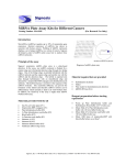
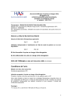
Signosis Innovative Plate Assay Solutions Cancer miRNA Profiling Plate Assay Kit I Catalog Number MA-0103 (For Research Use Only) Introduction miRNAs have been implicated in regulating various biological processes such as cell death and cell proliferation. They can act as oncogenes and tumor suppressor genes, playing a crucial role in tumorogenesis. The change in the expression level of miRNAs is associated with dysfunction of their corresponding protein-coding gene targets, many of which are involved in initiation and progression of cancer. For example, BCL2 oncogene is targeted by miR-15a and miR-16 target, PTEN tumor suppressor by miR-21, HOXD10 by miR-10b, Pak by miR-7, MYOD1 and ER by miR-206, and Her2 by miR125a. Signosis developed cancer miRNA plate assay kit I for quantitatively profiling the expression of seven well-known cancer related miRNAs, miRNA-15a, miR-16, miR-21, miR10b, miR-7, miR-125a and miR206. RNU48 is included for normalization. Diagram of miRNA plate array Principle of the assay Signosis’ proprietary miRNA plate array is a plate-based detection. In the assay, one miRNA molecule is flanked by a capture oligo and a biotinated detection oligo through two bridge oligos. One of the bridge oligos is partially hybridized with the miRNA molecule and the capture oligo and another one with the miRNA and the detection oligo. The hybrid is captured onto plate through hybridization with an immobilized oligo and detected by a streptavidin-HRP conjugate and chemiluminecscent substrate. This hybrid structure is sensitive to the sequence of the miRNA molecule. One nucleotide difference can prevent the formation of the hybrid and therefore miRNA isoform can be differentiated, which normally is hard to do with Northern blot. In addition, the sensitivity of the assay is higher than miRNA Northern blot assay. Materials provided with the kit One 96-well white plate (4oC) Streptavidin-HRP conjugate (4oC) Plate hybridization buffer (RT) 5x Plate hybridization wash buffer (RT) Block buffer (RT) 5x Detection wash buffer (RT) Substrate A (4oC) Substrate B (4oC) Substrate dilution buffer (RT) 8 different miRNA oligo mixes Material required but not provided Hybridization incubator Shaker Plate reader for chemiluminescent detection ddH2O (RNAase free) Reagent preparation before starting experiment Warm up Plate hybridization buffer and Hybridization Wash buffer at 45 oC before use. Dilute 30ml of 5x Plate Hybridization wash buffer with 120 ml of dH2O before use. Dilute 40ml of 5x Detection wash buffer with 160 ml of dH2O before use. Dilute 1000 times of streptavidin-HRP with block buffer before use at Step 10. Signosis, Inc. • 1700 Wyatt Drive Suite 10-12 • Santa Clara, CA 95054 • Tel 408 747 0771 • Fax 408 864 2182 Assay procedure Example of Data Analysis 1. Warm up the plate to room temperature, and arrange the appropriate number of the wells of the plate based on your experiment by removing the top foil sealing film with a blade. Keep the unused well sealed. Cancer miRNA Plate Assay I Analysis Normal Liver Breast cancer T47D 6000 5000 Make fresh 30X dilution of each oligo mix RLU 4000 Mix the following items in one well. 2ul -5 µl RNA (0.2µg-2 µg) 100 µl Plate hybridization buffer 4 µl diluted oligo mix 4ul Biotin Detection Oligo 2. Seal the wells with foil film securely and incubate the plate at 45 oC overnight. Ensure the numbers and letters on the plate are clearly visible from under foil seal by pressing the foil down on every single experimental well. Put an open container with water in the incubator to keep humidity and prevent evaporation from experimental wells. 3000 2000 1000 0 miR15 miR-16 miR-21 miR-10b miR-7 miR-125a miR-206 RNU48 Figure1: Cancer miRNA plate assay analysis of expression of seven genes. Expression of miRNAs was analyzed with 0.5ug total RNA from normal liver and T47D cells respectively. The assay was subjected to chemiluminescent plate reader. 3. Invert the plate over an appropriate container and expel the contents forcibly, and wash the plate 3 times by adding 200µl of pre-warmed 1x Plate Hybridization Wash Buffer. 4. Complete removal of liquid at each wash by firmly tapping the plate against clean paper towels. 5. Add 200µl of Block buffer incubate for 15 minutes at room temperature with gentle shaking. 6. Invert the plate over an appropriate container to remove block buffer. 7. Add 100 µl of diluted streptavidin-HRP conjugate to each well and incubate for 30 min at room temperature with gentle shaking. 8. Wash the plate 3 times with 1X Detection wash buffer. Complete removal of liquid at each wash by firmly tapping the plate against clean paper towels. 9. Freshly prepare the substrate solution: For the whole plate: 1ml Substrate A 1ml Substrate B 8ml Substrate dilution buffer 10. Add 95µl substrate solution to each well and incubate for 1 min. 11. Place the plate in the luminometer, and read. Set integration time to 1 second with no filter position. For the best results, read the plate within 5-20 minutes. Signosis, Inc. • 1700 Wyatt Drive Suite 10-12 • Santa Clara, CA 95054 • Tel 408 747 0771 • Fax 408 864 2182