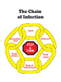
1
Infectious Diseases Resource for Emergency Service Workers Mar 2012 The Leeds, Grenville and Lanark District Health Unit www.healthunit.org Infectious Disease Program Community Health Protection Department 1-800-660-5853 (613)-345-5685 Brockville (613)-283-2740 Smiths Falls You can find this document at: www.healthunit.org/professionals/infectious/infectious.html The Leeds, Grenville and Lanark District Health Unit www.healthunit.org Infectious Diseases Resource for Emergency Service Workers. March 2012 TABLE OF CONTENTS Introduction / Objectives Section 1: Chain of Infection Section 2: Routine Practices and Additional Precautions Routine Practices / Risk Assessment Hand Hygiene Environmental Controls Cleaning Blood Spills Administrative Controls Policies and procedures/ Staff Education including Respiratory Etiquette Healthy workplace policies including Immunization programs Personal Protective Equipment Gloves / Masks / Respirators / Eye Protection / Gowns/Coveralls Additional Precautions Section 3: Notification Process by Emergency Service Worker Sample Designated Officer – Risk Assessment Form Section 4: Blood Borne Infections Hepatitis B Hepatitis C HIV/AIDS Exposure to Blood Borne Infections Emergency Service Guide to the Mandatory Blood Testing Act 2006 Section 5: Other Communicable Diseases and Emerging Issues Influenza Tuberculosis Meningococcal Disease Invasive Group A Streptococcal Disease Lyme Disease Bed Bugs Section 6: Antibiotic Resistant Organisms MRSA (Methicillin-Resistant Staphylococcus Aureus) VRE (Vancomycin Resistant Enterococcus) ESBL (Extended Spectrum Beta-Lactamase Producing Bacteria) C-Difficile (Clostridium Difficile) Section 7: APPENDICES APPENDIX 1 APPENDIX 2 APPENDIX 3 APPENDIX 4 APPENDIX APPENDIX APPENDIX APPENDIX 5 6 7 8 Clinical Syndromes Requiring the Use of Controls (including PPE) Pending Diagnosis Directions on the Preparation and Use of Chlorine Based Disinfectants Sample Procedures for Cleaning an Ambulance Advantages and Disadvantages of Hospital Grade Disinfectants and Sporicides Used for Environmental Cleaning Recommended Immunizations for all Emergency Service Workers Mandatory Blood Testing Act – Applicant Report Mandatory Blood Testing Act – Physician Report Screening Tool for Acute Respiratory Illness in Health Care Settings The Leeds, Grenville and Lanark District Health Unit www.healthunit.org Infectious Diseases Resource for Emergency Service Workers. March 2012 INTRODUCTION Emergency Service Workers (paramedics, fire fighters and police officers) may in the course of their duties, be exposed to communicable diseases. They are frequently called upon to react and provide the first level of care in working environments that are often uncontrolled, and in situations where they may risk their own safety to protect others. Within this working environment, it is possible for emergency service workers to be exposed to communicable diseases and to be unaware that they have been exposed. Training in recognizing and minimizing the risk of exposure will help reduce, but not eliminate, this risk. OBJECTIVES The objectives of this resource are as follows: • To educate Emergency Service Workers about the means of transmission of specific communicable diseases and the risk of occupational exposure • To encourage Emergency Service Workers to adopt practices and procedures to protect themselves and others • To educate Emergency Service Workers about the notification process The Leeds, Grenville and Lanark District Health Unit www.healthunit.org Infectious Diseases Resource for Emergency Service Workers. March 2012 SECTION 1: CHAIN OF INFECTION The spread of infection is best described as a chain with six links: • Infectious Agents • Reservoirs • Portals of Exit • Modes of Transmission • Portals of Entry • Susceptible Hosts Infectious Agents Bacteria, viruses, fungi and parasites are all Infectious Agents, which if given the opportunity can enter and survive in the body. Reservoirs A Reservoir is any person, animal, anthropod, plant soil or substance in which an infectious agent normally lives and multiplies. Infectious agents need reservoirs in order to survive, reproduce and be transmitted. Humans are the most common reservoirs of infectious agents. Portals of Exit Portals of Exit allows the infectious agent to escape their reservoir. Portals of exit from the human reservoir include: • Blood: open wound, needle puncture site, breaks in skin or mucous membranes • Respiratory Tract: nose and mouth (sneezing, coughing, breathing, talking), endotracheal tubes • Gastrointestinal Tract: mouth (saliva, vomitus), anus (feces/diarrhea), drainage tubes • Urinary Tract: urethral meatus Modes of Transmission The infectious agent travels to the host through Transmission. There are five routes of transmission: • • • • • Airborne Contact (direct and indirect) Droplet Vector Common Vehicles The Leeds, Grenville and Lanark District Health Unit www.healthunit.org Infectious Diseases Resource for Emergency Service Workers. March 2012 Airborne • • • Very small droplets generated by the person when he/she talks, coughs or sneezes These droplets stay suspended on dust particles or air The host needs to breathe in the infectious agent (e.g., chickenpox, tuberculosis, measles) Contact • • Direct contact is the transfer of microorganisms from direct physical contact between the infected/colonized person and the host (body surface to body surface) (e.g., MRSA, scabies) Indirect contact is the transfer of microorganisms to the host via an intermediate object (e.g., gloves not changed between clients) Droplet • • Large droplets are generated by the person when he/she talks, coughs or sneezes Droplets are projected in the air a short distance and deposited on the mucosa of the host: nose, eyes, mouth (e.g., bacterial meningitis, influenza) Vector • An insect/animal transmits the infectious agent to the host. (e.g., rabies, Lyme disease, West Nile virus) Common Vehicles • Common vehicle transmission refers to a single contaminated source such as food, medication, intravenous fluid, equipment, etc., which serves to transmit infection to multiple hosts. This can result in an explosive outbreak. Control is by maintenance of appropriate standards in the preparation of food and medications and in decontamination of equipment. Portals of Entry Infectious agents often enter the body through the same route they exited the reservoir (non-intact skin, respiratory tract). Susceptible Hosts The final link in the chain of infection is a susceptible host; someone at risk of infection. Infection does not occur automatically when the infectious agent enters the body. Some factors affecting the ability to fight off infection are age, underlying disease, immunocompromised status. The Chain of Infection Agent Susceptible Host Portal of Entry Reservoir STOP & THINK Portal of Exit Mode of Transmission Breaking The Chain of Infection nt Tre Un atme der nt lyi of ng Di sea tme a e r /T osis gn Dia /P Agent ses Susceptible ati Host on mu niz t rs Fi Reservoir Portal of Entry d Ai Portal of Exit ne ie o rs Pe g hin Mode of Transmission s a dw n Ha d Han n io at ol Is tal men n iron Env anitatio S g hin s a w Disinfection l na g Hy n tio a c du E Im y ic ol Han dwa Co Tr as h& tal n me tion n iro anita v S En on i t fec n i s Di ntr ol of shin g Ex Se creti cre on te tio Di ns sp os al W as & The Leeds, Grenville and Lanark District Health Unit www.healthunit.org Infectious Diseases Resource for Emergency Service Workers. March 2012 SECTION 2: ROUTINE PRACTICES & ADDITIONAL PRECAUTIONS Routine practices Infection prevention means breaking the chain of infection or interrupting the infectious disease process. Routine practices refer to infection prevention and control practices to be used with all patients during all care, to prevent and control transmission of microorganisms in all health care settings. • Assumes blood and body fluid of ANY client could be infectious • Recommends personal protective equipment and other infection control practices to prevent transmission • Decisions about personal protective equipment use is determined by the type of clinical interaction with the client The basic elements of Routine Practices are: Risk assessment, hand hygiene, environmental controls, administrative control, and personal protective equipment. Risk assessment • • • Done before each interaction with a client or their environment A dynamic process based on continuing changes in information Determines which interventions are required to prevent transmission during the interaction The risk assessment includes: a) assessing the risk of contamination of skin or clothing, exposure to blood, body fluids, secretions, excretions, tissues, exposure to non-intact skin, exposure to mucous membranes and exposure to contaminated equipment or surfaces AND b) recognition of symptoms of infection such as diarrhea and/or vomiting, cough, fever. Questions to Ask: Do I need protection for what I am about to do because there is a risk of exposure to blood and body fluids, mucous membranes, non-intact skin or contaminated equipment? (I must follow Routine Practices because there is a risk that I might expose myself to an infection that is transmitted via this route, or expose the patient to my microorganisms.) Do I need protection for what I am about to do because the client has undiagnosed symptoms of infection? (I must alert someone about the client who has symptoms so that a diagnosis may be made, and I must determine what organizational requirements are to be put in place to protect myself and others.) What are the organizational requirements for this client who has an identified infection? (I must follow the procedures proscribed for this infection to protect myself and others.) See Appendix #1 Clinical Syndromes Requiring the Use of Controls (including PPE) Pending Diagnosis. The Leeds, Grenville and Lanark District Health Unit www.healthunit.org Infectious Diseases Resource for Emergency Service Workers. March 2012 Hand hygiene When done correctly, hand hygiene is the best defense against germs and getting sick. Good hand hygiene is easy to learn and can reduce the spread of illness in both children and adults. When should you perform hand hygiene? Before you: • • • • Prepare or eat food Treat a cut or wound Look after someone who is sick Put in or take out contact lenses After you: • • • • • • • Go to the bathroom Handle uncooked food Blow your nose Handle garbage Handle an animal or waste Use the phone or computer Look after someone who is ill How should you wash your hands? 1. Wet hands with warm running water. 2. Add soap, and then scrub hands together to make a soapy lather. Do this away from the water for at least 15 seconds. 3. Wash the front and back of your hands, as well as in between fingers, around thumbs and under nails. 4. Rinse hands well, under warm running water. 5. Dry hands with a paper towel. 6. Turn off water using paper towel and dispose. What about alcohol based hand sanitizers? These are excellent at killing most germs. However, they must contain at least 70% alcohol. How do you use an alcohol based hand sanitizer? These should only be used if your hands are not visibly dirty. If hands are visibly dirty and soap and water is not available, wipe hands with a moistened towelette followed up with the use of hand sanitizer. They are widely used in the health care setting as an alternative to hand washing when access to water is inconvenient or unavailable. The Leeds, Grenville and Lanark District Health Unit www.healthunit.org Infectious Diseases Resource for Emergency Service Workers. March 2012 Steps to use alcohol based hand sanitizer: 1. 2. 3. 4. 5. 6. 7. 8. Apply enough sanitizer to open palms. Run hands together palm to palm. Rub in between and around fingers. Rub back of each hand with palm of other hand. Rub finger tips of each hand in opposite palm. Rub each thumb clasped in opposite thumb. Rub each wrist clasped in opposite hand. Keep rubbing hands until dry. Paper towels are not needed. Call Brockville 613-345-5685 Smiths Falls 613-283-2740 or 1-800-660-5853 Using Alcohol-Based Hand Rub These alcohol based solutions are very effective and easy on the hands but they are not a replacement for hand washing when visibly soiled Call Brockville 613-345-5685 Smiths Falls 613-283-2740 or 1-800-660-5853 The Leeds, Grenville and Lanark District Health Unit www.healthunit.org Infectious Diseases Resource for Emergency Service Workers. March 2012 Environmental controls Environmental controls are measures that are built into the infrastructure of the Emergency Service setting that have been shown to reduce the risk of infection to staff and patients. This includes patient care equipment that is in good repair and cleaning practices. Engineering controls such as point-of-care sharps containers are the preferred controls as they do not depend on individual health care provider compliance. Cleaning Blood Spills Recommendations: 1. Appropriate personal protective equipment should be worn for cleaning up a blood spill. Gloves should be worn during the cleaning and disinfecting procedures. If the possibility of splashing exists, the worker should wear a face shield and gown. For large blood spills, overalls, gowns or aprons as well as boots or protective shoe covers should be worn. Personal protective equipment should be changed if torn or soiled, and always removed before leaving the location of the spill, then practice hand washing. 2. The blood spill area must be cleaned of obvious organic material before applying a disinfectant, as hypochlorites (bleach) and other disinfectants are unable to kill the viruses until the blood is removed. 3. Excess blood and fluid capable of transmitting infection should be removed with disposable towels. Discard the towels in a plastic-lined waste receptacle. 4. After cleaning, the area should be disinfected with a low level chemical disinfectant (e.g., chemical germicides approved for use as “hospital disinfectants,” such as quaternary ammonium compounds) or sodium hypochlorite (household bleach). Concentrations ranging from approximately 500 ppm (1:100 dilution of household bleach) sodium hypochlorite to 5000 ppm (1:10 dilution of household bleach) are effective, depending on the amount of organic material (e.g., blood or mucus) present on the surface to be cleaned or disinfected. See Appendix 1 for directions on the preparation and use of chlorine-based disinfectants. Commercially available chemical disinfectants may be more compatible with certain medical devices that might be corroded by repeated exposure to sodium hypochlorite, especially 1:10 dilution. Manufactures’ recommendations for dilutions and temperatures of chemical disinfectants approved for use as hospital disinfectants must be followed. 5. For carpet or upholstered surfaces a low level disinfectant may be used. For home health care, a common supermarket disinfectant may be used. 6. Previous recommendations have suggested that sodium hypochlorite or chemical germicide should be left on the surface for 10 minutes. 7. The treated area should then be wiped with paper towels soaked in tap water. Allow the area to dry. 8. The towels should be discarded in a plastic lined waste receptacle. 9. Care must be taken to avoid splashing or generating aerosols during the clean up. 10. Hands must be thoroughly washed after gloves are removed. The Leeds, Grenville and Lanark District Health Unit www.healthunit.org Infectious Diseases Resource for Emergency Service Workers. March 2012 Administrative controls Administrative controls are measures that the health care setting puts into place to protect ESWs and patients from infection. Infection prevention and control education should be provided to all staff including: disease transmission, the risks associated with infectious diseases, the principles of Routine Practices as well as Additional Precautions, appropriate cleaning and/or disinfection of equipment and the environment and education about respiratory etiquette. Healthy workplace policies such as not coming into work when ill with symptoms of an infection should be clearly established. One of the most effective preventive measures to protect patients and ESWs from acquiring communicable diseases is immunization. (See Appendix 5 for immunization required for Emergency Service Workers.) Personal Protective Equipment The last control is at the level of the worker. Through the use of personal protective equipment, Emergency Service Workers can protect themselves and clients from the transmission of infectious agents. Gloves Gloves should be an adjunct to, not a substitution for, hand washing. Gloves are worn to: • Provide an additional protective barrier between Emergency Service Workers hands and blood, body fluids, secretions, excretions and mucous membranes • Reduce the potential transfer of microorganisms from infected patients to Emergency Service Workers and from client to client via Emergency Service Workers hands • Clean non sterile gloves should be worn: if exposure is anticipated to blood and body fluids capable of transmitting bloodborne infection if exposure is anticipated to potentially infectious material such as pus, feces, respiratory secretions or exudates of skin lesions when the Emergency Service Worker has non-intact skin on his or her hands • Sterile gloves must be worn for procedures in which the hands or the instruments being handled are entering a sterile body cavity or tissue • • • Worn gloves should be changed: between patient/client/resident contacts if a leak is suspected or the glove tears between care activities and procedures on the same patient after contact with materials that may contain high concentrations of microorganisms Hands must be washed after gloves are removed Remove potentially contaminated gloves prior to touching a clean environmental surface (e.g., lamps, blood pressure cuffs) The Leeds, Grenville and Lanark District Health Unit www.healthunit.org Infectious Diseases Resource for Emergency Service Workers. March 2012 Masks The need for masks during routine client care depends on the task being performed. Masks should fully cover the nose and mouth to prevent fluid penetration and/or large droplet transmission. Masks should be worn for: • activities that are likely to generate splashes or sprays of blood, body fluids, secretions or excretions • protection from acquiring infection by large droplets (bacterial meningitis, influenza) Surgical masks are considered adequate for this purpose. Respirators Respirators are protective devices that are worn over the mouth and nose, the entire face, or the entire head to protect the wearer from hazards that are present in the work environment. There are a number of different respirators, each designed to provide protection from particular hazards in the workplace. A particulate removing respirator is the kind of respirator most important to the issue of infection control. They are equipped with a filter to remove dusts, mists, fibres, fumes and droplets from the air. Currently, the Ministry of Health recommends using an N95 respirator for use for protection against infectious diseases (e.g., tuberculosis, SARS). Eye protection Masks and protective eye wear (e.g., goggles, safety glasses) or face shields should be worn to protect mucous membranes, non-intact skin and conjunctiva during procedures that are likely to generate splashes of blood or fluids capable of transmitting bloodborne pathogens (e.g., infant deliveries, forensic laboratory procedures). Eye glasses are not adequate protection. Gowns / Coveralls Long-sleeved gowns or coveralls should be worn during procedures that are likely to generate splashes of blood or fluid capable of transmitting bloodborne pathogens. Assessment of the specific risk will determine the type of gown required (e.g., fluid-resistant). Appendix E: PIDAC’s Routine Practices Fact Sheet for All Health Care Settings ROUTINE PRACTICES to be used with ALL PATIENTS Hand Hygiene Hand hygiene is performed using alcohol-based hand rub or soap and water: 9 Before and after each client/patient/resident contact 9 Before performing invasive procedures 9 Before preparing, handling, serving or eating food 9 After care involving body fluids and before moving to another activity 9 Before putting on and after taking off gloves and PPE 9 After personal body functions (e.g., blowing one’s nose) 9 Whenever hands come into contact with secretions, excretions, blood and body fluids 9 After contact with items in the client/patient/resident’s environment Mask and Eye Protection or Face Shield [based on risk assessment] 9 Protect eyes, nose and mouth during procedures and care activities likely to generate splashes or sprays of blood, body fluids, secretions or excretions. 9 Wear within two metres of a coughing client/patient/resident. Gown [based on risk assessment] 9 Wear a long-sleeved gown if contamination of skin or clothing is anticipated. Gloves [based on risk assessment] 9 Wear gloves when there is a risk of hand contact with blood, body fluids, secretions, excretions, non-intact skin, mucous membranes or contaminated surfaces or objects. 9 Wearing gloves is NOT a substitute for hand hygiene. 9 Remove immediately after use and perform hand hygiene after removing gloves. Environment and Equipment 9 All equipment that is being used by more than one client/patient/resident must be cleaned between clients/patients/residents. 9 All high-touch surfaces in the client/patient/resident’s room must be cleaned daily. Linen and Waste 9 Handle soiled linen and waste carefully to prevent personal contamination and transfer to other clients/patients/residents. Sharps Injury Prevention 9 NEVER RECAP USED NEEDLES. 9 Place sharps in sharps containers. 9 Prevent injuries from needles, scalpels and other sharp devices. 9 Where possible, use safety-engineered medical devices. Patient Placement/Accommodation 9 Use a single room for a client/patient/resident who contaminates the environment. 9 Perform hand hygiene on leaving the room. Images Developed By: Kevin Rostant The Leeds, Grenville and Lanark District Health Unit www.healthunit.org Infectious Diseases Resource for Emergency Service Workers. March 2012 Additional precautions Additional Precautions refer to infection prevention and control interventions to be used in addition to Routine Practices to protect staff and patients to interrupt transmission of an infectious agent that are suspected or identified in a patient. Additional Precautions are based on the mode of transmission (e.g., direct or indirect contact, airborne or droplet). There are three categories: Contact Precautions: Additional precautions by using gowns and gloves for contact with client or their environment such as medical equipment, environmental surfaces (i.e. used for MRSA, scabies). Droplet Precautions: Additional precautions by using surgical masks and eye protection when providing care within 2 meters of client (i.e. used for bacterial meningitis, influenza). Airborne Precautions: Additional precautions by using a fit tested N95 respirator (i.e. used for chicken pox, TB, measles). In addition to Routine Practices, the following elements comprise Additional Precautions: spatial separation and signage for patients, standardized PPE specific to the type of Additional Precautions, dedicated equipment, additional cleaning measures, limited transport of patients and effective communication about the Additional Precautions to other health care settings/facilities. This communication is very important for ESWs when transporting patients. The Leeds, Grenville and Lanark District Health Unit www.healthunit.org Infectious Diseases Resource for Emergency Service Workers. March 2012 SECTION 3: NOTIFICATION PROCESS FOR EMERGENCY SERVICE WORKERS The Emergency Service Workers Notification Process has been developed to assist Emergency Service Workers dealing with communicable disease exposures or potential exposures. When an Emergency Service Worker has a potential communicable disease exposure (such as needle-stick injury, exposure to blood and body fluids)*: • • • • • • • Emergency Service Worker reports incident to Designated Officer. Designated Officer assesses risk of exposure. (See sample Risk Assessment Form). If risk is not established, the Designated Officer provides counseling regarding the exposure and education on prevention. If risk is established, the Designated Officer contacts the Health Unit for further recommendations. The Health Unit reviews the exposure and may investigate further by contacting the hospital, the Emergency Service Worker, the client, etc. The Health Unit advises the Designated Officer and/or the Emergency Service Worker as soon as possible, of any specific actions that should be taken. The Health Unit will follow-up with the Designated Officer to review actions taken and review infection prevention and control practices. Designated Officer Risk Assessment: • • • No risk: Continue unrestricted work duties. Risk: Go to the emergency room for medical assessment. If physician confirms risk, they start appropriate follow-up o e.g., vaccination/antibiotics/return to work When the Health Unit is notified that an Emergency Service Worker may have been exposed to a communicable disease: • • • • The Health The Health exposure. The Health The Health Unit follows disease specific guidelines to ascertain appropriate contact follow-up. Unit notifies the Designated Officer or the Emergency Service Worker of a possible Unit provides recommendation for medical assessment and prophylaxis if needed. Unit counsels and provides education about Infection Prevention and Control practices. *Note: Emergency Service Workers can be assessed at the nearest Emergency Department following any exposure. SAMPLE DESIGNATED OFFICER – RISK ASSESSMENT FORM Designated Officer 1. Date of report Phone number: Emergency Service 2. Exposed Worker Information Name Date of Birth Sex Address Telephone (h) PC (w) Family Dr. 3. How did the exposure occur? Date and time of exposure: F needlestick/puncture by a sharp object; describe: visible blood on object?: F spray/splash in the eye by Type and relative amount of body fluid F spray/splash in the nose, mouth by Type and relative amount of body fluid F laceration of the skin by Type and relative amount of body fluid F non-intact skin exposed to Describe approximate size of area of non-intact skin and relative amount of body fluid F close contact with someone with a cough, possibly TB F close contact with someone suspected of having meningococcal disease F confined in a closed area (e.g. a vehicle, aircraft) with someone who was coughing Length of time: F giving mouth-to-mouth resuscitation to someone without using an appropriate mouthpiece F animal or human bite – skin broken? F Yes F No Comments Did the biter have blood in his/her mouth? F Yes F No F shared drinking glasses and other utensils F other (describe in detail) 4. How long was the contact/exposure? (e.g. the worker was in the same aircraft or vehicle for [length of time]; the worker was soaked with [type of body fluid] for at least [length of time] before washing it off). 5. What other information is available that will help assess exposure? (e.g. suspected diagnosis of the contact; location of the exposure such as a crack house, shooting gallery, homeless shelter, centre for the developmentally challenged, school, rock concert hall, etc.) Reference: http://www.publichealthgreybruce.on.ca/Communicable/Designated_Officers/Index.htm 6. What personal protective equipment (PPE) did the worker wear or use during the incident? F goggles F gloves, type F protective clothing F surgical mask F N95 mask F other (describe in detail) Was the PPE intact e.g. were the gloves torn? Did any body fluids soak through the protective clothing, coverall? 7. What is the worker’s immune status? Are his/her immunizations up-to-date for: F tetanus and diphtheria (every 10 years) F annual flu Date of most recent Date F MMR (measles, mumps, rubella) If born after 1970 and no history of having had measles or mumps, should have 2 doses of MMR given at least one month apart. Not to be given in pregnancy. Dates, if applicable #1 #2 Comment : F Varicella (Chicken Pox) Should be immune to chicken pox, either through vaccination or previous exposure to the disease. Vaccine not to be given in pregnancy. Comment : Has she/he received a full course of hepatitis B vaccine? Blood work done to check if immune F Yes F No F Yes F No Date Date of 3rd dose Result 8. Person who was the source of the blood/body fluid (if available): Name Date of Birth Address Sex PC Telephone (h) (w) Family Dr. Check all that apply: F injection drug user; snorts drugs F tattoos, body piercing F known hepatitis B, C or HIV positive F from a country with high rates of infection F other: history of blood transfusions prior to 1992, dialysis patient, multiple sexual partners F symptoms; explain: F taken to hospital? F Yes F No If Yes, when? Hospital name: 9. Recommendations 10. Outcome Date: Signature of Designated Officer: The Leeds, Grenville and Lanark District Health Unit www.healthunit.org Infectious Diseases Resource for Emergency Service Workers. March 2012 SECTION 4: BLOOD BORNE INFECTIONS Hepatitis B What is Hepatitis B? Hepatitis B is one of several viruses that attack the liver. Hepatitis B virus is found in the blood, vaginal secretions, semen and saliva¹ of an infected person. How is it spread? The most common modes of transmission are sexual, perinatal from mother to her newborn child before and during birth, percutaneous (injection drug use and needle stick injuries) and less frequently through close household contact where frequent sharing of personal items may occur. Contaminated blood or body fluids must penetrate the skin for an individual to become infected. Infection may occur due to infectious bodily fluids entering the body through a break in the skin or through exposure of mucous membranes (eyes, nose or mouth). Who is most at risk of exposure to Hepatitis B? • • • • • • • • • Injection drug users Sexually active homosexual or bi-sexual men Heterosexuals with multiple partners Household and sexual contacts of Hepatitis B Inmates of long-term correctional facilities Residents and staff of institutions for the developmentally disabled Populations from endemic areas Persons in occupations where exposure to blood or blood products is a risk, such as health care providers and emergency service workers Those receiving blood or blood products (testing of blood makes the risk extremely small) How is Hepatitis B diagnosed? There are blood tests available to determine if a person has been or is infected with Hepatitis B. What is progression of Hepatitis B? After an exposure, it usually takes between 45 to 160 days to develop signs and symptoms of the disease (incubation period). Hepatitis B infection may be detectable in the bloodstream as early as 30 days after exposure. About 50 percent of people who are infected will develop symptoms. Symptoms may include: fever, jaundice, fatigue and tenderness in the upper abdomen. Ninety percent of persons infected will go on to fight off the virus, develop protective antibodies and recover completely from the disease and develop lifelong immunity. One percent of infected individuals will die from the initial infection. Nine percent of cases go on to become chronic carriers. They do not fight off the virus. They remain The Leeds, Grenville and Lanark District Health Unit www.healthunit.org Infectious Diseases Resource for Emergency Service Workers. March 2012 infected and are infectious for long periods of time (possibly for life). These individuals are at a high risk for developing chronic liver disease and liver cancer. How is Hepatitis B treated? There is currently no treatment for acute Hepatitis B infection. For those who become chronic carriers, Interferon A may be offered for treatment. However, this has been found to be of limited effectiveness. Is there a vaccine? The following information is from the Canadian Immunization Guide, 7th edition (2006) prepared by the National Advisory Committee on Immunization (NACI). Published by the authority of the Minister of Health Canada. There is a vaccine available to prevent infection with Hepatitis B virus. The vaccination series comprises three doses of vaccine given at time 0, 1 month later, and 5 months after the second. It is very safe and over 90 percent effective. Hepatitis B immunization for Emergency Service Workers is recommended due to possible exposure to blood, blood products and bodily fluids that may contain the virus. When should post-serologic testing be done? Emergency Service Workers should be tested after receiving the vaccine series to ensure that they have developed an immune response. Post vaccination testing should be performed as soon as practical after 1 month but no longer than 6 months after completion of the vaccine series. A second series of vaccine is recommended for individuals who do not respond to the first series. Individuals who fail to respond after the second three- dose immunization series are unlikely to benefit from further immunization. If post vaccination testing has not been done in the specified time period, it should be conducted as part of the routine follow-up when a potential exposure occurs. For non-responders to the vaccine, or for those who have not received the full series of the vaccine and who receive a significant exposure, Hepatitis B Immune Globulin (HBIG) may be given after a potential exposure to prevent infection. It is most effective when given within 48 hours of the exposure. The usefulness of immune globulin decreases with time and is unknown after 7 days. HBIG is made from human plasma (component of blood) and has a high concentration of Hepatitis B antibodies (protective substances). HBIG provides effective short-term protection. Are booster doses of the Hepatitis B vaccine recommended? Routine booster vaccinations in persons with healthy immune systems are not recommended based on current evidence, which shows continuing protection. However, the studies of protective efficacy are ongoing and will determine if booster doses of the vaccine are needed in the future. Hepatitis B is a reportable disease. The Public Health Unit must be notified so that appropriate case and contact follow-up can be done. ¹ Hepatitis B Virus is present in saliva in much lower quantities in comparison to blood (approx. 1000 times lower in saliva) The Leeds, Grenville and Lanark District Health Unit www.healthunit.org Infectious Diseases Resource for Emergency Service Workers. March 2012 DECISION TREE Possible Exposure Hepatitis B Virus Exposure to blood or body fluids Assess risk: 1. Identify bodily fluids involved (significant if blood, semen, vaginal fluid or saliva) 2. Identify route of entry into Emergency Service Workers body (significant if broken skin or mucous membranes) Risk established No risk established Go to hospital emergency department for assessment The Leeds, Grenville and Lanark District Health Unit www.healthunit.org Infectious Diseases Resource for Emergency Service Workers. March 2012 Hepatitis C What is Hepatitis C? Hepatitis C is one of several viruses that attack the liver. Hepatitis C is spread by direct contact with infected blood. While body fluids are not considered to be a viable route, there may still be a risk if the body fluid contains even minute amounts of blood. How is Hepatitis C transmitted? Primarily, Hepatitis C is a disease of people who share drug equipment. Before blood was screened for the Hepatitis C virus (1989), transmission also occurred through blood transfusions. The average occurrence of infection after a needle stick injury with a needle contaminated with Hepatitis C virus is approximately 2 percent.² For Hepatitis C infected blood to cause infection, it must enter the body through a percutaneous injury (such as a needle stick injury), a break in the skin, or possibly through exposure to mucous membranes (eyes, nose or mouth). What are the other modes of transmission? Although the risk of sexual transmission of Hepatitis C is low, it does exist. The rate is estimated to be 1% to 3% from an infected person to his or her regular sexual partner over a prolonged period of time. Current data indicates that transmission from mother to infant before, during and after delivery is rare. When it occurs, the mechanism of transmission has not been established. How is Hepatitis C diagnosed? Hepatitis C is generally a chronic infection (life-long), for which there are very few symptoms for many years. Therefore, people with this disease may be carrying the virus and not know that they have it. Within 6 months of being infected with Hepatitis C, blood tests to assess the presence of the Hepatitis C virus will be positive in an infected person. Very few cases of Hepatitis C will present with acute illness. On the contrary, Hepatitis C will present itself many years after the initial infection with symptoms of liver disease. Often, these symptoms will be related to cirrhosis (scarring of the liver) and liver failure. The symptoms include fatigue, jaundice, abdominal pain and general malaise (feeling unwell). How is Hepatitis C treated? PEG-Interferon and Ribavirin have been used to treat Hepatitis C with variable success. Some people with Hepatitis C will eventually require a liver transplant. How can Hepatitis C be prevented? There are no drugs or vaccines that are effective either before or after exposure to prevent Hepatitis C. The only way to avoid infection is to protect oneself through common sense and routine practices. Immune globulin is not used in the prevention of Hepatitis C infection. ² British Columbia Medical Journal. “The latest on hepatitis C infection in BC.” Vol. 49, No. 8, October 2007, page(s) 447 & 456. BC Centre for Disease Control. DECISION TREE Possible Exposure to Hepatitis C Virus (HCV) Exposure to blood or body fluids Assess risk: 1. Identify bodily fluids involved (significant if blood or other bodily fluid contaminated with blood). 2. Identify route of entry into Emergency Service Workers body (significant if broken skin or mucous membranes). Some risk established See physician for baseline HCV test No risk established Positive (indicated infection prior to incident) Negative Retest in 6 months Positive Negative Retest in 6 months Positive Negative No infection Follow-up with family physician The Leeds, Grenville and Lanark District Health Unit www.healthunit.org Infectious Diseases Resource for Emergency Service Workers. March 2012 Human Immunodeficiency Virus (HIV) What is HIV? Human Immunodeficiency Virus (HIV) is a virus that breaks down the body’s defenses (immune system). When these defenses become weak and stop working, the body cannot fight off other infections and cancers. The person with a damaged immune system then becomes ill. AIDS stands for Acquired Immune Deficiency Syndrome. It is caused by the virus and is the terminal phase of this infection. Does everyone who has HIV have AIDS? Many people are infected with HIV that have no signs or symptoms of illness. Some people may have the virus and not know it. A person who is infected with the virus will eventually become ill. It can take anywhere from a few months to 10 or more years for this to happen. How is HIV spread? The virus is carried in blood, semen, vaginal fluids and breast milk. The virus must enter the body for a person to become infected with HIV. It cannot penetrate intact skin. The virus is not spread by ordinary everyday contact from: Telephones, toilet seats, swimming pools, whirlpools, touching intact skin, sharing eating utensils or via insect bites. HIV is spread mainly through sexual intercourse with an infected person or sharing needles, syringes and injecting equipment that contain blood infected with the virus. Also, the virus can be passed from an infected mother to her unborn child, during delivery or through breastfeeding. The risk of becoming infected by transfusions of blood, blood products, and transplanted organs and sperm donations is extremely low in Canada because strict testing procedures are in place. The average occurrence of infection, after a needle stick injury with a needle contaminated with HIV is approximately less than 0.5% (5 out of 1000 exposures).³ What can you do to protect yourself? Firstly, the most important step in protecting oneself is to understand how the virus is spread and to avoid the activities that could present a risk including: • Sexual intercourse with an infected person or a person at risk for HIV • Sharing needles, syringes or other infected equipment. Secondly, emergency service workers can reduce their risks of exposure to potentially infectious diseases by practicing routine practices and using personal protective equipment every time they may be exposed to blood or bodily fluids. There is no vaccine against HIV. The Leeds, Grenville and Lanark District Health Unit www.healthunit.org Infectious Diseases Resource for Emergency Service Workers. March 2012 How do you test for HIV? A simple blood test can be done to detect the presence of antibodies to HIV. When someone is infected with HIV, it can take up to 3 months for these antibodies to appear. If antibodies are found, the test is positive for HIV. What does a positive test mean? A positive test means that there are antibodies to HIV in the blood. A positive test means that the person has been infected with HIV and has the potential to pass it onto other people. A positive test does not mean that a person has AIDS. HIV positive clients are advised to practice safer sex and not share needles. They are advised to refrain from making any kind of blood, tissue, organs, sperm or ova donation and from sharing any personal hygiene articles such as razors or toothbrushes. Persons who may have been exposed to HIV should follow the same precautions during the period of waiting to find out whether they have been infected. What does a negative test mean? A negative test means that no antibodies to HIV were found in the blood at the time of testing. This does not mean that an infection is not present. It can take up to 3 months for the antibodies to appear after infection. This is why physicians and clinic nurses ask people to wait 3 months after their last risky exposure to be tested. ³ Control of Communicable Diseases Manual. 18 ed. David L. Heymann. Washington, D.C.: American Public Health Association, 2008. The Leeds, Grenville and Lanark District Health Unit www.healthunit.org Infectious Diseases Resource for Emergency Service Workers. March 2012 DECISION TREE Possible Exposure to HIV Exposure to blood or body fluids Assess risk: 1. Identify bodily fluid involved (significant if blood, semen, vaginal fluids, pleural fluid, synovial fluid, cerebrospinal fluid or amniotic fluid) 2. Identify route of entry into Emergency Service Workers body (significant if broken skin or mucous membranes) • risk established (high, medium, low) Some No risk established Go to nearest emergency department for possible treatment as soon as possible (ideally treatment should be initiated within 2 hours of the exposure but can be initiated at longer intervals) Establish baselines HIV test Positive (indicated infection prior to incident) follow-up with family physician Negative Retest in 6 weeks Positive Negative Positive Retest in 3 months Negative Positive Retest in 6 months Negative No infection Follow-up with family physician 4 4 For high risk exposures involving the initiation of PEP medications, testing at 12 months is also recommended by the BC Centre for Excellence in HIV/AIDS. The Leeds, Grenville and Lanark District Health Unit www.healthunit.org Infectious Diseases Resource for Emergency Service Workers. March 2012 EXPOSURE TO BLOOD BORNE INFECTIONS What body fluids are capable of transmitting blood borne infections? Body fluids capable of transmitting Hepatitis B virus, Hepatitis C virus and Human Immunodeficiency virus (HIV) from an infected source include: • Blood and all biological fluids visibly contaminated with blood • Semen and vaginal fluids (very low risk for Hepatitis C) • Amniotic, pleural, peritoneal, pericardial, synovial and cerebrospinal fluid • Saliva (for Hepatitis B, Hepatitis C and HIV if contaminated with blood and for Hepatitis B if it is not contaminated with blood) Note: Feces, nasal secretions, sputum, tears, urine and vomitus are not implicated in the transmission of Hepatitis B, Hepatitis C and HIV unless visibly contaminated with blood. What is a significant exposure? You can be exposed to Hepatitis B, Hepatitis C and / or HIV if you have direct contact with infected blood or body fluids in one of the following ways: • Needle stick of puncture/cut with a sharp object • Splash to eyes, nose or mouth • Prolonged or extensive contact of exposed skin that is cut, scratched or has sores, rashes or other skin conditions. A bite that breaks the skin is also included. What should I do if I have a significant exposure? 1. Cleanse the injury site immediately by: • Flushing the exposed mucous membrane or eye well with water • Washing the exposed skin with water and soap • Allowing bleeding at the site of injury and then wash it well with water and soap • Removing clothing that is contaminated with blood or body fluids 2. Report your injury to your employer when it happens. 3. Go to the nearest hospital emergency department as soon as possible. What is post-exposure prophylaxis (PEP)? Data published by the Centres for Disease Control and Prevention suggests that the post-exposure use of the medication zidovudine (AZT) may be protective against developing HIV infection.5 If the decision is made to give PEP medications for HIV to an exposed worker, then it should ideally be started with hours of exposure, preferably 1 – 2 hours. The interval after which there is no benefit from PEP is undefined. It is important that a thorough assessment of risk for occupational exposure to HIV takes place. In the majority of instances PEP will not be required. However, the potential benefits and risk of PEP The Leeds, Grenville and Lanark District Health Unit www.healthunit.org Infectious Diseases Resource for Emergency Service Workers. March 2012 medications should be discussed with the exposed emergency service worker. Hospitals with emergency department in Leeds, Grenville and Lanark District have established a system and protocol to provide counseling and prophylactic therapy at all times. The emergency services worker should be offered baseline testing for HIV antibody status. Without baseline data, any future claim for compensation for occupationally-acquired HIV illness could be jeopardized. 5 CDC Case Control Study of HIV Seroconversion in Health-Care Workers After Percutaneous Exposure to HIV-Infected blood, France, United Kingdom and US, January 1988- August 1994, MMWR, 44:020-933. The Leeds, Grenville and Lanark District Health Unit www.healthunit.org Infectious Diseases Resource for Emergency Service Workers. March 2012 EMERGENCY SERVICE WORKERS GUIDE TO THE MANDATORY BLOOD TESTING ACT, 2006 PURPOSE To require the taking of blood samples to protect victims of crime, emergency service workers, Good Samaritans and other persons. LEGAL AUTHORITY Ontario Regulation 449/07 The Mandatory Blood Taking Act 2006 BACKGROUND The Mandatory Blood Taking Act was proclaimed into force on August 10, 2007, repealing Section 22.1 of the Health Protection and Promotion Act. Ontario Regulation 449/07 (“Regulation”) to the Act was filed on August 10, 2007. This regulation sets out the process that an applicant must follow when applying to a medical officer of health to have a blood sample of another person analyzed if he/she came into contact with body fluids of another person. It also sets out procedures to be followed by medical officers of health, respondents, physicians who complete the physician report, laboratory personnel and the consent and capacity board. Who may apply Any person may apply to a medical officer of health to have a blood sample of another person analysed if the applicant came into contact with a bodily substance of the other person in any of the following circumstances: 1. As a result of being the victim of a crime. 2. While providing emergency health care services or emergency first aid to the person, if the person was ill, injured or unconscious as a result of an accident or other emergency. 3. In the course of his or her duties, if the person belongs to a prescribed class. 4. While being involved in a prescribed circumstance or while carrying out a prescribed activity. The Leeds, Grenville and Lanark District Health Unit www.healthunit.org Infectious Diseases Resource for Emergency Service Workers. March 2012 What are the prescribed classes The following are prescribed as classes of persons who may make an application under Section 2 of the Act: 1. Persons who are employed in a correctional institution, place of open custody or place of secure custody, as those terms are defined in the Ministry of Correctional Services Act. 2. Police officers as defined in the Police Services Act, employees of a police force who are not police officers, First Nations Constables and auxiliary members of a police force. 3. Firefighters, as defined in subsection 1 (1) of the Fire Protection and Prevention Act, 1997. 4. Paramedics and emergency medical attendants, as those terms are defined in the Ambulance Act. 5. Paramedic students engaged in field training. 6. Members of the College of Nurses of Ontario. 7. Members of the Ontario Medical Association (MBTA). FORMS All Mandatory Blood Testing Act forms, including French and Ontarians with Disabilities Act (ODA) compliant forms are available on the Service Ontario forms site at: http://www.forms.ssb.gov.on.ca/mbs/ssb/forms/ssbforms.nsf/?opendatabase&ENV=WWE For your convenience, direct links are provided here to the English versions: Applicant Report (Appendix 6) http://www.forms.ssb.gov.on.ca/mbs/ssb/forms/ssbforms.nsf/AttachDocsPublish/008-11002E~1/$File/008-11-002E.pdf Physician Report (Appendix 7) http://www.forms.ssb.gov.on.ca/mbs/ssb/forms/ssbforms.nsf/AttachDocsPublish/008-11001E~1/$File/008-11-001E.pdf Further information on the MBTA will be available shortly on the Ministry of Community Safety and Correctional Services website at http://www.mcscs.jus.gov.on.ca. If you have any questions related to the MBTA, please contact the Ministry at 416-325-7331. The Leeds, Grenville and Lanark District Health Unit www.healthunit.org Infectious Diseases Resource for Emergency Service Workers. March 2012 PROCESS For the Emergency Service Worker If you, as a person in a prescribed class, have come into contact with a bodily substance in the course of your work, you may apply to the Medical Officer of Health to require another person to submit to mandatory blood tests. You must complete the Applicant Report and following an examination by a physician, the physician must fill out the Physician Report. THE APPLICATION WHICH INCLUDES BOTH THE PHYSICIAN AND APPLICANT REPORTS MUST BE RECEIVED BY THE MEDICAL OFFICER OF HEALTH WITHIN 7 DAYS OF THE EXPOSURE. Base line testing for HIV; Hepatitis B and C should be drawn when you see your physician. A copy of these results must to be sent to the Medical Officer of Health once they are received. The Application must be delivered to the nearest health unit office. The application will be “time and date” stamped, and a nurse will quickly review the application with you to ensure that it has been completed correctly and that all the information that is required, is on the documents. A process, defined by law, will be followed once the application has been received. The Leeds, Grenville and Lanark District Health Unit www.healthunit.org Infectious Diseases Resource for Emergency Service Workers. March 2012 SECTION 5: OTHER COMMUNICABLE DISEASES and EMERGING ISSUES Respiratory Illnesses What are respiratory illnesses? Respiratory illnesses are a part of everyday life. They are impossible to avoid completely and for most of us they cause only minimal disruption to our lives. Bacteria or viruses too numerous to mention may cause respiratory illnesses. They cause symptoms such as fever, runny nose, coughing and sneezing. Some respiratory illnesses can get very serious, even life threatening in those at high risk: the young, the old and the immunocompromised. Respiratory illnesses may become life threatening when the virus has a greater capacity to cause harm (i.e., SARS). How can I protect myself from respiratory illnesses? Regardless of whether the cause is SARS, influenza or tuberculosis don’t put yourself at risk for illness. Be quick to protect yourself by hand washing, and wearing gloves and a mask. As soon as you come in contact with a person who has a cough or copious sputum production ask the right questions. See Appendix 5 Screening Tool for Influenza-like Illness. Place a mask on the person and yourself. Surgical masks are effective at reducing the amount of infectious agent spread by the infected person to the environment. The N95 mask will provide protection from the smaller airborne particles that may be spread. Don’t wait for diagnosis, stop the spread of infection! The Leeds, Grenville and Lanark District Health Unit www.healthunit.org Infectious Diseases Resource for Emergency Service Workers. March 2012 Influenza What is influenza? Influenza (the flu) is a serious illness. It is caused by influenza A and B viruses and occurs in Canada every year. Symptoms include fever, cough, headache, muscle soreness, sore throat and stuffy nose. Influenza can lead to pneumonia. It can also make other illnesses worse, especially chronic illness that affects the lungs and heart. How is it spread? Influenza spreads very easily from an infected person to others through coughing and sneezing. It is also spread by direct contact with contaminated surfaces or objects such as unwashed hands, clothes, toys, eating utensils, etc. after they have been contaminated by the virus. Who is at risk? People at greatest risk for serious infection, complications, hospitalization and/or death are children aged 6 – 23 months, people with chronic medical conditions and the elderly. Influenza virus is usually the predominant cause of serious (viral) respiratory infections. What is flu vaccine? Influenza vaccine provides adults and children with active immunity against the influenza virus. The vaccine cannot give you the flu because it does not contain the live virus. Vaccines allow your body to scope the enemy (influenza) before war to prepare its defense prior to attack. How well does the influenza vaccine against the flu? It protects about 70% of people who get a flu shot. There’s less chance of getting the flu, but if you do get it, you will be less sick than if you were not vaccinated. In elderly people this vaccine can prevent pneumonia in about 6 out of 10 people and can prevent death in 8 of 10 people. Less than 1 out of every 3 have some pain at the injection site after the needle is given. This lasts up to two days. Some people get muscle aches, fever and feel tired for a day or two after the needle. Do not get the flu vaccine if you have any of the following: • • • • • • Previous severe reactions to the influenza vaccine Severe allergy to eggs Allergy to thimerosal (preservative 0.01% added to prevent spoiling) An active neurological disorder or a past history of Guillan-Barre Syndrome Serious febrile illness (a mild infection without a fever is not a contraindication) Pregnancy and breastfeeding are not considered contraindications to the vaccine. The Leeds, Grenville and Lanark District Health Unit www.healthunit.org Infectious Diseases Resource for Emergency Service Workers. March 2012 Tuberculosis (TB) What is TB? TB is caused by a bacterium called mycobacterium tuberculosis. The bacteria enter the body by being inhaled into the lungs. TB can also spread to other parts of the body via the bloodstream. How is it spread? Almost exclusively, TB is spread by the airborne route. It can be spread from person to person when an infectious person coughs or sneezes. Fortunately, TB is not a highly infectious disease. Transmission usually requires close, frequent or prolonged exposure to an infected person. TB is only infectious if the disease is in the lungs (pulmonary TB) and the person is coughing the bacteria into the air. The bacteria are not filtered out by regular masks or stopped when the patient covers their mouth during coughing. What is TB infection? With TB infection, the person does not feel sick and they cannot spread the disease to others. Ninety percent of people who inhale TB-causing bacteria do not become ill. A skin test (Mantoux test) is able to detect TB infection. What is TB disease? About ten percent of people with TB infection go on to develop active tuberculosis disease (in which the bacteria are doing damage). The very young, the elderly, those with poor general health and the immune compromised (such as people with cancer or diabetes) become ill because their ability to fight infection is weaker. People with TB disease can spread the bacteria to other people. How do you test for TB? People who have had significant contact with the TB bacteria will have a positive skin test 8 weeks after the initial exposure. A negative result means that the person has not been infected with TB. If the test is positive, it means that the person has been exposed to the TB bacteria. It does not mean the person has active disease, is ill, or is infectious. A chest x-ray is needed to rule out a diagnosis of active pulmonary tuberculosis. What are the symptoms? Symptoms of active TB disease include fatigue, weight loss, loss of appetite, fever, cough and night sweats. A person with active TB disease may also cough a lot, cough up blood and have pain when coughing. How is TB treated? TB disease is curable. Usually, people are treated at home under their physicians’ supervision. In most cases, they are no longer contagious after 2 – 3 weeks of treatment. Taking medication can cure TB infection. The medication kills the bacteria before the person becomes ill. The Leeds, Grenville and Lanark District Health Unit www.healthunit.org Infectious Diseases Resource for Emergency Service Workers. March 2012 Meningococcal Disease What is meningococcal disease? Meningococcal disease is caused by a bacterium called Neisseria meningitidis. The bacteria can infect the bloodstream (called meningococcemia) and the lining of the brain and spinal cord (called meningitis). How is it spread? The bacteria that cause this disease are common and may live naturally in the nose and throat. Most people who are exposed to the bacteria do not become ill. However, a very small percentage of people who are exposed to the bacteria become seriously ill. The bacteria are spread from person to person through direct contact with saliva (kissing, sharing cigarettes or eating utensils) or through nasal secretions (coughing or sneezing). A person can spread the bacteria only while there are bacteria present in the nose and throat. These bacteria usually disappear within 24 hours after antibiotics have been started. As a precaution, preventive antibiotics are offered to persons who have had a significant exposure and may be at risk. Who is at risk? People who have had direct exposure to nose and mouth secretions of a person with meningococcal disease may be at risk. Significant meningococcal contacts include: • • • • • Household contacts of a case within the 7 days prior to the person becoming ill Daycare and nursery school contacts Medical and emergency personnel directly exposed to oral secretions of the case Intimate contacts Persons who may have shared cigarettes, food, or beverages with the case within 7 days before the onset of symptoms in the case, and until 24 hours after the person began treatment. What are the symptoms? Symptoms appear within 2 to 10 days after exposure to the bacteria, most commonly in less than 4 days. Initial symptoms include: fever, vomiting, severe headache, confusion and fatigue. Later symptoms 12 to 24 hours after onset may include: neck stiffness, seizures and a rash. If any of the above symptoms develop, immediate consultation with a family physician or the nearest Emergency Department is essential. Meningococcal disease is a reportable disease. The Public Health Unit must be notified so that appropriate case and contact follow-up can be done. What is the treatment? People having had a significant exposure to a case of meningococcal disease can receive preventive antibiotics, such as Rifampin. If there is uncertainty about the degree of exposure, the health unit can provide advice regarding any necessary action. The Leeds, Grenville and Lanark District Health Unit www.healthunit.org Infectious Diseases Resource for Emergency Service Workers. March 2012 DECISION TREE Possible Exposure to Meningococcal Disease Exposure to possible case Significant exposure if exposed to nose or mouth secretions Risk established No risk established Notify Medical Officer of Health Medical Officer of Health will contact hospital to confirm diagnosis of source patient Diagnosis is meningococcal disease Diagnosis is other than meningococcal disease Medical Officer of Health informs designated officer. Recommends preventive treatment. Medical Officer of Health will notify designated officer. No actions need to be taken. The Leeds, Grenville and Lanark District Health Unit www.healthunit.org Infectious Diseases Resource for Emergency Service Workers. March 2012 Invasive Group A Streptococcus What is GAS? Group A Streptococcus (GAS) is a bacterium usually found on the skin and in the nose and throat. People can have the bacterium and not feel sick. GAS may cause a mild infection like strep throat or impetigo (infection of skin). If can also cause more severe “invasive” infections like meningitis (infection of the lining of the brain). Two very serious diseases caused by GAS are necrotizing fasciitis and streptococcal toxic shock syndrome. How is GAS spread? These bacteria are spread through direct contact with mucus from the nose or throat of persons who are infected or through contact with infected wounds or sores on the skin. Treating an infected person with an antibiotic for 24 hours or longer generally eliminates their ability to spread the bacteria. Signs and symptoms of necrotizing fasciitis to look for include: • • • • • Fever Skin wound or redness on skin that gets much larger in just 1 to 3 days Skin changes from red to a dark purple color, skin starts to peel Blisters of fluid that form on skin Skin and muscles that become very painful and swollen Streptococcal toxic shock syndrome (STSS) is also a very serious infection. STSS happens in people who have a GAS infection that is so severe that their internal organs stop working. It is different from the toxic shock syndrome related to tampon use. The following persons are at higher risk for necrotizing fasciitis and STSS: • • • • • People with long-lasting illnesses like cancer, diabetes, and kidney disease Newborns and the elderly People who have just had surgery or an injury Substance abusers People with active chickenpox Exposure management: The routine use of antimicrobial prophylaxis for health care workers who are close contacts of GASassociated necrotizing fasciitis, toxic shock syndrome, meningitis, pneumonia, or any other form of invasive GAS is controversial and NOT generally recommended. If fluid from the nose, mouth or wound of the infected case did not contact a person’s mucous membranes or non-intact skin, that person was NOT exposed and does NOT need prophylactic antibiotics. Antimicrobial prophylaxis is NOT indicated for most health care workers who have been in contact with an infected patient. Antimicrobial prophylaxis may be considered on a case-by-case basis for health care workers who have been exposed. If indicated, prophylaxis should be given asap, preferably within 24 hours. See http://www.oha.com/Services/HealthSafety/Documents/Protocols/Group%20A%20Streptococcal%20Proto col%20Reviewed%20and%20Revised%20November%202010.pdf for more information about treatment. Reference: OHA, Group A Streptococcal Disease Surveillance Protocol for Ontario Hospitals, November, 2010. Public Health Division Lyme Disease This fact sheet provides basic information only. It must not take the place of medical advice, diagnosis or treatment. Always talk to a health care professional about any health concerns you have, and before you make any changes to your diet, lifestyle or treatment. Lyme Disease Lyme disease is an infection caused by the corkscrew-shaped bacteria, Borrelia burgdorferi. In Ontario, these bacteria are spread by the bite of blacklegged ticks (formerly called deer ticks), Ixodes scapularis. The blacklegged tick can be found sporadically throughout the province. What are ticks? Ticks are closely related to spiders. They are typically small when unfed, (1 to 5 mm in length), and all active stages feed on blood. They cannot fly and they move quite slowly. Ticks usually come in contact with people or animals by positioning themselves on tall grass and bushes. They may take several hours to find a suitable place on the host to attach to feed. Most tick bites are painless. The majority of bites will not result in disease because most ticks are not infected with the agent of Lyme disease. CDC Blacklegged ticks pass through three different life stages (larva-1, nymph-2, adult male-3, adult female-4). The nymphal stage typically occurs during the summer months and is the stage most likely to infect people with Lyme disease. This is due to their small size which prevents people from noticing them on their body. The ticks in the picture sit atop a dime for reference to their small size. R. Lindsay PHAC Catalogue # 014253 July/2009 © Queen’s Printer for Ontario An adult female blacklegged tick waiting on a blade of grass for host. In Ontario, blacklegged ticks are more commonly found in areas along the north shores of Lake Erie, Lake Ontario, and the St. Lawrence River. Locations with established blacklegged tick populations infected with the Lyme disease agent, include: Long Point Provincial Park, Turkey Point Provincial Park, Rondeau Provincial Park, Point Pelee National Park, Prince Edward Point National Wildlife Area, Wainfleet Bog Conservation Area, and in the St. Lawrence Islands National Park area. The precise boundaries of these established tick populations are difficult to define but it is anticipated that some of these populations will continue to expand into neighbouring areas. Blacklegged ticks are also known to feed on migratory birds and as a result, they can be transported throughout the province. Therefore, while the potential is low, it is possible for people to encounter blacklegged ticks, or to be infected with Lyme disease from the bite of an infected blacklegged tick, almost anywhere in the province. In general, blacklegged ticks infected with Lyme disease are much more common in the United States along the Atlantic seaboard from Maine to Virginia and in Minnesota and Wisconsin than they are in Ontario. How do ticks transmit Lyme disease? Blacklegged ticks are the only type of tick in Ontario that can consistently transmit Lyme disease in Ontario. Even with a bite from an infected blacklegged tick, there is only a small chance of getting Lyme disease. Ticks feed on blood by inserting their mouthparts (not their whole bodies) into the skin of a person, or an animal. Ticks feed slowly and their body gradually enlarges as it feeds, making it more visible. It usually takes from 3 to 7 days for a blacklegged tick to take a complete blood meal. Ticks are most likely to transmit infection after being attached for more than 24 hours of feeding (see attached figure) because the bacteria requires time to migrate from the tick’s gut to its salivary glands. Because of this delay, prompt detection and removal of ticks is one of the key methods of preventing Lyme disease. Probablility of Transmission 100 If a blacklegged tick has Lyme disease and it is removed quickly from the body, it is very unlikely that it has transferred Lyme disease to the human host. People who are bitten by a tick and who develop symptoms of infection should see their doctor. 75 50 25 0 0 24 48 72 96 Hours Tick Attached What are the symptoms? Early symptoms of Lyme disease usually occurs within one to two weeks, but can occur as soon as three days or as long as a month, after a tick bite. If you develop: fever, headache, muscle and joint pains, fatigue and a skin rash, especially one that looks like a red bull's eye (called erythema migrans), promptly seek medical advice. It is important to tell your doctor when and the geographical location of where you were bitten by a tick. Not all patients with Lyme disease will develop the bull’s eye rash. CDC A typical bull’s eye rash of a patient with Lyme disease. Not all rashes from Lyme disease will look like a typical bull’s eye. If you are bitten by a tick and/or if you get symptoms described in the previous section, see your doctor. 2 Treatment In most circumstances in Ontario, antibiotic treatment is not necessary if someone has been bitten by a tick. If a tick is discovered attached to a person, the tick should be promptly removed (see below). The tick can be identified through a doctor and/or public health unit. If the tick is a blacklegged tick, it will be tested for Lyme disease. Medical attention should be sought if any symptoms of early Lyme disease develop within 30 days of removal of the tick. In rare instances, antibiotic treatment may be recommended if the tick was attached for a long time (more than 24 hours), the person had been visiting an area where Lyme disease is relatively common, or the tick is not available for testing and the patient has symptoms of early Lyme disease. If symptoms of Lyme disease develop, antibiotics should prevent further complications. The earlier treatment is received, the better. If the initial infection is not treated, symptoms involving the heart, nervous system or joints can occur. How do I avoid ticks? Wear light-coloured clothing. It makes ticks easier to see and remove before they can attach to feed. Wear long pants and a long sleeved shirt. Wear closed footwear and socks. Tuck your pants into your socks. Use a tick repellent that has "DEET" (following the manufacturer's directions for use). Apply it to your skin and outer clothing. Avoid your eyes and mouth, as well as cuts and scrapes. Put a tick and flea collar on your pet and check them for ticks periodically. If you frequent the areas where blacklegged ticks are established, examine yourself thoroughly for ticks. It is important to do this each day. Pay special attention to areas such as groin, scalp and armpits. Use a mirror to check the back of your body or have someone else check it. What do I do if I find an attached tick? Prompt removal of ticks from your skin will help prevent infection, since transmission of the Lyme disease agent usually requires the tick to be attached for more than 24 hours. Using fine-tipped tweezers, carefully grasp the tick as close to your skin as possible. Pull it straight out, gently but firmly. Don't squeeze it. Squeezing the tick can cause the Lyme disease agent to be accidentally introduced into your body. Don't put anything on the tick, or try to burn the tick off. After the tick has been removed, place it in screw-top bottle (like a pill vial or film canister), and take it to your doctor or local health unit. They can send it to the Ontario Public Health Laboratory for identification. Establishing the type of tick may help to assess your risk of acquiring Lyme disease. It is important to remember where you most likely acquired the tick. It will help public health workers to identify areas of higher risk. Thoroughly cleanse the bite site with rubbing alcohol and/or soap and water. 3 Lyme disease testing Blood tests to support a diagnosis of Lyme disease are performed at the Ontario Public Health Laboratory. The diagnostic tests that are used are approved by federal regulators in Health Canada. The testing protocol follows the recommendations of the Canadian Public Health Laboratory Network, as well as the Centers for Disease Control (CDC) in the USA. The CDC and the Public Health Agency of Canada caution health care professionals and the public regarding the use of private laboratories offering Lyme disease testing in the USA, as these “forprofit” laboratories may not follow the same testing protocols as most Canadian provincial and federal or United States federal or state laboratories. For more information on this, please see the following link: http://www.cdc.gov/MMWR/preview/mmwrhtml/mm5405a6.htm Who can tell me more about Lyme disease? Talk to your doctor, or contact your local public health unit for more information. Lyme disease resources: Health Canada, It’s Your Health: Lyme Disease http://www.hc-sc.gc.ca/hl-vs/alt_formats/pacrb-dgapcr/pdf/iyh-vsv/diseases-maladies/lyme-eng.pdf Public Health Agency of Canada: Ticks and Lyme Disease http://www.phac-aspc.gc.ca/id-mi/tickinfo-eng.php Canada Communicable Disease Report: The rising challenge of Lyme borreliosis in Canada http://www.phac-aspc.gc.ca/publicat/ccdr-rmtc/08vol34/dr-rm3401a-eng.php Centers for Disease Control and Prevention (U.S.A.) http://www.cdc.gov/ncidod/dvbid/Lyme/index.htm 4 The Leeds, Grenville and Lanark District Health Unit www.healthunit.org Infectious Diseases Resource for Emergency Service Workers. March 2012 Emerging issue - BEDBUGS The Basics: • Bedbugs are small, red-brown insects (6mm in length when full grown – large enough to be seen by the naked eye.) They are flat and oval shaped. • Bedbugs feed on human blood at night, in darkness. They are unlikely to be active during the day. • Bedbugs hide in small spaces usually near the bed or where the client normally sleeps. • Bedbugs cannot fly or jump. • Bites caused by bedbugs often appear as itchy, red welts on the skin. • Bedbugs do not spread disease but scratching itchy bites may lead to skin infection. Mode of Transmission: • “Hitch hiking” when bugs climb into or on bags, clothing or other items. • “Migration” when bugs move by walking into other rooms or units in a building. How to Prevent Bedbug Hitchhikers: • When visiting, bring in as few items as possible and avoid placing bags close to furniture and walls. When this cannot be avoided, place items in a plastic bag that can be closed and inspect items for bedbugs on leaving. • Avoid sitting, especially on fabric-covered furniture. If possible, sit on wood or metal chairs as they are less likely to harbor bedbugs. • Inspect shoes, clothing and items taken into the home after leaving. • If you find any bedbugs on inspection, kill them immediately. • When you arrive home, place your clothin in the dryer on the ‘hot’ cycle for 30 minutes to kill any bugs that might be hitch hiking. • Follow any agency policy and procedure. Reference: Public Health Ontario – Regional Infection Control Networks, “Infection Prevention and Control Reference Tool for Health Care Providers in the community”. The Leeds, Grenville and Lanark District Health Unit www.healthunit.org Infectious Diseases Resource for Emergency Service Workers. March 2012 SECTION 6: ANTIBIOTIC RESISTANT ORGANISMS Methicillin-Resistant Staphylococcus Aureus (MRSA) What is MRSA? Staphylococcus aureus is a germ that lives on the skin and mucous membranes of healthy people. Occasionally S. aureus can cause an infection. When S. aureus develops resistance to certain antibiotics, it is called methicillin-resistant Staphylococcus aureus, or MRSA. How is MRSA spread? MRSA is spread from one person to another by contact, usually on the hands of caregivers. MRSA can be present on the caregiver’s hands either from touching contaminated material excreted by the infected person or from touching articles contaminated by the skin of a person with MRSA, such as towels, sheets and wound dressings. MRSA can live on hands and objects in the environment. Colonization and Infection: Colonization occurs when bacteria are present on or in the body without causing illness. MRSA can colonize the nose, skin and moist areas of the body. Infection occurs when bacteria get past the person’s normal defences and cause disease (e.g. skin bacteria getting into the bloodstream via an intravenous catheter). Infections with MRSA may be minor, such as pimples and boils, but serious infections may also occur, such as surgical wound infections and pneumonia. Risk Factors for MRSA Infection: MRSA infection usually develops in hospitalized patients who are elderly or very sick. Other factors that increase the risk for acquiring MRSA infection include: being colonized with MRSA; previous hospitalization or transfer between health care facilities; presence of an indwelling device. Prevention & Control of MRSA: • • • • PRACTICE GOOD HAND HYGIENE before and after patient or environment contact, which means the use of alcohol-based hand rub or soap and running water for at least 15 seconds. CONTACT PRECAUTIONS such as gloves and long-sleeved gowns are needed when providing direct care for colonized or infected patients in order to prevent the spread of the bacteria. A mask may be used to prevent your own colonization when rubbing your nose or eyes with your contaminated hands or gloves. Dedicated equipment or adequate cleaning and disinfecting of shared equipment, including transport equipment. Routine Cleaning (Appendix #3) plus clean/disinfect all horizontal and touched surfaces in the vehicle or ambulance. Floors should also be cleaned. Extra supplies left in the vehicle must be disinfected, sent for reprocessing or discarded. All items used to clean and disinfect (cloths, mop heads) must be laundered or discarded; they must not be used to clean any other area. References: PIDAC, Routine Practices and Additional Precautions in all Health Care Settings, Annex A: Screening, Testing and Surveillance for Antibiotic-resistant Organisms (ARO’s), July, 20ll.PIDAC, Best Practices for Environmental Cleaning for Prevention and Control of Infections in All Health Care Settings, December, 2009. The Leeds, Grenville and Lanark District Health Unit www.healthunit.org Infectious Diseases Resource for Emergency Service Workers. March 2012 Vancomycin Resistant Enterococcus (VRE) What is VRE? Enterococci are germs that live in the gastrointestinal tact (bowels) of most individuals and generally do not cause harm (this is termed “colonization”). Vancomycin-resistant enterococci (VRE) are strains of enterococci that are resistant to the antibiotic vancomycin. If a person has an infection caused by VRE it may be more difficult to treat. How is VRE Spread? VRE is spread from one person to another by contact, usually on the hands of caregivers. VRE can be present on the caregiver’s hands either from touching contaminated material excreted by an infected person or from touching articles soiled by faeces. VRE can survive well on hands and can survive for weeks on inanimate objects such as toilet seats, taps, door handles, bedrails, furniture and bedpans. VRE is easy to kill with the proper use of disinfectants and good hand hygiene. Risk Factors for VRE: People at risk for colonization or infection with VRE are usually hospitalized and have an underlying medical condition which makes them susceptible to infection. These conditions include patients with: previous hospitalization or transfer been health care facilities; critical illness in intensive care units; severe underlying disease or weakened immune systems; urinary catheters; exposure to a patient with VRE; antibiotic use, particularly vancomycin. Prevention & Control of VRE: • PRACTICE GOOD HAND HYGIENE before and after patient or environment contact, which means the use of alcohol-based hand rub or soap and running water for at least 15 seconds. • CONTACT PRECAUTIONS such as gloves and long-sleeved gowns are needed when providing direct care for colonized or infected patients in order to prevent the spread of the bacteria. • Use of dedicated equipment or adequate cleaning and disinfecting of shared equipment, including transport equipment (which must be cleaned and disinfected immediately after the patient leaves). • Stringent protocols are required for the cleaning and disinfection of areas contaminated with VRE and must be done immediately after the patient leaves. After cleaning, apply a low-level disinfectant to all surfaces in the vehicle/ambulance and ensure sufficient contact time with the disinfectant as per manufacturer’s instructions. • Remove all dirty/used items and discard extra supplies such as soap, glove box and paper towels. Use fresh cloths, mop, supplies and solutions to clean the area. • Use several cloths to clean and use each cloth one time only; do not dip a cloth back into disinfectant solution. There is to be no re-use of used cloths. References: PIDAC, Routine Practices and Additional Precautions in all Health Care Settings, Annex A: Screening, Testing and Surveillance for Antibiotic-resistant Organisms (ARO’s), July, 20ll.PIDAC, Best Practices for Environmental Cleaning for Prevention and Control of Infections in All Health Care Settings, December, 2009. The Leeds, Grenville and Lanark District Health Unit www.healthunit.org Infectious Diseases Resource for Emergency Service Workers. March 2012 Extended Spectrum Beta-Lactamase Producing Bacteria (ESBL) What are ESBLs? ESBL-producing bacteria are a group of bacteria that produce enzymes called ‘beta-lactamases’. These enzymes break down commonly used antibiotics so that the antibiotics don’t work and a different antibiotic may need to be used to treat the infection. Some people carry ESBL-producing bacteria but do not have an infection. How are ESBLs Spread? ESBL-producing bacteria can be spread to other people directly through touch, if hands are unwashed, or indirectly by contact with soiled equipment and, particularly urine-care equipment such as catheters and urinals. Risk Factors for ESBL: Risk factors for getting ESBL-producing bacteria include: • direct transfer from another health care facility including between facilities in the same health care group; • any health care facility admission in the past year; • a patient receiving home health care services or hemodialysis, a patient living in a communal living setting (e.g., shelter); • a patient who previously had an antibiotic-resistant organism (e.g., MRSA, VRE). ESBL-producing bacteria are becoming more common in the community. Prevention & Control of ESBLs: • PRACTICE GOOD HAND HYGIENE before and after patient or environment contact, which means the use of alcohol-based hand rub or soap and running water for at least 15 seconds. • CONTACT PRECAUTIONS such as gloves and long-sleeved gowns are needed when providing direct care for colonized or infected patients in order to prevent the spread of the bacteria. • Dedicated equipment or adequate cleaning and disinfecting of shared equipment, with particular attention to management of urinary catheters and associated equipment. • Routine Cleaning (Appendix #3) plus clean/disinfect all horizontal and touched surfaces in the vehicle or ambulance. Floors should also be cleaned. Extra supplies left in the vehicle must be disinfected, sent for reprocessing or discarded. All items used to clean and disinfect (cloths, mop heads) must be laundered or discarded; they must not be used to clean any other area. References: PIDAC, Routine Practices and Additional Precautions in all Health Care Settings, Annex A: Screening, Testing and Surveillance for Antibiotic-resistant Organisms (ARO’s), July, 20ll. PIDAC, Best Practices for Environmental Cleaning for Prevention and Control of Infections in All Health Care Settings, December, 2009. The Leeds, Grenville and Lanark District Health Unit www.healthunit.org Infectious Diseases Resource for Emergency Service Workers. March 2012 Clostridium Difficile (C-Difficile or C-Diff) What is Clostridium Difficile? C. difficile is one of the many germs (bacteria) that can be found in stool (a bowel movement). What is C. Difficile Infection? C. difficile infection occurs when antibiotics kill your good bowel bacteria and allow the C. diff bacteria to grow. When C. diff grows, it produces substances (toxins). These toxins can damage the bowel and may cause diarrhea. C. diff infection is usually mild but if severe, surgery may be needed and in extreme cases C. diff may cause death. C. difficile is the most common cause of infectious diarrhea in hospital. The main symptoms are: watery diarrhea, fever and abdominal pain or tenderness. Who gets C. Difficile Infection? C. difficile infection usually occurs during or after the use of antibiotics. Old age, presence of other serious illnesses and poor overall health may increase the risk of severe disease. How is C. Difficile Spread? When a person has C. difficile, the germs in the stool can soil surfaces such as toilets, handles, bedpans, or commode chairs. When touching these items our hands can become soiled. If we then touch our mouth we can swallow the germ. Our soiled hands also can spread the germ to other surfaces. The bacteria produce a spore which can survive in the environment for up to 70 days. They prefer to live in dry, dusty areas. How do you Prevent C. Difficile Infection? • • • • • • • • CLEANING HANDS is the most important way for everyone to prevent the spread of this germ. Hands should be washed after handling contaminated waste and prior to eating, feeding, or providing personal care. CONTACT PRECAUTIONS (gloves, gowns) should be started at the onset of diarrhea for any patient, to limit the spread of the illness to others. Specialized cleaning and disinfection practices are required for C. difficile. It is a spore-forming bacterium which can persist in the environment for months. Remove all dirty/used items from the vehicle and discard supplies such as soap, paper towels and glove box etc. Use fresh cloths, mop, supplies and solutions to clean the vehicle or ambulance. Use several cloths to clean. Use each cloth one time only; do not dip a cloth back into disinfectant solution. There is to be no re-use of used cloths. C. diff spores are only killed by sporicidal agents such as accelerated hydrogen peroxide (4.5%). In addition to routine cleaning (see Appendix #3), apply a sporicidal disinfectant to all surfaces in the vehicle/ambulance and ensure sufficient contact time with the disinfectant as per manufacturer’s instructions. Using fresh cloths, mop, supplies and solutions, re-clean and disinfect the area (a second time), using the above procedure. References: PIDAC, Best Practices for Environmental Cleaning for Prevention and Control of Infections in All Health Care Settings, December, 2009.PIDAC, Routine Practices and Additional Precautions, Annex C: Testing, surveillance and Management of Clostridium difficile In All Health Care Settings, May, 2010. The Leeds, Grenville and Lanark District Health Unit www.healthunit.org Infectious Diseases Resource for Emergency Service Workers. March 2012 APPENDIX 1 Clinical Syndromes Requiring the Use of Controls (including PPE) Pending Diagnosis • Acute diarrhea and/or vomiting of suspected infectious etiology: o o • Acute respiratory infection, undiagnosed: o o • GLOVES Rash suggestive of varicella or measles: o • SINGLE ROOM, FACIAL PROTECTION Undiagnosed rash without fever: o • FIT-TESTED N95 RESPIRATOR, NEGATIVE PRESSURE ROOM Suspected meningitis and/or sepsis with petechial rash: o • SINGLE ROOM/SPATIAL SEPARATION preferred, FACIAL PROTECTION, GLOVES GOWN if skin or clothing will come into direct contact with the patient or the patient’s environment Respiratory infection with risk factors and symptoms suggestive of Tuberculosis: o • GLOVES, SINGLE ROOM GOWN, if skin or clothing will come into direct contact with the patient or the patient’s environment and for pediatrics and incontinent/non-compliant adults NEGATIVE PRESSURE ROOM – only immune staff to enter Abscess or draining wound that cannot be contained: o o GLOVES GOWN if skin or clothing will come into direct contact with the patient Reference: Routine Practices and Additional Precautions in All Health care Settings, July, 2011, p. 36. The Leeds, Grenville and Lanark District Health Unit www.healthunit.org Infectious Diseases Resource for Emergency Service Workers. March 2012 APPENDIX 2 APPENDIX 1 Directions on the Preparation and Use of Chlorine Based Disinfectants To effectively clean and sanitize surfaces the following procedures must be followed: 1. Wash with warm water and detergent 2. Rinse with clean warm water 3. Sanitize (disinfect) A commercial disinfectant or household bleach may be used to kill harmful bacteria, viruses and parasites. If you use a commercial disinfectant please read and follow the label directions. Check with the Health Unit for permissible alternatives. BLEACH SOLUTIONS MUST BE MADE DAILY since bleach loses its strength and thus its effectiveness as it is exposed to air. When using household bleach, the following bleach solutions are recommended: ¼ cup BLEACH TO ONE GALLON (20 cups) OF WATER (15mls/1 litre). This standard solution can be used for routine, everyday cleaning and disinfecting of items and surfaces, such as diapering surfaces, table tops and toys. ONE PART BLEACH TO TEN PARTS WATER is a much stronger bleach solution and should be used for certain types of heavily contaminated surfaces or high-risk body secretions. Such situations would include: all blood spills or blood-contaminated items large amounts of vomitus or faeces. It is not needed for removing traces of faeces or urine from a change table or small amounts of “spit-up”. regular cleaning and disinfecting if the center has an outbreak of infectious disease STANDARD BLEACH SOLUTION ¼ cup bleach* Water 20 cups *or 15 mls bleach per 1 litre of water STRONG BLEACH SOLUTION 1 part bleach Water 10 parts water Canadian Pediatric Society, 1996. Well Beings Second Edition. The Leeds, Grenville and Lanark District Health Unit www.healthunit.org Infectious Diseases Resource for Emergency Service Workers. March 2012 APPENDIX 3 Sample Procedure for Cleaning an Ambulance Routine Clean Following Each Transport: • • • • • • • Place biomedical waste (e.g., dressings, bandages, contaminated sheets that are saturated with blood) in a clearly marked biohazardous waste receptacle Carefully dispose of sharps that are found during cleaning in appropriate sharps container Remove used linen/blankets for laundering Clean and disinfect/sterilize equipment used during the call Clean and disinfect the cab and patient compartment as required If the vehicle is heavily contaminated it will be taken out of service and deep cleaned Restock vehicle as required Deep Clean as Required and When Scheduled: Driver’s Compartment • Remove all equipment from the front of the vehicle • Clean and vacuum floor • Clean and disinfect all interior surfaces, including walls, doors, radio equipment, dash and windows Patient Compartment • Remove stretchers, clean and disinfect including mattress and belts; check for wear or damage • Remove wall suction, clean and disinfect • Remove contents of cupboards and shelves; clean and disinfect all surfaces • Clean, disinfect and dry all hard surface items before returning to cupboard or shelf; inspect for damage and expiration dates; repair/replace as needed • Sweep, vacuum, clean and disinfect floor • Clean and disinfect chairs, bench seats, seat belts • Clean and disinfect all interior surfaces, including ceiling and walls • Remove scuff marks • Check interior lighting • Empty, clean and disinfect waste containers • Clean interior windows Equipment Storage Compartment • Remove all equipment and sweep out compartment • Clean and disinfect compartment and restock Reference: Best Practices for Environmental Cleaning for Infection Prevention and Control in All Health Care Settings, December 2009, p. 85. The Leeds, Grenville and Lanark District Health Unit www.healthunit.org Infectious Diseases Resource for Emergency Service Workers. March 2012 APPENDIX 4 Advantages and Disadvantages of Hospital-grade Disinfectants and Sporicides Used for Environmental Cleaning (Reference: Adapted from Best Practices for Environmental Cleaning for Infection Prevention and Control in All Health Care Settings, December 2009, p. 131) Process Option Alcohols (7095%) Uses/Comments • External surfaces of some equipment (e.g. stethoscopes) • Noncritical equipment used for home health care Disinfection is achieved after 10 minutes of contact. Observe fire code restrictions for storage of alcohol. Chlorines • Hydrotherapy tanks, exterior surfaces of dialysis equipment, cardiopulmonary training mannequin, environmental surfaces • Noncritical equipment used for home health care • Blood spills Dilution of Household Bleach Undiluted: 5.25% Sodium hypochlorite, 50,000 Blood spill – major Dilute 1:10 with tap water to achieve 0.5% or 5,000 ppm chlorine Blood spill – minor: Dilute 1:100 with tap water to achieve 0.05% or 500 ppm chlorine Surface cleaning, soaking of items: Advantages/Comments • Non-toxic • Low cost • Rapid action • Non-staining • No residue • Effective on clean equipment/devices that can be immersed • • • Low Cost Rapid action Readily available in non hospital settings Sporicidal • Disadvantages/Comments • Evaporates quickly – not a good surface disinfectant • Evaporation may diminish concentration • Flammable – store in a cool well ventilated area; refer to Fire Code restrictions for storage of large volumes of alcohol • Coagulates protein; a poor cleaner • May dissolve lens mountings • Hardens and swells plastic tubing • Harmful to silicone; causes brittleness • May harden rubber or cause deterioration of glues • Inactivated by organic material • Contraindicated in the O.R. • Corrosive to metals • Inactivated by organic material; for blood spills, blood must be removed prior to disinfection • Irritant to skin and mucous membranes • Should be used immediately once diluted • Use in well-ventilated areas • Must be stored in closed containers away from ultraviolet light & heat to prevent deterioration • Stains clothing and carpets The Leeds, Grenville and Lanark District Health Unit www.healthunit.org Infectious Diseases Resource for Emergency Service Workers. March 2012 Process Option Chlorines Uses/Comments Dilute 1:50 with tap water to achieve 0.1% or 1,000 ppm chlorine (REF: Health Canada/PHAC: “Hand Washing, Cleaning, Advantages/Comments Disadvantages/Comments disinfection and Sterilization in Health Care. Table 7, page 17) Accelerated Hydrogen Peroxide 0.5% (7% solution diluted 1:16) • • • Isolation room surfaces Clinic and procedure room surfaces Low-level disinfection is achieved after 5 minutes of contact at 20° C. • • • • Monitoring not required, however test kits are available from the manufacturer Accelerated Hydrogen Peroxide 4.5% Hydrogen peroxide 3% (Non-antiseptic formulations) Quaternary ammonium compounds (QUATs) • • • Disinfection of toilet bowls, sinks, basins and commodes in washrooms of C. difficile patients . Following cleaning, sterility is achieved with a 4.5% solution after 10 minutes of contact. Do not use on medical devices or equipment or as a general environmental surface cleaner or disinfectant. • Noncritical equipment used for home health care • Floors, walls, furnishings Disinfection is achieved with a 3% solution after 30 minutes of contact. • Floors, walls and furnishings • Blood spills prior to disinfection • • • • • • • • Safe for environment Non-toxic Rapid action Available in a wipe Active in the presence of organic materials Excellent cleaning ability due to detergent properties Sporicidal Available in a gel format to ensure vertical surface adhesion during required contact time Safe for environment Non-toxis • Contraindicated for use on copper, brass, carbon-tipped devices and anodised aluminum • • Expensive Contraindicated for use on copper, brass, carbon-tipped devices and anodized aluminum, rubber, plastics Do not use on monitors Rapid action Safe for the environment Non-toxic • Non-corrosive, non-toxic, low irritant Good cleaning ability, usually have detergent properties May be used on food surfaces • • • • • • Contraindicated for use on copper, zinc, brass, aluminum Store in cool place, protect from light Do not use to disinfect instruments Limited use as disinfectant because of narrow microbicidal spectrum Diluted solutions may support the growth of microorganisms May be neutralized by various materials (e.g., gauze) The Leeds, Grenville and Lanark District Health Unit www.healthunit.org Infectious Diseases Resource for Emergency Service Workers. March 2012 APPENDIX 5 RECOMMENDED IMMUNIZATIONS FOR ALL EMERGENCY SERVICE WORKERS (ESW) VACCINE Tetanus & Diphtheria (Td) Hepatitis B Influenza Measles, Mumps Rubella (MMR) Varicella (chickenpox) INDICATIONS Following a complete primary series: Td booster every 10 years (plus one dose of Pertussis (Whooping cough) as an adult) Three‐dose series given at 0, 1, 6 months and post‐ immunization antibody testing to ensure protection (1 – 6 months after final dose) Recommended annually for protection against circulating strains ESWs born in 1970 or later without evidence of immunity to measles or mumps should receive 2 doses of MMR given 4 weeks apart. All women of reproductive age should have at least 1 documented dose of rubella vaccine or proof of immunity For those with no proof of immunity, prior vaccination, or history of chickenpox: 2 doses given 2 months apart The Leeds, Grenville and Lanark District Health Unit www.healthunit.org Infectious Diseases Resource for Emergency Service Workers. March 2012 APPENDIX 6 http://www.forms.ssb.gov.on.ca/mbs/ssb/forms/ssbforms.nsf/AttachDocsPublish/008‐11‐ 002E~1/$File/008‐11‐002E.pdf The Leeds, Grenville and Lanark District Health Unit www.healthunit.org Infectious Diseases Resource for Emergency Service Workers. March 2012 APPENDIX 7 http://www.forms.ssb.gov.on.ca/mbs/ssb/forms/ssbforms.nsf/AttachDocsPublish/008‐11‐ 001E~1/$File/008‐11‐001E.pdf The Leeds, Grenville and Lanark District Health Unit www.healthunit.org Infectious Diseases Resource for Emergency Service Workers. March 2012 APPENDIX 8 Case Finding/Surveillance Questionnaire for Acute Respiratory Infection (ARI) in all Health Care Settings 1. Do you have new/worse cough or shortness of breath? If ‘no’, stop here (no further questions) If ‘yes’, continue with next question 2. Are you feeling feverish*, or have you had shakes or chills in the last 24 hours? If ‘no’, take temperature; if >38oC, continue with next questions, otherwise stop (no further questions) If ‘yes’, take temperature and continue with next questions *NOTE: Some people, such as the elderly, and people who are immunocompromised, may not develop fever. If the answer to both questions 1 and 2 is ‘yes’, or if the answer to question 1 is ‘yes’ and the recorded temperature is >38oC, initiate Droplet and Contact Precautions and notify Infection Prevention and Control 3. Is either of the following true? Have you traveled within the last 14 days? Where**? Or Have you had contact in the last 14 days with a sick person who has traveled? Where**? **For a current list of Public Health Agency of Canada travel health notices, see: http://www.phac-aspc.gc.ca/tmp-pmv/pub-eng.php For additional information please consult with your local public health unit. Infection Prevention and Control should notify Public Health by phone when case has a positive travel history and/or there is a possible cluster/outbreak. Reference: Routine Practices and Additional Precautions, Annex B: Best Practices for Prevention of Transmission of Acute Respiratory Infection in All Health Care Settings, PIDAC, May, 2010.