1
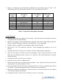
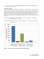
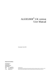
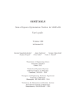
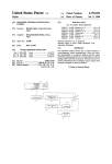
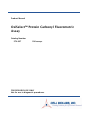
Product Manual OxiSelect™ Protein Carbonyl Fluorometric Assay Catalog Number STA-307 100 assays FOR RESEARCH USE ONLY Not for use in diagnostic procedures Introduction Protein oxidation is defined as the covalent modification of a protein induced either directly by reactive oxygen species (ROS) or indirectly by reaction with secondary by-products of oxidative stress. Oxidative modification of proteins can be induced in vitro by a wide array of pro-oxidant agents and occurs in vivo during aging and in certain disease conditions. There are numerous types of protein oxidative modification. The most common products of protein oxidation in biological samples are the protein carbonyl derivatives of Pro, Arg, Lys, and Thr. These derivatives are chemically stable and serve as markers of oxidative stress for most types of ROS. Traditionally, carbonyls have been measured spectrophotometrically through derivitization of the carbonyl group with dinitrophenylhydrazine, although this method has limited sensitivity. In response to this, a new fluorescent compound has been developed that binds specifically to carbonyls of oxidized proteins at 1:1 molar ratio. Upon binding with the protein carbonyl, the fluorescence produced is a direct measure of protein carbonyls and is more sensitive than the traditional spectrophotometric assay using DNPH. Cell Biolabs’ OxiSelect™ Protein Carbonyl Fluorometric Assay provides a convenient system to measure the protein carbonyl content in plasma, serum, cell lysates, tissues and purified proteins. Each kit provides sufficient reagents to perform up to 100 assays including unknown protein samples, assay blanks and standards. Assay Principle Protein carbonyls in protein samples (1-10 mg/mL) are first derivatized with the Protein Carbonyl Fluorophore. The Fluorophore binds to the protein carbonyl group in a 1:1 ratio. Proteins are then TCA precipitated and free Fluorophore is removed by washing the protein pellet with acetone. After dissolving the protein pellet in GuHCl, the absorbance of protein-fluorophore product is measured fluorometrically with a 485/538 nm filter set, and the protein carbonyl is subsequently calculated. Related Products 1. STA-303: OxiSelect™ Nitrotyrosine Protein Immunoblot Kit 2. STA-304: Protein Tyrosine Nitration Control (Nitrotyrosine-BSA) 3. STA-305: OxiSelect™ Nitrotyrosine ELISA Kit 4. STA-308: OxiSelect™ Protein Carbonyl Immunoblot Kit 5. STA-309: Oxidized Protein Immunoblot Control (Carbonyl-BSA) 6. STA-310: OxiSelect™ Protein Carbonyl ELISA Kit 7. STA-315: OxiSelect™ Protein Carbonyl Spectrophotometric Kit 8. STA-318: OxiSelect™ AOPP Assay Kit 9. STA-816: OxiSelect™ N-epsilon-(Carboxymethyl) Lysine (CML) Competitive ELISA Kit 10. STA-817: OxiSelect™ Advanced Glycation End Products (AGE) Competitive ELISA Kit 2 Kit Components 1. Protein Carbonyl Fluorophore (100X) (Part No. 230701): One 50 µL amber vial 2. 5X TCA Solution (Part No. 231502): One 50 mL bottle 3. Sample Diluent (10X) (Part No. 230702): One 20 mL bottle 4. Protein Solubilization Solution (Part No. 231504): One 15 mL bottle 5. Assay Diluent (4X) (Part No. 234505): One 50 mL bottle 6. Fluorophore Standard (Part No. 230703): One 20 µL amber vial of a 20 mM solution in DMSO Materials Not Supplied 1. 1 to 10 mg/mL protein samples such as plasma, serum, cell lysate, purified protein 2. Protein assay such as Pierce BCA or Bradford assay 3. Acetone 4. Streptomycin sulfate 5. 5 µL to 1000 µL adjustable single channel precision micropipettes with disposable tips 6. 50 µL to 300 µL adjustable multichannel micropipette with disposable tips 7. Bottles, flasks, and conical or microcentrifuge tubes necessary for reagent preparation 8. Centrifugal filter or concentrator 9. Reagents and materials necessary for sample extraction and purification 10. 96-well black or fluorescence microtiter plate 11. Fluorescent microplate reader capable of reading 480 nm (excitation) and 530 nm (emission) Storage Upon receipt, aliquot and store the Protein Carbonyl Fluorophore and Fluorophore Standard at -20ºC to avoid multiple freeze/thaw cycles. Store all other components at 4ºC until their expiration dates. Preparation of Reagents 1X TCA Solution: Dilute the 5X TCA Solution to 1X with deionized water. Stir to homogeneity. Store the 1X TCA at room temperature. Note: TCA Solution is highly corrosive. Use caution when handling. 1X Sample Diluent: Dilute the 10X Sample Diluent to 1X with deionized water. homogeneity. Store the 1X Sample Diluent at 4ºC. 1X Assay Diluent: Dilute the Assay Diluent 1:4 with deionized water. Mix to homogeneity. Store the 1X Assay Diluent at 4ºC. 3 Stir to 1X Protein Carbonyl Fluorophore: Prepare only enough for immediate applications. Prepare 1X Protein Carbonyl Fluorophore by diluting the 100X Protein Carbonyl Fluorophore stock solution 1:100 with 1X Sample Diluent. Do not store diluted solution. Preparation of Samples All samples should be assayed immediately or stored at -80°C. The assay can be used on cell lysates, tissue homogenates, serum, plasma, urine, as well as other biological fluids when applicable. Run proper controls as necessary. Always run a standard curve with samples. Use 1X Sample Diluent for dilutions and preparations of samples. Sample concentrations must be adjusted to 1-10 mg/mL prior to performing the assay. Note: For protein samples lower than 1 mg/mL, concentrate by centricon filtration or by mixing 0.8 vol of protein sample and 0.2 vol of 5X TCA solution, incubating 10 minutes on ice and centrifuging at 10,000 g. Dissolve the protein pellet in a small volume of 1X PBS. (Optional): High nucleic acid can erroneously contribute to higher estimation of carbonyls. To remove nuclei acid, add streptomycin sulfate or PEI to a final concentration of 1% and 0.5% respectively, incubate samples 30 minutes at room temperature and remove the nuclei acid precipitates by centrifuging at 6000 g for 10 minutes at 4ºC. Test supernatant accordingly. Cells or Tissues: Centrifuge cells at 1,500 x g for 5 minutes at 4°C. Remove media and resuspend in 1-2 mL cold 1X Sample Diluent. Homogenize or sonicate cells or tissues on ice. Centrifuge at 10,000 x g for 5 minutes at 4°C. Remove supernatant and store on ice. Store on ice or at -80°C for long-term storage. Prior to use, adjust the total protein concentration to 1-10 mg/mL with 1X Sample Diluent. Serum: Collect blood without using an anticoagulant. Allow blood to clot for 30 minutes at room temperature. Centrifuge blood at 2,000 x g for 15 minutes at 4°C. Carefully remove the top yellow serum layer without disturbing the white buffy layer. Store on ice or at -80°C for long-term storage. Prior to use, adjust the total protein concentration to 1-10 mg/mL with 1X Sample Diluent. Plasma: Collect blood using an anticoagulant such as citrate or heparin. Centrifuge blood at 1000 x g for 5 minutes at 4°C. Carefully remove the top yellow serum layer without disturbing the white buffy layer. Store on ice or at -80°C for long-term storage. Prior to use, adjust the total protein concentration to 1-10 mg/mL with 1X Sample Diluent. Urine: Urine normally has very low protein concentrations and is only recommended if it can be prepared in the 1-10 mg/mL range. Use 1X Sample Diluent for all dilutions. Preparation of the Fluorophore Standard Curve 1. Use the Fluorophore Standard to prepare a series of standards. Prepare standards just prior to use. Prepare a 25 μM solution of the Fluorophore Standard by diluting the stock 1:800 with 1X Assay Diluent. Vortex thoroughly. For example, add 2 μL of the 20 mM Fluorophore Standard stock to 1598 μL of 1X Assay Diluent. 4 2. Prepare a 1:2 dilution series of Fluorophore standards in the concentration range of 0 nM – 1,250 nM by diluting the 200 μM Fluorphore solution in 1X Assay Diluent (see Table 1). Standard Tubes 1 2 3 4 5 6 7 8 Fluorophore (µL) 50 of 25 μM solution 500 of Tube #1 500 of Tube #2 500 of Tube #3 500 of Tube #4 500 of Tube #5 500 of Tube #6 0 Assay Diluent (µL) 950 500 500 500 500 500 500 500 Fluorophore (nM) 1250 625 313 156 78 39 20 0 Table 1. Preparation of Fluorophore Standards. Assay Protocol 1. Determine the protein concentration of each sample. Adjust the protein concentration to between 1-10 mg/mL with 1X Sample Diluent. 2. Add 50 μL of sample to a microcentrifuge tube, and follow with a 50 μL of the freshly prepared 1X Protein Carbonyl Fluorophore solution to each sample tube. Vortex thoroughly. 3. Incubate samples overnight at room temperature and protected from light. 4. Add 400 μL of 1X TCA solution to each tube. Vortex thoroughly and incubate on ice for 10 minutes. 5. Centrifuge the tubes at 10,000 x g for 10 minutes. Remove and discard the supernatant. 6. Add 1 mL of acetone to each tube and vortex thoroughly in order to break up the pellet. If the pellet does not disintegrate upon vortexing, use a spatula or other small implement to manually break up the pellet. 7. Centrifuge the tubes at 10,000 x g for 10 minutes. 8. Repeat the acetone wash two additional times. 9. After the last wash, remove and discard the supernatant. Leave sample tube lids open, allow the sample pellet to dry out thoroughly for 1 hour. 10. After ensuring that all of the acetone has evaporated, add 50 μL of Protein Solubilization Solution to each tube. Vortex thoroughly and incubate 10 minutes at room temperature. 11. Add 450 μL of diluted Assay Diluent to each tube and vortex. Note that samples have been diluted 1:10 from their original concentration. 12. Centrifuge the tubes at 10,000 x g for 10 minutes to remove excess debris. 5 13. Determine the protein concentration of each sample. 14. Prepare a standard curve as outlined in Table 1. 15. Transfer 100 μL of each sample or prepared standards to a 96-well black fluorescence microtiter plate. 16. Read the fluorescence with a fluorescence plate reader with 480 nm excitation /530 nm emission filter settings. Calculation of Results The following figures demonstrate typical Protein Carbonyl Fluorometric Assay results. Fluorescence measurement was performed on SpectraMax Gemini XS Fluorometer (Molecular Devices) with a 485/538 nm filter set and 530 nm cutoff. One should use the data below for reference only. This data should not be used to interpret actual results. Figure 1. Protein Carbonyl Fluorophore Standard Curve. Calculations: 1. Determine the average fluorescence for each standard and sample. Subtract the value of 0 nM Fluorophore blank from itself and all other standard and sample values. These are the adjusted RFU values. 2. Graph the adjusted RFU values for the standards as a function of their corresponding concentration from Table 1. 6 3. Determine the carbonyl concentration of samples using the equation obtained from the linear regression of the standard curve. Calculation Example: Chemically oxidized and reduced BSA were prepared at 10, 5, and 1 mg/mL in 1X Sample Diluent. Samples were then derivatized with the Protein Carbonyl Fluorophore and processed according to the Assay Protocol. After TCA precipitation and wash, the protein pellet was dissolved in Protein Solubilization Solution and the protein concentration of the solubilized protein was determined by BCA assay. For example, for the 10 mg/mL Oxidized BSA sample, the solubilized protein concentration was 6.3 mg/mL and its adjusted RFU reading is 1015 after 10-fold dilution with Assay Diluent. Protein Carbonyl Concentration (nM) = [(1015 RFUs – 4.9382 (y intercept)) / 1.3797(slope)] x 10 (Dilution Factor) = 7321 nM Protein Carbonyl Content (nmol/mg) = 7321 nM / (6.3 mg/mL) = (7.321 nmol/mL) / (6.3 mg/mL) = 1.162 nmol/mg Figure 2. Protein Carbonyl Contents in Oxidized and Reduced BSA. 7 References 1. Buss, H, Chan, TP, Sluis, KB, Domigan, NM, and Winterbourn, CC. (1985) Free Radic Biol Med. 23: 361-6. 2. Cadenas, E., Boveris, A., Ragan, CI., and Stoppani, AO. (1977) Archives of Biochemistry & Biophysics 180: 248 257. 3. Hammer, M., Schweitzer, D., Richter, S., Konigsdorffer, E. (2005) Physiol. Meas. 26: N9-N12. 4. Reznick, AZ., and Packer, L. (1994) Methods Enzymol. 233: 263-357. 5. Talent, JM., Kong, Y., and Gracy, RW. (1998) Anal. Biochem. 263: 31 38. 6. Wakeyama, H., Takeshige, K., Takayanagi, R., and Minakami, S. (1982) Biochem J. 205: 593 601. Recent Product Citations 1. Lauritzen, K. H. et al. (2015). Impaired dynamics and function of mitochondria caused by mtDNA toxicity leads to heart failure. Am J Physiol Heart Circ Physiol. 309:H434-H449. 2. Sadowska-Bartosz, I. & Bartosz, G. (2015). Ascorbic acid and protein glycation in vitro. Chem Biol Interact. doi: 10.1016/j.cbi.2015.07.006. 3. Zabala, V. et al. (2015). Potential contributions of the tobacco nicotine-derived nitrosamine ketone (NNK) in the pathogenesis of steatohepatitis in a chronic plus binge rat model of alcoholic liver disease. Alcohol Alcohol. doi: http://dx.doi.org/10.1093/alcalc/agu083. 4. Rey, B. et al. (2014). Thyroid status affects membranes susceptibility to free radicals and oxidative balance in skeletal muscle of Muscovy ducklings (Cairina moschata). J Exp Zool A Ecol Genet Physiol. 321:415-421. 5. Tong, M. et al. (2014). Therapeutic reversal of chronic alcohol‐related steatohepatitis with the ceramide inhibitor myriocin. Int J Exp Pathol. 95:49-63. Warranty These products are warranted to perform as described in their labeling and in Cell Biolabs literature when used in accordance with their instructions. THERE ARE NO WARRANTIES THAT EXTEND BEYOND THIS EXPRESSED WARRANTY AND CELL BIOLABS DISCLAIMS ANY IMPLIED WARRANTY OF MERCHANTABILITY OR WARRANTY OF FITNESS FOR PARTICULAR PURPOSE. CELL BIOLABS’ sole obligation and purchaser’s exclusive remedy for breach of this warranty shall be, at the option of CELL BIOLABS, to repair or replace the products. In no event shall CELL BIOLABS be liable for any proximate, incidental or consequential damages in connection with the products. Contact Information Cell Biolabs, Inc. 7758 Arjons Drive San Diego, CA 92126 Worldwide: +1 858-271-6500 USA Toll-Free: 1-888-CBL-0505 E-mail: [email protected] www.cellbiolabs.com 2011-2015: Cell Biolabs, Inc. - All rights reserved. No part of these works may be reproduced in any form without permissions in writing. 8