1
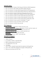
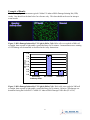
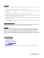
Product Manual OxiSelect™ Cellular UV-Induced DNA Damage Staining Kit (CPD) Catalog Number STA-327 96 assays FOR RESEARCH USE ONLY Not for use in diagnostic procedures Introduction Absorption of ultraviolet (UV) light produces two predominant types of DNA damage, cyclobutane pyrimidine dimers (CPD) and pyrimidine (6-4) pyrimidone photoproducts (6-4PP) (Figure 1). The result is a transition of C to T and CC to TT, which are the most frequent mutations of p53 in both human and mouse skin cancers. UV damaged DNA is usually repaired by nucleotide excision repair (NER) or base excision repair (BER). After UV exposure, cells activate p53 and stall the cell cycle for repair. If the damage is too severe, the cell will trigger apoptosis to get rid of DNA damaged, potentially mutant cells. Cell Biolabs’ OxiSelect™ Cellular UV-induced DNA Damage Staining Kit (CPD) is an immunofluorescence assay developed for rapid detection of CPDs in genomic DNA of cultured cells. Each kit provides sufficient reagents for up to 96 stainings in a 96-well plate. Figure 1: Structures of DNA lesions induced by UV Light Assay Principle Cells are first seeded in a 96-well tissue culture plate. Wells are then UV irradiated to induce DNA damage. After fixation and denaturation, cells containing CPD damage are probed with an anti-CPD antibody, followed by a FITC conjugated secondary antibody. The unbound secondary antibody is removed during a wash step, and stained cells can then be visualized with a fluorescence microscope. 2 Related Products 1. STA-320: OxiSelect™ Oxidative DNA Damage ELISA Kit (8-OHdG Quantitation) 2. STA-321: OxiSelect™ DNA Double-Strand Break (DSB) Staining Kit 3. STA-322: OxiSelect™ UV-induced DNA Damage ELISA Kit (CPD Quantitation) 4. STA-323: OxiSelect™ UV-induced DNA Damage ELISA Kit (6-4PP Quantitation) 5. STA-324: OxiSelect™ Oxidative DNA Damage Quantitation Kit (AP sites) 6. STA-325: OxiSelect™ Oxidative RNA Damage ELISA Kit (8-OHG Quantitation) 7. STA-326: OxiSelect™ Cellular UV-induced DNA Damage ELISA Kit (CPD) 8. STA-328: OxiSelect™ Cellular UV-induced DNA Damage ELISA Kit (6-4PP) 9. STA-329: OxiSelect™ Cellular UV-induced DNA Damage Staining Kit (6-4PP) 10. STA-351: OxiSelect™ Comet Assay Kit (3-Well Slides), 75 Assays 11. STA-353: OxiSelect™ Comet Assay Slides (3-Well), 25 Slides 12. STA-355: OxiSelect™ 96-Well Comet Assay Kit Kit Components 1. Anti-CPD Antibody, 100X (Part No. 232601): One 100 µL vial. 2. Secondary Antibody, FITC Conjugate, 100X (Part No. 232102): One 100 µL amber vial. 3. Denaturation Solution A, 100X (Part No. 232602): One 200 µL vial. 4. Denaturation Solution B, 100X (Part No. 232603): One 200 µL vial. 5. Assay Diluent (Part No. 310804): One 50 mL bottle. 6. 10X Wash Buffer (Part No. 250007): One 50 mL bottle. Materials Not Supplied 1. 96-well tissue culture plate 2. Cell line of interest 3. UV crosslinker, irradiator, or germicidal lamp 4. DPBS containing magnesium and calcium 5. 75% Methanol/25% Acetic Acid 6. 70% Ethanol 7. 10 µL to 1000 µL adjustable single channel micropipettes with disposable tips 8. 50 µL to 300 µL adjustable multichannel micropipette with disposable tips 9. Fluorescence microscope with FITC filter 3 Storage Store all kit components at 4ºC until their expiration dates. Preparation of Reagents 1X Wash Buffer: Dilute the 10X Wash Buffer Concentrate to 1X with deionized water. Stir to homogeneity. Anti-CPD Antibody and Secondary Antibody, FITC Conjugate: Immediately before use dilute the Anti-CPD Antibody 1:100 and Secondary Antibody 1:100 with Assay Diluent. Do not store diluted solutions. Denaturation Solution A: Immediately before use dilute the Denaturation Solution A 1:100 with 70% Ethanol. Do not store diluted solution. Denaturation Solution B: Immediately before use dilute the Denaturation Solution B 1:100 with DPBS (containing magnesium and calcium). Do not store diluted solution. Assay Protocol The following assay protocol is written for a 96-well format. Refer to the below table for the appropriate dispensing volumes of other plate formats. Note: using other plate formats will decrease the number of assays possible with this kit. Culture Dish DPBS during UV Irradiation (μL/well) 75% Methanol/ 25% Acetic Acid (μL/well) 70% Ethanol (μL/well) Denaturation Solution A (μL/well) DPBS during Washing (μL/well) Denaturation Solution B (μL/well) Assay Diluent Blocking (μL/well) 1X Anti-CPD Antibody Solution (μL/well) 1X Secondary Antibody, FITC Conjugate Solution (μL/well) Wash Buffer (μL/well) 96-well 48-well 24-well 12-well 6-well 100 200 400 800 1600 100 200 400 800 1600 100 200 400 800 1600 100 200 400 800 1600 200 400 800 1600 3200 100 200 400 800 1600 200 400 800 1600 3200 100 200 400 800 1600 100 200 400 800 1600 250 500 1000 2000 4000 Table 1. Dispensing Volumes of Different Plate Formats. 4 I. Cell Seeding 1. Harvest and resuspend cells in culture medium at 2-4 x 10⁵ cells/mL. Seed 100 µL in each well of a 96-well tissue culture plate and incubate overnight at 37ºC, 5% CO2 (cells should be > 80% confluent). II. UV Treatment, Fixation and Denaturation 1. Carefully remove medium from the wells by tilting the plate and aspirating from the edge. Gently add 100 µL of DPBS (containing magnesium and calcium) to each well, taking care not to dislodge the cells. 2. Perform UV irradiation to desired wells (removal of plate cover is recommended). Include wells without irradiation as a negative control. Samples should be performed in triplicate. 3. Aspirate the wells and add 100 µL of 75% Methanol/25% Acetic Acid to each well. Incubate 30 minutes at room temperature. 4. Aspirate the wells and add 100 µL of 70% Ethanol to each well. Incubate 30 minutes at room temperature. 5. Aspirate the wells and add 100 µL of Denaturation Solution A (see Preparation of Reagents) to each well. Incubate 5 minutes at room temperature. 6. Gently wash 3 times with 200 µL DPBS (containing magnesium and calcium). 7. Aspirate the wells and add 100 µL of Denaturation Solution B (see Preparation of Reagents) to each well. Incubate 10 minutes at room temperature. 8. Aspirate the wells and add 200 µL of Assay Diluent to each well. Block the wells 30 minutes at room temperature. III. CPD Immunofluorescence Staining 1. Aspirate the wells and add 100 µL of the diluted anti-CPD antibody (see Preparation of Reagents) to each well. Incubate at room temperature for 1 hour on an orbital shaker. 2. Wash microwell strips 4 times with 250 µL 1X Wash Buffer per well with thorough aspiration between each wash. After the last wash, empty wells and tap microwell strips on absorbent pad or paper towel to remove excess 1X Wash Buffer. 3. Add 100 µL of the diluted Secondary Antibody, FITC Conjugate (see Preparation of Reagents) to each well. Incubate at room temperature for 1 hour on an orbital shaker. 4. Wash microwell strips 4 times according to step 2 above. Proceed immediately to the next step. 5. Add 100 L of DPBS to each well. 6. View staining with a fluorescence microscope using FITC filter. 5 Example of Results The following figures demonstrate typical Cellular UV-induced DNA Damage Staining Kit (CPD) results. One should use the data below for reference only. This data should not be used to interpret actual results. Figure 2: DNA Damage Induced by UV Light in HeLa Cells. HeLa cells were seeded at 20K/well overnight, then exposed to light under a germicidal lamp for 30 minutes. Immunofluorescence staining of CPD damage was determined as described in the Assay Instructions. 1.6 1.4 OD 450nm 1.2 1.0 0.8 0.6 0.4 0.2 0.0 Secondary Antibody Alone UV Treated Untreated Figure 3: DNA Damage Induced by UV Light in HeLa Cells. HeLa cells were seeded at 20K/well overnight, then exposed to light under a germicidal lamp for 30 minutes. Relative CPD damage was determined using the OxiSelect™ Cellular UV-induced DNA Damage ELISA Kit (STA-326). 6 References 1. Lippke JA, Gordon LK, Brash DE, Haseltine WA. (1981) Proc Natl Acad Sci U S A. 78:3388– 3392. 2. Mitchell DL, Nairn RS. (1989) Photochem Photobiol. 49:805–819. 3. Ananthaswamy HN, Loughlin SM, Cox P, Evans RL, Ullrich SE, Kripke ML. (1997) Nat Med. 3:510–514. 4. Soehnge H, Ouhtit A, Ananthaswamy ON. (1997) Front Biosci. 2:D538–D551. 5. el-Deiry WS, Tokino T, Velculescu VE, Levy DB, Parsons R, Trent JM, Lin D, Mercer WE, Kinzler KW, Vogelstein B. (1993) Cell. 75:817–825. 6. Hermeking H, Lengauer C, Polyak K, He TC, Zhang L, Thiagalingam S, Kinzler KW, Vogelstein B. (1997) Mol Cell. 1:3–11. 7. Hill LL, Ouhtit A, Loughlin SM, Kripke ML, Ananthaswamy HN, Owen-Schaub LB. (1999) Science. 285:898–900. Recent Product Citation Núñez-Lozano, R. et al. (2015). Biocompatible films with tailored spectral response for prevention of DNA damage in skin cells. Adv Healthc Mater. doi: 10.1002/adhm.201500223. Warranty These products are warranted to perform as described in their labeling and in Cell Biolabs literature when used in accordance with their instructions. THERE ARE NO WARRANTIES THAT EXTEND BEYOND THIS EXPRESSED WARRANTY AND CELL BIOLABS DISCLAIMS ANY IMPLIED WARRANTY OF MERCHANTABILITY OR WARRANTY OF FITNESS FOR PARTICULAR PURPOSE. CELL BIOLABS’ sole obligation and purchaser’s exclusive remedy for breach of this warranty shall be, at the option of CELL BIOLABS, to repair or replace the products. In no event shall CELL BIOLABS be liable for any proximate, incidental or consequential damages in connection with the products. Contact Information Cell Biolabs, Inc. 7758 Arjons Drive San Diego, CA 92126 Worldwide: +1 858-271-6500 USA Toll-Free: 1-888-CBL-0505 E-mail: [email protected] www.cellbiolabs.com 2011-2015: Cell Biolabs, Inc. - All rights reserved. No part of these works may be reproduced in any form without permissions in writing. 7