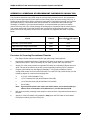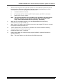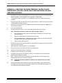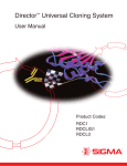
1
SIGMA-ALDRICH
FLAG® 96-well Immunoprecipitation System
User Manual
®
SIGMA-ALDRICH FLAG 96-well Immunoprecipitation System User Manual
FLAG and ANTI-FLAG are registered trademarks of Sigma-Aldrich Biotechnology LP. The product designations of pFLAG,
p3XFLAG, pFLAG-1, pFLAG-2, pFLAGSHIFT, pFLAG-CTS, pFLAG-ATS, pFLAG-MAC, pFLAG-CMV, YEpFLAG, and FLAGBAP are trademarks of Sigma-Aldrich Biotechnology LP.
All other trademarks are the property of their respective owners.
© 2010, Sigma-Aldrich Biotechnology LP. All rights reserved.
Date
2
Version #
®
SIGMA-ALDRICH FLAG 96-well Immunoprecipitation System User Manual
1.0
TABLE OF CONTENTS
1.0 Table of Contents................................................................................................................................3
2.0 System Components ..........................................................................................................................4
Immunoprecipitation Vector Kit (Product # COIP-P) ............................................................................4
FLAG 96-well Immunoprecipitation Kit (Product # HT-COIP-1) ...........................................................4
Immunoprecipitation Detection Kit (Product # COIP-D) .......................................................................4
3.0 Introduction .........................................................................................................................................5
4.0 FLAG 96-well Immunoprecipitation System Overview ...................................................................6
5.0 Immunoprecipitation Vector Kit (Product # COIP-P).......................................................................7
5.1 Reagents Provided (sufficient for 96 samples)............................................................................7
5.2 Additional Reagents and Equipment (Not Supplied) ...................................................................7
5.3 Expression Vectors......................................................................................................................8
5.4 Control Vectors ..........................................................................................................................10
5.5 Verification Primers ...................................................................................................................11
6.0 FLAG 96-well Immunoprecipitation Kit (Product # HT-COIP-1) ...................................................12
6.1 Reagents Provided (sufficient for 96 samples)..........................................................................12
6.2 Additional Reagents and Equipment (Not Supplied) .................................................................12
7.0 Immunoprecipitation Detection Kit (Product # COIP-D) ...............................................................13
7.1 Reagents Provided (sufficient for 96 samples)..........................................................................13
7.2 Additional Reagents and Equipment (Not Supplied) .................................................................13
8.0 Protocols for Studying Protein-Protein Interactions with the ANTI-FLAG M2 Plate..................14
8.1 Overview of the Analysis of Protein-Protein Interaction with the ANTI-FLAG M2 Plate by
ELISA or Western blotting .........................................................................................................14
8.2 General Notes ...........................................................................................................................15
8.2.1Plasmid DNA for transfection .............................................................................................15
8.2.2Transfection reagent ..........................................................................................................15
8.2.3Cell lysates used for co-immunoprecipitation assay..........................................................15
8.2.4Detection of endogenous binding partners during Western blotting or ELISA ..................16
8.3 Preparation of Cell Extract.........................................................................................................16
8.4 Immunoprecipitation with ANTI-FLAG M2 plate ........................................................................16
8.5 Detection of the Bound Protein by ELISA or Western Blotting..................................................17
8.5.1ELISA .................................................................................................................................17
8.5.2Western Blotting .................................................................................................................18
9.0 Troubleshooting Guide ....................................................................................................................19
10.0 References.........................................................................................................................................20
Appendix 1: Construction of fusion genes using standard cloning techniques................................21
Appendix 2: Screening of recombinant plasmids by colony PCR .......................................................22
Procedure for Screening Recombinant Plasmids...............................................................................22
Appendix 3: Capturing the Amino-terminal FLAG-BAP Fusion Protein with the ANTI-FLAG M2
plate and evaluating the pNPP substrate integrity ................................................................................24
Version #
3
Date
®
SIGMA-ALDRICH FLAG 96-well Immunoprecipitation System User Manual
2.0
SYSTEM COMPONENTS
Immunoprecipitation Vector Kit (Product # COIP-P)
•
pFLAG-CMV™-2 Expression Vector (10 µg)
•
pc-Myc-CMV-2 Expression Vector (10 µg)
•
pFLAG-CMV-2-p53 control plasmid (20 µg)
•
pc-Myc-CMV-2-Large T antigen control plasmid (10 µg)
•
pc-Myc-CMV-2-BAP control plasmid (10 µg)
•
Verification Primer-MF (2 nmol)
•
Verification Primer-MR (2 nmol)
•
Technical Bulletins
Storage Conditions: Store all components at -20°C
FLAG 96-well Immunoprecipitation Kit (Product # HT-COIP-1)
•
ANTI-FLAG M2 High Sensitivity Capture Plate (1 plate)
•
Amino-terminal FLAG-BAP Fusion Protein (2.5 µg)
•
10× Wash Buffer (50 ml)
•
Lysis Buffer (50 ml)
•
2× Sample Buffer (6 ml)
•
SealPlate Film (6)
•
User Manual
Storage Conditions: Store all components at 4°C
Immunoprecipitation Detection Kit (Product # COIP-D)
•
Monoclonal Anti-c-Myc, Clone 9E10, Alkaline Phosphatase Conjugate (0.25 ml)
•
Tris-Buffered Saline, pH 8.0, with 3% Nonfat Milk (1 packet)
•
SIGMA FAST™ p-Nitrophenyl Phosphate Tablets (5 tablet sets)
•
Technical Bulletins
Storage Conditions: Store all components at 4°C
Date
4
Version #
®
SIGMA-ALDRICH FLAG 96-well Immunoprecipitation System User Manual
3.0
INTRODUCTION
Researchers studying protein-protein interactions commonly use one of several different two-hybrid
systems. While these systems are of some use in determining protein binding partners, all of the
current two-hybrid systems generate false positives. Sigma's FLAG-96-well Immunoprecipitation Kit
provides a unique method to validate protein-protein interactions in a convenient 96-well format. This
system enables potential binding partners for the protein of interest to be tagged with FLAG and cMyc epitopes, followed by capture and detection using the ANTI-FLAG M2 High Sensitivity Capture
Plate. The system offers the following advantages compared to currently available techniques:
1.
Rapid 96-well format—The 96-well plate configuration is very effective at handling multiple
samples and greatly increases the throughput of the assay. Additionally, this is an ideal
system for optimizing in vitro binding conditions.
2.
No leaching of ANTI-FLAG M2 antibody—The ANTI-FLAG M2 antibody is covalently
coupled to the surface of the well and the elution conditions used are relatively mild, so very
little antibody leaches from the plate. This results in little to no interference from denatured
antibody heavy and light chain bands during Western blot analysis.
3.
Rapid protocol with quantitative results—Because an ELISA format can be used with this
system, the protein-protein interaction assay is rapid (less than 4 hours), sensitive, and
quantitative.
Version #
5
Date
®
SIGMA-ALDRICH FLAG 96-well Immunoprecipitation System User Manual
4.0
FLAG 96-WELL IMMUNOPRECIPITATION SYSTEM OVERVIEW
Figure 1. Outline of the procedure
Clone two potentially interacting proteins into the two mammalian epitope-tagged expression vectors provided in the
Immunoprecipitation Vector Kit. One vector generates an N-terminally tagged FLAG fusion protein while the other vector
produces an N-terminally tagged c-Myc fusion protein.
↓
Co-express the proteins in appropriate mammalian host cells
↓
Prepare cell extracts with Lysis Buffer
↓
Dilute the cell lysate with 1× Wash Buffer (if necessary)
↓
Incubate cell lysate in the 96-well ANTI-FLAG M2 High Sensitivity Capture Plate
↓
Wash the ANTI-FLAG M2 High Sensitivity Capture Plate with 1× Wash Buffer
↓
Analyze captured proteins by ELISA or Western blotting
Date
6
Version #
®
SIGMA-ALDRICH FLAG 96-well Immunoprecipitation System User Manual
5.0
IMMUNOPRECIPITATION VECTOR KIT (PRODUCT # COIP-P)
This kit includes the transient mammalian expression vectors pFLAG-CMV™-2 and pc-Myc-CMV-2,
which are designed for the construction and expression of N-terminally tagged fusion proteins with a
1
2
FLAG epitope (DYKDDDDK) or a c-Myc epitope (EQKLISEEDL) , respectively. The control plasmids
pFLAG-CMV-2-p53, pc-Myc-CMV-2-Large T antigen and pc-Myc-CMV-2-BAP are also supplied. The
control plasmids can be used as positive- and negative-binding partners in the immunoprecipitation
analysis (see Section 5.4). Figures 2 through 4 and Table I illustrate the features of the supplied
vectors.
5.1
Reagents Provided (sufficient for 96 samples)
Part
Product #
Amount
pFLAG-CMV™-2 Expression Vector
E7398
1 × 10 μg
pc-Myc-CMV-2 Expression Vector
P9236
1 × 10 μg
pFLAG-CMV™-2-p53 Control Plasmid
P9986
1 × 20 μg
pc-Myc-CMV-2-Large T antigen Control Plasmid
P9861
1 × 10 μg
pc-Myc-CMV-2-BAP Control Plasmid
P9736
1 × 2.5 μg
Verification Primer-MF
P2987
1 × 2 nmol
Verification Primer-MR
P3112
1 × 2 nmol
5.2
Additional Reagents and Equipment (Not Supplied)
•
Pipets (single- or multi-channel)
•
Thermal cycler
•
Pipette tips
•
PCR tubes or multi-well PCR plate
•
Taq DNA polymerase
Microcentrifuge tubes
•
•
Microcentrifuge
•
Restriction enzymes
•
Reagent reservoir
•
Phosphatase suitable for DNA
•
Vortexer
•
Competent E. coli cells
®
•
MilliQ or sterile water
•
LB-ampicillin agar plates
•
DNA plasmid purification kit
•
LB medium with 100 μg/ml Ampicillin
•
Transfection reagent
•
15 ml culture tubes
•
Agarose gel electrophoresis system
•
37°C incubator
Version #
7
Date
®
SIGMA-ALDRICH FLAG 96-well Immunoprecipitation System User Manual
5.3
Expression Vectors
Figure 2. Circular maps of the expression vectors
4679
4227
4688
Hind III
Not I
EcoR I
Cla I
Bgl II
EcoR V
Kpn I
Sal I
Xba I
Bam HI
Sma I
166
f1 IG
CMV
FLAG
pFLAG-CMV™-2
Hind III
Not I
EcoR I
Cla I
Bgl II
EcoR V
Kpn I
Sal I
Xba I
Bam HI
Sma I
166
4236
f1 IG
CMV
c-Myc
pc-Myc-CMV-2
4.7 kb
4.7 kb
hGH poly A
hGH poly A
1642
1651
SV40 ori
E.coli ori/b-lact
SV40 ori
2908
E.coli ori/b-lact
2917
Table I. Expression vector features
pFLAG-CMV™-2
(4679 bp)
pc-Myc-CMV-2
(4688 bp)
CMV promoter
166-916
166-916
Verification Primer-MF
825-854
825-854
Translational initiation
932-934
932-934
FLAG and c-Myc sequences
935-958
935-967
Multiple cloning region
956-1022
965-1031
hGH poly A
1023-1642
1032-1651
Verification Primer-MR
1080-1103
1089-1112
SV40 ori
1661-2005
1670-2014
pBR322 ori/beta-lactamase
2908-4092
2917-4101
f1 intragenic region
4227-4679
4236-4688
Feature
Note: The only difference between the two expression vectors is the epitope tags.
Date
8
Version #
®
SIGMA-ALDRICH FLAG 96-well Immunoprecipitation System User Manual
Figure 3. Nucleotide sequences of the epitope tags and multiple cloning region of
the pFLAG-CMV™-2 (upper) and pc-Myc-CMV-2 (lower) expression vectors
Version #
9
Date
®
SIGMA-ALDRICH FLAG 96-well Immunoprecipitation System User Manual
5.4
Control Vectors
The Immunoprecipitation Vector Kit provides positive and negative control vectors for coimmunoprecipitation (Figure 4). pFLAG-CMV™-2-p53 Control Plasmid expresses the N-terminallytagged FLAG fusion protein containing the wild-type C-terminal domain of mouse p53 (residues 733
390). pc-Myc-CMV-2-Large T antigen Control Plasmid expresses the N-terminally-tagged c-Myc
4
fusion protein containing the C-terminal domain of Large T-antigen (residues 86-708). pc-Myc-CMV2-BAP Control Plasmid expresses the N-terminally-tagged c-Myc fusion protein containing bacterial
alkaline phosphatase (BAP). Mammalian cells co-transfected with control plasmids pFLAG-CMV™-2p53 and pc-Myc-CMV-2-Large T antigen will express tagged fusion proteins that interact, providing a
5
positive control for the co-immunoprecipitation analysis. Cells co-transfected with pFLAG-CMV™-2p53 and pc-Myc-CMV-2-BAP express tagged fusion proteins that do not interact, demonstrating a
negative result in the co-immunoprecipitation analysis.
Figure 4. Circular maps of control vectors
5649
166
5197
Hind III
f1 IG
p53
coding
sequence
CMV
FLAG
Xba I
pFLAG-CMV™-2-p53
Control Plasmid
hGH poly A
5.6 kb
2612
SV40 ori
2631
E.coli ori/b-lact
6838
6133
166
6386
f1 IG
Large T
antigen
coding
sequence
CMV
c-Myc
pc-Myc-CMV-2-Large T antigen
Control Plasmid
6.8 kb
166
5681
Xba I
f1 IG
c-Myc
pc-Myc-CMV-2-BAP
Control Plasmid
Xba I
Sma I
6.1 kb
hGH poly A
hGH poly A
3801
Date
BAP
coding
sequence
CMV
3096
SV40 ori
E.coli ori/b-lact
Hind III
SV40 ori
5067
E.coli ori/b-lact
10
4362
Version #
®
SIGMA-ALDRICH FLAG 96-well Immunoprecipitation System User Manual
5.5
Verification Primers
Verification primer-MF and Verification primer-MR are supplied with this kit and allow the researcher
to screen for the presence, orientation, and reading frame of the DNA insert by colony PCR or DNA
sequencing (Table II).
Table II. Vector primers and sequences
Vector primers
Product #.
Sequence
Verification Primer-MF
P2987
5’ AAT GTC GTA ATA ACC CCG CCC CGT TGA CGC 3′
Verification Primer-MR
P3112
5’ TAT TAG GAC AAG GCT GGT GGG CAC 3′
Notes:
1.
Please see Appendix 1 for procedures involved in cloning recombinant plasmids.
2.
Please see Appendix 2 for procedures involved in screening recombinant plasmids by
Colony PCR.
3.
If the genes of interest are already cloned into expression vectors that contain FLAG or c-Myc
tags, it is not necessary to subclone them into the expression vectors provided in this kit.
Version #
11
Date
®
SIGMA-ALDRICH FLAG 96-well Immunoprecipitation System User Manual
6.0
FLAG 96-WELL IMMUNOPRECIPITATION KIT
(PRODUCT # HT-COIP-1)
The FLAG-96-well Immunoprecipitation Kit provides all of the unique reagents required to study
protein-protein interactions using a 96-well ANTI-FLAG M2 High Sensitivity Capture Plate (ANTIFLAG M2 plate). The ANTI-FLAG M2 plate is designed to capture recombinant FLAG fusion proteins
obtained from cell lysates of transfected cells.
6.1
Reagents Provided (sufficient for 96 samples)
Part
Composition
Product #
Amount
ANTI-FLAG M2 High Sensitivity
Capture Plate
—
P2983
1 plate
20 mM Tris-HCl (pH 7.4), 1 M NaCl, 1 mM DTT,
1.0% Triton® X-100
L1413
50 ml
10× Wash Buffer
500 mM Tris-HCl (pH 7.4), 1.5 M NaCl
W4511
50 ml
2× Sample Buffer
125 mM Tris-HCl (pH 6.8), 4% SDS, 20% (v/v)
glycerol, 0.004% bromphenol blue
B2556
6 ml
Lysis Buffer
Amino-terminal FLAG-BAP Fusion
Protein
—
A1974
2.5 μg
SealPlate Film
—
Z36, 965-9
5
6.2
Additional Reagents and Equipment (Not Supplied)
•
Pipets (single- or multi-channel)
•
Tissue culture incubator
•
Pipette tips
•
Mammalian protease inhibitor cocktail
•
Microcentrifuge tubes
•
Ice-cold PBS
•
Reagent reservoir
•
Cell scrapers
•
Microcentrifuge at 4°C
•
•
Vortexer
Kit for measuring total protein
concentration
•
MilliQ or sterile water
•
4°C incubator
•
Tissue culture plates
•
Plate washer
•
Tissue culture medium
•
Rotary shaker
•
Tissue culture cells, such as COS-7
Date
12
Version #
®
SIGMA-ALDRICH FLAG 96-well Immunoprecipitation System User Manual
7.0
IMMUNOPRECIPITATION DETECTION KIT
(PRODUCT # COIP-D)
The Immunoprecipitation Detection Kit provides all of the unique reagents required to detect proteinprotein interactions using a plate-based colorimetric assay. The substrate p-Nitrophenyl Phosphate
Disodium (pNPP) is hydrolyzed by Anti-c-Myc Alkaline Phosphatase-conjugated antibody to produce
a yellow-colored solution that can be read spectrophotometrically at 405 nm. Tris Buffered Saline (pH
8.0) with 3% Nonfat Milk is used to dilute the Anti-c-Myc Alkaline Phosphatase Conjugate. The pNPP
reaction is terminated with 3 N NaOH and then read at 405 nm. The SIGMA FAST™ pNPP tablet set
provided in the kit produces a ready-to-use solution after the addition of 5 ml of distilled or deionized
water.
7.1
Reagents Provided (sufficient for 96 samples)
Kit Components
Product #
Quantity
Monoclonal Anti-c-Myc, Clone 9E10, Alkaline Phosphatase Conjugate
A5963
1 × 0.25 ml
Tris Buffered Saline, pH 8.0, with 3% Nonfat Milk
T8793
1 packet
SIGMA FAST™ p-Nitrophenyl Phosphate Tablets
N1891
5 tablet sets
7.2
Additional Reagents and Equipment (Not Supplied)
•
Pipets (single- or multi-channel)
•
β-Mercaptoethanol
•
Pipette tips
•
Bulldog clamps
•
Microcentrifuge tubes
•
Boiling water bath
•
Reagent reservoir
•
SDS-PAGE gels and apparatus
•
Microcentrifuge at 4°C
•
Protein transfer apparatus
•
Vortexer
•
Protein transfer membrane
•
Plate washer
•
Tris-glycine buffer
•
Plate mixer
•
TBST
•
Rotary shaker
•
TBS
•
MilliQ or sterile water
•
Anti-mouse IgG peroxidase conjugate
•
3 N NaOH
•
•
50 ml conical tubes
ANTI-FLAG M2 monoclonal antibody
peroxidase (HRP) conjugate
•
Spectrophotometer capable of
measuring absorbance at 405 nm
•
ECL Plus™ Reagent
Version #
13
Date
®
SIGMA-ALDRICH FLAG 96-well Immunoprecipitation System User Manual
8.0
PROTOCOLS FOR STUDYING PROTEIN-PROTEIN
INTERACTIONS WITH THE ANTI-FLAG M2 PLATE
8.1
Overview of the Analysis of Protein-Protein Interaction with the ANTIFLAG M2 Plate by ELISA or Western blotting
Day 1: Plate cells
↓
Day 2: Transfect cells
↓
Day 3 or 4: Harvest cells with Lysis Buffer supplemented with mammalian protease inhibitor cocktail and prepare cell lysate
↓
Incubate cell lysate with the ANTI-FLAG M2 plate for 1 hour at 4°C
↓
(diluting the cell lysate is optional)
Wash the plate 4 times with 200 μl (per well) of the 1× Wash Buffer
↓
After this step, the plate can be analyzed by
ELISA or Western blotting
Analysis with ELISA:
Analysis with Western Blotting:
Add 200 μl of Anti-c-Myc Alkaline Phosphatase Conjugate
(diluted 1:100 in Blocking Buffer) to each well and incubate
for 1 hour at room temperature on a rotary shaker
Add 60 μl of 2× Sample Buffer (supplemented with
10% β-Mercaptoethanol) to each well and incubate at room
temperature for 30 minutes (with shaking on a vortexer)
↓
Wash the plate 4 times with 200 μl (per well) 1× Wash Buffer
↓
↓
Transfer the contents of each well to microcentrifuge tube
and boil for 5 minutes
Incubate each well with 200 μl pNPP substrate
↓
Incubate at room temperature for 15 to 30 min or until the
yellow color develops
↓
Stop the reaction with 3 N NaOH
↓
Measure A405 with a plate reader
Date
14
↓
Western Blotting
Version #
®
SIGMA-ALDRICH FLAG 96-well Immunoprecipitation System User Manual
8.2
General Notes
Please read this section carefully before proceeding with the experiment.
8.2.1
Plasmid DNA for transfection
To obtain maximum transfection efficiency, the DNA used for transfections must be of high quality
(free of protein, RNA, and chemical contamination, with an A260:A280 ratio of 1.8-1.9). To produce
high-quality plasmid DNA, we recommend Sigma’s GeneElute Endo-Free Plasmid Purification Kit
(Product # PLEX 15).
8.2.2
Transfection reagent
The quality of a transfection reagent dictates the sensitivity and specificity of an assay using the
FLAG 96-well Immunoprecipitation Kit. Generally, high transfection efficiency yields a more sensitive
assay, requiring a smaller sample volume. The Immunoprecipitation Vector Kit includes three control
plasmids for optimizing and monitoring the efficiency of transfection. To obtain high-efficiency
transfections, we recommend Sigma’s ESCORTII Transfection Reagent (Product # L6037) for
commonly used cell lines.
8.2.3
Cell lysates used for co-immunoprecipitation assay
1.
Expression level of the fusion proteins in the cell lysate. The concentration of the
expressed fusion protein is critical to the success of the co-immunoprecipitation analysis. The
expression of the FLAG and c-Myc fusion proteins should be verified by Western blotting with
ANTI-FLAG M2 antibody and anti-c-Myc antibody, prior to the co-immunoprecipitation
analysis. In general, a high concentration of fusion protein in the cell lysate will yield more
sensitive results. For each tag, the control plasmids supplied in the Immunoprecipitation
Vector Kit can be used as positive controls in transfection and Western blot analysis. FLAGp53 can be detected with ANTI-FLAG M2 antibody, while the c-Myc-Large T antigen and cMyc-BAP can be detected with monoclonal anti-c-Myc antibody 9E10.
2.
Amount of cell lysate used in the co-immunoprecipitation assay. The optimum amount
of cell lysate added to the wells of the ANTI-FLAG M2 plate may vary with each experimental
system and should be determined empirically. The ANTI-FLAG M2 plate-based immunoprecipitation is very sensitive, especially in ELISA. When analyzing the interaction of FLAGp53 and c-Myc-Large T antigen, the c-Myc-Large T antigen can be detected in as little as 5
μg of the cell lysate from co-transfected COS-7 cells (as compared to the negative control cell
lysate).
Note:
3.
Dilution of the cell lysate. The relative binding affinity for different protein complexes may
vary. Since high salt and detergent levels are present in the Lysis Buffer, it may be necessary
to dilute the cell lysate with 1× Wash Buffer to minimize disruption of the protein-protein
interactions.
Note:
4.
Version #
The volume of the cell lysate used should not exceed 200 μl since the wells of
the ANTI-FLAG M2 plate are coated up to the 200 μl level with the ANTI-FLAG
M2 antibody.
For the positive control interaction between FLAG-p53 and c-Myc-Large T
antigen, dilution of the cell lysate is not necessary.
Negative-binding protein partner. Inclusion of a negative-binding partner along with the
FLAG-tagged protein construct is critical for verifying the specific protein-protein interaction
observed. Over-expression of a fusion protein may cause nonspecific protein-protein
interactions between the proteins being tested. The c-Myc-BAP vector provided in the
Immunoprecipitation Vector Kit can be used as a negative-binding protein partner.
15
Date
®
SIGMA-ALDRICH FLAG 96-well Immunoprecipitation System User Manual
8.2.4
Detection of endogenous binding partners during Western blotting or ELISA
Although this kit and protocol are designed for detecting the c-Myc-tagged binding partner from cotransfected cell lysates using anti-c-Myc antibody, you may use the ANTI-FLAG M2 plate to capture
endogenous binding partners that interact with the FLAG-fusion protein from cells transfected only
with the FLAG-fusion protein. In this situation, optimum dilution and incubation times should be
determined for the specific antibodies used to detect the endogenous binding partners during
Western blotting or ELISA. Since the ANTI-FLAG M2 plate is coated with monoclonal ANTI-FLAG M2
antibody, either polyclonal antibody or antibody conjugates should be used for ELISA-based detection
to avoid problems with cross-reactivity.
Note:
This protocol has been optimized for analyzing the interaction of FLAG-p53 and c-MycLarge T antigen expressed in COS-7 cells. To ensure that the system works in your
hands, we recommend including the control plasmids provided in the
Immunoprecipitation Vector Kit in your experiment.
8.3
Preparation of Cell Extract
1.
Aspirate growth medium from the transfected cells to be assayed. Rinse the cells twice with
ice-cold PBS.
2.
Add 1 ml of ice-cold Lysis Buffer supplemented with mammalian protease inhibitors (10 μl per
1 ml Lysis Buffer, Product # P8340) to a 10-cm plate (you can adjust the volume of the Lysis
Buffer according to the size of the culture plate used).
3.
Scrape the cells off the plate with cell scrapers (Product # C2802) and transfer them to
microcentrifuge tubes.
4.
Incubate for 1 hour on ice, occasionally mixing with a vortexer.
5.
Microcentrifuge the cells for 15 minutes at 4°C and transfer the supernatant to a new tube. If
the supernatant is not to be used immediately, store the cell lysate at -70°C.
Note:
The supernatant may be viscous during transfer, due to the genomic DNA
released from the nuclei of lysed cells. Genomic DNA can be removed from the
supernatant with a 1 ml pipet tip prior to the transfer.
6.
Determine the total protein concentration of the supernatant using standard techniques. We
recommend using Sigma’s BCA Protein Assay. (Bicinchoninic Acid Kit, Cat. No BCA-1).
8.4
Immunoprecipitation with ANTI-FLAG M2 plate
Note:
For protein complexes where the interaction occurs at salt and detergent
concentrations different from the 1×
× Lysis Buffer, use the 1×
× Wash Buffer to
dilute the cell lysate. If dilution is required, first add the required amount of the
1×
× Wash Buffer to the plate and then add the cell lysate. The ANTI-FLAG M2
®
®
plate is resistant to the following detergents: 5% Tween -20, 5% Triton X-100,
0.1% Igepal CA-630, 0.1% CHAPS, and 0.2% digitonin. The ANTI-FLAG M2 plate
can also be used with 1.0 M NaCl.
1.
Rehydrate the plate by adding 200 μl of 1× Wash Buffer to each well.
2.
Incubate the plate for 2 minutes at room temperature.
Note:
3.
Date
If the entire plate is not used at once, cover the unused wells firmly with
SealFilm to prevent contamination from the used wells.
Remove the liquid from each well manually or with a plate washer. Tap the plate firmly onto a
paper towel until most of the solution is removed.
16
Version #
®
SIGMA-ALDRICH FLAG 96-well Immunoprecipitation System User Manual
4.
Incubate various amounts of cell lysate on the plate using a rotary shaker (with gentle
rocking) for 1 hour at 4°C. Cover the plate with the lid.
Note:
The ANTI-FLAG M2 plate is coated with 200 μl of ANTI-FLAG M2 antibody. We
recommend that the volume of cell lysate added to each well should not
exceed 200 μl.
5.
This kit includes Amino-terminal FLAG-BAP Fusion Protein as a positive control for immunoprecipitation on the plate. For a positive control, add 0.5 μg of FLAG-BAP fusion protein
(Product # A1974) diluted with 150 μl Lysis Buffer (Product # L1413) to a single well. This
control sample should be treated identically to all other samples.
6.
Wash each well on the plate 4 times with 200 μl of 1× Wash Buffer. The plate can be washed
either manually or with a plate washer. After washing, remove excess liquid by firmly tapping
the plate onto a paper towel.
Note:
8.5
After this step, the captured proteins can be analyzed by two methods: ELISA
(Section 8.5.1) or Western blotting (Section 8.5.2).
Detection of the Bound Protein by ELISA or Western Blotting
After the FLAG and c-Myc-tagged protein complexes have been captured (Section 8.4), they can be
detected using either ELISA or Western blotting.
8.5.1
ELISA
1.
Dilute Monoclonal Anti-c-Myc, Clone 9E10, Alkaline Phosphatase Conjugate (Product #
A5963) 1:100 with Blocking Buffer (Tris Buffered Saline [pH 8.0] with 3% Nonfat Milk). Add
200 μl of the diluted antibody to each well. Cover the plate with the lid to minimize
evaporation.
2.
Incubate the plate for 1 hour at room temperature on a rotary shaker with gentle agitation.
3.
During the incubation, prepare the pNPP substrate solution (provided in the Detection Kit)
and a 3 N NaOH Stopping Solution:
a.
Prepare enough pNPP solution for each well being used in the assay. To dissolve 1
tablet set, add 5 ml of MilliQ water to a sterile 50 ml conical tube and add one SIGMA
FAST™ p-Nitrophenyl Phosphate Tablet set (one buffer tablet and one pNPP tablet;
Product # N1891) to the water.
b.
Vortex until the tablets dissolve. The pNPP substrate solution should be used within
one hour.
4.
At the end of incubation, remove the liquid from each well and wash the wells 4 times with
200 μl each of 1× Wash Buffer.
5.
Add 200 μl of pNPP substrate to each well.
6.
Incubate the plate at room temperature for 15 to 30 minutes or until a yellow color develops.
Note:
7.
Stop each reaction with 50 μl of the 3 N NaOH Stopping Solution. Mix gently by pipetting.
Note:
8.
Version #
The time required to develop the yellow color will vary for different cell lysates.
The higher the concentration of fusion proteins in the cell lysate, the quicker
the yellow color will develop.
Avoid introducing bubbles into the wells. If bubbles are present, remove them
by gently tapping the plate prior to reading the absorbance.
Using a spectrophotometer, measure the absorbance of each well at 405 nm.
17
Date
®
SIGMA-ALDRICH FLAG 96-well Immunoprecipitation System User Manual
8.5.2
Western Blotting
1.
Add 60 μl of 2× Sample Buffer (supplemented with 10% β-mercaptoethanol) to each sample
well on the plate. Seal the plate with SealPlate Film (Product # Z36, 965-9) and clamp the
plate to a Vortex Genie 2 Mixer (or an equivalent device) with two bulldog clamps.
Notes:
1.
Warm the 2×
× Sample Buffer (Product # B2566) at 37°C for a few minutes before
use.
2.
Do not cover the plate with a plastic lid, since this combination may be too
thick for the bulldog clamps.
2.
Shake the plate for 30 minutes at room temperature on the vortexer at a setting of 3.
3.
Transfer the entire contents of each well to separate microcentrifuge tubes.
4.
Boil each sample for 5 minutes.
5.
Load approximately 50 μl of each sample onto a standard SDS-PAGE gel.
6.
Transfer the proteins from the gel to a membrane such as PVDF using Tris-Glycine Buffer
(25 mM Tris, 192 mM glycine, 15% v/v methanol [pH 8.3]; Product # T4904) and the settings
recommended by the manufacturer of your transfer apparatus.
7.
Block the membrane with Blocking Buffer at room temperature for 30 to 60 minutes.
8.
Transfer the membrane to a tray containing a 1:2,000 dilution of ANTI-FLAG M2 Monoclonal
Antibody Peroxidase (HRP) Conjugate (Product # A8592) or a 1:100 dilution (in Blocking
Buffer) of Monoclonal anti-c-Myc Unconjugated, Clone No. 9E10 (Product # M5546).
9.
Incubate at room temperature for 1 hour with gentle agitation.
10.
Wash the membrane three times (5 minutes per wash) with TBST at room temperature with
gentle agitation.
11.
For membranes probed with ANTI-FLAG M2 Monoclonal Antibody Peroxidase (HRP)
Conjugate, develop the membrane with ECL Plus™ Reagent according to the manufacturer’s
instructions.
12.
For membranes probed with Monoclonal anti-c-Myc Unconjugated, Clone No. 9E10, incubate
the membrane with anti-mouse IgG peroxidase conjugate (Product # A9044) as the
secondary antibody, diluted 1:80,000 in Blocking Buffer.
13.
Incubate the membrane at room temperature for 30 minutes with gentle agitation.
14.
Wash the membrane three times (10 minutes per wash) with TBST at room temperature with
gentle agitation.
15.
Rinse the membrane once with TBS (Product # T6664) to remove residual Tween-20.
16.
Develop the membrane with ECL Plus™ Reagent according to the manufacturer’s
instructions.
Date
18
Version #
®
SIGMA-ALDRICH FLAG 96-well Immunoprecipitation System User Manual
9.0
TROUBLESHOOTING GUIDE
Problem
Possible Cause
Solution
Precipitation of the cell lysate after mixing with
2× Sample Buffer for Western blotting.
You may experience a precipitation problem when
you mix undiluted cell lysate from certain
mammalian cell lines with 2× Sample Buffer. The
Lysis Buffer is very efficient at solubilizing proteins
from cells, which sometimes results in too much
protein in the lysate and causes aggregation.
Add 6 -8 M urea and 10% β-mercaptoethanol to
the 2× Sample Buffer and then mix with the cell
lysate. Heat at 100°C for 10 minutes before
loading on the SDS-PAGE gel.
Samples exhibit no signal or low signal in both
Western blotting and ELISA after capturing on
the ANTI-FLAG M2 plate.
The concentration of the fusion protein in the cell
lysates may be low.
Check the expression level of the fusion proteins
in the co-transfected cell lysate by Western
blotting using ANTI-FLAG M2 antibody to detect
the FLAG-tagged fusion protein or anti-c-Myc
antibody to detect the c-Myc-tagged fusion
protein. If expression level is low, optimize the
conditions for transfection with the control
plasmids.
High salt and detergent present in the Lysis Buffer
may interrupt protein-protein interactions.
Dilute the cell lysate 1- to 5-fold with 1× Wash
Buffer (e.g., 1 volume of cell lysate mixed with 1
to 4 volumes of 1× Wash Buffer) before
immunoprecipitation with ANTI-FLAG M2 plate.
In the ELISA analysis, the pNPP substrate may
have degraded.
Prepare a fresh stock of pNPP and use it within
1 hour. To determine the integrity of the
substrate, see Appendix 3.
Too much cell lysate added to the ANTI-FLAG M2
plate.
Dilute the cell lysate. You can do a titration
series of the cell lysate to be analyzed and find
the optimum amount of cell lysate to generate
an A405 reading between 0.5 and 1.0.
Incubation time with pNPP substrate was too long.
Decrease the incubation time with pNPP.
High background during ELISA.
It is unlikely that the background is contributed by
nonspecific interactions between the plate and
cellular proteins, since the ANTI-FLAG M2 plate is
highly specific. It is likely that the pNPP substrate
incubation time was too long or the substrate was
previously degraded.
Decrease the incubation time with the pNPP
substrate or prepare fresh substrate. To
examine the integrity of the substrate, please
see Appendix 3.
Low yield of eluted protein for Western blotting
analysis after capturing proteins on the ANTIFLAG M2 plate.
Loss of the 2× Sample Buffer (with 10% βmercaptoethanol) during the elution step
Do not vortex the plate too vigorously, which
may spill the 2× Sample Buffer Sample Buffer
(with 10% β-mercaptoethanol) into adjacent
wells. Set the vortexer at a speed that provides
adequate motion for covering the upper portions
of each well.
No β-mercaptoethanol in the 2× Sample Buffer
10% β-mercaptoethanol is critical for efficient
elution of bound proteins. Add this to your 2×
Sample Buffer.
Samples exhibit high signal in ELISA.
Version #
19
Date
®
SIGMA-ALDRICH FLAG 96-well Immunoprecipitation System User Manual
10.0 REFERENCES
1.
2.
3.
4.
5.
Date
Hopp, T.V., Prickett, K.S., Price, R.T., Libby R.T., March, C.J., Cerretti, D. Urdal, D.L. and
Conlon, P.J. Bio/Technology 6, 1204-1210 (1988).
Evan, G .I., Lewis, G.K., Ramsay, G., and Bishop, V.M. Mol. Cell. Biol. 5, 3610-3616 (1985).
Iwabuchi, K.M., Li, B., Bartel, P. and Fields, S. Oncogene 8, 1693-1696 (1993).
Li, B., and Fields, S., FASEB J. 7, 957-963 (1993).
Sambrook, J., Fritsch, E.F., and Maniatis, T. Molecular Cloning: A Laboratory Manual (Cold
Spring Harbor Laboratory, Cold Spring Harbor, NY) (1989).
20
Version #
®
SIGMA-ALDRICH FLAG 96-well Immunoprecipitation System User Manual
APPENDIX 1: CONSTRUCTION OF FUSION GENES USING STANDARD
CLONING TECHNIQUES
A brief outline of the recommended cloning technique is supplied below (for more information, please
5
refer to Sambrook et al., 1989). To test the interaction between protein X and Y, the gene for protein
X is cloned into the pFLAG-CMV™-2 vector and the gene for protein Y is cloned into the pc-MycCMV-2 vector.
1.
DNA encoding protein X and protein Y can be generated either by PCR amplification or by
cutting them out of another plasmid.
2.
Purify the DNA fragments with a DNA purification kit designed for PCR analysis. We
recommend using Sigma’s GenElute™ PCR DNA Purification Kit (Product # GEN-PCR).
3.
Digest both vectors with the appropriate restriction enzyme(s), dephosphorylate, and purify
the DNA.
4.
Ligate the vector and insert.
5.
Transform the ligation mixtures into competent E. coli cells.
6.
Plate the transformed E. coli on LB-Ampicillin agar plates.
7.
Screen clones using colony PCR or restriction analysis to identify those containing the correct
insert.
Version #
21
Date
®
SIGMA-ALDRICH FLAG 96-well Immunoprecipitation System User Manual
APPENDIX 2: SCREENING OF RECOMBINANT PLASMIDS BY COLONY PCR
This procedure describes colony PCR using the vector primers provided in the kit. We recommend
that you use one gene-specific primer and one of the vector primers to perform colony PCR (e.g., a
forward gene-specific primer and the Verification Primer-MR, or Verification Primer-MF and a reverse
gene-specific primer). This method allows the identification of recombinant clones with the right
orientation. In addition to your experimental sample, we recommend that you perform a positive
control PCR using one of the control plasmids as the template in conjunction with the vector primers.
This control reaction will confirm that the PCR was successful. If control plasmids and the vector
primers are used for the positive control PCR, the expected PCR fragments are shown in the table
below:
Product #
Sizes of Expected PCR fragment
(kb)
pFLAG-CMV™-2-p53 Control Plasmid
P9986
1.3 kb
pc-Myc-CMV-2-Large T antigen Control Plasmid
P9861
2.4 kb
pc-Myc-CMV-2-BAP Control Plasmid
P9736
1.7 kb
Plasmid
Procedure for Screening Recombinant Plasmids
1.
Pick single colonies from the LB-Ampicillin agar plate using a sterile pipet tip.
2.
Deposit the bacteria at the bottom of individual PCR tubes or the wells of a multiwell PCR
plates (Product # Z37, 490-3) containing 20 μl of water (Product # W1754) and vortex.
3.
Aliquot 5 μl of the colony mixture into separate PCR tubes or a multiwell PCR plate and set it
aside. The rest of the mixture can be left at room temperature until the PCR is completed.
4.
Prepare a PCR master mix with Sigma’s ReadyMix™ Taq (Product # P4600). Prepare
enough PCR master mix for all of the PCR tubes. For each PCR (30 μl total volume), mix the
following reagents in a sterile microcentrifuge tube:
a.
15 μl of 2× PCR ReadyMix™ Taq
b.
1 μl of Forward primer (20 μM Verification Primer-MF)
c.
1 μl of Reverse primer (20 μM Verification Primer-MR)
d.
8 μl of Water
Note:
If gene-specific primers are used in the PCR reaction, we recommend that you
make a stock concentration of 20 μM and use 1 μl of this stock per PCR.
5.
Mix gently and briefly centrifuge each sample to collect all of the components at the bottom of
the tube.
6.
Add 25 μl of the PCR master mix (prepared in Step 4) into the PCR tubes or the multiwell
PCR plate containing 5 μl of the colony mix.
Date
22
Version #
®
SIGMA-ALDRICH FLAG 96-well Immunoprecipitation System User Manual
7.
Start the PCR using a thermal cycler with a heated lid. The amplification parameters should
be optimized for each unique combination of primers, template, and thermal cycler. If the
vector primers are used, the cycling parameters should be:
a.
Incubate at 94°C for 5 minutes
b.
25 cycles of: 94°C for 40 seconds, 55°C for 50 seconds, and 72°C for 2 minutes.
c.
Extend at 72°C for 10 minutes
Note:
The proposed extension time is suitable for the amplification of PCR products
0.2 to 2 kb in length. You can alter the extension time in proportion to the
length of the expected PCR fragments (generally, 1 kb/minute).
8.
When the PCR is complete, analyze 15 μl of the amplified DNA from each PCR by
electrophoresis in an agarose/EtBr gel.
9.
When positive PCR products are identified, match them to the original PCR tubes or multiwell
PCR plates containing the colony mix.
10.
Take the positive colony mix and use it to inoculate 3 ml of fresh LB medium containing
100 μg/ml ampicillin in a 15 ml culture tube.
11.
Incubate the culture at 37°C overnight.
12.
Isolate plasmid DNA. We recommend using Sigma’s GenElute™ plasmid Purification Kit
(Product # PLN-70).
Note:
Version #
The vector primers can also be used for DNA sequencing.
23
Date
®
SIGMA-ALDRICH FLAG 96-well Immunoprecipitation System User Manual
APPENDIX 3: CAPTURING THE AMINO-TERMINAL FLAG-BAP FUSION
PROTEIN WITH THE ANTI-FLAG M2 PLATE AND EVALUATING THE pNPP
SUBSTRATE INTEGRITY
1.
Dilute the Amino-Terminal FLAG-BAP Fusion Protein (Product # A1974) or nontransfected
control cell lysate to a concentration of 2.5 ng/μl with 1× Wash Buffer.
2.
To rehydrate the wells, add 200 μl of 1× Wash Buffer to each of 6 wells and incubate for
2 minutes.
3.
Remove the solution from each well and firmly tap the plate on a paper towel until most of the
1× Wash Buffer has been removed.
4.
Add 200 μl of Amino-Terminal FLAG-BAP Fusion Protein solution (prepared in Step 1) into
each well (500 ng/well).
5.
Cover the plate with a lid.
6.
Incubate the plate at 4°C for 1 hour on an orbital shaker operating at 100 rpm.
Note:
During the incubation, prepare the pNPP substrate solution.
a.
Do not prepare the pNPP solution until the plate is incubating, as it must be used
within 60 minutes.
b.
Add 5 ml of MilliQ water to a sterile 50 ml conical tube.
c.
Add one SIGMA FAST™ p-Nitrophenyl Phosphate Tablet set (one buffer tablet and
one pNPP tablet; Product # N1891) to the water.
d.
Vortex until the tablets dissolve.
7.
Remove the solution from each well and firmly tap the plate on a paper towel until most of the
solution has been removed.
8.
Wash each well 4 times with 200 μl of 1× Wash Buffer.
9.
Remove the solution and tap the plate dry onto a paper towel.
10.
Add 200 μl of the pNPP solution to each well.
11.
Incubate at room temperature for 15 minutes.
12.
Immediately at the end of the incubation, add 50 μl of 3 N NaOH to each well (keep the
pipette tips submerged in the wells).
13.
Mix the solution several times by pipetting. This step stops the color development and
distributes the yellow color evenly in each well.
Note:
14.
Date
Avoid the formation of bubbles. If bubbles form, disrupt them with a clean
pipette tip.
Using a spectrophotometer, measure the A405 with a plate reader. If the control immunoprecipitation worked correctly and the pNPP solution is fresh (and prepared properly), the
A405 reading should be between 0.5 and 0.7.
24
Version #