1
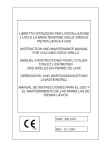
Other required appliances for urine analysis:
In case of visual reading
Urine analyzer DocUReader, LabUReader, LabUReader Plus, HandUReader or
LabUMat with instructions for use
Clean, detergent free and dry container for urine collection
Reagents Composition:
The reagents in the individual test fields are formulated to contain:
Test strip for semi-quantitative urine analysis from fresh urine. Use only with urine analyzer
DocUReader, LabUReader, LabUReader Plus, HandUReader or LabUMat or for visual
reading. The LabStrip U11 Plus is an in vitro diagnostic medical device for professional
laboratory use only in conformity with the Directive 98/79/EC.
Bilirubin:
Urobilinogen:
Ketones:
Ascorbic acid:
Glucose:
150 pcs urine test strips for rapid determination of Bilirubin, Urobilinogen, Ketones (Acetoacetic
Acid), Ascorbic acid, Glucose, Protein (Albumin), Blood, pH value, Nitrite, Leukocytes and
Specific Gravity in urine. Refer to the carton and label for specific parameter combination on the
product you are using.
Protein:
Blood:
Summary and Explanation:
pH:
Screening test for recognition of liver disease, biliary and hepatic obstructions, diabetes, and
haemolytic diseases, urological, and nephrological diseases associated with haematuria or
haemoglobinuria, diseases of the kidneys and urinary tract, pathological shifts in the pH value, as
well as for investigation of the sediment.
Clinical Utility:
Bilirubin: Intended to measure the levels of bilirubin conjugates in urine. Measurements of
urinary bilirubin and conjugates are used in the diagnosis and treatment of certain liver and bile
diseases.
Urobilinogen: Intended to detect and estimate urobilinogen (a bile pigment degradation product
of red blood cell haemoglobin) in urine. Estimations obtained by this device are used in the
diagnosis and treatment of liver diseases and haemolytic (red blood cells) disorders.
Ketones: Intended to detect ketones in urine. Identification of ketones is used for in the diagnosis
and treatment of acidosis (a condition characterized by abnormally high acidity of body fluids) or
ketosis (a condition characterized by increased production of ketone bodies) and for monitoring
patients with diabetes.
Ascorbic acid: Intended to measure the level of ascorbic acid (vitamin C) in urine.
Glucose: Intended to measure glucosuria (glucose in urine). Urinary glucose measurements are
used in the diagnosis and treatment of carbohydrate metabolism disorders including diabetes
mellitus and hyperglycemia.
Protein: Intended to identity protein in urine. Identification of urinary protein is used in diagnosis
and treatment of renal diseases.
Blood: Intended to detect occult blood in urine. Occult blood indicates serious urological or
kidney diseases.
pH: Intended to estimate the pH of urine. Estimations of pH are used to evaluate the acidity or
alkalinity of urine as it relates to numerous renal and metabolic disorders and in the monitoring of
patients with certain diets. Persisting high pH values indicate urinary tract infections.
Nitrite: Intended to identify nitrite in urine. Nitrite identification is used in the diagnosis and
treatment of urinary tract infections of bacterial origin.
Leukocytes: Intended to detect leucocytes in urine. Leucocytes indicate inflammatory diseases
of the kidney and the urinary tract, and suggest need of further investigation.
Specific Gravity: Intended to provide an estimation of renal ability of urine concentration or urine
dilution. The specific gravity of urine varies in accordance with the drinking quantity as well as
different disorders.
Principle of the Procedure:
Bilirubin: A red azo compound is obtained in the presence of acid by coupling of bilirubin with
diazonium salt.
Urobilinogen: The test is based on the coupling of urobilinogen with a stabilised diazonium salt
to a red azo compound.
Ketones: Acetone and acetoacetic acid react with sodium nitroprusside in alkaline solution to
give violet coloured complex (Legal’s test).
Ascorbic acid: The detection is based on the decolouration of Tillman’s reagent.
Glucose: The detection is based on the glucoseoxidase-peroxidase-chromogen reaction. Apart
from glucose no other compound in urine is known to give a positive reaction.
Protein: The test is based on the „protein error” principle of the indicator. The test is especially
sensitive in the presence of albumin. Other proteins are indicated with less sensitivity.
Blood: The detection is based on the pseudoperoxidative activity of haemoglobin and myoglobin,
which catalyze the oxidation of an indicator by organic hydroperoxide and chromogene producing
a green colour.
pH: The test paper contains indicators which clearly change colour between pH 5 and pH 9 (from
orange to green to turquoise).
Nitrite: The colour test is based on the principle of Griess reaction. Any degree of pink-orange
5
colouration should be interpreted as a positive nitrite test suggestive of ≥10 organisms/ml
urine.
Leucocytes: The test is based on the esterase activity of granulocytes. This enzyme splits
heterocyclic carboxylates. The component released reacts with a diazonium salt producing a
violet colour.
Specific Gravity: The test is based on a colour change of the reagent from blue-green to greenish
yellow depending on the concentration of ions in the urine. The test permits the determination of
urine density between 1.000 and 1.030.
Kit Components:
Each kit contains everything needed to perform 150 tests:
50 pcs LabStrip U11 Plus test strips,
Label with colour scale for visual reading,
1 pc Calibration card for checking and setting of analyzers LabUReader and
LabUReader Plus,
1 pc Calibration card for checking and setting of analyzers HandUReader and LabUMat,
1 pc Instructions for use
Angol-nemet_csikhaszn_ANA9201_4v1 1
Nitrite:
Leukocytes:
Specific Gravity:
Diazonium salt
Diazonium salt
Sodium nitroprusside
2,6-dichloro-phenol-indophenol
Glucose oxidase
Peroxidase
O-Tolidine hydrochloride
Tetra-bromophenol blue
Isopropylbenzol-hydroperoxide
Tetramethylbenzidine-dihydrochloride
Bromthymol blue
Methyl red
Sulfanilic acid
Tetrahydrobenzol[h]quinolon-3-ol
Carboxylic acid ester
Diazonium salt
Bromthymol blue
3.1 %
3.6 %
2.0 %
0.7 %
2.1 %
0.9 %
5.0 %
0.2 %
21.0 %
2.0 %
10.0 %
2.0 %
1.9 %
1.5 %
0.4 %
0.2 %
2.8 %
Concentrations given are based on reagent composition (w/w) at time of manufacture and may
vary within manufacturing tolerances.
Warning!
Every item in the package can be handled as household waste. As reactive materials are
present at very low quantity, the product does not come under the scope of the relevant
EU regulations for dangerous materials.
Keep away from swallowing, touching with skin or mucous membranes of chemicals!
For in vitro diagnostic use only!
The vial cap containing a non toxic, molecule-sieve based drying agent blocks the
test strips from air-humidity. In case of swallowing the drying agent chemicals, drink
substantial amount of liquid.
If you have any questions, please turn to your local distributor!
Arrangements for urine analysis:
Warning!
Use only urine analyzer DocUReader, LabUReader, LabUReader Plus, HandUReader or
LabUMat for LabStrip U11 Plus test strip urine analysis.
If you open a new LabStrip U11 Plus test strip package you can find a calibration card for
checking and setting of the urine analyzer LabUReader and LabUReader Plus and another
one for HandUReader and LabUMat. You can find the instructions for settings in the user
manual of the meter. Adjustment of the meter is not required until the start of next strip vial.
Follow the user manual of the urine analyzer DocUReader, LabUReader,
LabUReader Plus, HandUReader or LabUMat.
Specimen Collection and Preparation:
Collect urine in a clean, dry container which allows complete immersion of all the fields
on the test strip.
Do not add preservatives.
Test the specimen as soon as possible, with the sample well mixed but not centrifuged.
The use of fresh morning urine is recommended for optimal nitrite tests, as well as for
the valid determination of bilirubin and urobilinogen, since these compounds are
unstable when exposed to light and room temperature (+15 to +25 °C).
If immediate testing is not possible, the sample should be stored in the refrigerator
(+2 to +8 °C) and then brought to room temperature (+15 to +25 °C) before used in
the test.
Non-preserved urine at room temperature may undergo pH changes due to microbial
proliferation, which may interfere with protein determination.
If cleanly voided specimens are not collected from females, positive results for
leukocytes may be found due to contamination from outside the urinary tract.
Skin cleansers containing chlorhexidine may affect protein test results if specimen positive
contamination occurs.
Procedure and Notes:
Use only fresh, mixed, not centrifuged urine. First morning urine is recommended. Perform
the urine analysis in 4 hours after sample collection! Keep urine away from light.
Collect specimen in clean, rinsed containers, free of detergents. Do not use preservatives!
After removing the required number of strips, close immediately the container securely with
the vial cap containing drying agent.
Do not touch test areas of the reagent strip.
In case of instrumental reading
Read carefully the instructions for use of the analyzer DocUReader, LabUReader,
LabUReader Plus, HandUReader or LabUMat.
Immerse the test strip into urine for approx. 2 sec, so that all reagent areas are covered.
Remove excess urine from the strip by wiping the edge of the strip on the edge of the urine
container or on absorbent paper.
Place the test strip into the urine analyzer DocUReader, LabUReader, LabUReader Plus
or HandUReader according to the instructions for use. 1 minute after starting reading the
meter displays the result.
In case of reading with LabUMat, the meter feeds the strips and dips them into the sample
automatically then displays the result after 1 minute incubation period.
Immerse the test strip into the urine for approx. 2 sec, so that all reagent areas are covered.
Remove excess urine from the strip by wiping the edge of the strip on the edge of the
urine container or on absorbent paper.
To prevent interference from adjacent test areas, incubate the strip in horizontal position.
Compare the reagent areas on the strip with the corresponding color charts on the
container about 60 sec (between 60-120 sec for the Leucocyte test) after immersion. Do
not read later than 2 minutes.
Colours on the colour chart are representing the nominal values of the test fields. Actual
results are located around nominal values.
The white field between the specific weight and leukocyte test field is for instrument
measurements and is used to compensate for the intrinsic colour of urine.
Warning!
Do not take the strip out of the meter during reading procedure.
Prior to measuring always make sure that the process is performed according to the
instructions for use of the urine analyzer DocUReader, LabUReader, LabUReader Plus,
HandUReader or LabUMat.
Do not perform urine analysis at temperatures below +15˚C or above +35˚C.
Strips must be kept away from heat and direct sunlight.
Do NOT ever reuse test strips. After measurement completed eliminate the strip carefully.
Until usage, store the test strips in original packages. Strips in each vial should not be
mixed.
Diagnoses and therapies can not be derived from one single test result only, instead should
be based on all available medical diagnoses.
Never use the strip if more than 5 minutes spent from the moment of its removing from
the vial.
Biological risk!
Handle all specimens and strips as if they contamined infectious agents. When the assay
procedure is completed, dispose of specimens and strips carefully follow the relevant, local
instructions.
In rare occasions, the varying test conditions, due to the heterogenity of the different urine
(for reasons of different levels of activators, inhibitors, or different ion concentrations) may
cause variation in the intensity and contrast of the colors.
Not all cases of interference with every component of any medicines are known. The color
reaction of the pads might change. We, therefore, recommend another test at the end of any
medication with drugs.
Always follow the general working instructions for laboratories as well.
The test strips do NOT contain toxic materials!
Results:
Results are determined visually by direct comparison of reacted test fields with the colour chart
on the container label. Visual colour charts represent nominal test values for each test field-actual
values may vary around the nominal values.
The leucocyte and blood (erythrocyte) tests are not quantitative determinations, but serve
as screening methods for the presence of Leucocytes and blood (erythrocytes) in urine.
Microscopic examination of specimens with a positive Leucocyte or blood test result should be
performed if quantitative results are required.
Ascorbic acid may interfere with the glucose, nitrite, bilirubin and blood test results (see
Limitations below). If a positive Ascorbic acid result is found, either repeat the test at least
10 hours one day after discontinuation of Vitamin C administration or use a photometric test
unaffected by Ascorbic acid.
When using LabUReader, DocUReader, LabUReader Plus, HandUReader or LabUMat
please refer the user manual of those instrument.
System operation:
Each test strip has 11 measuring zones. These zones contain sensitive reagents. Colour of
the test field is changing as a result of chemical reaction of urine contact. The urine analyzer
DocUReader, LabUReader, LabUReader Plus, HandUReader or LabUMat detects the
colouration and displays the result.
Limitations of the Procedure:
Note: Diagnostic or therapeutic decisions should not be based on any single result or method.
Bilirubin: The reaction is unaffected by pH of urine. False low or negative results may be
simulated by large amounts of vitamine C or nitrite or by longer exposure of the sample to direct
light. Increased concentration of urobilinogen can reinforce the sensitivity of the field. Different
urine constituents (e.g. urine indicane) can lead to atypical coloration. For metabolites of drugs
see urobilinogen.
Urobilinogen: The reaction is unaffected by pH of urine. Higher concentration of formaldehyde
or exposure of the urine to light for a longer period of time may lead to lowered or falsely negative
results. Beetroot (excreted pigments) or metabolites of drugs which give a colour at low pH
(phenazopyridine, azo dyes, p-aminobenzoic acid or other medicaments which have a red
intrinsic coloration in acidic medium) may produce false positive results. Prolonged exposure
to light is to be avoided.
Ketones: Phthalein compounds and derivatives of anthrachinone interfere by producing a red
coloration in the alkaline range which may mask the coloration of ketones.
Ascorbic acid: As ascorbic acid already in low concentrations can disturb various test fields,
especially the glucose assay in low concentrations, the test must be repeated if the ascorbic acid
reaction is positive, however, at the earliest 10 hours after the last vitamin C intake (medication,
fruit and vegetables).
Glucose: High concentrations of ascorbic acid in urines with a low glucose concentration (up
to 100 mg/dl (5.5 mmol/l)) may inhibit the reaction and lead to lower or false negative results.
Repeat the test 10 hours one day after stopping the intake of vitamin C. Pay attention to the
ascorbic acid field. In addition an inhibitory effect is produced by gentisic acid, a pH value of <5
and high specific gravity. False positive reactions can also be produced by a residue of peroxide
containing cleansing agents or others.
2010.07.07. 9:28:19
Protein (albumin): Falsely positive results are possible in high alkaline urine samples
(pH >9) and in the presence of high specific gravity, after infusions with polyvinylpyrrolidone
(blood substitute) after intake of medicaments containing quinine and also by disinfectant
residues containing quaternary ammonium groups in the urine sampling vessel.
Blood: Microhaematuria does not affect the colour of urine and is only detectable by microscopic
or chemical tests. From a level approx. 25 Ery/μl and above, even at high concentrations of
ascorbic acid normally no negative results are observed. Falsely positive reactions can also be
produced by a residue of peroxide containing cleansing agents, activities of microbial oxidase
due to infections of the urogenital tract or by formaline.
For establishing an individual diagnosis, it is therefore indispensable to take into consideration
also the clinical manifestations.
The number of erythrocytes which are detected by sediment analysis may be lower than the result
of the test strip, because lysed cells are not detected by sediment analysis.
pH: No interferences are known.
Nitrite: Before testing the patient should ingest vegetable-rich meals, reduce fluid intake and
discontinue antibiotic and vitamin C therapy 3 days prior to the test. False positive results may
occur in stale urine samples, in which nitrite has been formed by contamination of the specimen
and in urines containing dyes (derivatives of pyridinium, beetroot). A negative result even in the
presence of bacteriuria can have the following reasons: bacteria not containing nitrate reductase,
antibiotic treatment diet with low nitrate content, high diuresis, high content of ascorbic acid or
insufficient incubation of the urine in the bladder.
Leukocytes: Strongly coloured compounds (e.g. nitrofurantoin) may disturb the colour of the
reaction. High concentrations of glucose, oxalic acid, drugs containing cephalexin, cephalothine
or thetracycline can lead to weakened reaction. False-positive reactions may be caused by
contamination of vaginal secretion. The number of leucocytes which are detected by sediment
analysis may be lower than the result of the strip, because lysed cells are not detected by
sediment analysis. Partial cytolysis intensifies the colour response, particularly in the region of
the maximum analytical sensitivity. Leucocyte esterase results may be positive in the absence
of observable cells if the leucocytes have lysed. False-positive reactions may be caused by
formaldehyde (preservative). Protein concentrations above 5 g/l or a high specific gravity may
diminish the colour response. Bacteria, trichomonas and erythrocytes do not, however, react with
the test field.
Specific Gravity: Highly acidic urines (pH <6) yield slightly elevated results whereas highly
alkaline urines (pH >8) yield diminished results. Glucose and urea do not interfere.
Expected values:
Bilirubin: Normally, no bilirubin is detectable in urine. Concentration of 0.5 mg/dl and more lead
to a colour of red-orange peach and indicate the early stage of a liver disease. The colour fields
correspond to the following bilirubin concentrations: neg. (negative), 1 (+), 3 (++), 6 (+++) mg/dl
or neg. (negative), 17 (+), 50 (++), 100 (+++) μmol/l. Values of 0.5-1 mg/dl bilirubun
are indicated.
Urobilinogen: The normal concentration of urobilinogen in urine goes from 0.1-1.8 mg/dl (1.730 μmol/l). Concentrations of >2 mg/dl (35 μmol/l) are considered to be pathological. Absolute
absence of urobilinogen in the urine, which is also pathological, cannot be demonstrated using
test strips. The colour fields correspond to the following urobilinogen concentrations: norm.
(normal), 2 (+), 4 (++), 8 (+++), 12 (++++) mg/dl or norm. (normal), 35 (+), 70 (++),
140 (+++), 200 (++++) μmol/l.
Ketones: Normally, the urine is free of ketones. Detectable concentrations of ketones can
originate from physiological stress (fasting, pregnancy, excessive sport). Phenylketones in higher
concentrations will produce variable colours. β-Hydroxibutyric acid is not detected. The colour
fields correspond to the following acetoacetic acid values: neg. (negative), 15 (+), 50 (++),
150 (+++) mg/dl or neg. (negative), 1.5 (+), 5 (++), 15 (+++) mmol/l. Values of
5 mg/dl acetoacetic acid or 50 mg/dl acetone are indicated.
Ascorbic acid: In the presence of ascorbic acid a colour change takes place from grey blue
to orange. The colour fields correspond to the following ascorbic acid concentrations: neg.
(negative), 20 (+), 40 (++) mg/dl or neg. (negative), 1.14 (+), 2.28 (++) mmol/l. Values of
5-10 mg/dl or 0.6-1.1 mmol/l are indicated.
Glucose: Normally, glucose cannot be detected in the urine although small amounts are
secreted also by the healthy kidney. Changes in the colorations less than 50 mg/dl (2.8 mmol/l)
are to be considered normal. The colour fields correspond to the following ranges of glucose
concentrations: norm. (normal), 50 (+), 150 (++), 500 (+++), 1000 (++++) mg/dl
or norm. (normal), 2.8 (+), 8 (++), 28 (+++), 56 (++++) mmol/l. Values of 40 mg/dl
glucose are indicated.
Protein (albumin): Normally, no protein is detectable in the urine of healthy subjects. Colour
intensities for the protein (albumin) field which match the intensity of the 0.3 g/l colour field are
to be classed as pathological. The colour fields correspond to the following ranges of albumin
concentrations: neg. (negative), 30 (+), 100 (++), 500 (+++) mg/dl or neg. (negative),
0.3 (+), 1.0 (++), 5.0 (+++) g/l. Values of approx. 15 mg/dl albumine are indicated.
Blood: Intact erythrocytes are reported by punctual colorations on the test pad, haemoglobin and
myoglobin are reported by a homogeneous green coloration. The color fields correspond to the
following values: neg. (negative), approx. 5-10 (+), approx. 50 (++), approx. 300 (+++) Ery/μl.
Values of approx. 5 erythrocytes/μl are indicated.
pH: Normal fresh urine of healthy people varies between 5 to 6 pH values. The colour fields
correspond to the following pH values: 5, 6, 7, 8, 9.
Nitrite: Negative results do not exclude significant bacteriuria (insufficient incubation, urinary
tract infections due to bacteria not containing nitrate reductase). Red or blue borders or edges
which may be present must not be interpreted as a positive result. Values of 0.05-0.1 mg/dl
nitrite are indicated.
Leucocytes: Urines of healthy subjects do not contain any leucocytes. Positive results,
even when constantly varying from negative to 25, are to be considered as clinically relevant.
Discolorations which no longer match the negative colour field are to be classed as positive.
The colour fields correspond to the following values: neg. (negative), approx.25 (+), approx. 75 (++),
approx. 500 (+++) leucocytes/μl. Values of 10-20 leucocytes/μl are indicated.
Specific Gravity: Normal fresh urine of healthy people varies between 1.015 to 1.025 values.
The colour scale has optimized at a pH of the urine of 6. The colour fields correspond to the
values 1.000, 1.005, 1.010, 1.015, 1.020, 1.025, 1.030.
Angol-nemet_csikhaszn_ANA9201_4v2 2
Measuring Ranges:
Parameters
neg.
+
++
+++
Bilirubin (mg/dl)
neg.
1
3
6
Urobilinogen (mg/dl)
norm
2
4
8
Ketones (mg/dl)
neg.
15
50
150
Ascorbic acid (mg/dl)
neg.
20
40
Glucose (mg/dl)
norm
50
150
500
Protein (mg/dl)
neg.
30
100
500
Blood (Ery/μl)
neg.
ca. 5-10
ca. 50
ca. 300
pH
5
6
7
8
Nitrite
neg.
pos.
Leukocytes (Leu/μl)
neg.
ca. 25
ca. 75
ca. 500
1000
1005
1010
1015
++++
12
Reagenzstreifen für Harnanalyse
1000
Teststreifen für semiquantitative Harnanalyse vom frischen Urin. Ausschliesslich zur Auswertung
in Urinanalysegeräten DocUReader, LabUReader, LabUReader Plus, HandUReader oder
LabUMat oder zum visuellen Ablesen. LabStrip U11 Plus Teststreifen nur zur professionellen
in-vitro-diagnostischen Anwendung nach Richtlinie 98/79/EC über In-vitro-Diagnostika.
9
Teststreifen für die schnelle Bestimmung von Bilirubin, Urobilinogen, Keton, Ascorbinsäure,
Glucose, Protein (Albumin), Blut, pH-Wert, Nitrit, Leukozyten, spezifischem Gewicht aus Harn.
Die Kombination der Parameter auf dem Streifen ist dem Packungsaufdruck zu entnehmen.
Compensation filed
Spoecific Gravity
1020
1025
1030
Storage and Stability:
Keep diagnostic test strips in tightly closed original tubes in a dry, dark and cool place (between
+2 to +30 °C). After removing the required number of strips, close immediately the container
securely. Do not remove the desiccant from the original cap
The strips must be kept away from moisture, direct sunlight, elevated temperature and
chemical fumes. Under proper conditions test strips are stable up to the stated expiry
date even after opening. Do not touch the test pads.
System quality control:
Each laboratory should evaluate according to its own working standards. Prepared leukocytes
suspensions can be used for the daily quality control of leukocyte tests.
For periodically checking your strips and system we offer the QC the Dipper (Quantimetrix
Corporation, REF: 1440-01), the Dropper (Quantimetrix Corporation, REF:1440-02) or
DipAndSpin (Quantimetrix Corporation, REF:1470-01) urine dipstick control solutions.
Dip the test strip into control solution instead of urine sample!
Read the chapter „Checking of the system” in the instructions for use of the urine
analyzer.
Specific Performance Characteristics:
The performance characteristics of LabStrip U11 Plus test strips are based on both clinical and
analytical studies. Sensitivity is dependent upon the colour perception of the reader, the presence
or absence of interfering specimens, and the lighting conditions for visual reading.
Each colour block on the chart corresponds to a range of analyte concentrations.
Bilirubin: In 90 % of urines tested, bilirubin concentrations of 0.5 mg/dl produced a positive
result. After a longer reaction time an unspecific yellow colour may develop, which can create
positive interference.
Urobilinogen: Based on the work of Kutter [10], a concentration of 1 mg/dl urobilinogen will
yield a positive result. The test is sufficiently sensitive so that normal specimens will produce a
slight pink colour.
Ketones: In 90 % of urines tested, acetoacetic acid at 8 mg/dl produced a positive result. The
test field reacts less sensitively with acetone. Hydroxybutyric acid is not detected.
Ascorbic acid: In 90 % of urines tested, ascorbic acid at 20 mg/dl yielded a positive result.
Glucose: The maximum sensitivity is 20 mg/dl. The test field response is adjusted so that
pathological glucose concentrations of 30 mg/dl (Fine) [11] are recognized. Sugars other than
glucose and other reducing substances do not react in this test. Possible interference by ascorbic
acid can be detected by the adjoining test field which reacts with the ascorbic acid.
Protein: In 90 % of urines tested, albumin concentrations of 12 mg/dl produced a positive result.
The test is more sensitive to albumin than to globulin, Bence-Jones proteins and mucoproteins.
A negative result does not exclude the presence of these other proteins.
Blood: The test permits the differentiation of intact erythrocytes from hemoglobin or myoglobin.
Erythrocytes react as diffuse patches on the field. The practical sensitivity of the test lies between
5 and 10 Ery/μl.
A study of 625 fresh urine specimens, comparing results with those using another test strip for
blood demonstrated a clinical specificity of 90.2 % and sensitivity of 81 %.
pH: pH values are determined to within 1 unit over the range from 5 to 9. Readings are not
affected by variations in the urine buffer concentration.
Nitrite: The maximum sensitivity is 0.05 mg/dl, which is equivalent to about 100,000 bacteria/ml.
In early morning urine, 90 % of all infections are detected by the nitrite test. Although most
uropathogenic bacteria are able to reduce nitrate to nitrite (e. g. Klebsiella, E. coli, Proteus,
Aerobacter, Citrobacter, etc.), results depend on the number of bacteria, the nitrate content, and
the retention time of the urine.
Leucocytes: In 90 % of urines tested, concentrations of 20 leucocytes/μl produced positive
results. Any pink coloration of the test field should be considered clinically significant. When
result from this method were compared with those using another test strip for leucocytes for 822
fresh urines, clinical specificity of 80 % and sensitivity of 89.2 % were determined.
Specific Gravity: In 86 % of 102 urines tested, the finds based on the colour chart were found
to be within the acceptable range of + or – one colour increment compared to the findings of
the reference refractometer.
Intra-assay
Within run precision was determined by using 10 replicas of two levels (normal, abnormal) of
control urine.The negative and positive values were correctly identified 100 % of time for all the
parameters.
Inter-assay
Between run precision was determined by using two levels (normal, abnormal) of control urine
in 10 independent assays and with three different lots of reagent over a 6 months period. The
negative and positive values were correctly identified 100 % of time for all the parameters.
Anwendung
Schnelltest zur Diagnostik und Früherkennung von Leberschäden, Verschlussformen,
Diabetes, hämolytischen, urologischen und nephrologischen Erkrankungen, die mit Hämaturie
und Hämoglobinurie verbunden sind. Erkrankungen im Bereich der Nieren und Harnwege,
pathologischen pH-Wert-Verschiebungen und zur Untersuchung des Sediments.
Klinische Bedeutung
Bilirubin: Zur Bestimmung von Bilirubin im Harn. Bestimmungen von Bilirubin und ihren
Konjugatum dienen zur Diagnose von Leber- und Gallenerkrankungen.
Urobilinogen: Zur Bestimmung von Urobilinogen im Harn. Die Bestimmung dient zur Diagnose
von Lebererkrankungen und gesteigertem Hämoglobinabbau infolge von hämolytischen
Erkrankungen.
Keton: Zur Bestimmung von Ketonkörpern im Harn. Die Bestimmung dient zur Diagnose von
Ketoacidose sowie zur Behandlung und Kontrolle von Diabetes-Patienten.
Ascorbinsäure: Zur Bestimmung von Ascorbinsäure (Vitamin C) im Harn.
Glucose: Zur Bestimmung von Glucose im Harn. Bestimmungen von Glucose im Harn dienen
zur Diagnose und Behandlung von Störungen des Kohlenhydratstoffwechsels, wie Diabetes
mellitus und Hyperglycaemie.
Protein (Albumin): Zur Bestimmung von Protein im Harn. Der Nachweis dient zur Diagnose
und Behandlung von Nierenerkrankungen.
Blut: Zur Bestimmung von okkultem Blut im Harn. Okkultes Blut im Harn weist auf Erkrankungen
des Urogenitalbereichs und der Niere hin.
pH-Wert: Zur Bestimmung des pH-Wertes im Harn. Die Bestimmung dient zur Bewertung der
Acidität oder Alkalität des Harns, die im Zusammenhang mit Stoffwechselstörungen auftreten
können, und zur Überwachung von Diäten. Anhaltend hohe pH-Werte deuten auf eine Infektion
des Urogenitalbereichs hin.
Nitrit: Zur Bestimmung von Nitrit im Harn. Nitrit im Harn deutet auf bakteriell verursachte
Infektionen des Urogenitalbereichs hin.
Leukozyten: Zur Bestimmung von Leukozyten im Harn. Leukozyten im Harm deuten auf
Entzündungen der Niere oder des Urogenitalbereichs hin.
Spezifisches Gewicht/Dichte: Zur Bestimmung der Dichte von Harn. Die Bestimmung
dient zur Kontrolle der Nierenfunktion und zur allgemeinen Bewertung der Konzentration der
Harnprobe. Je nach aufgenommener Flüssigkeitsmenge und äußeren Umständen kann die
Dichte des Harnes schwanken.
Testprinzipien
Bilirubin: Der Test basiert auf einer Kupplungsreaktion von Bilirubin mit Diazoniumsalz in
einem sauren Medium.
Urobilinogen: Das Testfeld für Urobilinogen enthält ein stabiles Diazoniumsalz. Durch
Kupplungsreaktion entsteht ein roter Azofarbstoff.
Keton: Es handelt sich um eine Variante der Probe nach Legal. Acetessigsäure und Aceton
reagieren mit Natrium-Nitroprussid in alkalischem Medium zu einem violetten Farbkomplex.
Ascorbinsäure: Der Nachweis beruht auf der Entfärbung von Tillmans-Reagenz.
Glucose: Der Nachweis basiert auf der Glucoseoxidase-Peroxidase-Chromogen Reaktion.
Auβer Glucose ist kein Harninhaltsstoff bekannt, der eine positive Reaktion liefert.
Protein (Albumin): Der Test beruht auf dem „Eiweißfehler“ des Indikators. Der Test reagiert
besonders empfindlich gegenüber Albumin. Andere Urinproteine reagieren weniger stark.
Blut: Die Pseudoperoxidase-Aktivität des Hämoglobins und Myoglobins führt in Anwesenheit
organischer Hydroperoxyde und eines Chromogens zu einem grünen Farbstoff.
pH: Das Testpapier enthält einen Mischindikator, der im pH-Bereich von 5 bis 9 deutlich
unterscheidbare Reaktionsfarben (von orange über gelb nach türkis) zeigt.
Nitrit: Farbtest auf Grundlage der Probe nach Grieß. Jede Rosafärbung gilt als positiv und
weist auf ≥105 Keime/ml Harn hin.
Leukozyten: Granulozytenesterasen spalten einen heterozyklischen Karbonsäureester, das
Spaltprodukt reagiert mit einem Diazoniumsalz zu einem violetten Farbstoff.
Spezifisches Gewicht/Dichte: Der Test beruht auf einem Farbumschlag des Wirkstoffes von
blaugrün nach grüngelb in Abhängigkeit der Konzentration ionischer Bestandteile im Harn. Der
Test erlaubt die Bestimmung der Harndichte zwischen 1.000 und 1.030.
Der Inhalt der Schachtel
Packungen mit 150 Teststreifen:
150 Stück LabStrip U11 Plus Teststreifen,
Farbenskala für visuelle Auswertung
1 Stück Kalibrierstreifen zur Kontrolle und zum Kalibrieren der Urinanalysegeräte
LabUReader und LabUReader Plus
1 Stück Kalibrierstreifen zur Kontrolle und zum Kalibrieren der Urinanalysegeräte
HandUReader und LabUMat
1 Stück Gebrauchsanleitung
2010.07.07. 9:29:39
Sonstige Mittel zur Urinanalyse
Urinanalysegeräte DocUReader, LabUReader, LabUReader Plus, HandUReader
oder LabUMat mit Gebrauchsanleitung
Trockenes, chemikalienfreies und sauberes Gefäß zum Auffangen von Urin.
Inhaltsstoffe
Die Reagenzien auf den einzelnen Testfeldern setzen sich wie folgt zusammen:
Diazoniumsalz
3.1 %
Diazoniumsalz
3.6 %
Nitroprussid-Natrium
2.0 %
2.6-Dichlorophenolindophenol
0.7 %
Glucoseoxidase
2.1 %
Peroxidase
0.9 %
O-Tolidin - Hydrochloride
5.0 %
Protein (Albumin):
Tetrabromphenolblau
0.2 %
Blut:
Isopropylbenzol-Hydroperoxide
21.0 %
Tetramethylbenzidin - Dihydrochloride
2.0 %
pH:
Bromthymolblau
10.0 %
Methylrot
2.0 %
Nitrit:
Sulfanilsäure
1.9 %
Tetrahydrobenzol[h]quinolon-3-ol
1.5 %
Leukozyten:
Karbonsäureester
0.4 %
Diazoniumsalz
0.2 %
Spezifisches Gewicht: Bromthymolblau
2.8 %
Die Prozentangaben basieren auf den Reagenz-Zusammensetzungen (w/w) zum
Herstellungszeitpunkt und können im Rahmen der Herstellungs-Toleranz variieren.
Bilirubin:
Urobilinogen:
Keton:
Ascorbinsäure:
Glucose:
Vorsicht!
Alle Mittel im Paket können im Haushaltsmüll entsorgt werden. Wegen geringes Anteils
an reaktiven Werkstoffen ist die EU Regelung über gefährliche Stoffe nicht anwendbar.
Verschlucken und Kontakt mit Haut und Schleimhäuten vermeiden.
Nur zur in-vitro-diagnostischen Anweisung.
Im Originalstopfen der Teststreifen gibt es einen nicht giftigen, saugfähigen Stoff
auf Molekularfilterbasis, der die Teststreifen vor Nässe schützt. Beim zufälligen
Verschlucken ergiebig Flüssigkeit trinken.
Bei Fragen wenden Sie sich bitte an Ihren Distributor.
Vorbereitung zur Harnanalyse:
Vorsicht!
Zur Harnanalyse mit Teststreifen LabStrip U11 Plus nur Harnanalysegerät DocUReader,
LabUReader, LabUReader Plus, HandUReader oder LabUMat verwenden.
In der Originalflasche LabStrip U11 Plus sind zwei Kalibrierstreifen zugepackt, jeweils ein
zum Kalibrieren der Geräte LabUReader und LabUReader Plus, bzw. HandUReader und
LabUMat. Zur Einstellung bitte die ausführliche Gebrauchsanleitung zum Gerät beachten. Das
Gerät ist beim Einsatz einer neuen Originalflasche neu zu kalibrieren.
Bitte die ausführliche Gebrauchsanleitung zum Harnanalysegerät DocUReader,
LabUReader, LabUReader Plus, HandUReader oder LabUMat beachten.
Probengewinnungen und Testvorbereitung
Eine frische Harnprobe in einem sauberen, trockenen Gefäß sammeln.
Keine Konservierungsmittel zufügen.
Den Test sollte so bald als möglich in der unzentrifugierten, gut durchmischten Probe
erfolgen.
Für optimale Nitrittestungen wird die Verwendung von frischem Morgenharn empfohlen,
genauso wie für die gültige Bestimmung von Bilirubin und Urobilinogen, da diese
Analyse bei Einfluss durch Licht und Raumtemperatur instabil sind (+15 bis +25 °C).
Wenn die Austestung nicht sofort durchgeführt werden kann, ist die Probe im Kühlschrank
(+2 bis +8 °C) aufzubewahren und vor Testung wieder auf Raumtemperatur (+15 bis
+25 °C) zu bringen.
Bei Raumtemperatur können im Harn ohne Konservierungsstoffe durch mikrobielle
Vermehrung pH-Veränderungen auftreten, die die Proteinbestimmung stören.
Falls die Entnahme von Harn bei Damen nicht einwandfrei durchgeführt wird, können
durch Kontaminationen vom äußeren Genitalbereich positive Ergebnisse für Leukozyten
erhalten werden.
Chlorhexidinhaltige Reinigungsmittel können bei Verunreinigung der Probe positive
Proteinergebnisse vortäuschen.
Durchführung
Nur gut gemischten, unzentrifugierten Harn, der nicht länger als 4 Stunden gestanden
hat, verwenden. Empfohlen wird der erste Morgenurin. Vor Licht schützen.
Zur Harnsammlung nur gut gespülte, saubere Gefäße verwenden. Keine
Konservierungsmittel zusetzen.
Stets nur die notwendige Anzahl an Teststreifen entnehmen. Packung nach der Entnahme
sofort wieder mit dem Originalstopfen fest verschließen.
Reaktionszone nicht berühren!
Instrumentelle Auswertung
Bei Auswertung mit DocUReader, LabUReader, LabUReader Plus, HandUReader
oder LabUMat bitte vorher die ausführliche Gebrauchsanweisung zum Gerät beachten.
Den Teststreifen ins Urinanalysegerät DocUReader, LabUReader, LabUReader Plus
oder HandUReader einlegen. Die Testergebnisse werden nach Ablauf von 60 Sekunden
auf dem Display angezeigt.
Im Urinanalysegerät LabUMat werden die Teststreifen automatisch weiterleitet und in die
Urinprobe getaucht. Die Testergebnisse werden nach Ablauf von 60 Sekunden auf dem
Display angezeigt.
Visuelle Auswertung
Teststreifen kurz (ca. 2 Sek.) in die Urinprobe eintauchen. Alle Testfelder benetzen.
Angol-nemet_csikhaszn_ANA9201_4v3 3
Überschüssigen Harn über die Kante des Streifens am Rand des Sammelgefäßes oder
auf saugfähigem Papier abstreifen.
Teststreifen während der Inkubationszeit waagerecht halten, um Interferenzen zwischen
den Reaktionszonen zu vermeiden.
Reaktionsfarben nach 60 Sek. (Leukozyten nach 60-120 Sek.) mit der Farbskala
vergleichen. Verfärbungen, die nur am Rand der Testfelder oder nach mehr als 2 Minuten
nach Testbeginn auftreten, sind ohne Bedeutung.
Die Farbfelder stellen Nennwerte dar. Istwerte schwanken um die Nennwerte.
Das Kompensationsfeld zwischen dem spezifischen Gewicht und den Leukozyten ist
frei von Chemikalien und dient ausschließlich der reflektrometrischen Auswertung der
Testfelder.
Vorsicht!
Grundsätzlich ist eine definitive Diagnose nicht auf der Basis einzelner Teststreifenresultate,
sondern erst im Zusammenhang mit anderen ärztlichen Befunden zu erstellen, und infolge
gezielter Therapie einzuleiten.
Biologische Gefahr
Entsorgen Sie die benutzten Teststreifen unter Beachtung der geltenden
Sicherheitsbestimmungen.
Durch die nicht konstante Zusammensetzung des Harns (z.B. wechselnder Gehalt von
Probe zu Probe an Aktivatoren oder Inhibitoren, wechselnde Ionekonzentration) sind die
Reaktionsbedienungen nicht immer gleich, so dass Intensität und Farbton in seltenen
Fällen variieren können.
Die Auswirkung von Medikamenten oder deren Metaboliten auf den Test ist nicht in allen
Fällen bekannt. Im Zweifelsfall wird deshalb empfohlen, den Test nach Absetzen der
Medikation zu wiederholen.
Für den Umgang mit Teststreifen sind die allgemeinen Arbeitsvorschriften für das Labor
zu beachten.
Die Teststreifen enthalten KEINE Giftstoffe.
Resultate
Alle Teststreifen können visuell ausgewertet werden, oder auch instrumentell, unter Verwendung
eines Harnanalysengerätes. Die visuellen Farbskalen ergeben Nominalwerte für jedes Testfeld,
die tatsächlichen Werte können von den Nominalwerten etwas abweichen.
Leukozytentest und Bluttest sind keine quantitativen Bestimmungen, sondern sie dienen
als Filterverfahren für den Nachweis von Leukozyten und Blut im Harn. Mit einem positiven
Ergebnis sollten mikroskopische Untersuchungen durchgeführt werden, wenn quantitative
Ergebnisse erforderlich sind.
Ascorbinsäure in hohen Konzentrationen kann insbesondere den Glucose-, Nitrite-, Bilirubinund Blutnachweis beeinflussen (Grenzen). Der Test muss bei positiver Ascorbinsäurereaktion
wiederholt werden, frühestens 10 Stunden nach der letzten Vitamin C-Aufnahme (Obst,
Gemüse, Medikation).
Näheres erfahren Sie bei Ihrem zuständigen Medizinproduktberater, wenn Sie LabUReader,
DocUReader, LabUReader Plus, HandUReader oder LabUMat benutzen.
Durchführung
Jeder Teststreifen hat 11 Reagenzzonen, die empfindliche chemische Stoffe enthalten. Wenn
diese Testfelder mit Urin benetzt werden, entsteht eine chemische Reaktion, wobei sich die
Farbe der Reagenzzone ändert. In den Harnanalysegeräten DocUReader, LabUReader,
LabUReader Plus, HandUReader oder LabUMat werden die Reaktionsfarben bestimmt und
die Messwerte ermittelt.
Grenzen
Grundsätzlich ist eine definitive Diagnose nicht auf der Basis einzelner Teststreifenresultate
gestellt werden.
Bilirubin: Die Reaktion ist pH-unabhängig. Falsch niedrige oder negative Resultate können
durch hohe Konzentrationen an Vitamin C oder Nitrit auftreten und durch längeres Stehen
am Licht. Erhöhte Urobilinogen-Konzentrationen können die Empfindlichkeit des Testfeldes
verstärken. Versch. Harnbestandteile (z.B. Harnindikant) können zu atypischen Verfärbungen
führen. Bzgl. Pharmakametaboliten siehe Urobilinogen.
Urobilinogen: Die Reaktion ist pH-unabhängig. Formaldehyd oder Sonnenlicht kann zu falsch
niedrigen oder negativen Werten führen. Rote Beete und Pharmakametabolite, die bei niedrigem
pH-Wert eine rote Färbung geben (Phenazopyridine, Azofarbstoffe, p-Aminobenzoesäure)
können falsch positive Ergebnisse verursachen. Sonnenlicht kann zu falsch negativen Werten
führen.
Keton: Phthaleinverbindungen und Anthrachinonderivate zeigen im alkalischen Bereich
rötliche Farbtöne, die den Nachweis überdecken können.
Ascorbinsäure: Da sich Ascorbinsäure die verschiedenen Testfeldern störend auswirkt, muss
der Test bei pos. Ascorbinsäurereaktion wiederholt werden, frühestens 10 Stunden nach der
letzten Vitamin C-Aufnahme (Obst, Gemüse, Medikation).
Glucose: Bis einer Glucosekonzentration von ca. 100 mg/dl (5,5 mmol/l) werden bei hohen
Ascorbinsäurekonzentrationen falsch negative Ergebnisse beobachtet. Der Test muss bei pos.
Ascorbinsäurereaktion frühestens 10 Stunden nach der letzten Vitamin C-Aufnahme wiederholt
werden. Hemmwirkung zeigen weiterhin Gentisinsäure, pH<5 und hohes spez. Gewicht.
Falsch positive Reaktionen können durch Reste peroxydhaltiger oder anderer Reinigungsmittel
hervorgerufen werden.
Protein (Albumin): Falsch positive Befunde können bei stark alkalischem Harn (pH>9) und
hohem spezifischem Gewicht, nach Infusionen mit Polyvinylpyrrolidon (Blutersatzmittel),
bei der Behandlung mit chininhaltigen Präparaten und durch Reste Desinfektionsmittel mit
quartären Ammoniumgruppen im Sammelgefäß auftreten.
Blut: Durch Mikrohämaturie wird die Farbe des Harns nicht beeinflusst, eine Bestimmung ist
daher nur mit chemischem Test oder mikroskopisch möglich. Ab einer Konzentration von ca.
25 Ery/μl oder höher werden auch bei hohen Ascorbinsäurekonzentrationen normalerweise
keine falsch negativen Ergebnisse beobachtet. Falsch positive Reaktionen können durch
Reste peroxydhaltiger oder anderer Reinigungsmittel, mikrobielle Oxidase-Aktivitäten bei
Urogenitaltraktinfektionen oder Formalin hervorgerufen werden. Die Aussagekraft eines
positiven Ergebnisses schwankt von Patient zu Patient, zur Erstellung einer individuellen
Diagnose ist daher das klinische Bild unerlässlich. Die Anzahl der im Sediment ermittelten
Erythrozyten kann niedriger sein als das Teststreifenresultat, da bereits lysierte Zellen im
Sediment nicht erfasst werden.
pH-Wert: Keine Interferenzen sind bekannt.
Nitrit: Vor der Untersuchung sollte der Patient gemüsereiche Nahrung zu sich nehmen,
die Flüssigkeitsaufnahme reduzieren und eine Antibiotica- oder Vitamin C-Therapie 3 Tage
vor Probennahme absetzen. Falsch positive Resultate können bei alten Urinen auftreten
(Nitritbildung auf Grund von Sekundärkontamination) und in Urinen, die Farbstoffe enthalten
(Pyridiniumderivate, rote Beete). Negative Anzeige bei vorliegender Bakterieurie kann folgende
Ursachen haben: Keime ohne Befähigung zur Nitratreduktion, Antibiotica-Therapie, nitratarme
Kost, starke Diurese, hoher Ascorbinsäuregehalt oder zu geringe Verweilzeit des Urins in der
Blase.
Leukozyten: Stark gefärbte Proben (z.B. Nitrofurantoin) können die Farbe auf dem Testfeld
beeinträchtigen, Glucose oder Oxalsäure in höheren Konzentrationen, Medikamente mit
Cephalexin, Cephalothin oder Tetracyclin können zu einer schwächeren Reaktion führen. Die
Anzahl der im Sediment ermittelten Leukozyten kann niedriger sein als das Teststreifenresultat,
da bereits lysierte Zellen im Sediment nicht erfasst werden. Granulozytenesterasen spalten
einen heterozyklischen Karbonsäureester, wenn die Leukozyten lysieren werden. Falsch
positive Reaktionen können durch Formaldehyd verursacht werden. Stark erhöhte (5 g/l)
Konzentrationen an Protein können die Farbreaktion abschwächen. Bakterien, Trichomonaden
und Erythrozyten reagieren dagegen nicht mit dem Testfeld.
Spezifisches Gewicht/Dichte: Stärker alkalischer (pH >8) Harn führt zu leicht erniedrigten,
stärker saurer (pH <6) Harn zu leicht erhöhten Befunden. Glucose und Harnstoff haben keinen
Einfluss.
Erwartungswerte
Bilirubin: Normalerweise ist Bilirubin im Harn nicht nachweisbar. Werte ab 0.5 mg/dl führen
zur rötlich-orangen Pfirsichfarbe und weisen auf das Frühstadium einer Lebererkrankung hin.
Die Farbfelder sind folgenden Konzentrationen zugeordnet: neg. (negativ), 1 (+), 3 (++),
6 (+++) mg/dl bzw. neg. (negativ), 17 (+), 50 (++), 100 (+++) μmol/l. Konzentrationen
ab 0.5-1 mg/dl Bilirubin werden angezeigt.
Urobilinogen: Die normale Urobilinogenkonzentration im Harn reicht von 0.1-1.8 mg/dl
(1.7-30 μmol/l), Konzentrationen >2 mg/dl (35 μmol/l) gelten als pathologisch. Die
Farbfelder entsprechen folgenden Urobilinogenkonzentrationen: norm. (normal), 2 (+), 4 (++),
8 (+++), 12 (++++) mg/dl bzw. norm. (normal), 35 (+), 70 (++), 140 (+++),
200 (++++) μmol/l.
Keton: Normalerweise enthält Harn keine Ketonkörper. Nachweisbare Ketonkonzentrationen
können durch physiologische Anstrengung (Fasten, Schwangerschaft, Sport) verursacht
werden. Phenylketone können in höheren Konzentrationen eine abweichende Färbung
ergeben. β-Hydroxibuttersäure wird nicht erfasst. Die Farbfelder sind folgenden
Acetessigsäurekonzentrationen zugeordnet: neg. (negativ), 15 (+), 50 (++), 150 (+++) mg/dl
bzw. neg. (negativ), 1.5 (+), 5 (++), 15 (+++) mmol/l. Konzentrationen ab 5 mg/dl
Acetessigsäure bzw. 50 mg/dl Aceton werden angezeigt.
Ascorbinsäure: Die Anwesenheit von Ascorbinsäure wird durch einen Umschlag von graublau
nach orange angezeigt. Die Farbfelder entsprechen: neg. (negativ), 20 (+), 40 (++) mg/dl
bzw. neg. (negativ), 1.14 (+), 2.28 (++) mmol/l. Konzentrationen von 5-10 mg/dl bzw.
0.6-1.1 mmol/l Ascorbinsäure wird angezeigt.
Glucose: Glucose ist normalerweise im Harn nicht nachweisbar, obwohl minimale Mengen
auch durch die gesunde Niere ausgeschieden werden. Farbänderungen schwächer als
50 mg/dl (2.8 mmol/l) sind als normal einzustufen. Die Farbfelder entsprechen folgenden
Konzentrationen: norm. (normal), 50 (+), 150 (++), 500 (+++), 1000 (++++) mg/dl
bzw. norm. (normal), 2.8 (+), 8 (++), 28 (+++), 56 (++++) mmol/l. Konzentrationen
ab 40 mg/dl Glucose werden angezeigt.
Protein (Albumin): Normalerweise ist Bilirubin im Harn nicht nachweisbar.
Farbwerte für Protein (Albumin), die eine Intensität des 0,3 g/l Farbfeldes erreichen, sind als
pathologisch zu bewerten. Die Farbfelder sind folgenden Albuminkonzentrationen zugeordnet:
neg. (negativ), 30 (+), 100 (++), 500 (+++) mg/dl bzw. neg. (negativ), 0.3 (+),
1.0 (++), 5.0 (+++) g/l. Konzentrationen ab ca. 15 mg/dl Albumin werden angezeigt.
Blut: Intakte Erythrozyten werden durch punktförmige Verfärbungen des Testfeldes Hämoglobin
bzw. Myoglobin durch eine homogene grüne Färbung angezeigt. Die Farbfelder entsprechen:
neg. (negativ), ca. 5-10 (+), ca. 50 (++), ca. 300 (+++) Ery/μl. Konzentrationen ab
5 Erythrozyten/μl werden angezeigt.
pH-Wert: Bei Gesunden liegt der pH-Wert des frischen Harns meist zwischen pH 5 und 6. Die
Farbvergleichsfelder entsprechen einem pH-Wert von: 5, 6, 7, 8, 9.
Nitrit: Negative Ergebnisse schließen eine signifikante Bakteriurie nicht aus (kurze Verweilzeit
des Harns in der Blase, Infektionen mit Bakterien ohne Nitratreduktase). Gelegentlich auftretende
rote oder blaue Ränder oder Ecken sind nicht als positive zu bewerten. Konzentrationen ab
0.05-0.1 mg/dl Nitrit werden angezeigt.
Leukozyten: Proben des Gesundens enthalten keine Leukozyten. Positive Ergebnisse, auch
wenn wiederholt zwischen negativ und 25, sind als klinisch relevant zu betrachten. Die
Farbvergleichsfelder entsprechen: neg. (negativ), ca .25 (+), ca. 75 (++), ca. 500 (+++)
Leukozyten /μl. Konzentrationen ab 10-20 Leukozyten /μl werden angezeigt.
Spezifisches Gewicht/Dichte: Der Normalwert liegt etwa zwischen 1.015 und 1.025. Die
Farbskala ist auf einen mittleren pH-Wert von Harn von 6 optimiert. Die Farbfelder sind
Konzentrationen von 1.000, 1.005, 1.010, 1.015, 1.020, 1.025, 1.030.
Messgrenzen
Parameter
neg.
+
++
+++
Bilirubin (mg/dl)
neg.
1
3
6
Urobilinogen (mg/dl)
norm
2
4
8
neg.
15
50
150
Keton (mg/dl)
Ascorbinsäure (mg/dl)
neg.
20
40
Glucose (mg/dl)
norm
50
150
500
Protein (mg/dl)
neg.
30
100
500
Blut (Ery/μl)
neg.
ca. 5-10
ca. 50
ca. 300
pH-Wert
5
6
7
8
Nitrit
neg.
pos.
Leukozyten (Leu/μl)
neg.
ca. 25
ca. 75
ca. 500
1000
1005
1010
1015
++++
12
1000
9
Kompensationsfeld
Spezifisches Gewicht
1020
1025
1030
2010.07.07. 9:29:39
Haltbarkeit
Teststreifen kühl und trocken aufbewahren (Lagertemperatur +2 bis +30 °C). Stets nur die
notwendige Anzahl an Teststreifen entnehmen. Packung nach der Entnahme sofort wieder fest
verschließen. Dose nach Entnahme sofort wieder mit dem Originalverschluss verschließen.
Teststreifen vor Licht und Feuchtigkeit schützen. Bei sachgemäßer Lagerung sind
die Teststreifen bis zum aufgedruckten Verfalldatum haltbar. Reaktionszone nicht
berühren.
Qualitätskontrollsystem
Jedes Labor sollte eigene Zielwerte für die adäquaten Leistungsstandards ermitteln. Testen Sie
jeden Tag stets negative und positive Kontrolllösungen.
Wie empfehlen Ihnen QC Dipper (Quantimetrix Corporation, REF: 1440-01), Dropper
(Quantimetrix Corporation, REF: 1440-02) oder DipAndSpin (Quantimetrix Corporation,
REF: 1470-01). Den Teststreifen in die Kontrolllösung statt des Urins eintauchen.
Bitte „Systemkontrolle” der ausführlichen Gebrauchsanweisung zum Harnanalysegerät
beachten.
Spezifische Leistungsangaben
Die Leistungsangaben des LabStrip U11 Plus Teststreifens basieren auf klinischen und
analytischen Studien. Die Empfindlichkeit ist von verschiedenen Faktoren abhängig, sowie den
Unterschieden in der Farbwahrnehmung, Anwesenheit oder Abwesenheit von normalerweise
im Harm anzutreffenden Inhibitoren und Matrixfaktoren.
Bilirubin: 90 % der Teste ergaben positive Resultate auf Bilirubinkonzentration ab 0.5 mg/dl.
Farbveränderungen, die nach Stellung auftreten, sind ohne Bedeutung.
Urobilinogen: Nach Kutter [10], führen Werte ab 1 mg/dl von Urobilinogen zu positiven
Resultaten.
Keton: 90 % der Teste ergaben positive Resultate auf Acetessigsäure ab 8 mg/dl. Der Test
reagiert nicht mit Aceton. Hydroxibuttersäure wird nicht erfasst.
Ascorbinsäure: 90 % der Teste ergaben positive Resultate auf Ascorbinsäure ab 20 mg/dl.
Glucose: Konzentrationen ab 20 mg/dl Glucose werden angezeigt. Die Empfindlichkeit der
Testfelder ermöglicht pathologische Glucosekonzentrationen von 30 mg/dl (Fine) [11] zu
erfassen. Außer Glucose ist kein Harninhaltsstoff bekannt, der eine positive Reaktion liefert.
Ascorbinsäure in hohen Dosen kann in Proben mit niedrigem Glucosegehalt die Reaktion
hemmen. Ascorbinsäurefeld beachten!
Protein (Albumin): 90 % der Teste ergaben positive Resultate auf Albuminkonzentrationen ab
12 mg/dl. Der Proteintest ist gegenüber Mucoproteinen und Globulinen weniger empfindlich.
Ein negatives Ergebnis schließt also das Vorhandensein dieser Proteine nicht aus.
Blut: Der Test ist gleichermaßen empfindlich für Myoglobin und Hämoglobin. Der Test erfasst
Werte ab 5 bis 10 Erythrozyten/μl Harn. Eine Studie von 625 frischen Harnproben, wobei
die Resultate mit denen von anderen Teststreifen verglichen wurden, ermittelt eine klinische
Spezifität von 90,2% und eine Empfindlichkeit von 81%.
pH-Wert: Die Farbskala erlaubt eine deutliche Differenzierung des pH-Wertes zwischen
pH 5 und 9. Die Werte werden nicht durch Änderungen der Urinpufferkonzentration
beeinträchtigt.
Nitrit: Der Nachweis erfasst Werte ab 0.05 mg Nitrit/dl Harn, das bedeutet 100.000 Keime/ml
Harn. In dem ersten Morgenurin wird 90 % der Ansteckung positives Nitritergebnis ergeben.
Das Prinzip dieses Tests beruht auf der Umwandlung von Nitrat (aus Nahrung) in Nitrit durch
gram-negative Bakterien im Harn. Der Test ist nitritspezifisch und reagiert keiner anderen
normalerweise im Harn ausgeschiedenen Substanz. Bakterien werden nicht aufgeführt.
Leukozyten: 90 % der Teste ergaben positive Resultate auf Konzentrationen ab 20 Leukozyten/μl.
Eine rosa Verfärbung am Testfeld wird als klinisch signifikant beurteilt. Klinische Empfindlichkeit
aus 822 Harnproben befunden zwischen 80 % und 89,2 %.
Spezifisches Gewicht/Dichte: Bei 86% von 102 Harnproben befunden die Werte am
Farbskala im Bereich von + oder – eins im Vergleich zu den durch die Refraktometer-Methode
gemessenen Werten.
Impräzision während der Analyse
Die Wiederholbarkeit während der Analyse wurde durch 10 wiederholte Messungen mit zwei
Harnkontrolllösungen (normal, abnormal) definiert. Die negativen and positiven Resultate
wurden zu 100% der Zeit auf alle Parameter richtig identifiziert.
Impräzision zwischen Analysen
Die Reproduzierbarkeit zwischen den Analysen wurde durch 10 unabhängige Messungen mit
zwei Harnkontrolllösungen (normal, abnormal) definiert. Die Messungen wurden 6 Monate
lang mit drei verschiedenen Chargen von Reagenzien duchgeführt. Die negativen and positiven
Resultate wurden zu 100% der Zeit auf alle Parameter richtig identifiziert.
Literature/Literatur
[1] Legal, E. A.: New Acetone Reaction and its Applicability for the Examination of Urine. Chem. Centr. 15:
652 (1983)
[2] Chertack, M. und Sherrick, J.: Evaluation of Nitroprusside Dip Test for Ketone Bodies. J. A. M. A. 167:
1621 (1958)
[3] Roe, J. H.: Chemical Determination of ascorbic, dehydroascorbic and diketogulonic Acids. Methods of
Biochemical Analysis, Vol 1: 115 (1954) ed. by d. Glick, Interscience Publisher, New York
[4] Comer, J.: Semiquantitative Specific Test Paper for Glucose in Urine. Anal. Chem. 28: 1748 (1956)
[5] Appel, W., Nurck, C. und Merkle, U.: A Rapid Test for Urinary glucose with an Ascorbic Acid Zone.
Medical laboratory 6: 29–39 (1979)
[6] Sorenson, S.: The Measurement of the Hydrogen Ion Concentration and Its Importance for Enzymatic
process. Biochem. Z. 21: 131 (1909)
[7] Vonderschmitt, D. und Scholer, A.: Teststreifen für Screening-Untersuchungen zum semiquantitativen
Nachweis von Proteinurinen. J. Clin. Chem. Biochem. 19: 997 (1981)
[8] Leonards, J.: Simple Test for Hematuria compared with Established Tests. J. A. M. A. 179: 807
(1962)
[9] Weltmann, O.: Method for the Simple Detection of Urinary Tract Infections. Wien. Med. Wschr. 72: 618
(1922)
[10] Kutter, D. und Humbel, R.: Quantitative Assay of Urinary Urobilinogen with p-Methoxybenzene
Diazoniumfluoroborate. Clin. chim. Acta 45: 61–66 (1922)
[11] Fine, J.: Glucose Content of Normal Urine. Brit. Med. J. 1: 1209–1214 (1965)
Stripping/ Streifen
REF
ANA-9901-1: 150 pieces of tests
Manufacturer/Hergestellt von:
77 ELEKTRONIKA Kft.
1116 Budapest, Fehérvári út 98.
HUNGARY/UNGARN
Tel: + 36 (1) 2061480
Fax:+ 36 (1) 2061481
E-mail: [email protected]
Homepage: www.e77.hu
Markings/Symbole
In vitro diagnostic medical device
In vitro Diagnostikum
REF
Catalogue Number
Artikelnummer
Lot Number
Chargenbezeichnung
Conformity with 98/79/EC (IVD) Directive
Dieses Produkt entspricht der Richtlinie IVDD 98/79/EC
Use by
Verwendbar bis
+30°C
+2°C
Temperature Limitation
Lagerung bei
Manufacturer
Hergestellt von
Keep away from sunlight
Teststreifen vor Licht und Feuchtigkeit schützen!
Consult instructions for use
Packungsbeilage beachten.
Caution
Vorsicht!
Biological Risks
Biologische Gefahr
150
Contains sufficient for 150 tests
Packungen mit 150 Teststreifen
Do NOT Reuse
Für den Einmalgebrauch.
Do not use if package is damaged
Nicht benützen, wenn die Verpackung beschädigt ist!
English language
Deutsche Sprache
ANA-9201-4 v2.2 GB/D 2010.06.
Angol-nemet_csikhaszn_ANA9201_4v4 4
2010.07.07. 9:29:40